UNIVERSITÀ DEGLI STUDI DI NAPOLI
“FEDERICO II”
Tesi di Dottorato
Effect of MGO accumulation on vascular function in
mouse experimental models in vitro and in vivo.
Coordinatore Prof. Giuseppe Cringoli Candidato Dott.ssa Immacolata Prevenzano Tutor Prof. Francesco Beguinot
Index List of abbreviations 11 List of figures 13 List of tables 15 Abstract 17 Background 1 Endothelial cell function. 19
2 Hyperglycemia and endothelial dysfunction. 20 3 Methylglyoxal metabolisms. 22 4 The glyoxalase pathway. 24
5 MGO and insulin resistance. 25 6 Effect of MGO accumulation on vascular function. 26
7 Role of MGO in development of 28
vascular complications. 7.1 Microvascular complications. 28
7.2 Macrovascular complications. 29
7.3 Bibliography. 32
Chapter 1 Effect of glyoxalase 1 gene deletion on glucose homeostasis and vascular function in mice. 1.1 Risk factors for the development of 44
complications in DM. 1.2 Rodent Models of DM and its related complications. 45
1.2.1 Mouse models of MGO accumulation. 47
1.3 Aim. 49
1.4 Results. 50
1.4.1 Metabolic characterization of Glo1KD mice. 50
1.4.2 Vascular function of Glo1KD mice. 55
1.5 Discussion. 57
1.6 Conclusion. 60
1.7 Material and Methods. 61
Index
Chapter 2 The role of miRNAs in MGO-induced insulin resistance in mouse aortic endothelial cells.
2.1 Insulin action. 73
2.2 Insulin action on endothelium. 74
2.3 Biogenesis of miRNAs. 75
2.4 Influence of miRNAs on insulin signalling pathway 77
and insulin resistance. 2.5 Aim. 79
2.6 Results. 80
2.6.1 Effect of MGO accumulation on 80
miRNAs expression in endothelial cells. 2.6.2 The role of miR-190a on MGO-mediated 81
insulin resistance in endothelial cells. 2.6.3 miR-190a down-regulation plays a role in 86
MGO-induced endothelial insulin resistance by increasing KRAS protein levels. 2.6.4 Effect of MGO on insulin pathway 88
phosphatase, targets of miRNAs down-regulated by MGO. 2.6.5 Effect of the modulation of the miR-214 89
and miR-126 on PHLPP2 protein levels in MAECs. 2.6.6 Effect of the modulation of miR-214 on 91
PHLPP2 levels in MAECs. 2.6.7 Role of miR-214 on MGO-mediated 95
endothelial insulin-resistance. 2.7 Discussion. 99
2.8 Conclusion. 104
2.9 Material and Methods. 105
List of abbreviations
ACE: Angiotensin-converting enzyme AGEs: Advanced glycation end-products AGO: Argonaute
Akt2 KO: Akt2 Knockout ALDH: Aldehyde dehydrogenase Ang II: Angiotensin II
AR: Aldose reductase DAG: Diacylglycerol
DHAP: Dydhdroxyacetone phosphate DKO: Double Knockout mice
DM: Diabetes mellitus ECs: Endothelial cells ED: endothelial dysfunction
eNOS: Endothelial nitric oxide synthase
ERK1/2: Extracellular signal-regulated protein kinase 1/2 E ET-1: Endothelin-1
FBS: Fetal bovine serum G3P: Glycerol- 3 phosphate
GFAT: glutamine fructose-6-phosphatase amidotrasferase GK: Goto-Kakizaki rats GlcNAc: N-acetilglucosamine Glo1 KD: Glo1-Knockdown Glo1: Glyoxalase 1 Glo2: Glyoxalase 2 GSH: Glutathione
GSIS: Glucose-stimulates insulin secretion GSK3: Glycogen syntase kinase
HbA1c: Glycated hemoglobin
ipGTT: intraperitoneal Glucose Tolerance Test IRS-1 KO: IRSKO mouse
IRS1: insulin receptor substrate ITT: Insulin Tolerance Test
KRAS: GTPase kirsten rat sarcoma homolog LDL: low-density lipoproteins
LIRKO: Liver-specific insulin receptor Knockout MAECs: Mouse aortic endothelial cells
MAPK: Mitogen-actived protein kinase MCP-1: Monocyte chemotactic protein-1 MGOO: Methylglyoxal
List of abbreviations
MGO-H1: Methylglyoxal hydroimidazolone microRNA: miRNAs
MOLD: 1,3-di(Nε-lysino)-4-methyl-imidazolium NAD: nicotinamide adenine dinucleotide
NADPH: nicotinamide adenine dinucleotide phosphate hydrogen NF-kB: Transcription factor nuclear factor kB
NO: Nitric oxide
PAI-1: Plasminogen activator inhibitor-1 PDK1: Phosphoinositide-dependent kinase-1 PHLPP1: protein phosphatase 1
PHLPP2: protein phosphatase 2 PI: Phosphatidyl inositol
PI3K: Phosphatidyl inositol 3 kinase PKB: Protein kinase B
PKC: Protein kinase C
PTEN: Phosphatase and tensin homolog PTP1B: Protein tyrosine phosphatase 1B RISC: RNA-induced silencing complex ROSs: Reactive oxygen species
SDH: Sorbitol dehydrogenase
SHR: Spontaneously hypertensive rats SNPs: Single nucleotide polymorphisms SOD: Superoxide dismutase
STZ: Streptozotocin
T2DM: Type 2 diabetes mellitus TNF-α: Tumor necrosis factor α: TP: miScript target protector
t-PA: tissue-type plasminogen activator VCAM-1: Vascular cell adhesion molecule-1 VSMC: Vascular smooth muscle cells
vWf: Von Willebrand factor WKY: Wistar-Kyoto rats
List of figures
1 Molecular pathways activated chronic hyperglycemia. 2 Pathways involved in MGOO formation and detoxification. 3 The Glyoxalase System. Glyoxalase system is
formed by two enzymes.
4 Sources of methylglyoxal (MGO) accumulation contributing to vascular dysfunction.
5 Major microvascular and macrovascular
complications associated with diabetes mellitus. 1.1 Expression levels of the Glo1 gene and
serum MGO concentration.
1.2 Glucose Tolerance Test (ipGTT). 1.3 Insulin Tolerance Test (ipTT).
1.4 Glucose-stimulated insulin secretion (GSIS) and fasting insulinemia.
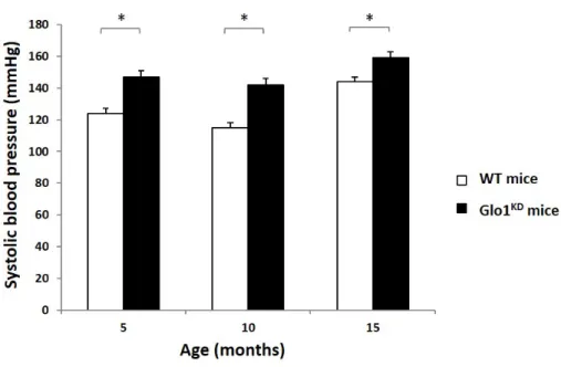
1.5 Measurement of systolic blood pressure.
1.6 Measurement of endothelium-dependent vasodilation. 2.1 Biogenesis of miRNA.
2.2 MicroRNA (miRNAs) targeting insulin signalling mediators. 2.3 MGO effect on miRNAs expression.
2.4 Effect of miR-190a inhibitor on insulin sensitivity in MAECs. 2.5 Effect of miR-190a mimic on insulin sensitivity in MAECs. 2.6 The role of KRAS in MGO-mediated effect.
2.7 Effect of MGO on miR-190a and vascular insulin sensitivity in vivo.
2.8 Effect of methylglyoxal (MGO) on miR-214 and
miR-126 target phosphatases of insulin signalling mediators. 2.9 Effect of miR-214 and miR-126 modulation on PHLPP2
protein levels in MAECs.
2.10 Effect of miR-214 mimic and miR-214 inhibitor on PHLPP2 levels in MAECs with or without MGO. 2.11 Effect of miR-214 on PHLPP2 regulation.
List of tables
Abstract
Methylglyoxal (MGO), a highly reactive dicarbonyl compound formed as by-product of glycolysis, is an ubiquitous metabolite of cellular metabolism. Therefore, it is produced in all cells, both under normal and pathological conditions. Under physiological circumstances, MGO is detoxified through the glyoxalase system, of which Glyoxalase 1 (Glo1) is the rate limiting enzyme. In pathological conditions, as in chronic hyperglycemia, high blood glucose levels lead to increased MGO accumulation. It is known that MGO plays a major role in endothelial cell damage and development of vascular disease. We have previously demonstrated that MGO induces endothelial insulin resistance both in vitro and in animal models. In the last few years, many evidence has provided a link between microRNAs (miRNAs) and diabetic complications. Indeed, miRNAs regulate cellular molecular pathways, including insulin signaling, thus controlling the pathophysiology of vascular bed. This study includes the investigation of two aspects of MGO effects on the pathophysiology of diabetes mellitus (DM) and its associated complications: 1. the evaluation of MGO accumulation on glucose homeostasis and vascular function in a mouse model knockdown for Glo1 (Glo1KD) and 2. the analysis of miRNAs contribution in MGO induced damaging effect on insulin responsiveness in mouse aortic endothelial cells (MAECs).
The results obtained in vivo demonstrated that the endogenous accumulation of MGO in mice with a reduced expression of Glo1 leads to an age-dependent development of glucose intolerance, in absence of hyperglycemia. Indeed, despite the reduced glucose tolerance at 10 months of age, Glo1KD mice have no differences in body weight and in the glucose levels, compared to WT mice, neither at 5 months nor at 10 months of age. While no alterations in the whole-body insulin-sensitivity have been observed by insulin tolerance tests, Glo1KD mice show a basal hyperinsulinemia and impaired glucose-stimulated insulin-secretion, compared to WT mice. Moreover, an increased systolic blood pressure accompanied by impaired endothelium-dependent vasodilation are already shown starting from 5 months of age in Glo1KD mice.
A deeper analysis of the molecular mechanisms involved in the endothelial dysfunction has been performed in vitro, in MAECs exposed to MGO, which we have previously demonstrated to display insulin resistance and an imbalanced production of vasoactive molecules: NO and ET-1. Our results demonstrate that MGO induces the down-regulation of 4 out of 84 diabetes-associated miRNAs. Among these, the reduced expression of miR-190a and miR-214 has been validated both in MAECs exposed to MGO and in aortae
Abstract
from Glo1KD mice. The inhibition of miR-190a and miR-214 impairs the insulin-induced activation of Akt1/eNOS pathway, whereas their overexpression prevents the MGO-induced insulin resistance in MAECs. In detail, we have identified the kinase KRAS and the phosphatase PHLPP2 as targets of miR-190a and miR-214, respectively. In MAECs increased KRAS levels result from the reduced expression of miR-190a and sustain the ERK hyperactivation, which is in turn responsible for the impairment of the insulin-stimulated IRS1/Akt/eNOS signal transduction in MAECs treated with MGO. Moreover, a reduced insulin-dependent activation of Akt in MGO-treated MAECs is fostered by higher protein levels of PHLPP2, which we validate here to be a direct target of miR-214.
In conclusion, our results demonstrate that Glo1 silencing is enough to induce MGO accumulation in vivo in Glo1KD mice, causing glucose intolerance and β-cell dysfunction, which are characteristic of T2DM pathogenesis, together with the impairment of hemodynamic function (i.e blood pressure and endothelial-dependent vasodilation), in a context of normoglycemia. Moreover, miR-190a and miR-214 play a role in the endothelial insulin-resistance induced by MGO in MAECs. Thus, representing potential biomarkers of vascular dysfunction. Further efforts in the development of pharmacological intervention to interfere with these pathogenic events will be useful to provide new therapeutic options aimed at preventing the onset and progression of vascular complications in diabetes.
Background 1. Endothelial cell function
The endothelium was considered to be a selective barrier to the diffusion of macromolecules from the vessel lumen to interstitial space. During the past years, different studies have highlighted additional roles for the endothelium. Indeed, the endothelium regulates vascular tone, cell-cell interaction, permeability and the coagulation system through the production of several factors, in response to various stimuli (Nigro et al. 2017, Schalkwijk et al. 2005). To carry out its above-mentioned functions, the endothelium produces components of the extracellular matrix such as collagen and a variety of regulatory mediators, including nitric oxide (NO), prostanoids, endothelin-1 (ET-1), angiotensin II (Ang II), tissue-type plasminogen activator (t-PA), plasminogen activator inhibitor-1 (PAI-1), von Willebrand factor (vWf), adhenosin molecular and cytokines (Schalkwijk et al. 2005). In physiological conditions, the endothelium actively decreases vascular tone, limits leucocyte adhesion and, thus, inflammatory activity in the vessel wall. It maintains vascular permeability to nutrients, hormones, other macromolecules and inhibits platelet adhesion and aggregation by producing prostacyclin and NO. Moreover, it limits the activation of the coagulation cascade by the thrombomodulin/protein C, heparan sulphate/antithrombin and tissue factor/tissue factor pathway inhibitor interactions and regulates fibrinolysis by producing t-PA and its inhibitor PAI-1. As mentioned above, the endothelium is able to synthesize NO, an important molecule that has vasodilator, platelet, anti-proliferative, permeability-decreasing and anti-inflammatory properties. NO inhibits leucocyte adhesion and rolling, as well as cytokine-induced expression of vascular cell adhesion molecule-1 (VCAM-1) and monocyte chemotactic protein-1 (MCP-1), effects that are at least in part attributable to the inhibition of the transcription factor nuclear factor κB (NF-κB) (Schalkwijk et al. 2005). Disturbing this tightly regulated equilibrium leads to endothelial dysfunction (ED). In particular, ED is the first step in the initiation, progression and clinical outcome of vascular complications, such as retinopathy, nephropathy and hypertension, that are the principal causes of mortality and morbidity of diabetic patients (Nigro et al. 2017). Anyway, the mechanisms leading to ED are not very completely clarified. Several studies have shown that hyperglycemia associated with impaired glucose tolerance and diabetes causes insulin resistance and ED (Du et al. 2001).
Background 2. Hyperglycemia and endothelial dysfunction
The hallmark of ED is represented by the impaired NO bioavailability. Additionally, ED is established when one or more of the following features occur: reduced endothelium-mediated vasorelaxation, hemodynamic deregulation, impaired fibrinolytic ability, enhanced turnover and /or overproduction of growth factors, increased expression of adhesion molecules and inflammatory genes, excessive generation of reactive oxygen species (ROSs), increased oxidative stress and enhanced permeability of the endothelial cells (ECs) layer (Taddei et al. 2003, Addabbo et al. 2009, Sena et al. 2013). The increased production of ROSs is due to the activation of nicotinamide adenine dinucleotide phosphate hydrogen (NADPH) oxidase, inactivation and reduced expression of the antioxidant enzymes, catalase and superoxide dismutase (SOD) or uncoupling of endothelial nitric oxide synthase (eNOS) (Dhananjayan et al. 2016). The increased ROSs production is one of the factors involved in the activation of transcription factors such as NF-κB. NF-κB is a key mediator that regulates multiple pro-inflammatory and pro-atherosclerotic target genes in ECs, vascular smooth muscle cells (VSMCs) and macrophages. Activation of NF-κB leads to an increased production of adhesion molecules, leukocyte-attracting chemokines and cytokines activating inflammatory cells in the vascular wall. In ED, a pro-thrombotic state is generated by the increased production of lesion-based coagulants, such as tissue factor, and the inhibitors of fibrinolysis, such as PAI-1. Vascular tone and remodeling are enhanced through reduced NO and an increased activity and production of vasoconstrictors (i.e. ET-1, angiotensin II, and prostanoids).
There are several factors contributing to ED, among which the most important are: hypertension, smoking, dyslipidemia and hyperglycemia (Sena et al. 2013). Hyperglycemia is one of major characteristic of type 2 diabetes mellitus (T2DM), a common metabolic disease with a high and growing prevalence (American Diabetes Association 2009). In both animal and human studies, it has been proven that hyperglycemia impairs endothelial function in both macro- and microvascular beds (Sena et al. 2013). Hyperglycemia causes vascular damage in different cell types of the vascular wall through the activation of several pathways including: 1) the increased flux of glucose and other sugars through the polyol pathway; 2) the augmented intracellular formation of advanced glycation end products (AGEs); 3) the increment of the expression of the receptor for AGEs (RAGE); 4) the activation of protein kinase C (PKC) isoforms; and 5) the
Background
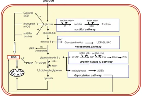
overactivation of the hexosamine pathway (Fig. 1). The activation of these biochemical ways promotes increased vascular oxidative stress, inflammation, apoptosis, atherogenesis and impaired endothelial function (Brownlee 2001).
Figure 1: Molecular pathways activated chronic hyperglycemia. (Schalkwijk and Stehouwer 2005).
In hyperglycemic condition, the excess of glucose can be metabolized to sorbitol and fructose by aldose reductase (AR) and sorbitol dehydrogenase (SDH), respectively. In the hexosamine pathway, fructose 6-phosphate is converted into glucosamine 6-phosphate by the enzyme glutamine fructose-6-phosphate amidotransferase (GFAT) and subsequently into N-Acetylglucosamine (GlcNAc). The mechanism responsible for the activation of PKC by hyperglycemia is related to de novo synthesis of the PKC activator, diacylglycerol (DAG) from a stepwise acylation of glycerol 3-phosphate (G3P). A major pathway activated by increased levels of the upstream glycolytic metabolite glyceraldehyde-3-phosphate is the AGEs non-enzymatic formation where the major intracellular AGEs precursor methylglyoxal (MGO) is generated from triose-phosphates fragmentation.
Background 3. Methylglyoxal metabolism
MGO is a highly reactive α-oxaldehyde, whose formation rate depends on the organism, tissue, cell metabolism and physiological condition (Igor et al. 2015). The endogenous MGO is derived from metabolic intermediates of carbohydrates, proteins and fatty acids (Fleming et al. 2011). In mammals, the glycolytic pathway is the principal source of MGO via fragmentation of triosephosphates G3P and dihydroxyacetone phosphate (DHAP) (Rabbani et al. 2016). Under normal physiological conditions, MGO is maintained at low levels. The latter are increased in conditions leading to higher triosephosphate levels, like happens in hyperglycemia when glucose metabolism is increased, in the impairment of pentose pathway with decreased G3P or in case of the increased anaerobic glycolysis occurring in hypoxia (Nigro et al. 2017). Although the production of MGO constitutes only 0.1% of glucotriose flux, its biological effect is important considering its high reactivity with proteins and nucleic acids. The irreversible reaction of MGO with proteins is directed to arginine residues forming hydroimidazolone adducts. The hydroimidazolone (MGO-H1) derived from MGO is the most abundant MGO-derived AGEs, as it accounts for >90% of all MGO adducts, equivalent to MGO-H1 residues in 1-5% of all proteins (Maessen et al. 2015).
Background
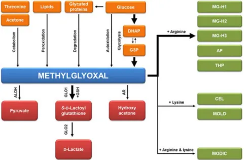
Figure 2. Pathways involved in MGO formation and detoxification. MGO is generated as a byproduct of glycolysis. Other sources of MGO are constitute by: autoxidation of glucose, catabolism of threonine and acetone, lipid peroxidation and degradation of glycated proteins. MGO is detoxified mainly by the glyoxalase system. Other minor pathways are aldehyde dehydrogenase (ALDH) and aldose reductase (AR). Otherwise, it may react with amino groups of proteins and other biomolecules to form 1-carboxyethyl-lysine (CEL) methylglyoxal-lysine dimer: 1,3-di(Nε-lysino)-4-methyl-imidazolium (MOLD), while it forms 2-ammonio-6-{(2-[(4-ammonio-5-oxido-5-oxopentyl)amino]-4-methyl-4,5-dihydro-1H-imidazol-5-ylidene) amino}hexanoate (MODIC) following dimer crosslink with arginine and lysine (Maessen et al. 2015).
Other minor contributors to MGO formation are from the oxidation of acetone (Beisswenger et al. 2006), the catabolism of ketone bodies that are increased in ketoacidosis (Lyles and Chalmers 1992), lipid peroxidation and degradation of glycated proteins and monosaccharides (Kalapos 1999, Thornalley et al. 1999) (Fig. 2). It is now known that MGO is observed at relative higher plasma levels (2–6 fold) in diabetic patients than in healthy subjects and are also found in many food products, beverages and cigarette smoke (Nigro et al. 2017). In detail, in physiological conditions, MGO levels is 50–150 nM in human plasma and 1–4 μM in human cells. Nevertheless, the contribution of exogenous MGO is still controversial. Several studies report that free MGO is rapidly degraded during digestion in the intestine and, thus, it does not influence plasma levels in vivo (Rabbani et al. 2016, Nigro et al. 2017), while others report that in rodents fed with
MGO-Background
supplemented diet there is a major brain and plasma MGO accumulation (Cai et al. 2014). When reactive dicarbonyl concentration increases beyond physiological levels, they produce protein and cell dysfunction leading to impaired health and disease (Rabbani et al. 2016).
Under physiological circumstances, MGO is detoxified by different mechanisms such us the glyoxalase, aldose reductase, aldehyde dehydrogenase and carbonyl reductase pathways (Maessen et al. 2015). Undoubtedly, the glyoxalase system, an ubiquitous enzymatic pathway, is the main detoxification system for MGO.
4. The glyoxalase pathway
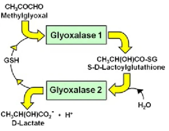
The glyoxalase pathway consists of two enzymes, glyoxalase 1 (Glo1) and glyoxalase 2 (Glo2) and a catalytic amount of glutathione (GSH) (Fig. 3).
Figure 3. The Glyoxalase System. Glyoxalase system is formed by two enzymes. Glyoxalase 1 catalyzes the conversion the MGO-GSH hemi-thioacetal to the thioester S-D-lactoyglutahione. Glyoxalase 2 catalyzes the hydrolysis of S-D-lactoylglutathione to form the end-product D-lactate (Rabbani et al. 2016).
In this process, Glo1 is the most crucial enzyme as it is defined the rate limiting enzyme by converting the MGO-GSH hemi thioacetal to the thioester S-D-lactoyglutahione (Sousa et al. 2013). Its activity is directly proportional to GSH concentration. Thus, when cellular GSH concentration declines, as in oxidative stress, there is an associated impairment of Glo1 activity. Glo2 is the second enzyme in the glyoxalase system and it catalyzes hydrolysis of S-D-lactoylglutathione to D-lactate and GSH (Thornalley 1993, Nigro et al. 2017). Glo1 gene is expressed in all tissues
Background
of prokaryotic and eukaryotic organisms (Aronsson and Mannervik 1978, He et al. 2000), and is the protein localized in the cytoplasmic compartment (Rabbani and Thornalley 2014). Several studies demonstrated that Glo1 activity can be modulated by both gene expression regulation and post-translational modifications (Ranganathan 1999). While genetic polymorphisms of Glo2 are extremely rare, different SNPs (single nucleotide polymorphisms) have been identified in the Glo1 gene that reduce the Glo1 activity and correlate with an increase of the diabetic neuropathy (Groener et al. 2013) and an increase risk of cardiovascular complications (Rabbani and Thornalley 2011).
5. MGO and insulin resistance
Insulin resistance is clinically defined as the inability of a known quantity of exogenous or endogenous insulin to produce a biological response, as the increase of glucose uptake and utilization (Gisela 2005). A widely accepted theory states that insulin resistance leads to T2DM, metabolic and cardiovascular disease and that MGO may contribute to the pathogenesis of insulin resistance (Nigro et al. 2017). We and others have provided evidence about the role of MGO on insulin-resistance in major target tissues for insulin action (Nigro et al. 2014). For instance, a short exposure of L6 muscle cells to MGO induces an inhibition of insulin-stimulated phosphorylation of protein Kinase B (PKB) and extracellular signal-regulated protein Kinase 1/2 (ERK1/2), without affecting insulin receptor tyrosine phosphorylation (Maessen et al. 2015). Moreover, it has been demonstrated that 3T3-L1 adipocyte treated with MGO, it shown an impairment of the insulin signalling, as indicated by decreased insulin-induced insulin receptor substrate (IRS-1) tyrosine phosphorylation (Jia and Wu 2007). Indeed, the incubation of the pancreatic INS1-E β-cells with MGO results in a glycogen synthase kinase (GSK3) mediated impairment of insulin secretion action (Maessen et al. 2015). In vivo, it has been demonstrated that MGO induces insulin resistance and salt sensitivity by increasing oxidative stress in Sprague–Dawley rats (Guo et al. 2009). In support of animal data and studies in animal models, a very recent human study in healthy overweight individuals demonstrates that with a diet low AGEs reduces the risk of T2DM by improving insulin sensitivity (De Courten et al. 2016). Insulin resistance is typically defined as decreased sensitivity or responsiveness to metabolic actions of insulin. However, diminished sensitivity to the vascular actions of insulin also plays an
Background
important role in the pathophysiology of insulin-resistant states (Baron et al. 1991, Natali et al. 1997). Indeed, endothelial insulin resistance is typically accompanied by reduced phosphatidylinositol 3 kinase (PI3K)/NO pathway and an intact or heightened mitogen-activated protein kinase (MAPK)/ET-1 pathway.
We have shown that MGO alters the sensitivity of the endothelium to insulin action both in vitro and in vivo. In particular, high levels of MGO cause an alteration in the release of two important vasoactive molecules by the endothelium: ET1 (vasoconstrictor action) and NO (vasodilatory action), highlighting MGO as an important culprit of endothelial dysfunction associated with insulin resistance (Nigro et al. 2014). Indeed, insulin resistant states are associated with metabolic abnormalities that include glucotoxicity, lipotoxicity and inflammation, thus contributing to the progression of long term endothelial dysfunction.
6. Effect of MGO accumulation on vascular function
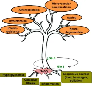
In the last decades, several studies have been focused on the central role of MGO in vascular defects (Shamsaldeen et al 20016), particularly on endothelial dysfunction, which is considered an important factor in the development of cardiovascular disease (Fig. 4).
Background
Figure 4. Sources of methylglyoxal (MGO) accumulation contributing to vascular dysfunction. Hyperglycemia, oxidative stress, inflammation and exogenous sources of MGO contribute both to the increase of MGO levels and the decrease of glyoxalase 1 (Glo1) activity. MGO-Glo1 imbalance leads to vascular dysfunction contributing to endothelial insulin-resistance, hypertension, atherosclerosis, microvascular complications, ageing and neuro-degeneration. (Nigro et al. 2017)
Brouwers et al. (2010) have demonstrated that Glo1 overexpression improved diabetic-induced impairment of NO-mediated relaxation. In vivo, exogenous administration of MGO to rats induces diabetes-like microvascular changes (Berlanga et al. 2005) and the impairment of endothelial function (Sena et al. 2012). Moreover, recent studies have shown that methylglyoxal induce vascular dysfunction in rat aorta, mesenteric arterial bed (Thilavech et al. 2017) and causes vascular contractile dysfunction in spontaneously hypertensive rats (SHR) (Mukohda et al. 2012). Dhar et al. have demonstrated a novel finding and a probable mechanism of increase in blood pressure, in particular they shown that MGO activates NF-κB through RAGE and thereby increases renin-angiotensin levels (Dhar et al. 2014). The MGO can impair the NO homeostasis and this has been related to endothelial dysfunction via a modulation of eNOS (Su et al 2013 and Turkseven et al 2014). Moreover, MGO and MGO-derived AGEs also play a harmful effect on microvascular function, contributing to the onset of nephropathy and neuropathy. Overexpression of Glo1 enzyme is important in the prevention of early renal impairment in rat models of
Background
diabetes (Brouwers et al. 2014), but also independently of hyperglycemia in apo E-/- mice. This is also confirmed by the evidence that MGO accumulation in Wistar normal rats impairs several renal disease markers progressively observed in diabetic Goto-Kakizaki rats (GK) (Rodrigues et al. 2014). Based on these findings, it became clear that an effective reduction of MGO accumulation is crucial for preserving vascular function.
7. Role of MGO in the development of vascular complications
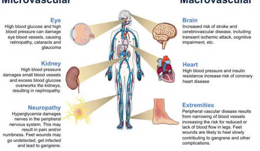
Hyperglycemia is a common feature in patients with diabetes mellitus (DM). DM is a common disease affecting more than 451 million people worldwide (Cho et al. 2018). One of the major concerns associated with diabetes relates to the development of micro-vascular (diabetic nephropathy, neuropathy, and retinopathy) and macro-vascular complication (coronary artery disease, peripheral arterial disease, and stroke), which greatly contribute to the high morbility and mortality associated with the disease (Fig. 5).
7.1 Microvascular complications
Results from both experimental and clinical studies on diabetes have demonstrated that hyperglycemia-induced MGO accumulation plays an important role in pathogenesis of microvascular complication.
Nephropathy
Diabetic nephropathy is the major cause of kidney disease and is a major risk factor for the development of macrovascular complications in patients effected by diabetic (Maessen et al. 2015). Indeed, it has been demonstrated that in cultured cells and kidneys from diabetic mice, increased glycolytic flux causes methylglyoxal modifications of the corepressor mSin3A, resulting in enhanced angiopoietin-2 expression, sensitizing microvascular endothelial cells to the proinflammatory effects of tumor necrosis factor (TNF-α) (Yao et al. 2007). Moreover, experimental studies in renal cells demonstrated that the MGO-induced inactivation of mitochondrial respiratory, chain may play an important role in renal cellular toxicity and development of diabetic nephropathy (Rosca et al.2002, Maessen et al. 2015).
Background
Retinopathy
Diabetic Retinopathy is a second common microvascular complication of DM, which causes serious damages in the retina, leading to increased vascular permeability, capillary microaneurysms, capillary degeneration and neovascularization (Forbes and Cooper 2013). Many clinical studies have demonstrated that, in both type 1 and type 2 diabetes (Fosmark et al. 2009, Fosmark et al. 2006), increased serum levels of the specific MGO-derived AGEs end product hydroimidazolone, is associated with the development of diabetic retinopathy (Fosmark et al. 2006). Moreover, overexpression of the Glo1 enzyme in diabetic rats prevents hyperglycemia-induced formation of MGO-derived AGEs in the neural retina and protects against retinal capillary degeneration over 6 months of diabetes (Queisser et al. 2010, Berner et al. 2012).
Neuropathy
Diabetic neuropathy is the most common complication of DM affecting as many as 50% of patients with type 1 and type 2 DM. In most cases, it is associated with foot ulcers, gangrene and limb amputation (Costa and Soares 2013, Chawla et al. 2016). The risk of diabetic neuropathy development is directly proportional to both the duration and magnitude of hyperglycemia. Several studies have confirmed the role played by glycation in diabetic neuropathy, however only a few studies focused specially on the importance of MGO and Glo1 (Jack and Wright 2012).
7.2 Macrovascular complications
Atherosclerosis
The atherosclerosis is a major cause of mortality in the people with diabetes (Grundy et al.1999). In most cases, the underlying process is a narrowing of arterial walls throughout the body (Maessen et al. 2015). In detail, it is a result from chronic inflammation and injury to the artery wall in the peripheral or coronary vascular system and of elevated levels of the low-density lipoproteins (LDL). Many studies have demonstrated that LDL can be modified by MGO, resulting in a change in both their physiological and biological properties (Schalkwijk et al 1998, Rabbani and Thornalley 2011, Turk et al. 2011, Brown et al. 2005). LDL play a significant role in the
Background
enhancement, development and progression of atherosclerosis, through a pathway that involves endothelial cell dysfunction.
Hypertension
MGO has been demonstrated to be involved in the development of other different macrovascular disease, such as the hypertension, but the exact mechanism is not yet fully understood. A link between MGO accumulation and hypertension has been demonstrated in rats with increased MGO levels in aortic and renal tissue (Guo et al. 2009, Wu and Juurlink 2002, Wang et al. 2004, Chang et al. 2005). Rats treated with MGO in drinking water show the increase systolic blood pressure and higher plasma levels of aldosterone, renin and catecholamines (Dhar et al. 2014). Similarly, a diet high in fructose, precursor of MGO, induces hypertension and renal injury in rats (Guo et al. 2012, Sánchez-Lozada et al. 2007). Although a dose of 1% MGO in drinking water alone has no effect on blood pressure for up to 4 weeks in Sprague-Dawley rats (Vasdev et al. 1998), surprisingly enough the same dose in combination with high-salt diet induced hypertension and enhances renal oxidative stress (Guo at al. 2009), suggesting that MGO causes hypertension only in rats with enhanced renal oxidative stress (Maessen et al.2015). Moreover, other studies showed that a significant increase in blood pressure coincided with elevated MGO levels in plasma and aorta of SHR in an age-dependent fashion, compared with age-matched Wistar–Kyoto rats (WKY) (Wang et al.2004, Wang et al. 2005). In the last few years, different mechanisms have been suggested as link between MGO and hypertension, as such it is known that MGO causes vascular Ca 2+ channel alterations, leading to increased cytosolic [Ca2+], peripheral vascular resistance and hypertension (Vasdev et al. 1998). Moreover, another study demonstrated that MGO-induced hypertension takes palace via angiotensin II type 1 receptor-mediates pathway (Chen et al. 2013). An improvement in blood pressure has been observed in SHR after treatment with aminoguanidine, a known scavenger of MGO (Wang et al. 2007, Wang et al. 2008).
Background
Figure 5. Major microvascular and macrovascular complications associated with diabetes mellitus. doi: 10.2210/rcsb_pdb/GH/DM/monitoring/complications
Bibliography 7.3 Bibliography
Addabbo F., Montagnani M., Goligorsky M.S. (2009) Mitochondria and reactive oxygen species. Hypertension. 53(6):885-92. doi: 10.1161/HYPERTENSIONAHA.109.130054
Aronsson A.C., Mannervik E. M. B. (1978) Glyoxalase I, a zinc metalloenzyme of mammals and yeast. Volume 81, Issue 4 Pages 1235-1240 https://doi.org/10.1016/0006-291X(78)91268-8
Baron A.D., Laakso M., Brechtel G., Edelman S.V. (1991) Mechanism of insulin resistance in insulin-dependent diabetes mellitus: a major role for reduced skeletal muscle blood flow. J Clin Endocrinol Metab. ;73(3):637– 643
Beisswenger B.G.K, Delucia E.M, Lapoint N., Sanford J. R., Beisswenger P. J. (2006) Ketosis Leads to Increased Methylglyoxal Production on the Atkins Diet. https://doi.org/10.1196/annals.1333.025
Berlanga J., Cibrian D., Guillén I., Freyre F., Alba J.S., Lopez-Saura P., Merino N., Aldama A.., Quintela A.M., Triana M.E., Montequin J.F., Ajamieh H., Urquiza D., Ahmed N. (2005) Methylglyoxal administration induces diabetes-like microvascular changes and perturbs the healing process of cutaneous wounds. JClin Sci (Lond). 109(1):83-95. DOI: 10.1042/CS20050026
Berner A.K., Brouwers O., Pringle R., Klaassen I., Colhoun L., McVicar C., Brockbank S., Curry J.W., Miyata T., Brownlee M., Schlingemann O., Schalkwijk C., Stitt A.W. (2012) Protection against methylglyoxal-derived AGEs by regulation of glyoxalase 1 prevents retinal neuroglial and vasodegenerative pathology. Diabetologia. 55(3):845-54. doi: 10.1007/s00125-011-2393-0
Brouwers O., Niessen P.M., Miyata T., Østergaard J.A., Flyvbjerg A., Peutz-Kootstra C.J., Sieber J., Mundel P.H.., Brownlee M, Janssen B.J., De Mey
Bibliography
J.G., Stehouwer C.D., Schalkwijk C.G. (2014) Glyoxalase-1 overexpression reduces endothelial dysfunction and attenuates early renal impairment in a rat model of diabetes. Diabetologia. 57(1):224-35. doi: 10.1007/s00125-013-3088-5
Brouwers O., Niessen P.M.., Haenen G, Miyata T., Brownlee M., Stehouwer C.D., De Mey J.G., Schalkwijk C.G. (2010) Hyperglycaemia-induced impairment of endothelium-dependent vasorelaxation in rat mesenteric arteries is mediated by intracellular methylglyoxal levels in a pathway dependent on oxidative stress. Diabetologia. 53(5):989-1000. doi: 10.1007/s00125-010-1677-0
Brown B.E., Dean R.T., Davies M.J. (2005) Glycation of low-density lipoproteins by methylglyoxal and glycolaldehyde gives rise to the in vitro formation of lipid-laden cells. Diabetologia. 48(2):361-9. DOI:10.1007/s00125-004-1648-4
Brownlee M. (2001) Nature. Dec 13;414(6865):813-20. Biochemistry and molecular cell biology of diabetic complications. DOI:10.1038/414813a Cai W., Uribarri J., Zhu L., Chen X., Swamy S., Zhao Z., Grosjean F.,
Simonaro C., Kuchel G.A., Schnaider-Beeri M., Woodward M., Striker G.E., Vlassara H. (2014) Oral glycotoxins are a modifiable cause of dementia and the metabolic syndrome in mice and humans Proc Natl Acad Sci U S A. 1;111(13):4940-5. doi: 10.1073/pnas.1316013111
Chang T., Wang R., Wu L. Methylglyoxal-induced nitric oxide and peroxynitrite production in vascular smooth muscle cells. Free Radical Biology and Medicine Volume 38, Issue 2, 15 January 2005, Pages 286-293https://doi.org/10.1016/j.freeradbiomed.2004.10.034
Chawla A., Chawla R., and Jaggi S. (2016) Microvasular and macrovascular complications in diabetes mellitus: Distinct or continuum? Indian J Endocrinol Metab. 20(4): 546–551. doi: 10.4103/2230-8210.183480
Bibliography
Chen X., Mori T., Guo Q., Hu C., Ohsaki Y., Yoneki Y., Zhu W., Jiang Y., Endo S., Nakayama K., Ogawa S., Nakayama M., Miyata T. & Ito S. (2013). Carbonyl stress induces hypertension and cardio–renal vascular injury in Dahl salt-sensitive rats Hypertension Research volume 36, pages 361–367 Cho N.H., Shaw J.E., Karuranga S., Huang Y., da Rocha Fernandes J.D.,
Ohlrogge A.W., Malanda B. (2018) IDF Diabetes Atlas: Global estimates of diabetes prevalence for 2017 and projections for 2045. Diabetes Res Clin Pract 138:271-281. doi: 10.1016/j.diabres.02.023
Costa P.Z., Soares R. (2013) Neovascularization in diabetes and its complications. Unraveling the angiogenic paradox. Life SciJun 13;92(22):1037-45. doi: 10.1016/j.lfs.2013.04.001
De Courten B., de Courten M.P., Soldatos G., Dougherty S.L., Straznicky N., Schlaich M., Sourris K.C., Chand V., Scheijen J.L., Kingwell B.A., Cooper M., Schalkwijk C.G., Walker K.Z., Forbes J.M. (2016) Diet low in advanced glycation end products increases insulin sensitivity in healthy overweight individuals: a double-blind, randomized, crossover trial. Am J Clin Nutr. 103(6):1426-33. doi: 10.3945/ajcn.115.125427
Dhananjayan R., Srivani Koundinya K. S, Malati T., Vijay Kumar Kutala. (2016) Endothelial Dysfunction in Type 2 Diabetes Mellitus Indian Journal of Clinical Biochemistry October, Volume 31, Issue 4, pp 372–379 doi: 10.1007/s12291-015-0516-y
Dhar I., Dhar A., Wu L., Desai KM. (2014) Methylglyoxal, a reactive glucose metabolite, increases renin angiotensin aldosterone and blood pressure in male Sprague-Dawley rats. Am J Hypertens. 27(3):308-16. doi: 10.1093/ajh/hpt281
Diabetes Care. 2009 Jan;32 Suppl 1: S62-7. Diagnosis and classification of diabetes mellitus. American Diabetes Association. doi: 10.2337/dc09-S062 Dionne E.M. Maessen, Coen D.A. Stehouwer, Casper G. Schalkwijk. (2015) The role of methylglyoxal and the glyoxalase system in diabetes and other
Bibliography
age-related diseases Clinical Science 128 (12) 839-861; DOI: 10.1042/CS20140683
Du X.L., Edelstein D., Dimmeler S., Ju Q., Sui C., Brownlee M. (2001) Hyperglycemia inhibits endothelial nitric oxide synthase activity by posttranslational modification at the Akt site.J Clin Invest.108(9):1341-8. 10.1172/JCI11235
Fleming T.H., Humpert P.M., Nawroth P.P., Bierhaus A. (2011) Reactive metabolites and AGE/RAGE-mediated cellular dysfunction affect the aging process: a mini-review.Gerontology 57(5):435-43. doi: 10.1159/000322087 Forbes J.M., Cooper M.E. (2013). Mechanisms of diabetic
complications.Physiol Rev. Jan;93(1):137-88doi: 10.1152/physrev.00045.2011
Fosmark D.S., Berg J.P., Jensen A.B., Sandvik L.., Agardh E, Agardh C.D., Hanssen K.F. Acta Ophthalmol. (2009) Increased retinopathy occurrence in type 1 diabetes patients with increased serum levels of the advanced glycation endproduct hydroimidazolone. 87(5):498-500. doi: 10.1111/j.1755-3768.2008.01300.x.
Fosmark D.S., Torjesen P.A., Kilhovd B.K., Berg T.J., Sandvik L., Hanssen K.F., Agardh C.D., Agardh E. (2006). Increased serum levels of the specific advanced glycation end product methylglyoxal-derived hydroimidazolone are associated with retinopathy in patients with type 2 diabetes mellitus. Metabolism 55(2):232-6 https://doi.org/10.1016/j.metabol.2005.08.017 Gisela W. (2005) Insulin and Insulin Resistance Clin Biochem Rev. 26(2): 19–
39
Groener J.B., Reismann P., Fleming T., Kalscheuer H., Lehnhoff D., Hamann A., Roser P., Bierhaus A., Nawroth P.P., Rudofsky G. (2013) C332C genotype of glyoxalase 1 and its association with late diabetic
Bibliography
complications. Exp Clin Endocrinol Diabetes. 121(7):436-9. doi: 10.1055/s-0033-1345124
Grundy S.M., Benjamin I.J., Burke G.L., Chait A., Eckel R.H., Howard B.V., Mitch W., Smith S.C., Sowers J.R. (1999) Diabetes and cardiovascular disease: a statement for healthcare professionals from the American Heart Association. Circulation. 7;100(10):1134-46.
Guo Q., Mori T., Jiang Y., Hu C., Ohsaki Y., Yoneki Y., Nakamichi T., Ogawa S., Sato H., Ito S. (2012) Losartan modulates muscular capillary density and reverses thiazide diuretic-exacerbated insulin resistance in fructose-fed rats. Hypertens Res. 35(1):48-54. doi: 10.1038/hr.2011.140
Guo Q., Mori T., Jiang Y., Hu C., Osaki Y., Yoneki Y., Sun Y., Hosoya T., Kawamata A., Ogawa S., Nakayama M., Miyata T., Ito S. (2009) Methylglyoxal contributes to the development of insulin resistance and salt sensitivity in Sprague-Dawley rats. J Hypertens. 27(8):1664-71. doi: 10.1097/HJH.0b013e32832c419a
He M.M., Clugston S.L., Honek J.F., Matthews B.W. (2000) Determination of the structure of Escherichia coli glyoxalase I suggests a structural basis for differential metal activation. Biochemistry. 39(30):8719-27. DOI: 10.1021/bi000856g
Igor A., Mireille B. and Pierre J. Magistretti (2015) Methylglyoxal, the dark side of glycolysis Front. Neurosci. https://doi.org/10.3389/fnins.2015.00023 Jack M.M. and Wright D.E. (2012) The Role of Advanced Glycation End products and Glyoxalase I in Diabetic Peripheral Sensory Neuropathy Transl Res. 159(5): 355–365 doi: 10.1016/j.trsl.2011.12.004
Jia X., Wu L. (2007) Accumulation of endogenous methylglyoxal impaired insulin signalling in adipose tissue of fructose-fed rats. Mol Cell Biochem. 306(1-2):133-9. Epub Jul 28. DOI:10.1007/s11010-007-9563-x
Bibliography
Kalapos M.P. (1999) Methylglyoxal in living organisms: chemistry, biochemistry, toxicology and biological implications. Toxicol Lett. 110(3):145-75. https://doi.org/10.1016/S0378-4274(99)00160-5
Lyles G.A., Chalmers J. (1992) The metabolism of aminoacetone to methylglyoxal by semicarbazide-sensitive amine oxidase in human umbilical artery. Biochemical Pharmacology Volume 43, Issue 7, 1 April, Pages 1409-1414 https://doi.org/10.1016/0006-2952(92)90196-P
Maessen D.E., Stehouwer C.D., Schalkwijk C.G. (2015) The role of methylglyoxal and the glyoxalase system in diabetes and other age-related diseases. Clin Sci (Lond). Jun;128(12):839-61. doi: 10.1042/CS20140683 Mukohda M., Okada M., Hara Y., Yamawaki H. (2012) Methylglyoxal
accumulation in arterial walls causes vascular contractile dysfunction in spontaneously hypertensive rats. J Pharmacol Sci. 2012;120(1):26-35 Natali A., Taddei S., Quinones G. A., Camastra S., Baldi S., Frascerra S., et al.
(1997) Insulin sensitivity, vascular reactivity, and clamp-induced vasodilatation in essential hypertension. Circulation 96(3):849–855
Nigro C., Leone A., Raciti G.A., Longo M., Mirra P., Formisano P., Beguinot F., Miele C. (2017) Methylglyoxal-Glyoxalase 1 Balance: The Root of Vascular Damage. Review. Int J Mol Sci doi: 10.3390/ijms18010188 Nigro C., Raciti G.A., Leone A.., Fleming TH., Longo M,.. Prevenzano
I., Fiory F., Mirra P., D'Esposito V., Ulianich L., Nawroth P.P., Formisano P., Beguinot F., Miele C. (2014) Methylglyoxal impairs endothelial insulin sensitivity both in vitro and in vivo. Diabetologia. 57(7):1485-94. doi: 10.1007/s00125-014-3243-7
Queisser M.A., Yao D., Geisler S. et al (2010) Hyperglycemia impairs proteasome function by methylglyoxal. Diabetes 59:670–67. doi: 10.2337/db08-1565
Bibliography
Rabbani N., Thornalley P.J. (2011) Glyoxalase in diabetes, obesity and related disorders. Semin Cell Dev Biol. May;22(3):309-17. doi: 10.1016/j.semcdb.2011.02.015
Rabbani N., Thornalley P.J. (2014) The critical role of methylglyoxal and glyoxalase 1 in diabetic nephropathy. 63(1):50-2. DOI: 10.2337/db13-1606 Rabbani N., Xue M., Thornalley P.J.(2016) Dicarbonyls and glyoxalase in disease mechanisms and clinical therapeutics. Glycoconj J. 33(4):513-25. doi: 10.1007/s10719-016-9705-z
Ranganathan S., Ciaccio P. J., Walsh E. S., Tew K. D. (1999) Genomic sequence of human glyoxalase-I: analysis of promoter activity and its regulation. Gene Volume 240, Issue 1, Pages 149-155 https://doi.org/10.1016/S0378-1119(99)00420-5
Rodrigues L., Matafome P., Crisóstomo J., Santos-Silva D., Sena C., Pereira P., Seiça R. (2014) Advanced glycation end products and diabetic nephropathy: a comparative study using diabetic and normal rats with methylglyoxal-induced glycation. J Physiol Biochem.70(1):173-84. doi: 10.1007/s13105-013-0291-2
Rosca M.G., Monnier V.M., Szweda L.I., Weiss M.F.( 2002) Alterations in renal mitochondrial respiration in response to the reactive oxoaldehyde methylglyoxal. Am J Physiol Renal Physiol. 283(1):F52-9 DOI: 10.1152/ajprenal.00302.2001
Sánchez-Lozada L.G., Tapia E., Jiménez A., Bautista P., Cristóbal M., Nepomuceno T., Soto V., Avila-Casado C., Nakagawa T., Johnson R.J., Herrera-Acosta J., Franco M. (2007) Fructose-induced metabolic syndrome is associated with glomerular hypertension and renal microvascular damage in rats. Am J Physiol Renal Physiol. 292(1): F423-9. DOI: 10.1152/ajprenal.00124.2006
Bibliography
Schalkwijk C.G, Stehouwer C.D. (2005) Vascular complications in diabetes mellitus: the role of endothelial dysfunction. Clin Sci (Lond) 109(2):143-59DOI: 10.1042/CS20050025
Schalkwijk C.G., Vermeer M.A., Stehouwer C.D,. te Koppele J., Princen H.M., van Hinsbergh V.W .(1998) Effect of methylglyoxal on the physico-chemical and biological properties of low-density lipoprotein.Biochim Biophys Acta. ;1394(2-3):187-98. https://doi.org/10.1016/S0005-2760(98)00112-X
Sena C.M, Matafome P., Crisóstomo J., Rodrigues L., Fernandes R., Pereira P., Seiça RM. (2012) Methylglyoxal promotes oxidative stress and endothelial dysfunction. Pharmacol Res. 65(5):497-506. doi: 10.1016/j.phrs.2012.03.004
Sena C.M., Pereira A.M., Seiça R. (2013) Endothelial dysfunction - a major mediator of diabetic vascular disease. Biochim Biophys ActaDec 1832(12):2216-31doi: 10.1016/j.bbadis.2013.08.006
Shamsaldeen Y.A., Mackenzie L.S., Lione L.A., Benham C.D. (2016) Methylglyoxal, A Metabolite Increased in Diabetes is Associated with Insulin Resistance, Vascular Dysfunction and Neuropathies. Curr Drug Metab.;17(4):359-67
Sousa S. M, Gomes R.A., Ferreira A.E., Ponces Freire A., Cordeiro C. (2013) The glyoxalase pathway: the first hundred years….and beyond. Biochem J. 453(1):1-15 doi: 10.1042/BJ20121743
Su Y., Qadri S.M.., Hossain M, Wu L., Liu L. (2013) Uncoupling of eNOS contributes to redox-sensitive leukocyte recruitment and microvascular leakage elicited by methylglyoxal. Biochem Pharmacol. 86(12):1762-74. doi: 10.1016/j.bcp.2013.10.008
Taddei S., Ghiadoni L., Virdis A., Versari D., Salvetti A. (2003) Mechanisms of Endothelial Dysfunction: Clinical Significance and Preventive
Non-Bibliography
Pharmacological Therapeutic StrategiesCurrent Pharmaceutical Design Volume 9, Issue 29 DOI: 10.2174/1381612033453866
Thilavech T., Abeywardena M. Y., Adams M., Dallimore J., Adisakwattana S. (2017) Naturally occurring anthocyanin cyanidin-3-rutinoside possesses inherent vasorelaxant actions and prevents methylglyoxal-induced vascular dysfunction in rat aorta and mesenteric arterial bed. Biomedicine and pharmacotherapy volume 95, pages 1251-1259 Doi: 10.1016/j.biopha.2017.09.053
Thornalley P. J., Langborg A., Minhas H. S. (1999) Formation of glyoxal, methylglyoxal and 3-deoxyglucosone in the glycation of proteins by glucose. Biochemical Journal Nov 08, ,344(1)109-116; DOI: 10.1042/bj3440109
Thornalley P.J. (1993) The glyoxalase system in health and disease. Molecular Aspects of Medicine Volume 14, Issue 4, Pages 287-371. https://doi.org/10.1016/0098-2997(93)90002-U
Turk Z., Čavlović-Naglića M., Turk Nikša (2011) Relationship of methylglyoxal-adduct biogenesis to LDL and triglyceride levels in diabetics Turk Life Sciences Volume 89, Issues 13–14, 26 Pages 485-490 https://doi.org/10.1016/j.lfs.2011.07.021
Turkseven S., Ertuna E., Yetik-Anacak G., Yasa M. (2014) Methylglyoxal causes endothelial dysfunction: the role of endothelial nitric oxide synthase and AMP-activated protein kinase α. J Basic Clin Physiol Pharmacol. (1):109-15. doi: 10.1515/jbcpp-2013-0095
Vasdev S., Ford CA.., Longerich L., Parai S., Gadag V., Wadhawan S. (1998) Aldehyde induced hypertension in rats: prevention by N-acetyl cysteine. Artery 1998;23(1):10-36
Wang X., Chang T., Jiang B., Kaushik D., Wu L. (2007) Attenuation of hypertension development by aminoguanidine in spontaneously
Bibliography
hypertensive rats: role of methylglyoxal. Am J Hypertens. 20(6):629-36. DOI:10.1016/j.amjhyper.2006.12.003
Wang X., Desai K., Clausen J. T., and Wu L. (2004) Increased methylglyoxal and advanced glycation end products in kidney from spontaneously hypertensive rats. Kidney International, Vol. 66, pp. 2315–2321 https://doi.org/10.1111/j.1523-1755.2004.66034.x
Wang X., Jia X., Chang T., Kaushik D., Wu L. (2008) Attenuation of hypertension development by scavenging methylglyoxal in fructose-treated rats Journal of Hypertension volume 26 - Issue 4 p 765–772. doi: 10.1097/HJH.0b013e3282f4a13c
Wang X., Kaushik D., Chang T.; Wu L.(2005) Vascular methylglyoxal metabolism and the development of hypertension Journal of Hypertension: Volume 23 - Issue 8 - p 1565–1573 doi: 10.1097/01.hjh.0000173778.85233.1b
Wu L., Juurlink B.H. Hypertension. (2002) Increased methylglyoxal and oxidative stress in hypertensive rat vascular smooth muscle cells. 39(3):809-14
Yao D., Taguchi T., Matsumura T., Pestell R., Edelstein D., Giardino I., Suske G., Rabbani N., Thornalley P. J., Sarthy V. P., Hammes H.Peter and Brownlee M. (2007) High Glucose Increases Angiopoietin-2 Transcription in Microvascular Endothelial Cells Through Methylglyoxal Modification of mSin3A. The Journal of Biological Chemistry 282, 31038-31045. http://www.jbc.org/cgi/doi/10.1074/jbc.M704703200
Chapter 1
Effect of glyoxalase 1 gene deletion on glucose homeostasis and
vascular function in mice
Nigro C., Leone A., Longo M., Prevenzano I., Fleming T.H., Nicolò A., Parrillo L., Spinelli R., Formisano P., Nawroth P.P., Beguinot F., Miele C. (2018) Methylglyoxal accumulation de-regulates HoxA5 expression, thereby impairing angiogenesis in glyoxalase 1 knock-down mouse aortic endothelial cells. Biochim Biophys Acta Mol Basis Dis. pii: S0925-4439(18)30391-0. doi: 10.1016/j.bbadis.2018.10.014.
Effect of MGO accumulation in mice
1.1 Risk factors for the development of complications in DM
Beyond the classification of DM, the presence of chronically elevated blood glucose levels is implicated in the progression of diabetes complications. For this reason, the primary goal of therapeutic treatment is to reduce hyperglycemia. Nonetheless, while most of the literature claims the benefits of glycemic control in the prevention of vascular complications (Cefalu 2006), additional evidence suggests that the risk of complications may be decreased further if glycated hemoglobin (HbA1c) is reduced below levels currently accepted as clinical goal (Stratton et al. 2000). In support of this possibility, Khaw et al (2001) demonstrated that a reduced HbA1c level is associated with a lower rate of cardiovascular disease, even in non-diabetic subjects. At the same time a clinical study on HbA1c contribution to retinopathy in type 1 diabetic patients shows that lowering HbA1c levels is not enough to prevent the increased incidence of events in the long term (Lind et al. 2010). Moreover, it is known that as blood pressure levels increase in diabetics there is a parallel increase in cardiovascular disease, diabetic retinopathy and nephropathy (Rask-Madsen and King et al. 2013). Many studies have demonstrated clear benefits in lowering blood pressure. Indeed, in the UKPDS trial, tight blood pressure control with angiotensin-converting enzyme (ACE) inhibitors or -blockers significantly reduced diabetes-related events and diabetes-related deaths (U.K Prospective Diabetes Study Group 1998, Katherine L Bate and George Jerums 2003). In addition, other studies demonstrated that it is important to control also the levels of dyslipidemia to reduce the risk of development of diabetic complications. Indeed, two placebo-controlled trials have shown that treatment with statins reduces the risk of a major cardiovascular event by 37% in patients with type 2 diabetes without clinically apparent cardiovascular disease (Heart Protection Study Collaborative Group. 2002, Colhoun et al. 2004, Marshall and Flyvbjerg 2006). A recent study found that the risk of diabetic complication can be reduced through a multifactorial approach, for example reducing the smoke or administration the low-dose of aspirin (Marshall and Flyvbjerg 2006). Therefore, all these studies suggest that other risk factors besides poor glycaemia control may be important for the onset of vascular complications (Bots et al. 2016).
Effect of MGO accumulation in mice 1.2 Rodent Models of DM and its related complications
Studies in animal models have provided significant advances to the knowledge of cardiovascular complications in understanding to which extent insulin resistance, hyperinsulinemia and hyperglycemia may individually contribute to vascular and cardiac dysfunction in diabetes. Among the commonly used models, the ob/ob mouse is a diabetic animal with genetic mutations on the leptin gene. As a consequence of this defect, ob/ob mice develop hyperphagia and obesity, reduced glucose tolerance, severe hyperinsulinemia, insulin resistance and impaired would healing (Potenza et al. 2011). The db/db mice, carrying a mutation on the gene encoding for the leptin receptor in C57b/KsJ strain, show hyperphagia, obesity and early insulin resistance. This model rapidly develop hyperglycemia, diabetic nephropathy and ketosis as their β-cells are unable to maintain high levels of insulin secretion required for survival (Maessen et al. 2015).
In addition, several diabetic models have been utilized to understand the relationship between metabolic control and cardiovascular health. eNOS KO/lepr (db/db) double knockout (DKO) mice develop obesity, hyperglycemia and also hypertension (Potenza et al. 2011). The cp-cp (ceruloplasmin) rats spontaneously develop the pathophysiological characteristic resembling the human metabolic syndrome, for this reason are use as model of diabetes. In detail the cp/cp rats are homozygous for autosomal recessive cp gene and develop atherosclerosis, ischemic myocardial lesions and microvascular renal dysfunction not accompanied by hypertension (Russell et al. 1998). The GK rats show neonatal β-cell mass deficiency, which is responsible for the basal hyperglycemia. Indeed, at 8 weeks of age, sustained hyperglycemia is accompanied by both severely impaired insulin release and insulin resistance (Miralles and Portha 2001). This animal model exhibits endothelial dysfunction as early as 4 months of age and has provided important informations on the relationship between changes in β cells mass and the occurrence of diabetic vascular complications.
Moreover, also the Zucker Diabetic Fatty (ZDF) rats, selectively inbred for hyperglycemia, can be placed in this category. Indeed, at 10 weeks of age, ZDF rats show more than a 4-fold increase in blood glucose levels that
Effect of MGO accumulation in mice
remain high throughout their entire lifespan. They show coronary and aortic endothelial dysfunction that are involved in atherogenesis and vascular alterations occurring in this model (Otlam et al. 2008, Chinen et al. 2007). SHR rapresent a genetic model of hypertension, in which the defects in endothelial insulin signalling with impaired PI3-Kinase-dependent and augmented MAPK-dependent activities precede several disturbances of the metabolic syndrome (Potenza et al. 2005). For this reason, they are used in the studies of endothelial function.
Further rodent models have been generated to study the role of insulin-signalling mediators in the pathogenesis of DM. In detail, the deletion of IRS-1 in the IRSKO mouse (IRS-1 KO) leads to β-cell hyperplasia and insulin resistance, mainly localized to skeletal muscle tissue. Hypertension onset, secondary to impaired endothelium-dependent relaxation, and hypertriglyceridemia development, secondary to impaired activation of lipoprotein lipase in insulin resistant adipose tissue, have been observed in these mice. Akt2 knockout in mice results in glucose intolerance, hyperinsulinemia, insulin resistance and, under some conditions, overt diabetes. Akt 2 is predominantly expressed in pancreatic β-cells, skeletal muscle and brawn fat, but also in platelets. Indeed, Akt2 knockout mice (Akt 2 KO) display defects in platelet aggregation and thrombus formation (Woulfe et al. 2004). Moreover, liver-specific insulin receptor knockout (LIRKO) mice exhibit a dramatic elevation in blood glucose and the loss of gluconeogenesis regulation. It has been demonstrated that LIRKO mice show marked hypercholesterolemia and develop severe atherosclerosis at 12 weeks, when fed with atherogenic diet (Biddinger et al. 2011).
In the absence of appropriate diabetic/atherosclerotic models, exposure to chemicals (e.g streptozotocin) as well as to diabetogenic or atherogenic diets has been used to evaluate the impact of hyperglycemia, obesity, insulin resistance and hypercholesterolemia on vascular complication development in wild type and genetically modified rodents (Potenza et al. 2011). Schreyer et al. have demonstrated the atherogenic diet containing 1.25% cholesterol, 15% fat, and 0.5% cholic acid induces atherosclerosis. Moreover, also a diabetogenic diet containing 35.5% fat (58% of calories, primarily lard) and 36.6% carbohydrate (primarily sucrose)) may induce vascular lesions by altering both lipid and glucose metabolism, while a high fat/high sucrose diet used to induce obesity and diabetes in C57BL/6 mice, may provide an
Effect of MGO accumulation in mice
important tool for the study of diabetes accelerated atherosclerosis (Schreyer et al. 1998). In rats the administration of streptozotocin (STZ), after a primary administration of nicotinamide adenine dinucleotide (NAD), produces a T2DM model at a rate of 75-80%, which develops a mild and stable hyperglycemia without changes in plasma insulin (Masiello P. et al. 1998). Combination of STZ administration in animals with a genetic insulin resistant background (e.g. in ZFR model) or under a high fat or high fructose diet produces models that develop overt hyperglycemia in the presence of normal blood insulin and, hence, are regarded as more appropriate for T2DM studies (Chatzigeorgiou et al. 2009). Moreover, other studies have shown that the long-term administration of diets containing front 40% to 60% of lipids promotes metabolic disorders in animal models (Flanagan et al., 2008); and induces adipocyte hypertrophy (Barbosa-da-Silva et al. 2012), T2DM, hypertriglyceridemia (Fraulob et al. 2010) and liver steatosis in mice (Aguila et al. 2003, Barbosa-da-Silva et al. 2013).
Therefore, the use of the appropriate animal models can provide interesting data needed to clarify the pathophysiological mechanisms responsible for the onset of diabetic complications.
1.2.1 Mouse models of MGO accumulation
To prove the effect of MGO on vascular function, animal models that allow to observe the systemic implications of an imbalanced accumulation/detoxification ratio of MGO have been generated. Among these, the chronic administration of a MGO solution has been performed in rodents in many studies. Dhar et al (2011) have been demonstrated that chronic MGO infusion by minipumps causes pancreatic β-cell dysfunction and induces DM in Sprague-Dawley rats. Others have been demonstrated that high MGO levels induce vascular contractile dysfunction in arterial wall of SHR (Mukohda et al. 2012). We have previously demonstrated that intraperitoneal administration of MGO to C57BL/6 mice impairs whole-body insulin sensitivity and induced endothelial insulin resistance (Nigro et al. 2014). However, it is important to note that exogenous sources of MGO are only partially absorbed and not completely accumulated in tissues as free MGO, thus limiting the interpretation of these studies. The measurement of tissue and plasma levels of MGO in experimental models is, therefore,
Effect of MGO accumulation in mice
necessary to ensure the patho-physiological relevance of the animal models. To bypass these limitations, a possibility is to modulate Glo1 activity and/or its expression. Indeed, MGO accumulation may be induced by reducing Glo1 activity, through the chemical Glo1 inhibitor “SpBrBzGSHCp2” or silencing Glo1 expression (Nigro et al. 2017). Interestingly, it has been shown that non-diabetic mice knock-down for Glo1 (Glo1 KD) expression show an increase of MGO modified proteins and oxidative stress, causing alteration in kidney indistinguishable from those caused by diabetes (Giacco et al. 2014). Bierhaus et al (2012) demonstrated that this mouse model develops the thermal and medical hyperalgesia, signs of peripheral neuropathy. Moreover, the beneficial effect of MGO detoxification have been proved by studies using Glo1 overexpressing models. Interestingly, Glo1 over-expression in STZ-induced diabetic mice prevents diabetes-induced oxidative stress and the development of kidney pathology, despite unchanged levels of diabetic hyperglycemia (Giacco et al. 2014). Moreover, Glo1 overexpression ameliorates renal ischemia-reperfusion injury (Kumagai et al. 2009), reduces endothelial dysfunction and attenuates early renal impairment in rat model of diabetes (Brouwers et al. 2014).
This is in line with a recent clinical study reporting that pharmacological induction of Glo1 activity improves insulin sensitivity and glycemic control in obese patients (Xue et al. 2016). Furthermore, Glo1 has been linked to coronary artery disease (Makinen et al. 2014) and hypertension (Wilson et al. 1991) in epidemiological studies. A recent study obtained in Glo1-tg mice has shown that Glo1 overexpression is able to prevent the MGO-mediate increase of inflammation in diabetes, which leads to endothelial cell loss and, contributes to the development of diabetic cardiomyopathy (Vulesevic et al. 2016). Moreover, the overexpression of Glo1 reduces age-related glycative and oxidative stress in the vasculature and attenuates endothelial dysfunction through the modulation of eNOS phosphorylation from early aging. This proves that the regulation of glycative stress by enhanced Glo 1 activity counteracts physiological vascular aging. Therefore, the regulation of glycative stress may represent a versatile target for the prevention of vascular aging and its associated complications (Jo-Watanabe et al. 2014, Nigro et al. 2017).
Effect of MGO accumulation in mice 1.3 Aim
DM is a common metabolic disorder that is strongly associated with vascular complications. Several studies demonstrated that diabetes is linked to macro- and microvascular complications, which are responsible for high mortality and morbidity of the disease. For this reason, it is considered an important public health problem.
Hyperglycemia represents the main feature of DM and one of the major cause of plasma and intracellular MGO accumulation. In physiological conditions, MGO is efficiently detoxified by the glyoxalase system, however its levels increase under pathological conditions, such as hyperglycemia contributing to cardiovascular complication associated with diabetes. Indeed, several studies carried out in models of hyperglycemia have proved the toxic effect of MGO and its derived AGEs on micro and macrovascular function (Chinen et al. 2013), on insulin sensitivity of muscle, adipose tissue and β-cells. However, the specific effect of a physiological accumulation of the MGO a tissue damage has not been yet clarified. To this aim, it is necessary to use an optimal experimental model to investigate the pathological processes by which MGO favors the onset of DM and its associated complication.
This work aims to investigate the impact of endogenous MGO accumulation on metabolic and vascular functions in Glo1-knockdown mice (Glo1KD).
Effect of MGO accumulation in mice 1.4 Results
1.4.1 Metabolic characterization of Glo1KD mice
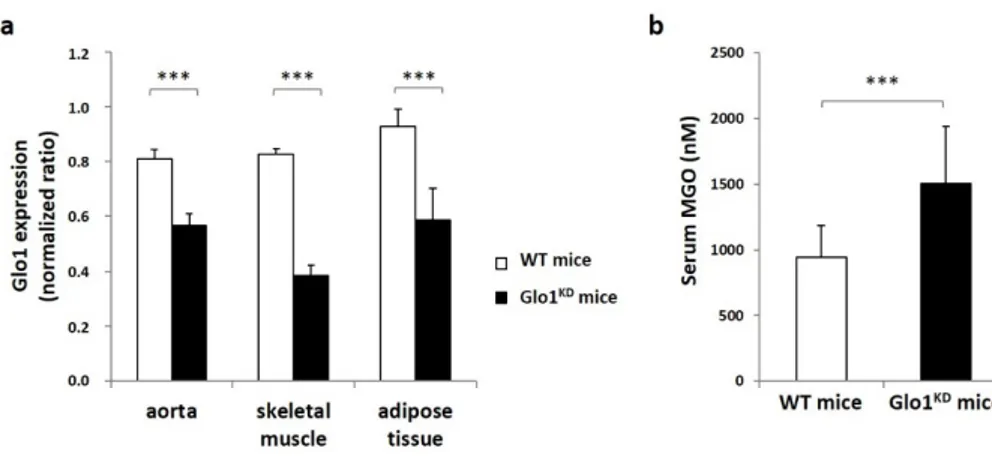
Glo1KD mice and their wild type littermates (WT mice) were used to evaluate the effect of MGO accumulation on glucose tolerance. In order to validate this experimental model, the first step was to evalaute the Glo1 expression in different tissues by Real Time-PCR. As expected, Glo1KD mice show a significant reduction in tissue expression of the Glo1 gene compared to WT mice (Figure 1.1A). In detail, the absolute quantification revealed that Glo1KD mRNA levels are reduced from 30% to 50% in vascular tissue, skeletral muscle and adipose tissue isolated from Glo1KD mice compared to WT mice. To test if the partial deletion of Glo1 expression was enough to induce MGO accumulation, endogenous MGO levels were measuread by HPLC and resulted to be increased by 1.5-fold in the serum from Glo1 KD mice compared to WT mice (figure 1.1 b).
Figure 1.1 Expression levels of the Glo1 gene and serum MGO concentration. (a) The
expression of Glo1 gene was measured in aorta, muscle (quadriceps) and adipose tissue (epididymal fat) of Glo1KD and WT mice. Glo1 expression was normalized on the number
of GAPDH molecules, used as a housekeeping gene. (b) Serum MGO concentrations were measured by HPLC. Statistical analysis was performed by Student t-test, where p≤0.05 was considered statistically significant (*** p≤0.001).
Effect of MGO accumulation in mice
Metabolic characterization of Glo1KD mice have been evaluated during lifespan. As shown in table 1.1, the data obtained indicate that Glo1KD mice show no significant variation in body weight, food intake, fasting and fed glycaemia neither at 5 nor at 10 months of age, indicating that Glo1KD mice are normoglycemic and, thus, do not display a diabetic phenotype. Glucose levels have been measured during intraperitoneal Glucose Tolerance Tests (ipGTT) in order to evaluate the ability of the mouse to restore glucose levels at normal values after a bolus of glucose.
Table 1.1. Metabolic parameters
5 months 10 months
Variable WT mice Glo1KD mice Test T WT mice Glo1KD
mice Test-T Body weight (g) 25.4±0.8 24.5±0.7 n.s. 32.6±1.5 35.1±1.8 n.s. Fold intake 2.99±0.04 2.9±0.13 n.s - - n.s Fasting glycaemi a (mg/dl) 105.9±5.3 97.1±3.5 n.s. 114.6±8.4 106.4±6.0 n.s Fed glycaemi a (mg/dl) 138.9±4.1 145.6±6.9 n.s. 143.6±3.1 137.3±4.4 n.s.
Effect of MGO accumulation in mice
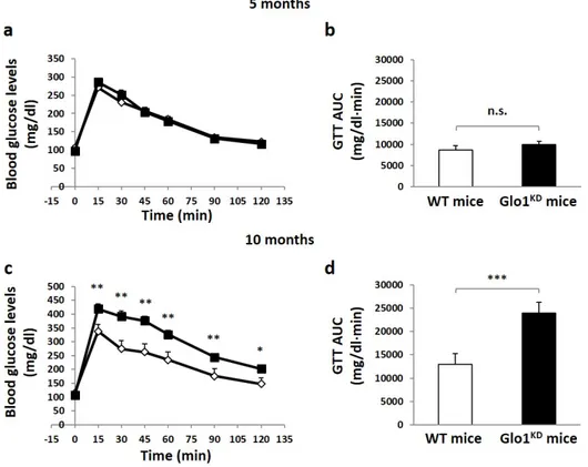
Figure 1.2 Glucose Tolerance Test (ipGTT). (a, c) The graph shows the curves related to
blood glucose levels of WT mice and GloKD mice within 2 hours after intraperitoneal
administration of a glucose bolus (2 g / kg) at 5 and 10 months. Blood glucose was measured by the use of a portable blood glucometer at the indicated times. (b, d) The bars of the graph analyze the area under the glucose curve of Glo1KD and WT mice. Statistical
analysis was performed by Student's t-test (* p≤0.05; ** p≤0.01; *** p≤0.001).
No differences in glucose tolerance have been revealed at 5 months of age. By contrast, Glo1KD mice develop a reduced glucose tolerance at 10 months of age, compared to WT mice. This is indicated by both the calculation of the area under curve (WT 12990±1595 vs KD 23944±2300) and by the single time points of glucose measurements performed during the entire test, when from 15 to 120 minutes following glucose bolus, blood glucose levels are higher in Glo1 KD mice compared to WT mice (figure 1.2 c).
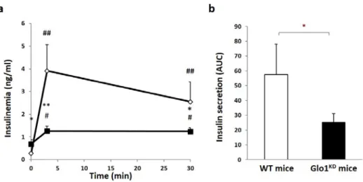
Effect of MGO accumulation in mice
Figure 1.3 Insulin tolerance test (ipITT)
(a) The graph shows the curves related to blood glucose levels of WT mice and GloKD
mice within 2 hours after intraperitoneal administration of a insulin (0,75 U/kg). Blood glucose was measured by the use of a portable blood glucometer at the indicated times. (b) The bars of the graph analyze the area under the glucose curve of Glo1KD and WT
mice. Statistical analysis was performed by Student's t-test (* p≤0.05; ** p≤0.01; *** p≤0.001).
Next, insulin sensitivity has been evaluated by means of the insulin tolerance test (ITT). The data obtained show that insulin sensitivity is not affected in Glo1KD mice compared to WT mice (figure 1.3). In light of the impairment of glucose tolerance pancreatic function has been tested by glucose-stimulated insulin secretion (GSIS).