Novel Pharmacological and Magnetic
Resonance Strategies to Enhance Boron
Neutron Capture Therapy (BNCT) Efficacy
in the Clinical Treatment of Malignant Glioma
Paola Porcari
1, Silvia Capuani
2and Francesco Saverio Pastore
31
Sapienza University of Rome, Rome,
2Sapienza University of Rome, CNR-IPCF UOS Rome, Physics Department, Rome
3University of Rome Tor Vergata, Neuroscience Department,
Institute of Neurosurgery, Rome,
Italy
1. Introduction
High-grade glioma, such as anaplastic astrocitomas (AA, WHO grade 3) and glioblastomas multiforme (GBM, WHO grade 4) are extremely aggressive and highly infiltrative brain tumours (Kleihues & Cavenee, 2000; Louis et al., 2007). In most cases they recur locally after applying the standard multimodality treatment based on surgical resection, followed by radiotherapy and/or chemotherapy. Despite advances in medicine, malignant gliomas continue to carry a dismal prognosis, even though a modest increase (by 4.5 months) in median survival and quality of life has been achieved. The main limitation to the effectiveness of surgery and radiotherapy in patients suffering from high-grade glioma is that these techniques, based on the geometric definition of tumour volume, are not suitable to eradicate tumour infiltrating cells within normal brain tissue. Moreover adjuvant chemotherapy has little effect on prolonging survival in patients with GBM (Stupp et al., 2005). As a consequence, novel therapeutic approaches, based on a better understanding of cancer biology, are needed. To this end, experimental therapies such as gene therapy (Mischel et al., 2003), antiangiogenic therapy (Van Meir et al., 2010), monoclonal antibodies (Zhu et al. 2010), cancer immunotherapy (Keunen et al., 2011), vaccines (Hickey et al., 2010), boron neutron capture therapy (BNCT) (Barth et al., 1992, 2005) and radioimmunotherapy (Joensuu, 2000) are under investigation. Among these, BNCT represents a promising adjuvant therapy for malignant glioma, and for other forms of cancer such as head/neck cancer. It is a binary form of radiation therapy based on the selective accumulation of boronated compounds within tumour cells which are then irradiated by low-energy thermal neutrons. The nuclear reaction that occurs between the stable isotope, 10B, and thermal
neutrons, yields high-energy alpha particles and recoiling lithium nuclei which release most of their ionizing energy within a few microns (about one cell diameter), therefore limiting radiation damage only to 10B-containing cells. Thus, BNCT can be considered as a
through boron compounds which selectively accumulate within them. Given this selectivity of BNCT, infiltrating tumour cells, as well as subclinical lesions, can be targeted by 10B
compounds and underwent the therapeutic effect.
For effective BNCT a large amount of 10B nuclei (about 109 atoms of 10B per cell) (Barth &
Soloway, 1997) should be selectively accumulated within tumour cells, whilst 10B
concentration levels in blood and in normal brain tissues should be the lowest. At the same time, a tumour-to-brain (T:Br) 10B concentration ratio of at least 3:1 must be achieved to
ensure the optimal therapeutic dose to the tumour. Furthermore a sufficient number of thermal neutrons (thermal neutron fluences should be greater than 1012 ncm-2) must be
captured by 10B atoms into the target volume during irradiation (Barth & Soloway, 1997).
BNCT therapy has been evaluated for safety and efficacy in several centres around the world. So far, no severe effects of BNCT-related toxicity have been observed (Phase I), whilst little evidence of therapeutic effectiveness has been evaluated (Phase II) in patients with GBM (Diaz, 2003). Presently the therapy is under experimentation in Phase II clinical trials, while a randomized Phase III study has not yet been justified because of previous disappointing results.
Currently, the boron carrier most widely used for clinical purpose is the boronated derivative of the essential amino acid L-phenylalanine, p-boronophenylalanine (BPA). Due to its poor solubility at physiological pH, it is administrated as a complex with fructose (BPA-fr complex). It is widely accepted that BPA is actively transported across the blood brain barrier (BBB) into the normal glia, while its uptake within the tumour is due to an increased rate of L-amino acid transport across the tumour cell membrane (Wittig et al., 2000). In addition, BPA accumulation within tumour cells increases during the cell cycle (S-phase) so that its use in treating aggressive GBM might be an advantage. Furthermore it has been demonstrated that pre-treatment with L-tyrosine (Papaspyrou et al. 1994), or other molecules targeted by L or A amino acid transport systems, can enhance intracellular BPA accumulation (Wittig et al., 2000). Previous in vitro (Wittig et al., 2000) and in vivo (Capuani et al., 2008, 2009) studies have demonstrated that preloading with L-DOPA (a well-known molecule with chemical structure similar to those of L-tyrosine and BPA) improves the accumulation of BPA. As a consequence, more interest is being devoted to the potential clinical application of L-DOPA preloading. Indeed, due to the wide clinical experience with the administration of L-DOPA for the treatment of Parkinson’s disease, its use as a potential enhancer of BPA accumulation in BNCT clinical trials could be immediately applied. The main limitations for BNCT effectiveness are due to: a) insufficient 10B intake within
tumour cells, even if the most efficient methods of 10B administration are utilised; b) the lack
of reliable imaging methods for monitoring the bio-distribution of 10B-carriers in order to
estimate both the effectiveness of the carrier and the optimal timing for neutron irradiation (that is when T:Br 10B concentration ratio achieves the maximum value whilst at the same
time the 10B concentration in blood is the lowest).
Our research work has focused on developing solutions to overcome these BNCT limitations, in order to make it a clinically useful treatment modality in the near future. Specifically, we have evaluated in vivo (using C6 glioma model) the pharmacokinetics of BPA, and the effect of L-DOPA preloading on BPA accumulation both in the tumour and normal tissues. Pharmacokinetic data were helpful in determining the optimal irradiation time, as well as to develop computational strategies in order to define as accurately as possible the radiation dose released within tumour and surrounding healthy tissues.
In order to determine the best fitting curve of BPA pharmacokinetics (used to extrapolate the BPA concentration over time after infusion), in vivo monitoring of 10B-carrier was
performed using nuclear magnetic resonance (NMR), either by imaging (MRI) or by spectroscopy (MRS). In both cases, the fluorinated analogue of BPA, F-labelled BPA, was investigated using 19F-MRI and 19F-MRS (Porcari et al., 2006, 2008, 2009). All previously
mentioned methodologies and strategies designed to overcome some fundamental limitations of BNCT therapy have been developed to ensure a straightforward transfer from pre-clinical to clinical applications.
2. Principles of boron neutron capture therapy
BNCT is an experimental, radio-therapeutic modality able to biologically target malignant cells. It theoretically allows a selective delivery of the radiation damage within an infiltrating cancer cell while preserving the surrounding healthy tissues.
BNCT has been preferentially employed in clinical trials designed for the treatment of GBM (Henriksson et al., 2008; Yamamoto et al., 2008, 2011; van Rij et al., 2005). This high grade tumour of the central nervous system is highly malignant and extremely infiltrative, characterized by rapid tumour growth with a wide microscopic invasion of malignant cells within the normal parenchyma. It is extremely resistant to all current therapies, including surgery, chemotherapy, radiotherapy, immunotherapy and gene therapy. Despite advances in medicine, its prognosis is still very poor with a median survival time of less than one year (Ohgaki & Kleihues, 2005). Thus, GBM remains one of the challenges to be faced by physicians and scientists worldwide. BNCT holds therapeutic promise for these incurable tumours. Clinical interest in BNCT has also been focused on high-grade gliomas (Chanana et al., 1999), as well as the treatment of malignant meningiomas (Tamura et al., 2006) and cutaneous melanomas (Mishima & Kondoh, 2000). More recently, interest has been extended to other forms of neoplasms such as head-neck cancers (Kankaanranta et al., 2007), liver (Wittig et al., 2008a) and lung tumours (Suzuki et al., 2007), as well as undifferentiated thyroid cancers (Dagrosa et al., 2007).
The treatment is binary and is carried out following two distinct phases. Firstly, an intravenous infusion of 10B-enriched compounds is given to the patient, allowing the
selective uptake of 10B-carriers within neoplastic cells. Subsequently, when the T:Br 10B
concentration ratio has achieved its highest value (at least 3:1) and, at the same time, the 10B
concentration in blood is the lowest, the patient is irradiated with low energy (E < 0.4eV, thermal) or higher energy (0.4eV ≤ E ≤ 10 keV, epithermal) neutrons.
BNCT is based on the nuclear reaction 10B(n,)7Li (1) (Sauerwein, 1993) that occurs when
the stable isotope, 10B (characterized by a high thermal neutron capture cross section)
captures a thermal neutron (nth) to yield 11B* in an unstable form, which decays in highly
cytotoxic alpha (4
2H) and lithium ( 7
3Li) particles (Fig. 1.a). Due to their high Linear Energy Transfer (LET) (ICRU, 1998), these heavy charged particles release along their paths (comparable with the cell diameter, 5-9 m) a great density of ionization responsible for elevated Relative Biological Effectiveness (RBE) (ICRU, 1998). Thus, the dose delivered and the radiation damage is mostly confined within the 10B-loaded
4 7 2 3 10 11 * 5 th 5 4 7 * 2 3 + + 2.79MeV (6%) + + + 2.31MeV (94%) H Li B n (0.025eV) B H Li 7 * 3 + γ(0.48MeV) Li (1)
Fig. 1. 10B(n,)7Li reaction (a); cellular damage of BNCT (b).
The effectiveness of the therapy is dependent on two conditions:
a high number of 10B atoms must be selectively localized within neoplastic cells (at least
20-35 μg/g ~109 atoms of 10B per targeted cell) (Barth et al., 1996; Barth & Soloway,
1997);
a sufficient number of thermal neutrons has to reach and be captured by the 10B atoms
into the target volume during irradiation (thermal neutron fluences should be greater than 1012 n·cm-2) (Barth et al., 1996; Barth & Soloway, 1997).
Although theoretically only a few alpha particles, releasing their energy within a cancer cell, assure the cell death, both of the above conditions should be satisfied because of the low probability of interaction between a single 10B atom with a thermal neutron.
The therapeutic gain of BNCT is strictly dependent on the achievable T:Br 10B concentration
ratio between tumour and normal tissue. It has been established that the higher the T:Br 10B
concentration ratio is, the better the therapeutic gain of BNCT. Moreover, the tolerance dose of normal tissues should not be exceeded. It is mainly dependent on the neutron capture reactions, 1H(n,γ)2H and 14N(n,p)14C (2) (Soloway et al., 1997), that occur when hydrogen, 1H, and nitrogen, 14N, isotopes (with relative abundances of 3% and 10%, respectively, in
normal tissue) capture thermal neutrons yielding low LET γ rays and high LET protons (2), respectively. 1 2 1 2 2 th 14 14 14 15 14 th H(n, ) H H + n H H +γ(2.23MeV) N(n,p) C N + n N C + p(0.63MeV) (2)
Due to the small neutron capture cross-sections of 1H and 14N (σH = 0.332 and σN = 1.82
barns; 1 barn = 10-24 cm2) compared with that of 10B (σB = 3838 barns), the dose released
within surrounding healthy tissues is, in most of cases, much smaller than that delivered within the tumour, even though its value is dependent on neutron fluences. Thus, the upper limit of the neutron fluences is determined by the normal tissue tolerance dose for protons
and γ rays. As a consequence, for the best therapeutic result the T:Br 10B concentration ratio
should be as high as possible.
In order to satisfy the previous conditions, intensive investigations have been performed since the introduction of BNCT in most of the research centres worldwide. Considerable efforts have been directed towards the design and synthesis of new efficient boron agents, as well as in developing strategies to maximize the tumour boron uptake whilst minimizing, at the same time, 10B levels in blood and in normal brain. The disappointing outcomes of early
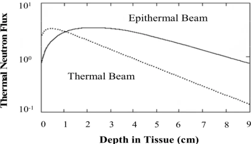
BNCT clinical trials in the United States (Slatkin, 1991) were mainly due to the inability of thermal neutrons to deliver therapeutic neutron fluences to deep-sited brain tumours. To overcome this, the use of higher energy epithermal neutron beams was pursued because of their greater tissue penetrating properties. Indeed, when epithermal neutrons penetrate tissues, they are slowed down into the thermal neutron range (Seppälä et al., 2002, Coderre et al., 1997) by means of collisions with atoms (Fig. 2). Epithermal neutrons, therefore, allow delivery of therapeutic fluences of thermal neutrons at greater depths in the brain without reflecting the scalp or doing a craniotomy as required by using thermal fluences.
0 1 2 3 4 5 6 7 8 9
Depth in Tissue (cm)
Thermal Beam
Epithermal Beam
Th
e
r
m
a
l
N
e
u
tr
o
n
F
lu
x
10-1 100 101 0 1 2 3 4 5 6 7 8 9Depth in Tissue (cm)
Thermal Beam
Epithermal Beam
Th
e
r
m
a
l
N
e
u
tr
o
n
F
lu
x
10-1 100 101Fig. 2. Variation of the thermal neutron fluence with tissue depth using a thermal or epithermal neutron beam (Coderre & Morris, 1999).
Currently, nuclear reactors are the only sources of neutrons for clinical BNCT. The neutrons are produced by the fission process in the core of the reactor and are classified according their energy as thermal (Eth < 0.025eV), epithermal (0.4eV < Eepi < 10 keV) or fast (Efast > 10k
eV). Since it is highly unlikely that the reactors can be sited in the main medical centres, alternative sources of thermal and epithermal neutrons for BNCT are being sought (Blue & Yanch, 2003). Among these, low-energy proton accelerators with low z targets are the most attractive.
At present several reactors creating optimal epithermal neutron beams for BNCT are being used clinically worldwide. They include the Massachusetts Institute of Technology Reactor (MITR) (Busse et al., 2003) in the USA, the Kyoto University Research Reactor (KURR) and JRR4 at the Japan Atomic Energy Research Institute (Nakagawa, 2003) in Japan, and the RA-6 CNEA reactor in Bariloche (Riley et al., 2008), Argentina. In Europe there are several clinical BNCT nuclear reactors: the FiR1 clinical reactor in Helsinki (Finland) (Joensuu et al.,
2003), the LVR-15 reactor at the Nuclear Research Institute in Rez (Czech Republic) (Burian et al., 2004) and the clinical reactor at Studsvik Medical AB (Sweden) (Capala et al., 2003).
2.1 Boron agents
Since its inception, the development of boron delivery agents for BNCT therapy has been one of the most important topics to fulfil. For BNCT to be successful 10B carriers should
satisfy the following requirements:
selectivity for malignant cells (with preferential 10B intracellular localization) compared
with blood and contiguous normal tissue;
achievement of tumour boron concentrations of at least 20-35 μg10B/g (approximately
109 boron atoms per cell);
permanence (at a constant concentration) within tumour during the BNCT radiation procedure and rapid clearance from both blood and normal tissues. This is necessary to estimate the radiation dose delivered to tumour, brain and vascular endothelium; minimal systemic toxicity in order to achieve adequate tumour concentrations in vivo
assuring, at the same time, favourable T:Br and tumour-to-blood (T:Bl) concentration ratios (at least 3:1);
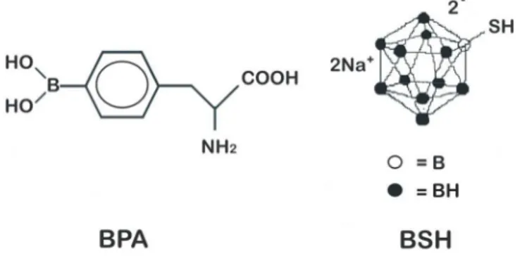
So far, two 10B carriers have been used clinically: the polyhedral borane, sodium
borocaptate (Na2B12H11SH or BSH) (Fig. 3), and the dihydroxyboryl derivate of
phenylalanine, boronophenylalanine (C9H12BNO4 or BPA) (Fig. 3).
Both compounds are characterized by low toxicities, selective tumour cell uptake, long tumour persistence and safety after their intravenous (i.v.) administration.
Fig. 3. Chemical structures of BPA and BSH.
Moreover, it has been demonstrated that either BPA or BSH may be able to target both proliferating and non-proliferating cells. This is of major importance for GBM treatment because of the relatively small percentage of GBM cells in the proliferative status at any time. Previously, BPA and BSH have been employed as boron agents in clinical trials designed for brain tumour treatments in the United States of America (Chanana et al.,1999; Coderre et al.,1999), in Europe (Phase I) (Capala et al., 2003; Joensuu et al., 2003, Burian et al., 1994) and in Japan (Phase II) (Nakagawa et al., 2003). The results of these trials confirmed the therapeutic efficacy of BNCT and provided the basis of the subsequent experimental clinical trials. However, the design of BNCT clinical protocols carried out using both mentioned 10
B-carriers was also influenced by the findings on animal model studies (Nakagawa et al., 2007; Smith et al., 1996).
In recent years, the clinical use of BPA for the GBM treatment has aroused great interest (Coderre et al., 1997; Capala et al., 2003; Joensuu et al., 2003) because of its encouraging results in experimental brain tumour therapy. Due to its low solubility in aqueous solutions at physiological conditions (pH ~ 7.4) it is administered as a complex with fructose (BPA–fr complex) (Yoshino et al., 1989). It was observed that BPA can be selectively accumulated either in the main tumour mass or in the microscopic cluster of tumour cell invading the normal parenchyma, even though the measured 10B concentration in the isolated cluster was
only about 50% of that obtained in the main tumour mass (Smith et al., 1996). This result is of great importance for the efficacy of BNCT because the isolated clusters represent potential sites of tumour re-growth. Furthermore the ability of BPA to target the microscopic cluster within the normal brain suggests that it is actively transported through the BBB.
Although the details of BPA accumulation into tumour cells are not completely understood, it is accepted that it is due to an elevated rate of amino acid transport across the tumour cell membrane (Wittig et al., 2000). Furthermore, there is evidence that BPA accumulation is enhanced by a pre-treatment with molecules vehiculated through L or A amino acid transport systems (Wittig et al., 2000). The increase of BPA intracellular accumulation was also demonstrated in mouse melanoma cells (Papaspyrou et al., 1994) using L-tyrosine pre-administration.
In addition, it has also been demonstrated that BPA accumulation within tumour cells increases during the cell cycle (S phase) (Nichols et al., 2002) so that its use in the treatment of aggressive brain gliomas might be an advantage.
3. Main limitations for BNCT effectiveness
BNCT is one of the most complex therapeutic modalities used to treat malignant brain tumours. Its success or failure is highly dependent on a combination of several chemical, physical and biological factors. Up to now, BNCT has been trialled to investigate its safety and efficacy in several centres worldwide. However, to date, the results of Phase I and II clinical trials have not shown therapeutic responses to justify Phase III trials.
These disappointing results are mainly due to the following limitations.
The first limitation was mostly due to insufficient uptake of 10B-labelled compound within
tumour cells even though the most advanced methods of 10B administration were used
(Chanana et al., 1999; Elowitz et al., 1998).
Normally, the 10B uptake within brain tumours may be influenced by several factors such as
the BBB permeability to the 10B-carrier, the plasma concentration profile of the 10B-agent
(which is dependent on either the drug dose or the way of administration), the blood flow within tumour as well as the drug lipophilicity.
So far, some strategies have been proposed to improve BNCT effectiveness by increasing BPA and BSH tumour intake. Some of these, including the use of pharmacological agents such as mannitol (Barth et al., 2000) or Cereport (RMP-7) (Yang et al., 1997) to disrupt the BBB, have been experimented on animal models (Barth et al., 2000; Yang et al., 1997). Although the results showed an increase in T:Br and T:Bl indices, the potential toxicities were not completely investigated. Moreover these methodologies have been classified as invasive, so numerous investigations are needed before considering them as potential applications in future clinical trials.
The second main limitation for BNCT effectiveness was due to the lack of efficient imaging methods to monitor the spatial bio-distribution of 10B-labelled compounds and their
pharmacokinetics, in order to estimate the efficacy of the carrier and the optimal timing of neutron irradiation. This ideal time is when the 10B concentration in tumour is higher than
the concentration in blood and surrounding healthy tissues to prevent damage to these regions. Previous studies carried out in order to estimate brain-to-blood (Br:Bl), T:Br and T:Bl 10B concentration ratios using kinetic models (Ryynanen et al., 2000; 2002) and
Inductively Coupled Plasma-Atomic Emission Spectrometry (ICP-AES) (Laakso et al., 2001) techniques, gave different results.
Up to now several techniques (Wittig et al., 2008b) have been used to determine the spatial distribution and pharmacokinetics of 10B agents (Elowitz et al., 1998; Ryynanen et al 2000,
2002, Laakso et al., 2001, Kabalka et al., 2003, Wang et al., 2004). Among these, MRI and MRS provide useful methods for non-invasive and non-destructive real-time monitoring of 10B
compounds during BNCT treatment in vivo. Given the low sensitivity of the 10B NMR
method (Bendel et al., 2001; Bendel 2005) and the intense proton background signal that makes 1H-MRS (Zuo et al., 1999) and Magnetic Resonance Spectroscopy Imaging (MRSI)
(Bendel et al., 2005) techniques problematic in vivo, new strategies to detect BPA by NMR are in progress.
4. Strategies to improve the efficacy of the therapy
In order to make BNCT a clinical useful treatment modality in the near future, our work aims at investigating solutions to overcome the main limitations to the efficacy of the current methodology.
Firstly, with the aim of improving the effectiveness of the therapy by increasing BPA tumour intake, the strategy used was to assess the effect of L-DOPA pre-loading on BPA accumulation within the tumour.
L-DOPA is a well-known molecule with a chemical structure similar to those of L-tyrosine and BPA. Its use as a potential enhancer of BPA accumulation was suggested by previous encouraging results obtained on both mouse melanoma (Papaspyrou et al., 1994) and 9L rat gliosarcoma cells by pre-administration of L-tyrosine (Wittig et al., 2000). The enhancement of BPA accumulation in 9L rat gliosarcoma cells has been also replicated by using pre-treatment with both molecules targeted by L and A aminoacid transport system. These findings suggest that the substrate-coupled antiport (exchange) mechanism of these transporters is enhanced by the preloading of specific aminoacids. Previous in vitro (Wittig et al., 2000) and in vivo (Capuani et al., 2008; 2009) studies have demonstrated that L-DOPA preloading improves the accumulation of BPA in the tumour. Specifically it was demonstrated in vivo (Capuani et al., 2008; 2009) that L-DOPA pre-administration on C6 glioma model gave rise to an increase of BPA tumour accumulation of 2.7 times with respect to those of controls. Conversely, no significant difference was evaluated by using the High Performance Liquid Chromatography (HPLC) method in both blood and normal brain between L-DOPA preloaded rats and controls. These findings are of fundamental importance for their impact on potential clinical applications. Indeed, the introduction of L-DOPA as a potential enhancer of BPA accumulation in BNCT clinical trials could be of immediate application because of its established clinical use as a treatment for Parkinson’s disease.
Then, with the aim of investigating the pharmacokinetic behaviour of 10B carriers and their
boron bio-distribution, both of them essential to evaluate the efficiency of the carrier and the optimal irradiation time, a novel approach to detect BPA was proposed. The strategy used was to map the fluorinated analogue of BPA (19F–BPA–fr complex) (Fig. 4) using 19F NMR in a way
similar to Positron Emission Tomography (PET) studies (Kabalka et al., 2003; Wang et al., 2004). The feasibility of the method was previously demonstrated in vitro (Porcari et al., 2006).
Fig. 4. Chemical structures of 19F–BPA and 19F–BPA–fr complex.
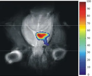
Specifically, selective bio-distribution (Fig. 5.) (Porcari et al., 2008; 2009) of 19F–BPA–fr
complex in C6 tumour-bearing rats as compared with normal brain has been demonstrated using 19F MRI. In addition, a better understanding of 19F–BPA pharmacokinetic was
achieved because of the correlation between the results obtained by using both 19F MRI and 19F MRS methodologies. Indeed the correlation between 19F MR monitoring on rat brain over
4h after 19F–BPA–fr complex infusion and the quantification of 19F spectra collected from
blood samples showed a maximum uptake of 19F–BPA in C6 glioma at 2.5h after infusion.
Thus, 2.5h after infusion is the optimal time of neutron irradiation according to previous results (Hsieh et al., 2005) obtained by using PET measurements of 19F–BPA.
These findings suggest the potential future application of 19F MRI and 19F MRS using 19F–
BPA in clinical trials. Indeed, the correlation of both techniques allows the mapping with the high spatial resolution characteristics of MRI of the distribution of 10B compounds and at the
same time to follow the pharmacokinetic of 10B agents. Moreover, since 19F NMR can be
performed using an 1H MR scanner by suitably tuning RF coils, only minor improvements
in the MRI clinical scanner are required for future clinical applications.
Fig. 5. 19F–BPA–fr complex bio-distribution map in C6 glioma model at 2.5h after infusion (Porcari et al., 2008)
5. Conclusion
It is apparent from previous sections that BNCT is one of the most complex therapeutic modalities for brain tumour treatment. Due to the lack of progress in developing more effective treatments for high grade gliomas, the main challenge for BNCT in the near future, is to become a clinically useful treatment modality. Our work in optimizing the therapy has just this aim. Our research has been focused in overcoming some of the major limitations of BNCT effectiveness.
Firstly, in order to improve 10B accumulation within the tumour, we demonstrated, both in
vitro and in vivo, the potential of L-DOPA to enhance tumour uptake of BPA (Capuani et al.,
2008, 2009). The most interesting findings of this work were the increased BPA tumour uptake in vitro, with C6 glioma cells, as well as in vivo, with C6 glioma model. Indeed, the L-DOPA preloading increased the BPA intracellular accumulation in C6 glioma cells 5 times with 4-hours of L-DOPA incubation (Capuani et al., 2008). The BPA tumour uptake in C6 glioma model (Capuani et al., 2008) increased 2.7 times. Interestingly, there was no increasing of BPA uptake in a normal brain. These stimulating results encourage the potential use of L-DOPA in BNCT of brain tumours because of L-DOPA ability to induce a significant enhancement of BNCT effectiveness without remarkable associated side effects. Moreover, the use of L-DOPA in BNCT clinical trials could be also facilitated because of its long-standing clinical use as a treatment for Parkinson’s disease.
In order to determine the optimal irradiation time improving the BNCT efficiency, 19F MR
imaging and spectroscopy methodologies were proposed for investigating the pharmacokinetics and bio-distribution of BPA (Porcari et al., 2008; 2009). The correlation between both imaging and spectroscopic results obtained on glioma model highlights a better understanding of 19F–BPA uptake either in the tumour or in systemic circulation
confirming evidence of maximum BPA uptake within the tumour at 2.5 hours after infusion (Porcari et al., 2008). These results demonstrate that both 19F MRI and 19F MRS are feasible
and practical methodologies with potential future clinical application. Indeed, 19F NMR can
be performed with an 1H MR clinical scanner with only minor hardware and software
improvements. Both of the solutions proposed to improve BNCT effectiveness will help the therapy to overcome its main hindrances to become a clinically useful modality in the near future.
6. Some standard abbreviations and symbols
10B Boron-10 isotope
BBB Blood Brain Barrier
BNCT Boron Neutron Capture Therapy
BPA p-boronophenylalanine
BPA-fr complex p-boronophenylalanine-fructose
complex
Br:Bl brain-to-blood
BSH sodium borocaptate
14C Carbon-14 isotope
γ rays gamma rays
Emission Spectrometry
19F-BPA fluorinated analogue of BPA
19F-BPA-fr complex 19F-BPA-fructose complex
19F-MRI Fluorine Magnetic Resonance Imaging
19F-MRS Fluorine Magnetic Resonance
Spectroscopy
19F-NMR Fluorine Nuclear Magnetic Resonance
GBM Glioblastomas Multiforme
1H, 2H Hydrogen isotopes
4
2H Alpha particle
HPCL High Performance Liquid
Chromatography
7Li Lithium-7 isotope
L-DOPA L-3,4-dihydroxyphenylalanine
LET Linear Energy Transfer
MRI Magnetic Resonance Imaging
MRS Magnetic Resonance Spectroscopy
MRSI Magnetic Resonance Spectroscopy
Imaging
nth thermal neutron
14N, 15N Nitrogen isotopes
NMR Nuclear Magnetic Resonance
p proton
PET Positron Emission Tomography
σ cross-sections
RBE Relative Biological Effectiveness
T:Bl tumour-to-blood T:Br tumour-to-brain
WHO World Health Organization
7. Acknowledgment
We acknowledge support from Italian Institute of Technology under the Seed project BACT-MOBIL.
8. References
Barth, R.F., Coderre, J.A., Vicente, M.G. & Blue, T.E. (2005). Boron neutron capture therapy of cancer: current status and future prospects. Clinical Cancer Research, Vol. 11, No. 11, (June 2005), pp. 3987-4002, ISSN 1557-3265
Barth, R.F., & Soloway, A.H. (1997). Boron neutron capture therapy of brain tumors-current status and future prospects. Journal of Neuro-Oncology, Vol. 33, No. 1-2, (May 1997), pp. 3-7, ISSN 0167-594X
Barth, R.F., Soloway, A.H. & Brugger, R.M. (1996). Boron neutron capture therapy of brain tumors: past history, current status, and future potential Cancer Investigation, Vol. 14, No 6, pp. 534-50, ISSN 0735-7907
Barth, R.F., Soloway, A.H., Fairchild, R.G. & Brugger, R.M. (1992). Boron neutron capture therapy for cancer: Realities and prospects. Cancer, Vol. 70, No. 12, (December 1992), pp. 2995-3007, ISSN 0008-543X
Barth, R.F., Yang, W., Rotaru, J.H., Moeschberger, M.L., Boesel, C.P., Soloway, A.H., Joel, D.D., Nawrocky, M.M., Ono, K. & Goodman, J.H. (2000). Boron neutron capture therapy of brain tumurs: Enhanced survival and cure following blood-brain barrier disruption and intracarotid injection of sodium borocaptate and boronophenylalanine. International Journal of radiation, oncology, biology, physics, Vol. 47, No. 1, (April 2000), pp. 209-18, ISSN 0031-9155
Bendel, P. (2005). Biomedical application of 10B and 11B NMR. NMR in Biomedicine, Vol. 18,
No. 2, (April 2005), pp. 74-82, ISSN 0952-3480
Bendel, P., Koudinova, N. & Salomon, Y. (2001). In vivo imaging of the neutron capture therapy agent BSH in mice using 10B-MRI. Magnetic Resonance in Medicine, Vol. 46,
No. 1, (July 2001), pp. 13-7, ISSN 1522-2594
Bendel, P., Margalit, R. & Salomon, Y. (2005). Optimized 1H MRS and MRSI methods for the
in vivo detection of boronophenylalnine. Magnetic Resonance in Medicine, Vol. 53,
No. 5, (May 2005), pp. 1166-71, ISSN 1522-2594
Blue, T.E. & Yanch, J.C. (2003). Accelerator-based epithermal neutron sources for boron neutron capture therapy of brain tumors. Journal of Neuro-Oncology, Vol. 62, No. 1-2, (March-April 2003), pp. 19-31, ISSN 0167-594X
Burian, J., Marek, M., Rataj, J., Flibor, S., Rejchrt, J., Viererbl, L., Sus, F., Honova, H., Petruzelka, L., Prokes, K., Tovarys, F., Dbaly, V., Benes, V., Kozler, P., Honzatko, J., Tomandl, I., Mares, V., Marek, J. & Syrucek, M. (2004). Report on the first patient group of the phase I BNCT trial at the LVR-15 reactor. (2004) Proceeding of the 11th
World Congress on Neutron Capture Therapy, pp. 27-32, Boston, USA, October 11-15,
2004
Busse, P.M., Harling, O.K., Palmer, M.R., Kiger, W.S., Kaplan, J., Kaplan, I., Chuang, C.F., Goorley, J.T., Riley, K.J., Newton, T.H., Santa, Cruz, G.A., Lu, X.Q. & Zamenhof, R.G. (2003). A critical examination of the results from the Harvard-MIT NCT program phase I clinical trial of neutron capture therapy for intracranial disease.
Journal of Neuro-Oncology, Vol. 62, No. 1-2, (March-April 2003), pp. 111-21, ISSN
0167-594X
Capala, J., Stenstam, B.H., Sköld, K., Munck af Rosenschöld, P., Giusti, V., Persson, C., Wallin, E., Brun, A., Franzen, L., Carlsson, J., Salford, L., Ceberg, C., Persson, B., Pellettieri, L. & Henriksson, R. (2003). Boron neutron capture therapy for glioblastoma multiforme: clinical studies in Sweden. Journal of Neuro-Oncology, Vol. 62, No. 1-2, (March-April 2003), pp. 135-44, ISSN 0167-594X
Capuani, S., Gili, T., Bozzali, M., Russo, S., Porcari, P., Cametti, C., D'Amore, E., Colasanti, M., Venturini, G., Maraviglia, B., Lazzarino, G. & Pastore, F.S. (2008). L-DOPA preloading increases the uptake of borophenylalanine in C6 glioma rat model: a new strategy to improve BNCT efficacy. International Journal of Radiation,
Oncology, Biology, Physics, Vol. 72, No. 2, (October 2008), pp. 562-7, ISSN
Capuani, S., Gili, T., Bozzali, M., Russo, S., Porcari, P., Cametti, C., Muolo, M., D'Amore, E., Maraviglia, B., Lazzarino, G. & Pastore, F.S. (2009). Boronophenylalanine uptake in C6 glioma model is dramatically increased by L-DOPA preloading.
Applied Radiation and Isotopes, Vol. 67, Suppl. 7-8, (March 2009), S.34-6, ISSN
0969-8043
Chanana, A.D., Capala, J., Chadha, M., Coderre, J.A., Diaz, A.Z., Elowitz, E.H., Iwai, J., Joel, D.D., Liu, H.B., Ma, R., Pendzick, N., Peress, N.S., Shady, M.S., Slatkin, D.N., Tyson, G.W. & Wielopolski, L. (1999). Boron neutron capture therapy for glioblastoma multiforme: interim results from the phase I/II dose-escalation studies. Neurosurgery, Vol. 44, No. 6, (June 1999), pp. 1182-93, ISSN 0148-396X
Coderre, J.A., Elowitz, E.H., Chandha, M.D., Bergland, R., Capala, J., Joel, D.D., Liu, H.B., Statkin, D.N. & Chanana, A.D. (1997). Boron neutron capture therapy for glioblastoma multiforme using p-boronophenyalanine and the epithermal neutrons: trial design and early clinical results. Journal of Neuro-Oncology, Vol. 33, No. 1-2, (May 1997), pp. 141-52, ISSN 0167-594X
Coderre, J.A. & Morris, G.M. (1999). The radiation biology of boron neutron capture therapy. Radiation Research, Vol. 151, No. 1, (January 1999), pp. 1-18, ISSN 0033-7587
Dagrosa, M.A., Thomasz, L., Longhino, J., Perona, M., Calzetta, O., Blaumann, H., Rebagliati, R.J., Cabrini, R., Kahl, S., Juvenal, G.J. & Pisarev, M.A. (2007). Optimization of boron neutron capture therapy for the treatment of undifferentiated thyroid cancer. International Journal of Radiation,
Oncology, Biology, Physics, Vol. 69, No. 24, (November 2007), pp. 1059-66, ISSN
0031-9155
Diaz, A.Z. (2003). Assessment of the results from the phase I/II boron neutron capture therapy trials at the Brookhaven National Laboratory from a clinician's point of view. Journal of Neuro-Oncology, Vol. 62, No. 1-2, (March-April 2003), pp. 101-9, ISSN 0167-594X
Elowitz, E.H., Bergland, R.M., Coderre, J.A., Joel, D.D., Chadha, M. & Chanana, A.D. (1998). Biodistribution of p-boronophenylalanine in patients with glioblastoma multiforme for use in boron neutron capture therapy. Neurosurgery, Vol. 42, No. 3, (March 1998), pp. 463-93, ISSN 0148-396X
Henriksson, R., Capala, J., Michanek, A., Lindahl, S.A., Salford, L.G., Franzén, L., Blomquist, E., Westlin, J.E., Bergenheim, A.T. & Swedish Brain Tumour Study Group (2008). Boron neutron capture therapy (BNCT) for glioblastoma multiforme: a phase II study evaluating a prolonged high-dose of boronophenylalanine (BPA). Radiotherapy and Oncology, Vol. 88, No. 2, (March 2008), pp. 183-91, ISSN 0167-8140
Hickey, M.J., Malone, C.C., Erickson, K.L., Jadus, M.R., Prins, R.M., Liau, L.M. & Kruse, C.A. (2010). Cellular and vaccine therapeutic approaches for gliomas. Journal of
translational medicine, Vol 14, No 8, pp.100, ISSN 1479-5876
Hsieh, C.H., Chen, Y.F., Chen, F.D., Hwang, J.J., Chen, J.C., Liu, R.S., Kai, J.J., Chang, C.W., & Wang, H.E. (2005). Evaluation of pharmacokinetics of
4-borono-2-(18)F-fluoro-L-phenylalanine for boron neutron capture therapy in a glioma-bearing rat model with hyperosmolar blood-brain barrier disruption. Journal of Nuclear Medicine, Vol. 46, No. 11, (November 2005), pp. 183-91, ISSN 1858-65
International Commission on Radiation Units and Measurements. (1998). ICRU Report No. 60
- Fundamental Quantities and Units for Ionizing Radiation, In ICRU Publications, (Ed.),
ISBN 09-13394599, Bethesda, Maryland, U.S.A
Joensuu, H. (2000). Novel cancer therapies: more efficacy, less toxicity and improved organ preservation. Annals of Medicine, Vol. 32, No. 1, (February 2000), pp. 31-3, ISSN 0785-3890
Joensuu, H., Kankaanranta, L., Seppälä, T., Auterinen, I., Kallio, M., Kulvik, M., Laakso, J., Vähätalo, J., Kortesniemi, M., Kotiluoto, P., Serén, T., Karila, J., Brander, A., Järviluoma, E., Ryynänen, P., Paetau, A., Ruokonen, I., Minn, H., Tenhunen, M., Jääskeläinen, J., Färkkilä, M. & Savolainen, S. (2003). Boron neutron capture therapy of brain tumors: clinical trials at the finnish facility using boronophenylalanine. Journal of Neuro-Oncology, Vol. 62, No. 1-2, (March-April 2003), pp. 123-34, ISSN 0167-594X
Kabalka, W. G., Nichols, T. L., Smith, G. T., Miller, L. F., Khan, M. K. & Busse, P. M. (2003). The use of positron emission tomography to develop boron neutron capture therapy treatment plans for metastatic malignant melanoma. Journal of
Neuro-Oncology, Vol. 62, No. 1-2, (March-April 2003), pp. 187-95, ISSN
0167-594X
Kankaanranta, L., Seppälä, T., Koivunoro, H., Saarilahti, K., Atula, T., Collan, J., Salli, E., Kortesniemi, M., Uusi-Simola, J., Mäkitie, A., Seppänen, M., Minn, H., Kotiluoto, P., Auterinen, I., Savolainen, S., Kouri, M., & Joensuu, H. (2007). Boron neutron capture therapy in the treatment of locally recurred head and neck cancer. .
International Journal of Radiation, Oncology, Biology, Physics, Vol. 69, No. 2, (October
2007), pp. 475-82, ISSN 0031-9155
Keunen, O., Johansson. M., Oudin. A., Sanzey. M., Rahim, S.A., Fack, F., Thorsen, F., Taxt, T., Bartos, M., Jirik, R., Miletic, H., Wang, J., Stieber, D., Stuhr, L., Moen, I., Rygh, C.B., Bjerkvig, R. & Niclou, S.P. (2011). Anti-VEGF treatment reduces blood supply and increases tumor cell invasion in glioblastoma. Proceedings of the National
Academy of science of the United States of America, Vol 108, No. 9, (March 2011), pp
3749-54, ISSN:0027-8424
Kleihues, P. & Cavenee, W.K. (2000). Pathology and genetics of tumours of the nervous system,
World Health Organization classification of tumours. In Kleihues, P., Cavenee, W.K.,
(Ed.), IARC Press, ISBN 9283224094, Lyon, France
Laakso, J., Kulvik, M., Ruokonen, I., Vahatalo, J., Zilliacus, R., Farkkila, M. & Kallio, M. (2001) Atomic emission method for total boron in blood during neutron capture therapy. Clinical Chemistry, Vol. 47, No. 10. (October 2001), pp. 1796–803 ISSN 0009-9147
Louis, D.N., Ohgaki, H., Wiestler, O.D., Cavenee, W.K., Burger, P.C., Jouvet, A., Scheithauer, B.W. & Kleihues, P. (2007). The 2007 WHO classification of tumours of the central nervous system. Acta Neuropathologica, Vol. 114, No. 2, (July 2007), pp. 97-109, ISSN 1432-0533
Mischel, P.S., Shai, R., Shi, T., Horvath, S., Lu, K.V., Choe, G., Seligson, D., Kremen, T.J., Palotie, A., Liau, L.M., Cloughesy, T.F. & Nelson, S.F. (2003). Identification of molecular subtypes of glioblastoma by gene expression profiling. Oncogene, Vol. 22, No. 15, (April 2003), pp. 2361-73, ISSN 1476-5594
Mishima, Y. & Kondoh, H. (2000). Dual control of melanogenesis and melanoma growth: overview molecular to clinical level and the reverse. Pigment Cell Research, Vol. 13, Suppl. 8, pp. 10-22, ISSN 0893-5785
Nakagawa, N., Akai, F., Fukawa, N. Fujita, Y., Suzuki, M., Ono, K. & Taneda, M. (2007). Early effects of boron neutron capture therapy on rat glioma models. Brain Tumor
Pathology, Vol. 24, No. 1, (May 2007), pp. 7-13, ISSN 1433-7398
Nakagawa, Y., Pooh, K., Kobayashi, T., Kageji, T., Uyama, S., Matsumura, A. & Kumada, H. (2003). Clinical review of the Japanese experience with boron neutron capture therapy and a proposed strategy using epithermal neutron beams. Journal of
Neuro-Oncology, Vol. 62, No. 1-2, (March-April 2003), pp. 87-99, ISSN 0167-594X
Nichols, T.L, Kabalka, G.W., Miller, L.F., Khan, M.K. & Smith, G.T. (2002). Improved treatment planning for boron neutron capture therapy for glioblastoma multiforme using fluorine-18 labeled boronophenylalanine and positron emission tomography.
Physics in Medicine and Biology, Vol. 29, No. 10, (October 2002), pp. 2351-8, ISSN
1361-6560
Ohgaki, H. & Kleihues, P. (2005). Population-based studies on incidence, survival rates, and genetic alterations in astrocytic and oligodendroglial gliomas. Journal of
Neuropathology and Experimental Neurology, Vol. 64, No. 6, (June 2005), pp. 479-89,
ISSN 0022-3069
Papaspyrou, M., Feinendegen, L.E. & Müller-Gärtner, H.W. (1994). Preloading with L-tyrosine increases the uptake of boronophenylalanine in mouse melanoma cells.
Cancer Research, Vol. 54, No. 24, (December 1994), pp. 6311-4, ISSN 1538-7445
Porcari, P., Capuani, S., Campanella, R., La Bella, A., Migneco, L.M. & Maraviglia, B. (2006). Multi-nuclear MRS and 19F-MRI of 19F-labelled and 10B-enriched
p-boronophenylalanine-fructose complex to optimize boron neutron capture therapy: phantom studies at high magnetic fields. Physics in Medicine and Biology, Vol. 51, No. 12, (June 2006), pp. 3141-54, ISSN 1361-6560
Porcari, P., Capuani, S., D'Amore, E., Lecce, M., La Bella, A., Fasano, F., Campanella, R., Migneco, L.M., Pastore, F.S. & Maraviglia, B. (2008). In vivo 19F-MRI and 19F-MRS of 19F-labelled boronophenylalanine-fructose complex on a C6 rat glioma model to
optimize boron neutron capture therapy. Physics in Medicine and Biology, Vol. 53, No. 23, (December 2008), pp. 6979-89, ISSN 1361-6560
Porcari, P., Capuani, S., D'Amore, E., Lecce, M., La Bella, A., Fasano, F., Migneco, L.M., Campanella, R., Maraviglia, B. & Pastore, F.S. (2009). In vivo 19F MR imaging and
spectroscopy for the BNCT optimization. Applied Radiation and Isotopes, Vol. 67, Suppl. 7-8, (March 2009), S.365-8, ISSN 0969-8043
Riley, K.J., Binns, P.J., Harling, O.K., Kiger, W.S., González, S.J., Casal, M.R., Longhino, J., Larrieu, O.A. & Blaumann, H.R. (2008). Unifying dose specification between clinical BNCT centers in the Americas. Medical Physics, Vol.35, No. 4, (April 2008), pp. 1295-8, ISSN 0094-2405
Ryynanen, P., Kangasmaki, A., Hiismaki, P., Coderre, J. A., Diaz, A. Z., Kallio, M., Laakso, J., Kulvik, M., & Savolainen, S. (2002). Non linear models for the kinetics of 10B in blood after BPA-fructose complex infusion Physics in Medicine and Biology, Vol. 47, No. 7, ( March 2002), pp. 737-45, ISSN 1361-6560
Ryynanen, P., Kortesnieni, M., Coderre, J. A., Diaz, A. Z., Hiismaki, P. & Savolainen, S. (2000). Models for estimation of the 10B concentration after BPA-fructose complex
infusion in patient during epithermal neutron irradiation in BNCT International
Journal of Radiation, Oncology, Biology, Physics, Vol. 48, No. 1 , ( November 2000), pp.
1145-54, ISSN 0031-9155
Sauerwein, W. (1993). Principles and history of neutron capture therapy. Strahlentherapie und
Onkologie, Vol. 169, No. 1, (January 1993), pp.1-6, ISSN 0179-7158
Seppälä, T., Auterinen, I., Aschan, C., Serén, T., Benczik, J., Snellman, M., Huiskamp, R., Ramadan, U.A., Kankaanranta, L., Joensuu, H. & Savolainen, S. (2002). Dose planning with comparison to in vivo dosimetry for epithermal neutron irradiation of the dog brain. Medical Physics, Vol.29, No. 11, (November 2002), pp. 2629-40, ISSN 0094-2405
Slatkin, D.N. (1991). A history of neutron capture therapy of brain tumours. Postulation of a brain radiation dose tolerance limit. Brain, Vol. 114, No. Pt4, (August, 1991), pp. 1609-29, ISSN 0006-8950
Smith, D.R., Chandra, S., Coderre, J.A. & Morrison, G,H. (1996). Ion microscopy imaging of 10B from p-boronophenylalanine in a brain tumor model for boron neutron capture therapy. Cancer Research, Vol. 56, No. 19, (October 1996), pp. 4302-6, ISSN 1538-7445
Soloway, A.H., Barth, R.F., Gahbauer, R.A., Blue, T.E. & Goodman, J.H. (1997) The rationale and requirements for the development of boron neutron capture therapy of brain tumors. Journal of Neuro-Oncology, Vol. 33, No. 1-2, (May 1997), pp. 9-18, ISSN 0167-594X
Stupp, R., Mason, W.P., van den Bent, M.J., Weller, M., Fisher, B., Taphoorn, M.J., Belanger, K., Brandes, A.A., Marosi, C., Bogdahn, U., Curschmann, J., Janzer, R.C., Ludwin, S.K., Gorlia, T., Allgeier, A., Lacombe, D., Cairncross, J.G., Eisenhauer, E., Mirimanoff, R.O., European Organisation for Research and Treatment of Cancer Brain Tumor & Radiotherapy Groups; National Cancer Institute of Canada Clinical Trials Group & et al. (2005). Radiotherapy plus concomitant and adjuvant temozolomide for glioblastoma. The New
England Journal of Medicine, Vol. 352, No. 4, (March 2005), pp. 987-996, ISSN
1533-4406
Suzuki, M., Sakurai, Y., Hagiwara, S., Masunaga, S., Kinashi, Y., Nagata, K., Maruhashi, A., Kudo, M. & Ono, K. (2007). First attempt of boron neutron capture therapy (BNCT) for hepatocellular carcinoma. Japanese Journal of Clinical Oncology, Vol. 37, No. 5, (May 2007), pp. Vol. 352, No. 4, (March 2005), pp. 987-996, ISSN 0368-2811
Tamura, Y., Miyatake, S., Nonoguchi, N., Miyata, S., Yokoyama, K., Doi, A., Kuroiwa, T., Asada, M., Tanabe, H. & Ono, K. (2006). Boron neutron capture therapy for recurrent malignant meningioma. Case report. Journal of Neurosurgery, Vol. 105, No. 6, (December 2006), pp. 898-903, ISSN 00223085
Van Meir, E.G., Norden, A.D., Hadjipanayis, C.G., Shu, H.K., Wen, P.Y. & Olson, J.J. (2010). Exciting New Advances in Neuro-Oncology: The Avenue to a Cure for Malignant Glioma. CA: A Cancer Journal for Clinicians, Vol. 60, No.3, (May-June 2010), pp. 166-93, ISSN 1542-4863
van Rij, C.M., Wilhelm, A.J., Sauerwein, W.A. & van Loenen, A.C. (2005). Boron neutron capture therapy for glioblastoma multiforme. Pharmacy world & Science, Vol 27, No. 2, pp 92-5, ISSN 2210-7703
Wang, H.E., Liao, A.H., Deng, W.P., Chang, P.F., Chen, J.C., Chen, F.D., Liu, R.S., Lee, J. S. & Hwang, J.J. (2004). Evaluation of 4-Borono-2-18F-Fluoro-L-Phenylalanine-Fructose as a probe for boron neutron capture therapy in a glioma-bearing rat model. Journal
of Nuclear Medicine, Vol. 45, pp. 302-8, ISSN 0161-5505
Wittig, A., Malago, M., Collette, L., Huiskamp, R., Bührmann, S., Nievaart, V., Kaiser, G.M., Jöckel, K.H., Schmid, K.W., Ortmann, U. & Sauerwein, W.A. (2008a) Uptake of two
10B-compounds in liver metastases of colorectal adenocarcinoma for extracorporeal
irradiation with boron neutron capture therapy (EORTC Trial 11001). International
Journal of Cancer, Vol. 122, No. 5, (March 2008), pp. 1164-71, ISSN 1097-0215
Wittig, A., Michel. J., Moss. R.L., Stecher-Rasmussen. F., Arlinghaus, H.F., Bendel, P., Mauri, P. L., Altieri, S., Hilger, R., Salvadori, P.A., Menichetti, L., Zamenhof, R. & Sauerwein, W.A.G. (2008b). Boron analysis and boron imaging in biological materials for Boron Neutron Capture Therapy. Critical Reviews in Oncology/.
Hematology, Vol. 68, No. 1, (October 2008), pp. 66-90, ISSN 1040-8428
Wittig, A., Sauerwein, W.A. & Coderre, J.A. (2000). Mechanisms of transport of p-borono-phenylalanine through the cell membrane in vitro. Radiation Research, Vol. 153, No. 2, (February 2000), pp. 173-80, ISSN 1938-5404
Yamamoto, T., Nakai, K. & Matsumura, A. (2008). Boron neutron capture therapy for glioblastoma. Cancer Letters, Vol. 262, No. 2, (January 2008), pp. 143-52, ISSN 0340-3835
Yamamoto, T., Nakai, K., Nariai, T., Kumada, H., Okumura, T., Mizumoto, M., Tsuboi, K., Zaboronok, A., Ishikawa, E., Aiyama, H., Endo, K., Takada, T., Yoshida, F., Shibata, Y. & Matsumura, A. (2011). The status of Tsukuba BNCT trial: BPA-based boron neutron capture therapy combined with X-ray irradiation. Applied Radiation and
Isotopes, Vol. 15, (February 2011), [Epub ahead of print], ISSN 0969-8043
Yang, W., Barth, R.F., Rotaru, J.H., Moeschberger, M.L., Joel, D.D., Nawrocky, M.M., Goodman, J.H. & Soloway, A.H. (1997). Boron neutron capture therapy of brain tumors: enhanced survival following intracarotid injection of sodium borocaptate with or without blood-brain barrier disruption. International Journal of Radiation,
Oncology, Biology, Physics, Vol. 37, No. 3, (February 1997), pp. 663-72, ISSN
0031-9155
Yoshino, K., Suzuki, A., Mori, Y., Kakihana, H., Honda, C., Mishima, Y., Kobayashi, T. & Kanda, K. (1989). Improvement of solubility of p-boronophenylalanine by complex formation with monosaccharides. Strahlentherapie und Onkologie, Vol. 165, No. 2-3, (Febrary-March 1989), pp.127-9, ISSN 1439-099X
Zhu, X., Fujita, M., Snyder, L.A. & Okada, H. (2010). Systemic delivery of neutralizing antibody targeting CCL2 for glioma therapy. Journal of Neuro-Oncology, [Epub ahead of print], (Nov 2010), ISSN 0167-594X
Zuo, C.S., Prasad, P.V., Busse, P., Tang, L. & Zamenhof, R.G. (1999). Proton nuclear magnetic resonance measurement of p-boronophenylalnine (BPA): a therapeutic agent for boron neutron capture therapy. Medical Physics, Vol.26, No. 7, (July 1999), pp. 1230-
ISBN 978-953-307-646-1 Hard cover, 464 pages
Publisher InTech
Published online 22, September, 2011 Published in print edition September, 2011
InTech Europe
University Campus STeP Ri Slavka Krautzeka 83/A 51000 Rijeka, Croatia Phone: +385 (51) 770 447 Fax: +385 (51) 686 166 www.intechopen.com
InTech China
Unit 405, Office Block, Hotel Equatorial Shanghai No.65, Yan An Road (West), Shanghai, 200040, China Phone: +86-21-62489820
Fax: +86-21-62489821
Management of CNS Tumors is a selected review of Central Nervous System (CNS) tumors with particular emphasis on pathological classification and complex treatment algorithms for each common tumor type. Additional detailed information is provided on selected CNS tumor associated disorders.
How to reference
In order to correctly reference this scholarly work, feel free to copy and paste the following:
Paola Porcari, Silvia Capuani and Francesco Saverio Pastore (2011). Novel Pharmacological and Magnetic Resonance Strategies to Enhance Boron Neutron Capture Therapy (BNCT) Efficacy in the Clinical Treatment of Malignant Glioma, Management of CNS Tumors, Dr. Miklos Garami (Ed.), ISBN: 978-953-307-646-1, InTech, Available from: http://www.intechopen.com/books/management-of-cns-tumors/novel-pharmacological-and-magnetic-resonance-strategies-to-enhance-boron-neutron-capture-therapy-bnc