Molecules 2016, 21, 1530; doi:10.3390/molecules21111530 www.mdpi.com/journal/molecules Review
The Chemistry and Pharmacology of Citrus Limonoids
Roberta Gualdani 1, Maria Maddalena Cavalluzzi 2, Giovanni Lentini 2,* andSolomon Habtemariam 3,*
1 Department of Chemistry ”U. Shiff”, University of Florence, Via della Lastruccia 3, Florence 50019, Italy;
2 Department of Pharmacy-Drug Sciences, University of Studies of Bari Aldo Moro, Via E. Orabona n. 4,
Bari 70126, Italy; [email protected]
3 Pharmacognosy Research Laboratories & Herbal Analysis Services, University of Greenwich,
Central Avenue, Charham-Maritime, Kent ME4 4TB, UK
* Correspondence: [email protected] (G.L.); [email protected] (S.H.); Tel.: +39-080-544-2744 (G.L.); +44-208-331-8302 (ext. 8424) (S.H.)
Academic Editor: Vassilios Roussis
Received: 17 October 2016; Accepted: 10 November 2016; Published: 13 November 2016
Abstract: Citrus limonoids (CLs) are a group of highly oxygenated terpenoid secondary metabolites
found mostly in the seeds, fruits and peel tissues of citrus fruits such as lemons, limes, oranges, pumellos, grapefruits, bergamots, and mandarins. Represented by limonin, the aglycones and glycosides of CLs have shown to display numerous pharmacological activities including anticancer, antimicrobial, antioxidant, antidiabetic and insecticidal among others. In this review, the chemistry and pharmacology of CLs are systematically scrutinised through the use of medicinal chemistry tools and structure-activity relationship approach. Synthetic derivatives and other structurally-related limonoids from other sources are include in the analysis. With the focus on literature in the past decade, the chemical classification of CLs, their physico-chemical properties as drugs, their biosynthesis and enzymatic modifications, possible ways of enhancing their biological activities through structural modifications, their ligand efficiency metrics and systematic graphical radar plot analysis to assess their developability as drugs are among those discussed in detail.
Keywords: citrus limonoids; tetranortriterpenoids; lead compound; structure-activity relationships;
ligand efficiency metrics; developability; antimicrobial; anticancer; antioxidant; antiinflammatory; insecticidal; antidiabetic
1. Citrus Limonoids: An Introduction
1.1. Complexity and Evolution of Citrus Limonoid Research
Citrus limonoids (CLs) are a crowded family of polycyclic secondary metabolites found mostly in seeds [1], fruits [2], and peel [3–7] tissues of citrus fruits, including lemons, limes, oranges (both sour and sweet), pumellos, grapefruits, bergamots, and mandarins [5]. CLs are regarded as highly oxygenated triterprenoids as they contain relatively high numbers of oxygen atoms (7–11) in their structures. CLs are found as both free aglycones and corresponding β-D-glucosides, the former mostly occurring in seeds while the latter are formed during fruit maturation. The tissue distribution of aglycones and corresponding glucosides reflect their solubility in water with relatively more insoluble compounds, free CLs, being more abundant in seeds and fruit peel [5]. The formation of CL β-D-glucosides is catalyzed by a specific limonoid glucosyltransferase that reduces the concentration of bitter limonoid aglycones in citrus fruits thus conferring agreeable taste to citrus fruit juices and other products [1,8]. Indeed, several CLs gradually confer alkaloid-like taste to citrus fruit juices after processing (delayed bitterness [9]). With the notable exception of some ceremonial alcoholic
beverages (social lubricants such as cocktails), the quinine-like bitter taste is generally not favoured by humans. Thus, early studies on CLs focused on enhancing the commercial value of fruit juices by employing debittering methods that reduce the content of these secondary metabolites [1]. In the last decade, delayed bitterness has continued to attract interest from several research groups and a number of papers have been dedicated to studies on mechanisms underlying delayed bitterness [10–14], possible ways of reducing [15–18] or modulating [19–23] the formation of bitter limonoids [1] in citrus fruits and juice products. On the other hand, evaluation of biological activities of CLs have disclosed great potential for these phytochemicals that furnished the rational basis of traditional medicinal uses of CL-containing folk remedies [24–29] as well as modern nutraceutical products [29,30]. Obviously, taste is a biological answer to xenobiotics just like any other pharmacological effect and during studies on delayed bitterness, a new limonoid possibly endowed with peculiar biological activities was discovered (see Section 3). Thus, considering the two sets of studies (bitterness vs other pharmacological effect) as being separated is nothing more than an escamotage suggested by the sake of simplicity and the need to reduce the dimensionality of treatise. Indeed, this review will not consider details of recent literature concerning the problem of delayed bitterness and its negative economic impact on the citrus industry and hence the reader may refer to the above cited references. In the following sections, we systematically review the limonoids of the limonin (1, Figure 1) group (and corresponding β-D-glucosides) [8] which were studied in the last decade (2005–2016), focusing on analytical and medicinal chemistry aspects. Newly isolated limonoids will be reported and a wide (but not exhaustive) overview on recent findings concerning limonoid bioactivity will be given. Further references focusing on limonoid biological activities may be found in several excellent previous reviews [1,15,29–36]. Most of the references reviewed herein derive from Reaxys database. Further references were obtained through browsing PubMed, ScienceDirect and Google by substance chemical names. Patent literature was not considered.
O O O R O O O O H H Limonin (1) R = O O O R2 O R1 O O O H H Nomilin (2) R1 = Ac; R2 = H O O H O O O O O Obacunone (3) Limonexic acid (5) R = O O H O Isolimonexic acid (6) R = O O O H O O O O O H H O H Ichangin (8) O O O O O O O H H Deoxylimonin (10) O O O OH O O O O H H Isoobacunoic acid (9) O O O OH O Deacetylnomilin (4) R1 = H; R2 = O O H O O Citrusin (7) R1 = Ac; R2 = Aglycones Figure 1. Cont.
O CO2H O O O O O H H R Limonin 17-β-D-glucoside (11) H O O H O O CO2H O R Obacunone 17-β-D-glucoside (12) O CO2H R O H O H O O H H O O H
Deacetylnomilinic acid 17-β-D-glucoside (13)
O CO2H R O H O O O H H O O H O
Nomilinic acid 17-β-D-glucoside (14)
O CO2H O OH O O O H H R
Isoobacunoic acid 17-β-D-glucoside (15)
Glucosides (R = ) O O OH OH O H O H
Figure 1. Structures of citrus limonoids (CLs) of the limonin (1) biosynthetic group (aglycones and corresponding β-D-glucosides) studied in the stated period (2005–2016).
1.2. Chemistry and Classifications
The chemistry of CLs both in a historical perspective [29] and for structural classification purposes [37] has been masterfully treated in recent reviews. Briefly, CLs are classified in the enlarged class of the terpene family of natural products. Terpenes (from terpentin, old spelling of turpentine, C10H16 [38]) are natural products formally deriving from head-tail polymerization of isoprene (C5H8) units [39]. The two common isoprene building blocks: isopentenyl pyrophosphate (IPP) and dimethylallyl pyrophosphate (DMAPP) enzymatically-polymerise to form triterpene precursors like that of squalene. Thus, six isoprene units are needed to form the triterpene squalene (C30H50), the closest biosynthetically CL-related hydrocarbon. The processing of such a triterpene skeleton through reactions including cyclization, aromatisation, rearrangements, and oxygenation can give rise to around 200 different sub-structural groups, including the highly oxygenated subgroup of CLs. Since an isobutyl terminal moiety is missing in all CLs, they are generally named tetranortriterpenoids, where the prefix “tetranor-“ indicates the loss of four carbon atoms [40].
Nature gave a formidable representation of her fantasy when developing limonoid structures. The variety and complexity of scaffolds found in the limonoid family constitute the foundation for the wide range of biological activities shown by these compounds and represent a challenge for researchers who want to study structure-activity relationships (SAR) or, more modestly, delineate relationships based on shared structural features. In the latter case, it is a common practise to relate the structure of limonoids to an archetypal limonoid scaffold from which actual limonoids may be formally derived modifying cycles and/or introducing or modifying functional groups and substituents. This reference compound (16, Figure 2) consists of four rings condensed in steroid-like manner and designated A through D [37]. At the C-17 position, a furan ring is attached while five methyls are positioned at C-4, two β- and one α-hydrogens at C-8, C-10, and C-13 respectively. The compounds 13-epi-24,25,26,27,30-pentanordammarane and 13-epi-24,25,26,27,30-pentanorlanostane
may be considered compound 16’s closest parent hydrides (cf. the dammarane (17) and lanostane (18) structures in Figure 2). H H H O O H O A D 17
Beema and Corey's 'protolimonoid' ('archelimonoid') (16) H H H H 17 20 21 25 26 27 24 30 13 Dammarane (17) H H H H 17 20 21 25 26 27 24 30 13 Lanostane (18)
Figure 2. Structures of reference compounds for citrus limonoids (CLs).
The term “protolimonoid” [37,41] to indicate reference molecular entities like 16 should be discouraged since properly called protolimonoids (also named protomeliacins or melianes [42]) are (a) naturally occurring compounds; (b) bear an intact (eventually oxygenated) dammarane-like C-17 side chain [43]; and (c) seem to be biogenetically related to limonoids as biochemical precursors [42–44], while the synthetic compound 16 [41] that bears a furan ring at position C-17 has not been detected in the plant kingdom.
During the biosynthetic processes, all CLs of the limonin group (Figure 1) have undergone oxidative fission at ring A. On this basis, they are defined as A-secolimonoids, with the prefix “seco-“ (from Latin secare, to cut) denoting ring cleavage. Several of them have also undergone bond cleavage at the ring D. Counterintuitively, CLs present more rings than compound 16. This happens for two reasons: (1) in all CLs but 10, an oxirene ring is fused on the D ring; (2) oxidative cleavage of C-4,C-5- and C-16,C-17-bonds, and eventual further oxidation generate carboxyl and hydroxyl groups that are condensed to generate lactones rings. This is why CLs are generally referred to as mono- or dilactones. In congeners 5–7, the furan ring appended to the C-17 position is further oxidised to give hydroxybutenolide moieties which confer mild acidic properties. Table 1 offers an overview of the main structural features of the stated compounds.
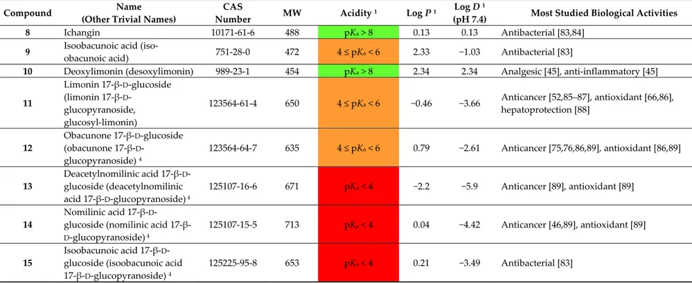
Where free carboxylic groups are present, higher acidic properties arise. This is the case of isoobacunoic acid (9) and all β-D-glucosides which are formally obtained in a two-step route including ring D lactone opening and subsequent condensation with β-D-glucose. Thus, compounds
1–15 may be classified in increasing order of acidity, from neutral (1–4, 8, 10) through the most acidic
congeners (dicarboxylic acid β-D-glucosides). Table 2 reports pKa value ranges covering the acidity of compounds 1–15 together with other main physico-chemical features for the stated compounds. Most studied biological activities (discussed in detail in Section 3) are given as well. As expected, aglycones present the highest log P values while glucosides show negative log D7.4 values thus indicating high hydrophilicity at the physiological pH. The unbalanced solubility profile of the latter may explain the relatively low number of studies on their respective biological activities which are hampered by difficult isolation from natural sources and poor pharmacokinetics.
Table 1. Overview of the main structural features of compounds 1–15.
O
OR
6R
1CH
R
3O
O
O
Z
X Y
O
H
H
R
5R
2R
4 n' n'' 1–15Compound Name Molecular
Formula n′ n′′ R
1 R2 R3 R4 R5 R6 X Y Z Main Functional
Groups Notes
1 Limonin C26H30O8 2 1 O Cycle bond Cycle
bond O–CH=CH Dilactone -
2 Nomilin C28H34O9 2 1 Cycle bond OAc Cycle bond H Cycle bond O–CH=CH Dilactone, acetic ester - 3 Obacunone C26H30O7 1 1 Cycle bond - Cycle bond H Cycle
bond O–CH=CH Dilactone
Dehydrated analog of 4 4 Deacetylnomilin C26H32O8 2 1 Cycle bond OH Cycle bond H Cycle
bond O–CH=CH Dilactone
Deacetyl-derivative of 2
5 Limonexic
acid C26H30O10 2 1 O Cycle bond
Cycle bond CO–O– CHOH Dilactone, pseudoacid Hydroxybutenolide analog of 1 6 Isolimonexic
acid C26H30O10 2 1 O Cycle bond
Cycle bond
CHOH–O– CO
Dilactone,
pseudoacid Constitutional isomer of 5
7 Citrusin C28H34O11 2 1 Cycle bond OAc Cycle bond H Cycle bond CHOH–O– CO Dilactone, acetic ester, pseudoacid Hydroxybutenolide analog of 2
8 Ichangin C26H32O9 2 1 OH OH Cycle bond Cycle
bond O–CH=CH Dilactone Spiro analog of 1
9 Isoobacunoic acid C26H32O8 2 1 O H H Cycle bond O–CH=CH Lactone, carboxylic acid Product of formal reductive cleavage of 1
10 Deoxylimonin C26H30O7 2 0 O Cycle bond Cycle
Table 1. Cont.
Compound Name Molecular
Formula n′ n′′ R
1 R2 R3 R4 R5 R6 X Y Z Main Functional
Groups Notes
11 Limonin
17-β-D-glucoside C32H42O14 2 1 O Cycle bond
Oglc 1 H O–CH=CH Lactone, carboxylic acid 17-β-D-Glucopyranoside of 1 12 Obacunone 17-β-D-glucoside C32H42O13 1 1 Cycle bond - Cycle bond H Oglc 1 H O–CH=CH Lactone, carboxylic acid 17-β-D-Glucopyranoside of 3 13 Deacetylnomilinic acid 17-β-D-glucoside C32H46O15 2 1 OH OH H H Oglc
1 H O–CH=CH Dicarboxylic acid
17-β-D-Glucopyranoside of hydrolyzed 4 14 Nomilinic acid 17-β-D-glucoside C34H48O16 2 1 OH OAc H H Oglc 1 H O–CH=CH Dicarboxylic acid,
acetic ester Ac-derivative of 13
15 Isoobacunoic acid
17-β-D-glucoside C32H44O14 2 1 O H H
Oglc
1 H O–CH=CH Dicarboxylic acid
17-β-D-Glucopyranoside of 9
1 Oglc: O-β-D-glucopyranosyl.
Table 2. Overview of the main physico-chemical properties and biological activities of compounds 1–15.
Compound Name
(Other Trivial Names)
CAS
Number MW Acidity
1 Log P 1 Log D 1
(pH 7.4) Most Studied Biological Activities
1
Limonin (citrolimonin, dictamnolactone, evodin, obaculactone)
1180-71-8 470 pKa > 8 1.66 1.66
Analgesic [45], anticancer [46–59], anti-inflammatory [45,60–62], antibacterial [63,64], antioxidant [4,65,66], antiviral [67], larvicidal [68,69] 2 Nomilin 1063-77-0 514 pKa > 8 2.47 2.47 Anticancer [46,47,58,59,70], anti-hyperglycemic [71], anti-inflammatory [25,62], antioxidant [4,65,66], antiviral [67], larvicidal [68,69], osteoclastogenesis inhibition [72] 3 Obacunone (casimirolide, obacunon, tricoccin S3) 753-03-1 454 pK a > 8 2.91 2.91 Anticancer [46,47,58,73–75], antibacterial [76,77] anti-hyperglycemic [78], antioxidant [66], larvicidal [79,80] 4 Deacetylnomilin (isolimonin,
deacetylnomilinate) 3264-90-2 472 pKa > 8 1.57 1.57 Anticancer [58,70], antioxidant [66]
5 Limonexic acid (limonexin,
shihulimonin A, substance X) 2 99026-99-0 502 6 ≤ pKa ≤ 8 −0.93 −0.7
Anticancer [28,48,52,81], antioxidant [28], hepatoprotection [82]
6 Isolimonexic acid 3 113164-90-2 502 6 ≤ pKa ≤ 8 −0.93 −0.7 Anticancer [48,52]
Table 2. Cont.
Compound Name
(Other Trivial Names)
CAS
Number MW Acidity
1 Log P 1 Log D 1
(pH 7.4) Most Studied Biological Activities
8 Ichangin 10171-61-6 488 pKa > 8 0.13 0.13 Antibacterial [83,84]
9 Isoobacunoic acid
(iso-obacunoic acid) 751-28-0 472 4 ≤ pKa < 6 2.33 −1.03 Antibacterial [83]
10 Deoxylimonin (desoxylimonin) 989-23-1 454 pKa > 8 2.34 2.34 Analgesic [45], anti-inflammatory [45]
11 Limonin 17-β-D-glucoside (limonin 17-β-D -glucopyranoside, glucosyl-limonin) 123564-61-4 650 4 ≤ pKa < 6 −0.46 −3.66 Anticancer [52,85–87], antioxidant [66,86], hepatoprotection [88] 12 Obacunone 17-β-D-glucoside (obacunone 17-β-D -glucopyranoside) 4 123564-64-7 635 4 ≤ pKa < 6 0.79 −2.61 Anticancer [75,76,86,89], antioxidant [86,89] 13 Deacetylnomilinic acid 17-β-D -glucoside (deacetylnomilinic acid 17-β-D-glucopyranoside) 4 125107-16-6 671 pKa < 4 −2.2 −5.9 Anticancer [89], antioxidant [89] 14 Nomilinic acid 17-β-D -glucoside (nomilinic acid
17-β-D-glucopyranoside) 4
125107-15-5 713 pKa < 4 0.04 −4.42 Anticancer [46,89], antioxidant [89]
15
Isoobacunoic acid 17-β-D -glucoside (isoobacunoic acid 17-β-D-glucopyranoside) 4
125225-95-8 653 pKa < 4 0.21 −3.49 Antibacterial [83]
1 Predicted data generated using the ACD/Labs Percepta Platform—PhysChem Module (ACD/Labs, Toronto, ON, Canada); 2 Experimental; 3 pKa was assumed to
1.3. Biosynthesis and Enzymatic Modifications
The biochemistry of CLs has been adequately reviewed in the past [8,15,90] and will not herein be considered in detail. A graphical overview of the main biosynthetic routes involving the CLs is presented in Figure 3. The farthest CL precursor is obviously acetyl coenzyme A (18) which gives mevalonic acid (19) through a three-step biosynthetic route. Three further steps are needed to obtain isopentyl diphosphate (IPP, 20) or its isomer dimethylallyl diphosphate (DMAPP). Three C5 units of IPP/DMAPP furnish carbons to constitute the scaffold of farnesyl diphosphate (FPP, 21) and two C15 units of the latter finally give squalene (SQ, 22), the closest hydride to CLs. SQ is oxidized to give a reactive epoxide, (S)-2,3-oxidosqualene (also named 2,3-epoxysqualene or squalene epoxide, 23), which undergoes cyclization generating a prolanosterol cation 24. Through a series of 1,2-shifts of hydrides and methyls, this intermediate may generate several triterpenes including lanosterol (25) and the two epimers euphol (26) and tirucallol (27). The latter lanostadienols are both considered as the ultimate biogenetic precursors of CLs. Indeed, dramatic oxidation of the C-17 side chain of 26 and
27 generate several protolimonoids which finally give nomilin (2) and deacetylnomilinic acid (28),
the two CLs likely precursors of all limonoids of the limonin group. The proposed biogenetic relationships between compounds 1–15 are graphically summarized in Figure 3 and briefly described in the next section. The reader may also find further details on the biosynthesis of these secondary metabolites in excellent recent reviews [37,43].
O OH O H O H O P O O H O P O O H OH O O-CoA O P O OH OPO O H OH Farnesyl diphosphate (FPP, 21) Acetyl CoA (18) Mevalonic acid (19) Isopenthyl diphosphate (IPP, 20)
Squalene (SQ, 22) O (S)-2,3-Epoxysqualene (23) O H H H Lanosterol (25) O H H H H H + Prolanosterol cation (24) O H H Euphol (20R, 26) Tirucallol (20S, 27) 20 Protolimonoids 2 3 O O O H O H O O H H O O H O Deacetylnomilinic acid (28) H+ H O O H H O O O O O O H Obacunoic acid (29) O O O H O O O H H O O H O O Nomilinic acid (30) 8 4 1 10 5 7 9 6
Figure 3. Precursors of citrus limonoids and proposed biogenetic relationships between congeners of the limonin (1) group.
2. Limonin Congeners: A Medicinal Chemistry Perspective 2.1. Medicinal Chemistry Tools
One must appreciate that a simple overview of biological activities documented for CLs over the last decade would not be sufficient to scrutinise the compounds’ therapeutic and/or lead potential. Hence, a comprehensive medicinal chemistry tools and analysis methodology are employed herein to assess the various limonin biosynthetic products. Degraded limonoids (e.g., isobenzofuranones sharing structural features with limonoids [91]) and other tetranortriterpenoids extracted from different genera and/or not belonging to the limonin biosynthetic group will not be considered and hence readers are rather directed to relevant recent reviews [15,37,92–101].
From the medicinal chemistry point of view and to envisage best CLs candidates as lead compounds, ligand efficiency metrics (LEM) need to be calculated. LEM [102] are composite parameters [103] ever increasingly used to set expectations about lead compound developability [104]. The most relevant LEM indices adopted herein along with their corresponding brief explanations [105] are summarised below:
• Lean ring index (LRI) = heterocycles/carbocycles ratio. This index was introduced since the number of aromatic and heteroaromatic rings has been found to correlate with an increased risk of attrition in development [106]. In particular, the following scale in the detrimental effects of rings has been observed [107]: carboaromatics >> heteroaromatics > carboaliphatics > heteroaliphatics, with the latter generally being beneficial. Thus, the higher LRI, the better the developability. • Potency efficiency index percentage (PEI%) = (potency/MW Da) × 100, where potency is
generally given in the form of –Log (half maximal inhibitory concentration) (pIC50) or –Log (half maximal effective concentration) (pEC50) for the activity actually concerned. We adopted this index as a slight modification of the well-known binding efficiency index (BEI) = pIC50/MW kDa [104] since PEI% values allow clearer graphical representations than the former. Since an idealized compound should have a BEI value of 27, the corresponding PEI% value would be 2.7.
• Ligand efficiency index (LEI) = potency/number of non-hydrogen atoms (“heavy atoms”, HA). For the sake of graphical clarity, we adopted the descriptor LE (ligand efficiency) corresponding to the product LEI × 1.37 (LE = ∆G°/HA = −2.303 RT/HA ≈ 1.37 potency/HA). An ideal lead compound should have LE > ~0.3 kcal per mole per HA [102].
• Fraction sp3 (Fsp3 = number of sp3 hybridized carbons/total carbon count). Lovering [108] suggested this descriptor as a measure of complexity since he noted that drug candidates nearest to clinical use generally present high Fsp3 and chirality centers. 0.36 is the suggested lowest allowed limit value [109].
• Lipophilic ligand efficiency (LLE = potency − cLog P, also referred to as lipophilic efficiency, LipE). LLE is simply the difference between potency and the most popular lipophilicity descriptor (cLog P), and may be considered as a measure of the specificity of binding. Proposed acceptable values of LLE for drug candidates are >~5 [102].
• Ligand efficiency dependent lipophilicity (LELP = cLog P/LE). LELP combines aspects of LE and LLE into a single index and may be considered as the price of ligand efficacy paid for with Log P [110]; it should be <7.5 [102].
By using these analysis parameters, recent attempts undertaken to enhance the biological activities of natural limonoids through structural modification (i.e., semisynthetic analogue preparation and bio-evaluation) are analysed in the forthcoming text.
2.2. Citrus Limonoids: Sources, Main Structural Features, and Developability As Therapeutic Agents
2.2.1. Limonin (1, Figure 1)
Known in the past also as obaculactone, evodin, or dictamnolactone from the names of some of its natural sources (Phellodendron amurense bark—Phellodendri Cortex, Ōbacu in Japanese [111]—
considered as the standard bearer of CLs and continues to be the most studied congener of the class. Limonin (1) is normally extracted from the Citrus genus plant fruits and hence it’s early trivial name, citrolimonin [112]. “Limonin”, in turn, recalls limon—the transliterated form of the old English word
lymon which came from old French limon (“citrus fruit”; via Provençal, from Persian limu passing
through Arabian laimun, generic terms for citrus fruits, cognate with Sanskrit nimbu) [113]. When 1 was discovered [114], its bitter taste and some preliminary assays disguised it as an alkaloid probably suggesting to Berlays [115] the stem—in(e) which was commonly used for quinine-like compounds. Limonin has also recently been found in Citrus genus plant bark [25,116,117], and root [118], tissues. In addition to the above reported plants, 1 has been isolated from numerous non-Citrus species such as the endemic Brazilian species Raulinoa echinata [119], Glycosmis parva (a wild small shrub distributed in Thailand) [120], Poncirus trifoliata (a tree that is widely distributed in China) [121], and
Dictamnus angustifolius [27,122,123].
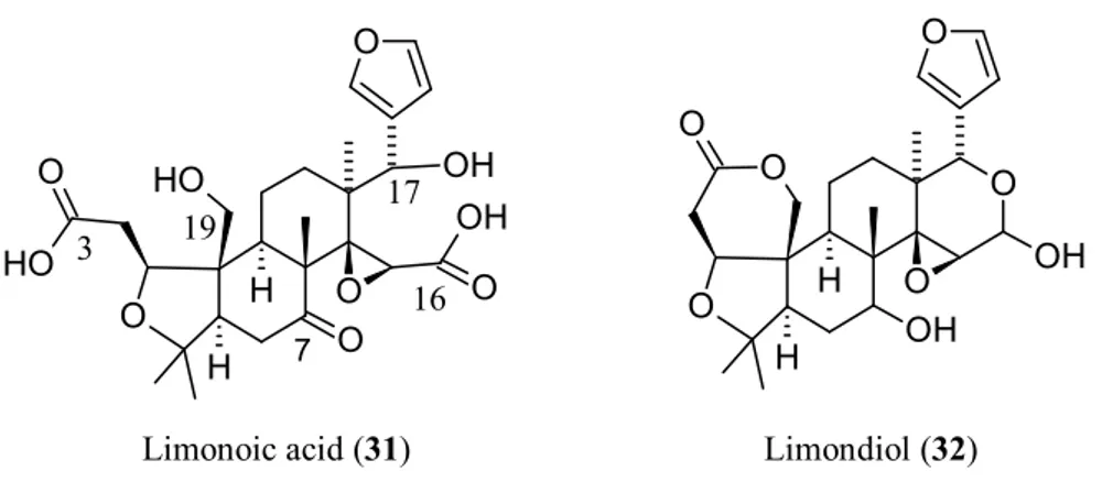
From a biosynthetic point of view (Figure 3), 1 has nomilin (2) as its most accredited parent compound in an enzymatic route possibly passing through obacunone (3), obacunoic acid (29), and ichangin (8). Despite extensive radioactive tracer work, the definitive proof on all four involved steps has not yet been given. Alternatively, isoobacunoic acid (9) was once considered as a possible precursor of 1. Unfortunately, radioactive 9 has not been shown to be converted to 1 in Citrus [8]. Structurally, 1 may be considered as the dilactone derivative of limonoic acid (31, Figure 4) and hence can be given semisystematic names such as limonoic acid di-δ-lactone, limonoic acid 3,19:16,17-dilactone, or limonoate A-ring, D-ring dilactone. On the other hand, 1 also relates to the oxidation product, limondiol (32, Figure 4), and thus may also be named as 7,16-dioxo-7,16-dideoxylimondiol.
O O O O OH OH O O H H O OH O O H O O O O H H OH O H 19 3 16 17 7
Limonoic acid (31) Limondiol (32)
Figure 4. Structures of limonoic acid (31) and limondiol (32).
When dissecting the fused hexacyclic scaffold of 1, the isochromene (2-benzopyran) system is the higher priority parent component, followed by isobenzofurane, pyran, and oxyrene rings, in decreasing order of priority. In comparison with the archetypal limonoid scaffold 4,4,8-trimethyl-17-(furan-3-yl)androstane (16, Figure 2), two out of four fused carbocycles (cycles A and D) are replaced by a furopyran and a pyran system, respectively, which are condensed to a surviving naphthalene core.
Due to its good number of chiral centres (nine) and its high fraction sp3 (Fsp3 = 0.73), 1 is one of the most complex CLs. The unusually high number of heterocycles (LRI = 2.5) together with its relatively low lipophilicity (cLog P = 1.66) render the ligand metrics profile of 1 relatively good. This consideration together with the relatively high availability of 1 in adequate amount to facilitate the preparation of semisynthetic analogues endorse 1 as a lead compound and justify both the plethora of biological activities reported for 1 (cf. Table 2 and Section 3) and its universally recognised role of flagship member of the natural polyoxygenated triterpene family [37].
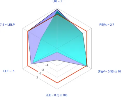
The most studied pharmacological activities of 1 are related to its possible application as an anticancer agent. Generally 1 displays antiproliferative activity in the low micromolar range. Thus, the structure of 1 is relatively redundant when considered in relation to its displayed potencies (LE < 0.37 Kcal per mol per HA; PEI% < 2.7). In Figure 5 a spider plot based on LEM calculated for 1 is shown where the IC50 value for in vitro aromatase inhibition (5.22 μM [46]) was used as a measure of potency.
Figure 5. Radar plot of ligand efficiency metrics as a graphical tool to assess developability of limonin (1) as a drug. Sub-optimal property space corresponds to the inner red hexagon showing sides marked with “zero”. Ideally, good lead compounds would be represented by areas wider than this inner hexagon. This spider plot indicates the properties of 1 that need improvement. Legend: Fsp3: fraction
sp3; LRI: heterocycles/carbocycles ratio; PEI: potency efficiency index; LE: ligand efficiency; LLE:
lipophilic ligand efficiency; LELP: ligand efficiency dependent lipophilicity; see text for details.
The radar plot indicates that 1 may be considered as a fairly good lead compound with the tactics of molecular simplification (i.e., reduction of molecular weight and HA) and introduction of nitrogen atoms (possibly improving potency) being obviously suggested. Both tactics have been recently explored with the former having inspired the synthesis of an ever increasing number of degraded limonoids [37,91]: a detailed examination of these compounds is however beyond the scope of this review. The latter tactic has been adopted in a relatively smaller number of studies. The straightest way to introduce nitrogen atoms in the limonin structure is represented by oxime derivatization of its C-7 carbonyl group. Patil and co-workers obtained limonin oxime (compound 33, Figure 6) and its O-methyl derivative 34 [46]. Both compounds were about twice as potent as 1 in the aromatase inhibition assay. Incidentally, defuranlimonin (35, a limonin analogue with a carboxyl group appended at the C-17 position in lieu of the furan ring; Figure 6) was as active as 1 while defuran nomilin (36) was about six times more potent than 2. A similar trend was observed when studying the effect of CLs on biofilm formation in E. coli where 33, 34, and 35 were more active than 1 [63]. Furthermore, 34 showed higher induction of the detoxicant liver enzyme glutathione S-transferase (GST) than the one displayed by 1 [49]. These findings suggest the possibility of improving potency by introducing nitrogen atoms in the CL scaffold and underlay the structural redundancy of CLs. However, pitfalls in this generalization should always be taken into account. When inhibition of cell-cell signalling was investigated in Vibrio harvey, 1 resulted to be more active than its derivatives 33–
35 [63]. The variability of the in vitro study results is one of the challenging aspects in the study of
CL activities and was also noted when studying proapoptotic [89] and cytotoxic effects [81] of CLs on eukaryotic cells. Puzzling as it is, the finding of varying effects of CLs on different cell lines suggests the possibility that mixtures of CLs, such as those found in citrus fruits, may prove more effective with respect to the isolated components when trying to face highly varying human cancers [81] or polybacterial human diseases.
A series of limonin (1) and deoxylimonin (10) oxime derivatives of the general structure 37 (Figure 7) were obtained through an oxime derivatization procedure, and subsequently evaluated as anti-inflammatory (ear swelling induced by xylene test in mice) and analgesic (acetic acid-induced
writhing and tail-immersion tests in mice) agents [45]. In this case, tertiary amine moieties were introduced onto C-7 position in order to obtain water-soluble derivatives, possibly endowed with higher bioavailabilities than those displayed by the parent CLs 1 and 10. The latter were chosen as parent compounds to evaluate the role played by the epoxide ring in the above activities. Generally,
1 and its derivatives were more active than the corresponding deoxygenated congeners, thus
underlying the relevant contribution of the oxirene moiety to analgesic and anti-inflammatory activities. Whether this contribution stems from epoxyde reactivity towards protein nucleophilic side chains [3] however remains to be demonstrated. Incidentally, these findings are in agreement with the above general observation on LRI, Fsp3, and the number of chirality centers as reliable predictors of drug developability. As expected, all derivatives were more water-soluble than the parent compounds. Water-solubility was obviously directly related to pKa values and inversely related to Log D7.4. The most interesting compound was the limonin derivative bearing an O-diethylaminoethyl side chain (38) since it was more potent than acetylsalicylic acid and naproxen in the analgesic and the anti-inflammatory assays, respectively.
O O O O O N O O H H OR Limonin oxime (33) R = H Limonin methoxime (34) R = Me O O O O O O O OH O H H Defuran limonin (35) O O O O O O O H H O O OH Defuran nomilin (36)
Figure 6. Semisynthetic analogs of limonin (1); and nomilin (2) endowed with anti-aromatase (33–36) [46]; and anti-biofilm (33–35) [63] properties.
On' O O O O N O O H H O N R2 R1 37
( )
n'' n' = 0, 1 n'' = 2, 3 R1 = R2 = Me, Et R1, R2 = -(CH 2)5-, -(CH2)2O(CH2)2 -O O O O O N O O H H O N 38Figure 7. Water-soluble analogs of limonin (1); and deoxylimonin (10) endowed with analgesic and anti-inflammatory properties [45].
Generally, 1 and its glucoside 11 share the same activities regardless of the test used (cf. Table 2). However, relevant nuances in favour of 11 have been reported. As an example, both the aglycon and its glucoside are more effective against neuroblastoma cells (SH-SY5Y) compared to colon carcinoma cells (Caco-2) while having no effect on normal cells (epithelial Chinese hamster ovary cells, CHO) [58]. However, 1 showed a slower rate of induction of caspase 3/7 activity in comparison to 11. Furthermore, while micromolar concentrations of both 1 and 11 arrested cell growth, biochemical and morphological data showed that 11 induced a more rapid cell death. Incidentally, the higher resistance of Caco-2 cells may be related to high expression of the ABC transporter P-gp [3,51], an efflux pump that impedes cytoplasmic accumulation of lipophilic compounds.
2.2.2. Nomilin (2, Figure 1)
Nomilin (2) is characterized by a seven-membered oxepine ring and may be considered as the product of an acetate equivalent addition to the structure of obacunone (3). The trivial name of 2 is an anagram of “limonin” while its semisystematic name is 1-acetyloxy-1,2-dihydroobacunoic acid ε-lactone, and is based on the trivial name for the product formally obtainable by hydrolysis of 3, obacunoic acid (29). However, 2 precedes 3 and 29 in the proposed biogenetic relationships between CLs (Figure 3). Indeed, it has been hypothesized that trans-elimination of the acetyl group of 2 gives
3, and the hydrolytic opening of the Α ring lactone of 3 gives 29. The enzymes responsible for these
two steps have been isolated from bacterial cells, but they have not yet been found in Citrus species [8]. The direct precursor of 2 is known to be deacetylnomilinic acid 28.
Radioactive tracer work on CLs is indebted to 2 since radiolabelled nomilin can be easily prepared using labelled acetate and in turn converted into other labelled CLs [8]. Compounds 1–4 are the major CLs and 2 has also recently been found in the barks of Citrus genus [7,25] and in root bark of Dictamnus angustifolius [122,123]. When biological activities are considered, 2 shares the same profile of 1 (Table 2) displaying comparable potencies in a range from five-fold lower to five-fold higher values than the ones shown by 1 [46,67,77]. Since 2 has higher MW, HA, and cLog P values than 1, the latter seems to be preferable as a lead compound (Figure 8). For these comparative analysis, IC50 value from in vitro aromatase inhibition (18.86 μM [46]) was used as a measure of potency.
Figure 8. Radar plot of ligand efficiency metrics as a graphical tool to assess developability of limonin (1, purple area) and nomilin (2, blue-green area) as drugs. Sub-optimal property space corresponds to the inner hexagon showing red sides marked with “zero”. Ideally, good lead compounds would be represented by areas wider than this inner hexagon. This spider plot indicates that 1 should be preferred to 2 as a lead compound. Legend: Fsp3: fraction sp3; LRI: heterocycles/carbocycles ratio; PEI:
potency efficiency index; LE: ligand efficiency; LLE: lipophilic ligand efficiency; LELP: ligand efficiency dependent lipophilicity; see text for details.
2.2.3. Obacunone (3, Figure 1)
This α,β-unsaturated ε-lactone is also known as obacunon (cf. 1 for ethymology), casimirolide (from Casimiroa edulis, Mexican apple) or tricoccin S3 (since initially believed to be cognate of tricoccins—secondary metabolites isolated from Cneoraceae species such as C. tricoccon). Compound
3 may be obtained by refluxing 2 with a mixture of acetic anhydride and pyridine [124] and may be
considered as the product of intramolecular condensation of obacunoic acid 29 (obacunoic acid, ε-lactone; limonoic acid 3,19:16,17-dilactone, limonoate D-ring-lactone). Indeed, 29 was obtained by heating 3 in a 0.1 M NaOH solution [124].
Radioactive tracer work demonstrated the biosynthetic route relating 3 to 2, as a product, and to
29, as a precursor [8] (Figure 3). In the last decade, 3 has been isolated from non-Citrus species, including Phellodendron amurense [125], Dictamnus dasycarpus [27,126,127], Dictamnus angustifolius [27,122,123], and Harrisonia perforata (the only species of the genus Harrisonia growing in Thailand) [128].
The anticancer activity of 3 has been mostly attributed to the α,β-unsaturated ketone functional group in the A ring [15]. The high lipophilicity of 3 (cLog P = 2.91, Table 2) should also be taken into account. In fact, this property is generally positively related to cytotoxicity since the kinetics of drug uptake in cancer cells is higher with increased lipophilicity [129]. However, 3 rarely came so far as the most active potential anticancer CL of the limonin group [46] in the stated period, when anticancer activity is concerned. Thus, the price paid in term of Log P to gain activity through lipophilicity was not a good investment with 3 and its LEM profile acknowledges it being inferior to 1 as a lead compound for anticancer design (Figure 9). For this analysis, the IC50 value for in vitro aromatase inhibition (28.04 μM [46]) was used as a measure of potency.
Figure 9. Radar plot of ligand efficiency metrics (LEM) as a graphical tool to assess developability of limonin (1, blue area) and obacunone (3, yellow area) as anticancer agents. Sub-optimal property space corresponds to the inner hexagon showing red sides marked with “zero”. Ideally, good lead compounds would be represented by areas wider than this inner hexagon. This spider plot indicates that 1 should be preferred to 3 as a lead compound. Legend: Fsp3: fraction sp3; LRI:
heterocycles/carbocycles ratio; PEI: potency efficiency index; LE: ligand efficiency; LLE: lipophilic ligand efficiency; LELP: ligand efficiency dependent lipophilicity; see text for details.
The tactics of nitrogen introduction via oxime derivatization of 3 has recently led to a series of obacunone oxime and its ester derivatives were obtained (Figure 10) [80]. Furthermore, the larvicidal activity of these compounds against oriental armyworm (Mythimna separata Walker), a lepidopteran pest, has been evaluated [80]. Rewardingly, several of the studied compounds were more active than the lead 3, with the two chlorobenzoyl derivatives 40 and 41 displaying 60% higher larvicidal activity than that of the parent compound 3.
H O O H OO N O O OH Obacunone oxime (39) H O O H OO N O O O O R
Obacunone oxime esters (40, R = o-Cl; 41, R = m-Cl )
Figure 10. Structures of obacunone oxime (39) and two of its ester derivatives (40,41) endowed with high larvicidal activity [80].
2.2.4. Deacetylnomilin (4)
At the time of its early isolation from orange seeds, deacetylnomilin (4) was erroneously considered as an isomer of limonin (1) and called isolimonin [130,131]. Indeed, 4 may be considered as the formal product of reductive cleavage of the A ring lactone of 1 and subsequent isomerization of the so-obtained carboxylic acid (isoobacunoic acid, 9) to give a β-hydroxy-ε-lactone ring (4-hydroxyoxepin-2-one) in lieu of the tetrahydrofuran one. Thus, two hydrogen atoms should be added to 1 structure in order to obtain 4 (see Table 1). The relationship between 4 and 2 is reflected in their corresponding semisystematic names since 4 is also known as 1,2-dihydro-1-hydroxyobacunoic acid, ε-lactone. A straighter structural linkage relates 4 to deacetylnomilinic acid
28, which is most likely the initial precursor of all limonin related CLs discussed in this review [8]
(see Figure 3).
The physico-chemical profile of 4 shows a definite overlapping with that of 1. No surprise, then, if the two CLs present similar activities and potencies of action [3,47,51,58,77]. Paralleling what has been observed for 1 and its glucoside 11, the glucoside of 4 (12) sharply reduced cancer cell viability as compared to aglycone [58] thus corroborating the hypothesis that glucosides may be better active apoptosis-inducing agents [132].
2.2.5. Pseudoacids: Limonexic Acid (5), Isolimonexic Acid (6), and Citrusin (7)
Limonexic acid (5) may be considered as a product of oxidation of the furan ring of 1 to give a Υ-hydroxybutenolide moiety appended at C-17. Since limonoids with oxidized furan rings are relatively rare in the Rutaceae, this 21-hydroxy, 23-oxo analogue of 1 has long been considered as an artefact caused by photooxidation occurring during extraction of 1 from its natural sources. Indeed, photooxidative degradation of the furane ring may cause limonin glucoside instability in vitamin B2 -containing beverages exposed to light [133]and oxidative cleavage is a commonly used approach to obtain degraded limonoids [92]. When the constitutional isomer of 5, isolimonexic acid 6, was isolated from Tetradium glabrifolium [134], the possibility that 5 and 6 were real secondary metabolites was considered as probable since spontaneous photooxidation reactions may hardly generate stereochemically homogeneous, isolated isomers such as the above butenolides [119]. Unfortunately, an error occurred in the graphics of the first work reporting on 6 [134] and this generated some confusion in the literature to the point that the Chemical Abstracts Service (CAS) gave two different CAS numbers to the above isomers but these numbers correspond to the same structure while PubChem attributes the same SMILES notation to 5 and 6. In 2001, Gai et al. reported on the isolation of “a new compound” from Evodia rutaecarpa, 21-(R and S)-hydroxy-23-oxo-20-en-limonin, and named it shihulimonin A (Shihu is a word frequently found in the Chinese folk medicine but is commonly related to Dendrobium genus) [135]. Indeed 5 was early isolated by Emerson and generally referred to as ‘Emerson’s substance X’ [131]. The agreeing works of several research groups [136–138] definitively demonstrated that 5 has the structure reported in Figure 1 while 6 is the 23-hydroxy, 21-oxo isomer of 5, notwithstanding contrasting literature graphics [25,28,118,120]. The hemiacetal C-atom (C-21 and C-23 for 5 and 6, respectively) is labile in solution and this is why no stereochemically notations are generally reported about them. Incidentally, the stem -exic is used in
lieu of -oic to indicate butenolide pseudoacids. Probably this derives from the fact that “exic” in Japanese stays for “eic”, thus indicating the α,β-unsaturated nature of the pseudoacid ring.
Limonexic acid (5) has been recently found also in Citrus genus plant barks [25,117], root [118], and flower [28] tissues. Other natural sources of 5 in the stated period were Raulinoa echinata Cowan (a spiny shrub endemic of Brazil) [139,140] and Glycosmis parva (a wild small shrub distributed in Thailand; a mixture of 5 and 6 was obtained) [120]. Isolimonexic acid (6) has been isolated also from
Citrus genus plant root [118].
A relatively less explored pseudoacid is citrusin (7). Biogenetically derived from nomilin (2), this Υ-hydroxybutenolide is to 2 as isolimonexic acid (6) is to 1. Citrusin has been recently found also in
Citrus genus plant bark [25]. The biosynthetic relationship between the pseudoacids 5–7 and the
corresponding putative parent compounds 1 and 2 is to be demonstrated.
Considering the presence of one more chiral centre, the absence of aromatic ring, and low lipophilicity (cLog P < 0), pseudoacids 5–7 are probably the most interesting CLs as lead compounds for further semisynthetic analogue development. In particular, an introduction of an aromatic ring would be well tolerated and should in principle allow increased activity. In Figure 11, the LEM profile of 5 is reported in comparison with that of 1. The IC50 value for in vitro aromatase inhibition (20.02 μM [46]) was used as a measure of potency. The studies conducted so far suggest that 5–7 are not superior to 1 or 2 (cf. Section 3).
Figure 11. Radar plot of ligand efficiency metrics (LEM) as a graphical tool to assess developability of limonin (1, blue area) and limonexic acid (5, magenta area) as anticancer agents. Sub-optimal property space corresponds to the inner hexagon showing red sides marked with “zero”. Ideally, good lead compounds would be represented by areas wider than this inner hexagon. This spider plot indicates that 5 should be preferred to 1 as a lead compound. Legend: Fsp3: fraction sp3; LRI:
heterocycles/carbocycles ratio; PEI: potency efficiency index; LE: ligand efficiency; LLE: lipophilic ligand efficiency; LELP: ligand efficiency dependent lipophilicity; see text for details.
Ichangin (8) (from C. ichangensis) has also been recently isolated from the stem bark and leaves of the South-Americaan plant, Raputia heptaphylla [141]. It may be considered as the product of hydrolytic cleavage of the oxygen-containing portion of the isobenzofuran nucleus of 1 (A’ ring). The removal of A’ ring creates a spiro junction at the C-10. Indeed, 8 has been proposed as the immediate precursor of 1 though the step involving ichangin (Figure 3) remains hypothetical [8].
Isoobacunoic (or iso-obacunoic) acid (9) is the constitutional isomer of 4 and 29. Its semisystematic name is 19-deoxylimonoic acid δ-lactone. In the last decade, 9 was also isolated from the root bark of Dictamnus angustifolius [90,122,123] and structurally, isoobacunoic acid (9) to deacetylnomilinic acid (28) is the same as 1 is to 8. Indeed, it was once considered as an intermediate
between obacunoic acid (29) and 1 but labelled isoobacunoic acid has never been shown to be converted to 1.
Deoxylimonin (or desoxylimonin) (10) may also named
14,15-deepoxy-14,15-didehydrolimonoic acid, di-δ-lactone. As previously discussed, 10 is generally less interesting than its epoxidised analogue 1. A few reports on the biological activities published in the last 10 years (Table 2) will be reviewed in Section 4.
2.2.6. Newly Identified Limonin Related Limonoids
In 2008, two research groups separately reported on the isolation and identification of the C-17 limonin (1) epimer, named epilimonin (41), which displays the furan ring at C-17 in a β-orientation rather than the α-orientation as in 1 [10,142]. In particular, Glabasnia and Hofmann [10] investigated the hydrolytic liberation of 1 from its precursor limonin 17β-D-glucopyranoside (11) in orange juice samples. When the aqueous glucoside solution was adjusted to pH 1.5 and incubated for 4 h at 60 °C, a new compound was isolated and purified by semi-preparative high pressure liquid chromatography (HPLC), and its chemical structure was determined by tandem mass spectrometry (MS-MS) and 1D/2D-NMR experiments. The Authors concluded that 41 was unequivocally identified for the first time. A reaction pathway showing the formation of the two epimers from (11), favoured by C-17 furan ring through anchimeric assistance was also proposed (Figure 12) [10]. In 2003, while studying the human bioavailability of limonoid glucosides, a metabolite with similar chromatographic behavior and mass spectrum as 1 was identified, but it was not characterized at that time [143]. In 2008, the unknown compound was isolated by fractional crystallization monitoring each step by means of HPLC coupled to photodiode array and evaporative light scattering detectors (HPLC-PDA-ELSD). This metabolite was finally identified as the C-17 epimer of 1 through side-by-side comparison of the corresponding physical properties, including MS, IR, 1H- and 13C-NMR [142].
O CO2H O O O O O H H O Glc Limonin 17-β-D-glucoside (11) H+ glucose O O O O O O H H O O H + H+ O O O O O O O O H H
42 Limonin (1) and epilimonin (41) Figure 12. Formation of limonin (1) and its epimer epilimonin (41) from limonin 17β-D-glucopiranoside (11) via a furan-3-ylidene cationic intermediate (42); Glc = β-D-glucopyranosyl.
Three new pseudoacids were identified in the last decade, including two nomilin- and one ichangin-related limonoids. By extraction from C. sudachi peels and separation through HPLC, 21,23-dihydro-23-hydroxy-21-oxodeacetylnomilin (43, Figure 13) and 3-O-methyl-21,23-dihydro-23-hydroxy-21-oxonomilinic acid (44) were identified by spectroscopic (IR, 1H-NMR, 13C-NMR, heteronuclear multiple bond correlation (HMBC), nuclear Overhauser effect spectroscopy (NOESY) and spectrometric (high resolution fast-atom bombardment mass spectrometry (HR-FAB-MS) analyses. However, as suggested by the authors, it is possible that 44 is an artefactual derivative of
O O O O O H H O O O H O O H O O O O O H H O H O O O H O O O 21,23-dihydro-23-hydroxy-21-oxodeacet
ylnomilin (43) 3-O-methyl-21,23-dihydro-23-hydroxy-21-oxonomilinic acid (44) Figure 13. Structures of two new pseudoacids isolated from Citrus sudachi.
The third new pseudoacid, ichanexic acid (45, Figure 14), was identified in sour orange (C. aurantium L.) together with isolimonic acid (46), with the latter being isolated for the first time in its native form and not as the methyl ester. Ichanexic acid (45) is structurally related to ichangin (8) as isolimonexic acid (6) is to 1. Isolimonic (or isolimonoic acid, 46) is a constitutional isomer of 8. The purity of the isolated compounds were analyzed by TLC and HPLC and their structures were identified by one-dimensional (1H-, 13C-) and two-dimensional (1H-H and 1H-3C) NMR experiments. Axial configuration was assumed for C-18 methyl group and the stereochemistry of endocyclic methine and methylene protons were assigned by nuclear Overhauser enhancement (NOE) experiments supporting the proposed structures [144].
O O O O O H H O H O O H O O H O ichanexic acid (45) O O O O O H H O O O H O H isolimonic acid (46)
Figure 14. Structures of ichanexic acid (45) and isolimonic acid (46).
In the final analysis, it is worth reviewing some rather puzzling new limonoids such as the (E)-isomer of obacunoic acid (29) which was recently isolated from the root bark of Dictamnus
angustifolius [122]. The geometry of the double bond was established on the basis of the vinylic
protons’ coupling constant in the 1H-NMR analysis vis-a-vis those previously reported for the the β-D-glucoside of trans-obacunoic acid [145]. Hence, 29 was a novel limonoid named by Sun and co-workers as dictangustone A [122]. In a recent review article [90], limonoic acid (31) was erroneously reported as a limonoid isolated from Dictamnus dasycarpus although the original studies presented it as a neutral limonoid isomeric to limonin (1) [127,146]. Another recent review on the antioxidant activity of CLs has reported “millington acid 17-β-D-glucoside (NAG)” and “deacetylation millington acid 17-β-D-glucoside (DNAG)” as limonoids [31]. Indeed, “(downtown) milligton acid” can be found in some Internet pages to indicate nomilinic acid (30) but, despite our efforts, we could not find how this curios name was conceived and given to 30.
3. Isolation and Identification of Limonoids from the Citrus Genus
In the last decade, several articles have been published on the isolation and identification of limonoids from various C. species. A wide spectrum of analytical methods were used, including the well-known thin layer chromatography (TLC), nuclear magnetic resonance (NMR), mass
spectrometry (MS), high pressure liquid chromatography (HPLC), and capillary electrophoresis (CE) techniques. Advanced complementary analytical methods included tandem MS (MS/MS), liquid chromatography/electrospray ionization MS (LC-ESI-MS), atmospheric pressure chemical ionization MS (APCI-MS), and collisionally activated dissociation (CAD) MS/MS. These techniques have been well reviewed in the earlier literature [1,36,147–149]. In this section, we focus on the isolation and identification of both new and known limonoids by using improved techniques and/or from varieties of Citrus never investigated until that time. Moreover, attention is paid to metabolomic studies and investigation on secondary metabolite pathways. For the isolation and identification of limonoids from C. species by using conventional techniques, the reader may refer to other useful reports [4,116,117,150].
3.1. Limonoid Aglycones
Several studies were aimed at the identification of limonoid aglycones in Citrus fruits as they are known to be responsible for the gradual bitterness observed in citrus juices. The limonin (1) content of the Iranian orange juice concentrates (OJCs), for example, was determined by reversed-phase HPLC and spectrophotometric analysis. With regards to the HPLC analysis, the reconstituted OJC samples were injected into a column with only simple filtration through a nylon filter without extraction. Both methods showed that most samples contained high amounts of (1), which implies that the Iranian local orange cultivars (i.e., rasmiye shomaal) should be categorized in the bitter oranges group [151]. Through a combination of HPLC and ESI/MS, as many as 11 limonoid aglycones have been isolated and identified in the fruit peel and seeds of C. pyriformis Hassk. In particular, 1 and deacetylnomilin (4) have been isolated from fruit peel, whilst nomilin (2) and ichangin (8) were obtained from defatted seeds after chromatographic separation. The isolated compounds were identified by MS (electron ionization (EI), chemical ionization (CI and ESI), 1D- and 2D-NMR experiments (attached proton test (APT), correlation spectroscopy (COSY), heteronuclear single-quantum correlation (HSQC), HMBC, and NOESY) and comparison with literature data as well as authentic substances. Quantitative and qualitative analysis of CLs from C. pyriformis were also determined by using LC-ESI/MS leading to the tentative structural assignements of seven more aglycones [3]. Furthermore, while comparing the chemical profiles of C. wilsonii Tanaka (CWT) and
C. medica L. (CML), 1, 2, and obacunone (3) were unequivocally characterized among 25 compounds
identified through TLC and HPLC-quadrupole time-of-flight-QTOF-MS. The quantitative results obtained by the HPLC coupled with diode array detector (HPLC-DAD) method demonstrated that (2) was the most dominant constituent in CML and also indicated that there were significant differences in chemical composition between the two species [152]. In a study dedicated to the simultaneous quantification of coumarins, flavonoids, and limonoids in Fructus Citri Sarcodactylis by HPLC-DAD, 1 and 2 were identified among the 11 major bioactive components [153]. With the aim of screening new sources of 1 and 2 among C. species, reversed-phase HPLC analyses were also performed on different citrus cultivar and, in some cases, on different fruit tissues [65,154–156].
Only two studies reported the isolation of limonexic acid (5) in the last decade (two further studies reporting on the isolation of both 5 and its β-D-glucoside are treated in Section 3.3 [48,52]. After isolation from C. aurantium var. amara, 5 was identified through MS and NMR spectra [28]. It was also isolated from the stem bark of C. medica L. var. sarcodactylis SWINGLE, together with 1 and
2 which were also isolated from the root bark, and identified by comparison of their spectroscopic
data (UV, IR, NMR, and MS) with those reported in the literature [25]. Lastly, a method has been reported for the identification of limonoid A ring lactones in citrus samples. In fact, since only indirect chemical and biochemical techniques [157–159]orMS detection were available, Breksa III et al. finally developed a rapid and reliable LC-ESI-MS method for the direct quantification of limonoate A Rring lactone (LARL, 47, Figure 15) and nomilinoate A ring lactone (NARL, 48, Figure 15) in a wide variety of citrus juices. These limonoid A-ring lactones were isolated by solid-phase extraction from juice samples and analyzed by negative ion LC-ESI-MS while their concentrations were established by fluorescence spectroscopy [160].
O CO2H O O O O O H H OH
Limonoate A ring lactone (LARL, 47) O OH O O O O O H H O OH O
Nomilinoate A ring lactone (NARL, 48)
Figure 15. Structures of limonoate A-ring lactone (LARL, 47) and nomilinoate A-ring lactone (NARL, 48).
3.2. Limonoid Glucosides
Rapid and simple separation methodologies are essential to obtain pure citrus limonoid glucosides for biological evaluations [161]. On the other hand, until the last decade, no method of separation for aglycones from glucosides was available [162]. In this regard, two studies reported on a reversed-phase flash chromatography technique developed for the separation and isolation of limonoid glucosides whose identities were then confirmed by a suitable spectrometric method. First, Raman et al. [161] purified several limonoid glucosides extracted from defatted seed powder of grapefruit (C. paradisi Macf.) by reversed-phase flash chromatography. The procedure yielded two glucosides with purity higher than 90% which were subsequently identified by ESI-MS as nomilin 17-β-D-glucopyranoside (49, Figure 16) and nomilinic acid 17-β-D-glucopyranoside (14) [161]. Four years later, a decigram-scale method was developed by Breska III et al. for the separation of limonin 17-β-D-glucopyranoside (11) from contaminating bitter 1 through C18 flash chromatography. The identities of the two limonoids were then confirmed by LC-MS analysis [163]. With regards to their identification, a rapid and selective LC-MS method has been applied to characterize CLs in citrus juices, extracts, and partially purified liquid samples, and estimate their relative concentrations without the need to treat or dilute the samples. A phenyl stationary phase, as an alternative to C18 was employed in most of the HPLC and LC-MS analysis undertaken [162].
Nomilin 17-β-D-glucoside (49) O CO2H R O O O O H H O O Deacetylnomilin 17-β-D-glucoside (50) O CO2H R O OH O O H H O O CO2H R O H O O O O H H O H Ichangin 17-β-D-glucoside (51) R = O O OH OH O H O H
Figure 16. Structures of limonoid glucosides isolated from C. species through recently developed methodologies.
3.3. Limonoid Aglycones and Glucosides
Despite the number of analytical methods reported for either CL aglycones or glucosides quantification, until 2007, there was no method for the simultaneous quantification of CL aglycones and glicosides in citrus fruits and seeds. Vikram et al. finally developed a reversed-phase HPLC method coupled with DAD for their simultaneous quantification. By using a C18 column and a binary solvent system (3 mM phosphoric acid/acetonitrile), five limonoid aglycones (1, 2, 8, 9, and 46) and two glucosides (11 and 13) were simultaneously identified in four varieties of citrus fruits and seeds, namely Rio Red Grapefruit, navel orange, valencia orange, and tangerines. Determining the limonoid concentrations directly from the peak area, 1 and 11 were found to be the predominant limonoid aglycone and glucoside, respectively, in all the tested samples [164]. By using the same method, three limonoid aglycones (8, 9, and 46) and two limonoid glucosides (13 and 15) were purified from sour orange (C. aurantium L.) seeds. This time, the identities of the purified compounds were also confirmed by positive APCI-ESI-MS spectra for aglycones and negative ion APCI for glucosides and the spectra were compared with published data. It is noteworthy that a cation H+ exchange column was employed to separate limonoids from flavonoids [83]. Moreover, the soft ionization technique (CID) was coupled with the above described reversed-phase HPLC, and limonoid aglycones and glucosides were simultaneously identified from complex citrus samples. In particular, 1, 2, 4, 8, and
46 were identified by positive ion CID MS/MS, whereas the glucosides 11–15 were identified by
negative ion CID MS [165]. On the other hand, RP-HPLC-PDA-MS was used for the qualitative and quantitative characterization of limonoid aglycones and glucosides in juice, peels, pulps, and seeds of two bergamot cultivars (Fantastico and Femminello). Limonoid aglycones were the most abundant in seeds and peels (70% and 80% of the total, respectively), while limonoid glucosides were predominant in juices and pulps (61% and 76% of the total, respectively), thus reflecting their corresponding lipophilicity. Calibration curves were built by using pure limonoids isolated from bergamot seeds and juice through 2D-HPLC/PDA/MS preparative system [5].
A simple and rapid colorimetric method, amenable to the simultaneous analysis of multiple sample with a plate reader, has been proposed by Breksa III et al. [166] for estimating the total 1 and
11 concentrations in citrus juices. Until that time, no methods for the spectrophotometric
determination of limonoid glucosides were known, since the above-reported Abbasi’s spectrophotometric method is specific for limonin aglycone [151]. The new method is based on the formation of red to orange colored derivatives resulting from the treatment of 1, 11, or a fruit extract with 4-(dimethylamino)benzaldehyde (DMAB) in the presence of perchloric and acetic acids. Limonoate A-ring lactone (LARL, 47) was also tested and found to mirror the properties of 1, likely due to the rapid conversion of 47 into 1 when treated with the acidic indicator solution [166]. By using this colorimetric method, the first analysis of total limonoid content in sour orange juices was also performed.
CL glucosides, 47, and 48 were also analyzed by LC-MS as previously reported [160]. The results obtained suggest that sour oranges at maturity are not distinctly different from their sweet orange counterparts in terms of LARL and NARL concentrations, and therefore may share common genomic sequences that encode for the limonoid biosynthetic pathways. On the contrary, there is a significant variability within and across C. species with regard to their total limonoid glucosides concentrations which are not entirely dependent upon genetic background. Indeed, environment factors such as location, plant health, cultivation practices, and fruit maturity are contributing factors for limonoids variability in concentrations. In general, when compared to other citrus juices, the limonoid glucoside content in the sour orange juices is much lower than that of sweet oranges and, among the limonoid glucosides present, nomilin related glucosides, 13, 14, and deacetylnomilin 17-β-D-glucoside (50), were predominant [167]. Finally, in a project aimed at using renewable natural resources, Minamisawa et al. [168] proposed in 2014 the extraction of large amounts of limonoids from waste yuzu (C. junos) seeds. By using reversed-phase HPLC and LC-MS, four limonoid aglycones (1–4) and seven limonoid glucosides; 11–14, 49, 50, and ichangin glucoside (51, Figure 16) of the limonin group were identified from the yuzu seed extracts. Their amounts were found to be higher than those found in other citrus fruits [168]. The isolation and identification of both limonoids aglycones and
glucosides by routinary HPLC and spectrometric or spectroscopic analyses is reported in several recent papers [48,52,169].
3.4. Metabolomic Analysis
Metabolic profiling has become an invaluable tool to identify as many metabolites as possible in biological systems. It is well known that metabolites are the downstream products of gene expression and, as such, subjected to a thorough selection process [170]. Therefore, secondary metabolites can be used either as quality traits or as markers for the selection and/or certification of different fruit sources [171] or for the physiological evaluation of plant genotypes [172]. In 2014, two different MS ionization methods, direct analysis in real time MS (DART-MS) and HPLC-ESI-MS, were explored by Pan et al. [172] to profile the metabolites in the fruit flesh of “Anliu” sweet orange and its bud mutant ‘Hong Anliu’. A total of 133 metabolites were tentatively identified, representing thus by far the most comprehensive metabolomic analysis in citrus. Among them, four limonoids were identified: 1, 2, 3, and 8. In general, more ions were detected in the LC-ESI-MS experiments, clearly indicating that the matrix effects play an important role in the DART measurements. Conversely, the major advantage of the DART-MS is in that this technique can perform sensitive and comprehensive analyses for metabolites in intact biological samples without the need of any sample preparation. This study provided a comprehensive assessment of metabolites in orange fruits and also revealed metabolomics differences in fruits between two isogenic orange genotypes [172].
Limonin with 84 of its metabolites were also identified in a polar extract from lemon (C. limon) by liquid chromatography-quadrupole time of flight-tandem mass spectrometry (LC-QTOF-MS) that provided high mass resolution of the parent ion and their fragments for each metabolite. Thus, the tentative identification of all the compounds was based on MS/MS spectra by comparison with databases and without reference standards [173]. A reversed-phase liquid chromatography method coupled to a QTOF-MS was developed to analyze the metabolite profiles of juices from 12 commercial varieties grouped into blonde and navel types, mandarins, lemons, and grapefruits. Several limonoids were identified by analysis of such mass spectra and it has been demonstrated that all orange and grapefruit varieties showed high contents of 1 and 11. The rest of the identified limonoids were highly abundant in oranges and, in particular, Sucrenya cultivar showed a specific accumulation of 3 and 47 [170]. Similarly, non-targeted HPLC-ESI-QTOF-MS based metabolomic analysis was performed by Wang et al. [2] to study more thoroughly than before the tissue-specific metabolism in citrus. Particularly, four fruit tissues (flavedo, albedo, segment membrane, and juice sacs) and different C. species (lemon, pumello and grapefruit, sweet orange and mandarin) were investigated. More than 54 metabolites, including 1 and 2, were putatively identified and a differential accumulation patterns of them in various tissues and species was revealed. In particular,
1 and 2 were mainly found in the segment membrane; the lowest levels were detected in flavedo.
Furthermore, lemon showed the highest abundance of both bitter metabolites, while the least accumulation was observed in orange [2].
A simple and rapid ultra-performance liquid chromatography coupled with quadrupole time-of-flight mass spectrometry method (UPLC-Q-TOF-MS) was applied to assess the chemical compositions of C. reticulata Blanco cv. Ponkan for the first time. According to the chromatography retention time, UV spectra, exact molecular weight, and high energy fragment ions combined with the information of reference standards or literature, a total of 32 bioactive compounds were unambiguously identified or tentatively characterized in Ponkan peel methanol extract. The compounds included 1, 2, 14, 46. Furthermore, 14 and 46 were characterized for the first time in Ponkan peel. It is noteworthy that the method is very fast since the well separation of chemical components in Ponkan peel was completed in 7 min [7].
3.5. Metabolic Transformations of Secondary Metabolites
In 2014, Ren et al. [174] carried out a comparative investigation on the metabolism of 1 and 3 in five different species of liver microsomes (human, monkey, dog, rat, and mouse liver microsomes) and in zebrafish through ultra-high performance liquid chromatography (UHPLC) coupled with a


![Figure 6. Semisynthetic analogs of limonin (1); and nomilin (2) endowed with anti-aromatase (33–36) [46]; and anti-biofilm (33–35) [63] properties](https://thumb-eu.123doks.com/thumbv2/123dokorg/5479296.62409/12.892.141.765.412.618/figure-semisynthetic-analogs-limonin-nomilin-endowed-aromatase-properties.webp)