Development and Clinical Evaluation of
MatrixMANDIBLE Subcondylar Plates System
(Synthes)
Roberto Cortelazzi, MD
1,2Mario Altacera, MD
1Monica Turco, MD
1Viviana Antonicelli, MD
2Michele De Benedittis, MD
21Department of Maxillo-facial Surgery, General Regional Hospital “F. Miulli,” Bari, Italy
2Gradute School of Maxillo-facial Surgery, University of Bari, Bari, Italy
Craniomaxillofac Trauma Reconstruction 2015;8:94–99
Address for correspondence Viviana Antonicelli, MD, Chirurgia Maxillo-facciale, Ospedale Regionale“Miulli,” Strada Provinciale per Santeramo, Acquaviva delle Fonti 70021, Italy
(e-mail: [email protected]).
Fractures of the mandibular condyle still account for a signi fi-cant amount of all mandibular fractures, according to recent clinical reviews from 25 to 45%.1,2In a randomized study in 2005,3 Loukota et al divided condylar fractures into head fractures (8%), neck fractures (32%), and subcondylar fractures (60%). Despite controversies between functional or surgical indications,4–8treatment’s aims must be functional restoration with a mouth opening> 40 mm, absence of pain during function, preservation of mandibular excursions, and restora-tion of occlusion with facial symmetry.9Open reduction and internal rigidfixation (ORIF) in subcondylar and condylar neck fractures has become the standard option, owing to technical progresses in osteosynthesis methods (three-dimensional [3D]–shaped condylar plates) and introduction of
endoscopic-assisted procedures.10–12Although encouraging biomechanical and clinical data, ORIF of condylar fractures is still prone to complications, such as plate bending and screws loosening, resulting in inadequate stability, at a rate of 4 to 20%.13–16 Authors report their personal clinical experience in plating technique of subcondylar and condylar neck fractures using specific MatrixMANDIBLE Subcondylar Plates System (Synthes, Soletta, Switzerland), a specialized osteosynthesis system developed during the past 4 years.
Materials and Methods
Data were collected between 2009 and 2013 with a mean of 12 patients treated per year and a total of 62 patients, with an
Keywords
►
subcondylar
►
osteosynthesis
►
internal rigid
fixation
►
condylar plate
Abstract
In this article, authors report the different steps of development and clinical validation
of MatrixMANDIBLE Subcondylar Plates (Synthes, Soletta, Switzerland), a specialized
osteosynthesis system developed by Synthes during the past 4 years. Between 2009 and
2013, a total of 62 patients were treated for subcondylar and condylar neck fractures via
a preauricular or retromandibular/transparotid approach. The MatrixMANDIBLE
Sub-condylar Plates System consists of a Trapezoidal Plate, a three-dimensional (3D) 4-hole
1.0-mm plate for smaller fracture areas, the Lambda Plate, a 7-hole 1.0-mm linear plate
which mimics the two miniplates technique, and the Strut Plate, a 3D 1.0-mm plate with
great versatility of employment. All devices satisfy the principles of a functionally stable
osteosynthesis as stated by Champy et al. None of the plates broke and no macroscopic
condylar displacement was noted on radiological follow-up. Clinical and functional
parameters assessed at 6 months postoperative (mandibular range of motion, pain,
dental occlusion) were almost restored. MatrixMANDIBLE Subcondylar Plates System
(Synthes) has proved to provide sufficient mechanical stiffness and anatomically
accurate fracture reduction to avoid major postoperative drawbacks of subcondylar
and condylar neck fractures.
received
December 10, 2013 accepted after revision July 9, 2014
published online October 31, 2014
Copyright © 2015 by Thieme Medical Publishers, Inc., 333 Seventh Avenue, New York, NY 10001, USA.
Tel: +1(212) 584-4662.
DOI http://dx.doi.org/ 10.1055/s-0034-1395382. ISSN 1943-3875.
age range of 17 to 63 years and a male:female ratio of 3:1. Pathogeneses of fractures were road accidents (48%), fist-fights (26%), accidental falls (16%), and sport traumas (10%). ORIF was performed in severely displaced/dislocated subcon-dylar and consubcon-dylar neck fractures with dental malocclusion or vertical reduction of the ramus height, monolateral or bilat-eral condylar fractures, and isolated or complex mandibular/ facial fractures. Exclusion criteria were total edentulism of one or both alveolar ridges and severe comorbidities. To reach the condyle area, the preauricular (25 cases) and the retro-mandibular/transparotid (37 cases) extraoral approaches were used, depending on the height of the fracture line (high-neck, low-neck, subcondylar fractures). Patients had at least 6 and 36 months maximum of follow-up period (with a mean follow-up of 21 months), consisting of clinical and radiographic evaluations. Clinical parameters monitored in the postoperative period were stability of occlusion, mea-surement of maximum interincisal distance, protrusion, lat-erotrusion, and mandibular deviation on mouth opening, persistence of pain during function, or chronic infection. Postoperative radiographic assessment included panoramic radiographs or computed tomographic scan. During 2009 and 2010, ORIF of subcondylar and condylar neck fractures were performed using two 4-hole 1.0-mm miniplates with mono-cortical screws, one plate placed parallel to the condylar axis along the posterior border of the ramus and the other parallel to the sigmoid notch, as stated by different authors in the literature.15,17–26At the beginning of 2010, a new plating device for condylar fractures was introduced, the Matrix-MANDIBLE Subcondylar Trapezoidal Plate (Synthes), a 4-hole 1.0-mm plate precontoured tofit the convex anatomy of the subcondylar region that progressively replaced the two mini-plates technique in the surgical practice (►Figs. 1and2). At the end of 2010, another plate design was presented, the MatrixMANDIBLE Subcondylar Lambda Plate (Synthes), a 7-hole 1.0-mm plate which mimicked the two miniplates technique, with a straight segment parallel to the posterior border of the ramus and an anterior curved arm aligned to the sigmoidal notch (►Figs. 3and4). From 2010 to 2013, these two condylar plating systems were both used in subcondylar and condylar neck fractures, addressing a great variety of fractures with some differences according to specific indica-tions. With its particular design, the Lambda Plate could address a large fracture area, although needing wider expo-sition through a retromandibular/transparotid approach.
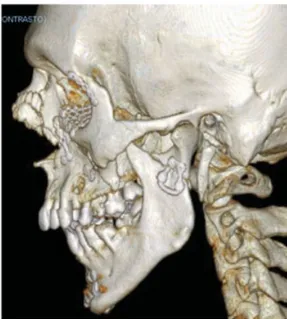
Fig. 1 Displaced subcondylar fracture in a complex facial fracture.
Fig. 2 Anatomic restoration of subcondylar region through a Trape-zoidal Plate.
Fig. 3 Severely dislocated subcondylar fracture: lost relation between condyle and glenoid fossa with malocclusion.
Fig. 4 Accurate anatomic fracture reduction and rigidfixation through a Lambda Linear Plate.
Linear holes arrangement in the cranial segment of the plate facilitatedfixation of high-neck condylar fractures with small condylar fragments. The feasibility of bending both arms or cut some holes allowed an easy anatomic adaptation to the fracture area. On the contrary, the reduced size of the Trapezoidal Plate allowed placement using different surgical approaches (preauricular or retromandibular/transparotid). The specific design easily fit the convex anatomy of the subcondylar region but its position was limited by the neck width. In 2013, a new condylar plate design, the Matrix-MANDIBLE Subcondylar 1.0 mm Strut Plate (Synthes) was introduced. With its particular plate design, the newly devel-oped condylar device proved to have great versatility of employment and became the system of primary choice in authors’ surgical practice (►Figs. 5and6). The Strut Plate was designed according to the two miniplates concept: the straight 3-hole segment has to be parallel to the posterior border of the ramus, aligned with the condylar head, while the 2-hole segment follows the rim of the sigmoid notch. Moreover, its size smaller than the Lambda Plate allowed placement through all surgical approaches. Its lightly curved profile and the possibility of bending the superior holes independently allowed a comfortably adaptation to the anat-omy of the condylar neck and subcondylar region.
Results
Postoperative clinical and radiological results were generally comparable using all kinds of condylar device. The operation time for ORIF of the condylar fractures ranged variably, between 60 and 120 minutes, depending on the surgical approach and the degree of condylar displacement/disloca-tion. No anatomic misalignment of the fracture was observed or no plate fracture occurred in the follow-up period. Post-operative wound healing complications were observed in 25% of patients (16 patients): temporary salivary leakage through the surgical wound was observed in 10 patients who received
a retromandibular/transparotid approach, healed with sterile compressive dressings. Six patients developed postoperative infections (four patients who received a preauricular ap-proach and two patients who received a retromandibular/ transparotid approach); the most common isolated pathogen was Staphylococcus epidermidis and infections were treated with irrigations and antibiotics systemic administration. Screw loosening was observed in three patients who devel-oped postoperative infections: the screws were removed but nonetheless fracture healing and bone consolidation were complete in all cases. Clinical and functional evaluation revealed a mean maximal mouth opening of 41 mm at 6 months follow-up period (minimum value 35 mm, maxi-mum 48 mm); mandibular protrusion and laterotrusion were almost normal (a mean value of 5 mm at 6 months postoper-atively) with a slight lateral deviation to the fractured side on maximum mouth opening (1–2 mm) in 12 of the 62 patients. Although a transient slight malocclusion in the immediate postoperative period in 11 patients, which disappeared with functional elastics therapy, pretraumatic occlusion was gen-erally restored. Despite initial pain during function, at mean follow-up period, it generally disappeared. A total of 18 patients (11 who received the retromandibular/transparotid and 7 who received the preauricular approach) suffered from transient facial nerve palsy, of both the frontal branch and the zygomatic one, which spontaneously disappeared in a mean time of 3 to 4 weeks, probably due to wider surgicalfields or retractors’ soft tissues compression.
Discussion
A proper surgical treatment of condylar neck and subcondylar fractures is mandatory to avoid long-term severe drawbacks, such as asymmetry, growth deficiency involving the orbit, the maxilla and the mandible if occurring during childhood, temporomandibular joint (TMJ) dysfunction with pain, mal-occlusion with retrognathia, open bite, reduced protrusion and laterotrusion, orfibroosseous or osseous ankylosis. The treatment outcome of condylar neck and subcondylar
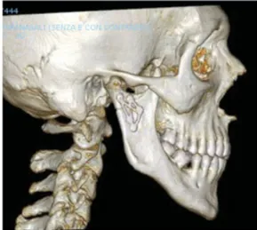
Fig. 5 Subcondylar fracture with displacement: loss of vertical dimension and open bite.
Fig. 6 Reduction andfixation using a Strut Plate: restoration of ramus height and occlusion.
fractures has gained considerably from ORIF.6,27–30 Never-theless, internal rigidfixation has to be sufficient stable to fulfil the principles of functionally stable osteosynthesis as stated by Champy et al.31–33The osteosynthesis device has to be rigid enough to withstand physiologic masticatory forces applied on the condylar region and the plate design has tofit the strain pattern occurring in the condylar region during function: tensile strain lines along the rim of the sigmoid notch and compressive strain lines along the posterior border of the ramus, parallel to the condylar axis.26The use of a single straight 4-hole miniplate, placed vertically, aligned to the condylar axis, where the bone of the ramus is thickest, is not adequate.8,14,16,34–40 It neither respects the principle of functionally stable osteosynthesis nor prevents secondary displacement with fracture line diastasis along the sigmoid notch and plate fracture. It has been clearly demonstrated both clinically and experimentally.15,16,20,23,34,36,41–46Even the alternative solutions advocated by some authors6,14,47–52 of a more rigid plate or a more solidfixation by means of bicortical screws or dynamic compression plate lead to treatment failure.41,53,54Moreover, the use of heavy plates is usually not applicable to the small condylar segment. The two 4-hole miniplates technique with monocortical screws has proved to be the most reliable and functionally stable osteosynthesis procedure for condylar neck and subcondylar fractures and is considered the standard practice.15,17–26 Wagner et al in 2002 experimentally proved its biomechani-cal superiority.42The anterior plate is placed along the tensile strain lines following the rim of the sigmoid notch and protects the posterior plate from mechanical strains. The posterior plate,fixed along the condylar axis at the posterior border of the ramus and almost strain free, retains the reduced correct position in the frontal and horizontal planes and prevents slippage or rotation of the condylar fragment. However, it could be sometimes difficult to insert four screws in the smaller condylar segment and can be particularly challenging in minimally invasive approaches to the condylar fracture. In the effort to overcome these limitations, specially designed 3D plates have been proposed and are available. Square and rectangular 3D plates were introduced by Farm-and et al in the 1990s for condylar fractures treatment55,56as the best mechanical compromise to ensure primary stabili-zation of condylar fractures. They were resumed in 2006 by Meyer et al43as a 3D rectangular plate (Profile 2.3, Leibinger, Freiburg, Germany). Unlike the two miniplates technique, 3D plates seemed to improve osteosynthesis stability thanks to the mechanical connection between the two plate’s arms. Moreover, their smaller size can avoid excessive soft tissue stripping during surgery and improves handling in minimally invasive surgical approaches. However, the square and rect-angular plate geometry grossly respects the two miniplates concept in withstanding masticatory forces and partially satisfies the principles of functionally stable osteosynthesis as stated by Champy et al.31–33The anterior arm being parallel to the posterior one, it does not sufficiently support the tensile strains that develop parallel to the boundary of the sigmoid notch. An incorrect transmission of forces may de-rive.26Several solutions have been proposed in the literature to
overcome these problems. In 2007, Meyer et al introduced the 3D 4/9-hole trapezoidal osteosynthesis plate (Modus TCP 2.0 Medartis, Basel, Switzerland) fixed with 2.0 monocortical screws, specifically designed to improve plate biofunctionality in stabilizing subcondylar and condylar neck fractures.57 Ow-ing to this plate’s shape modification, the anterior plate arm could follow as close as possible the tensile strains developing along the rim of the sigmoid notch, while the posterior arm could parallel the posterior border of the ramus with its compressive strain lines. Meyer et al published the results of an experimental protocol: standardized subcondylar fractures were produced on fresh dentate human mandibles, stabilized with either a 4- or a 9-hole 3D Trapezoidal Plate and loaded into a test bench reproducing maximum static biting forces. Results were evaluated by visual analysis of the macroscopic fracture line displacement and photoelastimetric analysis of the pre- and postsurgical strain patterns. No macroscopic fracture displacement was noted; the preoperative strain pattern in the subcondylar area, disrupted by the produced fracture, was perfectly restored by the 3D Trapezoidal Plate owing to a physiologic transmission of forces. The biomechan-ical required principles of functionally stable osteosynthesis were satisfied. The 3D design improves mechanical stability and needs minimal soft tissue stripping. Moreover, unlike the two miniplates technique, only two monocortical screws, both in the 4- and 9-hole plates, are necessary in the fractured condyle. The two different 4- or 9-hole variations allow adaptation to various situations: common subcondylar and high-neck fractures for the 4-hole plate, whose size respects the mean condylar neck width; comminuted, low-neck, and subcondylar fractures for the 9-hole plate, which allows greater adaptation to particular situations (poor bone quality, poor primary reduction, multiple fractures lines) and some bending possibilities. First in 2007 and then in 2011, Lauer et
al58,59 presented their delta-shaped condylar miniplate
(Modus Trauma 2.0 Condylus Fragment Plate, Medartis, Frei-burg, Germany) and the Trilock Delta Condyle Trauma Plate (Medartis)fixed with 2.0 mm monocortical screws. The Delta Plate was initially tested in biomechanical and clinical studies confirming it could sufficiently neutralize tension and com-pression forces and produce great stability, combined with the advantage of a smaller plate.57–60 In the effort to further ameliorate results, a locking system was introduced, the Tri-lock Delta Condyle Trauma Plate. Resembling an external fixture, it makes thorough plate bending superfluous because intimate plate adaption to the bone contour is no longer necessary, preventing from excessive stripping. Its greater primary stability across the fracture gap reduces micromove-ments and screws loosening. In an experimental study with porcine hemimandibles,59the biomechanical behavior of Delta Plate and Trilock Delta Condyle Trauma Plate was compared with the two miniplates system. Results revealed generally better performances of the Delta System, and of the locking system in particular, compared with the two miniplates tech-nique. Authors report their experience in the treatment of subcondylar and condylar neck fractures using the Synthes MatrixMANDIBLE Subcondylar Plates System. Introduced in 2010 and gradually enriched through years, it consists of
implants specifically designed for fractures of the subcondylar and condylar neck region to address different fracture pat-terns. All devices satisfy the principles of functionally stable osteosynthesis as stated by Champy et al31–33: the plate’s anterior arm has to perfectly follow the tensile strain lines along the rim of the sigmoid notch and acts as a tension resisting plate; the plate’s posterior arm has to be located along the axis of the condyle neck, free from any harmful bending strain, and essentially maintains the reduction preventing slippage or rotation of the condylar fragment. The thickness of all plates in the system is 1.0 mm, complying with the thickness of the plate proposed by Seemann et al.61Moreover, the 3D shape of the Trapezoidal and Strut Plates with their connection arms provides greater internal stability and more optimal leverage. The 7-hole 1.0-mm Lambda Plate gives the possibility to address a large fracture area through a wide extraoral access. With its 2-hole linear cranial segment and the feasibility of bending both anterior and posterior arms or cutting some holes it allows a greater anatomic adaptation. The Trapezoidal and the Strut Plate merge both the 3D plate concept and the two miniplates technique. The 4-hole 1.0-mm Trapezoidal Plate stabilizes a smaller fracture area but does not need excessive soft tissues stripping. It is precontoured to adapt to the convexity of the condylar neck and subcondylar region but the neck width can be a position limiting factor. After the introduction of the 4-hole 1.0-mm Strut Plate this plate has been widely used in various different situations owing to its greater versatility. Its reduced size allows for employment through different extraoral approaches with minimal soft tissues dissection. It has a precontoured curved profile to anatomically fit the fracture area, which can be further improved by a considerable bending chance.
Conclusion
Clinical data collected by the authors and the widely studied biomechanical background in the literature suggest that Synthes Subcondylar Plate System fulfils the principles of a functionally stable osteosynthesis and is suitable for ORIF of subcondylar and condylar neck fractures. The wide range of plate designs allows adequate stabilization of different fracture patterns although the 4-hole 1.0-mm Strut Plate proves greater versatility. A further clinical long-term evaluation is mandatory to determine the plates’ behavior in vivo.
References
1 Gassner R, Tuli T, Hächl O, Rudisch A, Ulmer H. Cranio-maxillo-facial trauma: a 10 year review of 9,543 cases with 21,067 injuries. J Craniomaxillofac Surg 2003;31(1):51–61
2 Stacey DH, Doyle JF, Mount DL, Snyder MC, Gutowski KA. Manage-ment of mandible fractures. Plast Reconstr Surg 2006;117(3): 48e–60e
3 Loukota RA, Eckelt U, De Bont L, Rasse M. Subclassification of fractures of the condylar process of the mandible. Br J Oral Maxillofac Surg 2005;43(1):72–73
4 Walker RV. Condylar fractures: nonsurgical management. J Oral Maxillofac Surg 1994;52(11):1185–1188
5 Baker AW, McMahon J, Moos KF. Current consensus on the management of fractures of the mandibular condyle. A method by questionnaire. Int J Oral Maxillofac Surg 1998;27(4):258–266 6 Ellis E III, Simon P, Throckmorton GS. Occlusal results after open or
closed treatment of fractures of the mandibular condylar process. J Oral Maxillofac Surg 2000;58(3):260–268
7 De Riu G, Gamba U, Anghinoni M, Sesenna E. A comparison of open and closed treatment of condylar fractures: a change in philoso-phy. Int J Oral Maxillofac Surg 2001;30(5):384–389
8 Haug RH, Assael LA. Outcomes of open versus closed treatment of mandibular subcondylar fractures. J Oral Maxillofac Surg 2001; 59(4):370–375, discussion 375–376
9 Zachariades N, Mezitis M, Mourouzis C, Papadakis D, Spanou A. Fractures of the mandibular condyle: a review of 466 cases. Literature review, reflections on treatment and proposals. J Cra-niomaxillofac Surg 2006;34(7):421–432
10 Hochban W, Ellers M, Umstadt HE, et al. Zur operativen reposition undfixation von unterkiefergelenkfortsatzfrakturen von enoral. Fortschr Kiefer Gesichtschir 1996;41:80–85
11 Jacobovicz J, Lee C, Trabulsy PP. Endoscopic repair of mandibular subcondylar fractures. Plast Reconstr Surg 1998;101(2):437–441 12 Sandler NA. Endoscopic-assisted reduction andfixation of a man-dibular subcondylar fracture: report of a case. J Oral Maxillofac Surg 2001;59(12):1479–1482
13 Raveh J, Vuillemin T, Lädrach K, Roux M, Sutter F. Plate osteosyn-thesis of 367 mandibular fractures. The unrestricted indication for the intraoral approach. J Craniomaxillofac Surg 1987;15(5): 244–253
14 Ellis E III, Dean J. Rigidfixation of mandibular condyle fractures. Oral Surg Oral Med Oral Pathol 1993;76(1):6–15
15 Hammer B, Schier P, Prein J. Osteosynthesis of condylar neck fractures: a review of 30 patients. Br J Oral Maxillofac Surg 1997;35(4):288–291
16 Klotch DW, Lundy LB. Condylar neck fractures of the mandible. Otolaryngol Clin North Am 1991;24(1):181–194
17 Pape HD, Hauenstein H, Gerlach KL. Surgical care of condylar fractures using miniplates: indication, technic and 1st results and limits. Fortschr Kiefer Gesichtschir 1980;25:81–83
18 Krenkel C. Biomechanics and Osteosynthesis of Condylar Neck Fractures of the Mandible. Carol Stream, IL: Quintessence Publish-ing Co.; 1994
19 Choi BH, Kim KN, Kim HJ, Kim MK. Evaluation of condylar neck fracture plating techniques. J Craniomaxillofac Surg 1999;27(2): 109–112
20 Choi BH, Yi CK, Yoo JH. Clinical evaluation of 3 types of plate osteosynthesis for fixation of condylar neck fractures. J Oral Maxillofac Surg 2001;59(7):734–737, discussion 738
21 Devlin MF, Hislop WS, Carton ATM. Open reduction and internal fixation of fractured mandibular condyles by a retromandibular approach: surgical morbidity and informed consent. Br J Oral Maxillofac Surg 2002;40(1):23–25
22 Schön R, Gutwald R, Schramm A, Gellrich NC, Schmelzeisen R. Extraoral and intraoral endoscopically assisted management of collum fractures. Mund Kiefer Gesichtschir 2002;6(4):236–240 23 Rallis G, Mourouzis C, Ainatzoglou M, Mezitis M, Zachariades N.
Plate osteosynthesis of condylar fractures: a retrospective study of 45 patients. Quintessence Int 2003;34(1):45–49
24 Suzuki T, Kawamura H, Kasahara T, Nagasaka H. Resorbable poly-L-lactide plates and screws for the treatment of mandibular condylar process fractures: a clinical and radiologic follow-up study. J Oral Maxillofac Surg 2004;62(8):919–924
25 Vesnaver A, Gorjanc M, Eberlinc A, Dovsak DA, Kansky AA. The periauricular transparotid approach for open reduction and inter-nalfixation of condylar fractures. J Craniomaxillofac Surg 2005; 33(3):169–179
26 Meyer C, Kahn JL, Boutemi P, Wilk A. Photoelastic analysis of bone deformation in the region of the mandibular condyle during mastication. J Craniomaxillofac Surg 2002;30(3):160–169
27 Haug RH, Brandt MT. Traditional versus endoscope-assisted open reduction with rigid internalfixation (ORIF) of adult mandibular condyle fractures: a review of the literature regarding current thoughts on management. J Oral Maxillofac Surg 2004;62(10): 1272–1279
28 Schön R, Gutwald R, Schramm A, Gellrich NC, Schmelzeisen R. Endoscopy-assisted open treatment of condylar fractures of the mandible: extraoral vs intraoral approach. Int J Oral Maxillofac Surg 2002;31(3):237–243
29 Schneider M, Erasmus F, Gerlach KL, et al. Open reduction and internalfixation versus closed treatment and mandibulomaxillary fixation of fractures of the mandibular condylar process: a ran-domized, prospective, multicenter study with special evaluation of fracture level. J Oral Maxillofac Surg 2008;66(12):2537–2544 30 Eckelt U, Schneider M, Erasmus F, et al. Open versus closed
treatment of fractures of the mandibular condylar process-a prospective randomized multi-centre study. J Craniomaxillofac Surg 2006;34(5):306–314
31 Champy M, Lodde JP. Syntheses mandibulaires. Localisation des syntheses en fonction des contraintes mandibulaires. Rev Stoma-tol 1976;77:971–976
32 Champy M, Wilk A, Schnebelen JH. Die Behandlung von Mandibula-frakturen mittels Osteosynthese ohne Ruhigstellung nach der Technik von F.X. Michelet. Zahn Mund Kieferheilk 1975;63:339–341 33 Champy M, Lodde JP, Jaeger JH, Wilk A. Osteosyntheses
mandibu-laires selon la technique de Michelet, I: bases biomecaniques. Rev Stomatol 1976;77:569–576
34 Sargent LA, Green JF Jr. Plate and screwfixation of selected condylar fractures of the mandible. Ann Plast Surg 1992;28(3):235–241 35 Lambert S, Reychler H, Micheli B, Pecheur A. Treatment of fractures
of the mandibular condyle. Rev Stomatol Chir Maxillofac 1995; 96(2):96–104
36 Nehse G, Maerker R. Indications for various reconstruction and osteosynthesis methods in surgical management of subcondylar fractures of the mandible. Fortschr Kiefer Gesichtschir 1996; 41:120–123
37 Newman L. A clinical evaluation of the long-term outcome of patients treated for bilateral fracture of the mandibular condyles. Br J Oral Maxillofac Surg 1998;36(3):176–179
38 Undt G, Kermer C, Rasse M, Sinko K, Ewers R. Transoral miniplate osteosynthesis of condylar neck fractures. Oral Surg Oral Med Oral Pathol Oral Radiol Endod 1999;88(5):534–543
39 Ellis E III, Throckmorton GS, Palmieri C. Open treatment of condylar process fractures: assessment of adequacy of reposition-ing and maintenance of stability. J Oral Maxillofac Surg 2000; 58(1):27–34, discussion 35
40 Hyde N, Manisali M, Aghabeigi B, Sneddon K, Newman L. The role of open reduction and internalfixation in unilateral fractures of the mandibular condyle: a prospective study. Br J Oral Maxillofac Surg 2002;40(1):19–22
41 Haug RH, Peterson GP, Goltz M. A biomechanical evaluation of mandibular condyle fracture plating techniques. J Oral Maxillofac Surg 2002;60(1):73–80, discussion 80–81
42 Wagner A, Krach W, Schicho K, Undt G, Ploder O, Ewers R. A 3-dimensionalfinite-element analysis investigating the biomechan-ical behavior of the mandible and plate osteosynthesis in cases of fractures of the condylar process. Oral Surg Oral Med Oral Pathol Oral Radiol Endod 2002;94(6):678–686
43 Meyer C, Serhir L, Boutemi P. Experimental evaluation of three osteosynthesis devices used for stabilizing condylar fractures of the mandible. J Craniomaxillofac Surg 2006;34(3):173–181
44 Iizuka T, Lindqvist C, Hallikainen D, Mikkonen P, Paukku P. Severe bone resorption and osteoarthrosis after miniplate fixa-tion of high condylar fractures. A clinical and radiologic study of thirteen patients. Oral Surg Oral Med Oral Pathol 1991;72(4): 400–407
45 Ziccardi VB, Schneider RE, Kummer FJ. Wurzburg lag screw plate versus four-hole miniplate for the treatment of condylar process fractures. J Oral Maxillofac Surg 1997;55(6):602–607, discussion 608–609
46 Sugiura T, Yamamoto K, Murakami K, Sugimura M. A comparative evaluation of osteosynthesis with lag screws, miniplates, or Kirschner wires for mandibular condylar process fractures. J Oral Maxillofac Surg 2001;59(10):1161–1168, discussion 1169– 1170
47 Ellis E III. Condylar process fractures of the mandible. Facial Plast Surg 2000;16(2):193–205
48 Ellis E. Discussion about:” A biomechanical evaluation of mandib-ular condyle fracture plating technique” by Haug et al. J Oral Maxillofac Surg 2002;60:80–81
49 Koberg WR, Momma WG. Treatment of fractures of the articular process by functional stable osteosynthesis using miniaturized dynamic compression plates. Int J Oral Surg 1978;7(4):256–262 50 Troulis MJ, Kaban LB. Endoscopic approach to the ramus/condyle
unit: Clinical applications. J Oral Maxillofac Surg 2001;59(5): 503–509
51 Laverick S, Jones DC. Letter about“Open reduction and internal fixation of fractured mandibular condyles by a retromandibular approach: surgical morbidity and informed consent” by Devlin et al. Br J Oral Maxillofacial 2002;40:453–454
52 Brandt MT, Haug RH. Open versus closed reduction of adult mandibular condyle fractures: a review of the literature regarding the evolution of current thoughts on management. J Oral Max-illofac Surg 2003;61(11):1324–1332
53 Kellman RM. Endoscopically assisted repair of subcondylar frac-tures of the mandible: an evolving technique. Arch Facial Plast Surg 2003;5(3):244–250
54 Lachner J, Clanton JT, Waite PD. Open reduction and internal rigid fixation of subcondylar fractures via an intraoral approach. Oral Surg Oral Med Oral Pathol 1991;71(3):257–261
55 Farmand M. Experiences with the 3D miniplate osteosynthesis in mandibular fractures. Fortschr Kiefer Gesichtschir 1996;41:85–87 56 Farmand M, Dupoirieux L. The value of 3 dimensional plates in maxillofacial surgery. Rev Stomatol Chir Maxillofac 1992;93(6): 353–357
57 Meyer C, Martin E, Kahn JL, Zink S. Development and biomechani-cal testing of a new osteosynthesis plate (TCP) designed to stabilize mandibular condyle fractures. J Craniomaxillofac Surg 2007;35(2): 84–90
58 Lauer G, Pradel W, Schneider M, Eckelt U. A new 3-dimensional plate for transoral endoscopic-assisted osteosynthesis of condylar neck fractures. J Oral Maxillofac Surg 2007;65(5):964–971 59 Haim D, Müller A, Leonhardt H, Nowak A, Richter G, Lauer G.
Biomechanical study of the Delta plate and the TriLock Delta condyle trauma plate. J Oral Maxillofac Surg 2011;69(10): 2619–2625
60 Lauer G, Haim D, Proff P, et al. Plate osteosynthesis of the mandibular condyle. Ann Anat 2007;189(4):412–417
61 Seemann R, Schicho K, Reichwein A, Eisenmenger G, Ewers R, Wagner A. Clinical evaluation of mechanically optimized plates for the treatment of condylar process fractures. Oral Surg Oral Med Oral Pathol Oral Radiol Endod 2007;104(6):e1–e4