UNIVERSITÀ DEGLI STUDI DI SALERNO
Dipartimento di Farmacia
PhD Program
in
Drug Discovery and Development
XXX Cycle — Academic Year 2017/2018
PhD Thesis in
Molecular basis of
cardiomiopathy
Candidate Supervisor
Michela Pecoraro
Prof. Ada Popolo
Co-Tutor
Prof.Aldo Pinto
I should apologize to my self for believing I was not
ABSTRACT
The normal heart rhythm is guaranteed by an important intercellular junctions system, named Gap Junction (GJs). Each GJs consists of two units called connexons formed by six specific trans-membrane proteins named connexins (Cx). Recent reports suggest the presence of Cx43 in the inner mitochondrial membrane where it plays an important cardioprotection mechanism. Alterations in Cx43 expression and distribution were observed in several myocardium disease; i.e. in hypertrophic cardiomyopathy, heart failure and ischemia. Thus, in this doctoral study, we investigated the role of Cx43, and in particular of mitochondrial Cx43, in different cardiomyopaties models.
At first, we have investigated the involvement of mitochondrial Cx43 in an in vitro model of chemical hypoxia. Hypoxia was induced by adding Cobalt Chloride (CoCl2) on H9c2
cardiomyoblast cell line, both in absence and in presence of Radicicol, an Hsp 90 inhibitor that blocks Cx43 translocation to the mitochondria. Our results showed that CoCl2 reduces the
expression of Cx43 on the cell membrane and, moreover, it increases Cx43 expression at mitochondrial level, where it is involved in the regulation of reactive oxygen species production,
calcium storage and mitochondrial membrane depolarization. Furthermore, in an in vivo Doxorubicin (DOXO)-induced cardiotoxicity in a short-term mouse model we have studied the modulation of Cx43 expression/activity and its dysregulation. Our results showed that DOXO is able to induce significant changes in calcium homeostasis and alterations in Cx43 expression and localization. These effects are evident even in the heart of mice that received a single DOXO-administration. Finally, we have investigated if the pretreatment with Diazoxide (DZX), an opener of mitochondrial KATP-channels, attenuates
DOXO-induced cardiotoxicity in a short-term mouse model. Our results demonstrate that DZX represents a promising protective intervention against DOXO-induced cardiotoxicity, by reducing the calcium homeostasis alteration and trying to restore the major cardiac parameters altered by DOXO treatment. This is in agreement with our hypothesis that mitochondrial Cx43 and DZX are involved in the cardioprotection mechanism.
Index
LIST OF ABBREVIATIONS
... ..1
INTRODUCTION
... ..3
AIM OF WORK
...17
CHAPTER I: Role of mitochondrial Connexin 43
in a chimical model of hypoxia
...19
1.1 Introduction ... 19
1.2 Material and Methods ... 21
1.2.1 Experimental Protocols ... 21
1.2.2 Cell Culture ... 21
1.2.3 MTT assay ... 22
1.2.4 Mitochondrial protein extraction and Western blot analysis for mitochondrial Cx43 ... 23
1.2.5 Total protein extraction and Western blot analysis for procaspase 3 and caspase 9... 24
1.2.6 Immunofluorescence Analysis with Confocal Microscopy ... 25
1.2.7 Measurement of Intracellular calcium Signaling ... 26
1.2.8 Measurement of Mitochondrial Superoxide Evaluation with MitoSOX Red ... 27
1.2.9 Measurement Mitochondrial Membrane Depolarization with TMRE ... 28
1.2.10 Cytochrome c release detection by cytofluorometry ... 29
1.2.11 Analysis of apoptosis ... 29
1.3 Results ... 31
1.3.1 CoCl2-induced chemical hypoxia ... 31
1.3.2 CoCl2 increased mitochondrial Cx43 expression ... 33
1.3.3 The inhibition of Cx43 translocation on mitochondria increased Mitochondrial Superoxide Production ... 35
1.3.4 CoCl2 induces calcium homeostasis alteration ... 36
1.3.5 The inhibition of Cx43 Translocation Enhanced Mitochondrial Membrane Depolarization ... 38
1.3.6 Inhibition of Cx43 translocation to mitochondria enhances CoCl2 -induced apoptotic response ... 39
1.4 Discussion ... 41
CHAPTER II: Involvement of Connexin 43 in Calcium
Impairment induced by Doxorubicin
in a short-term mouse model ... 45
2.1 Introduction ... 45
2.2 Materials and Methods ... 49
2.2.1 Materials ... 49
2.2.2 Animals ... 49
2.2.3 Experimental Protocols ... 49
2.2.4 Echocardiogram ... 51
2.2.5 Histological staining ... 51
2.2.6 Protein Extraction and Western Blot Analysis ... 51
2.2.7 Mitochondrial Protein Extraction and Western Blot Analysis for Mitochondrial Cx43 and pCx43 ... 52
2.2.8 Primary Cardiomyocytes Isolation and Measurement of Intracellular Ca2+ Signaling ... 53
2.2.9 Immunohistochemical Analysis ... 55
2.3 Results ... 56
2.3.1 Cardiac Functions ... 56
2.3.2 Histopathological examination ... 57
2.3.3 Doxorubicin Administration Alters Calcium Homeostasis ... 58
2.3.4 Doxorubicin Administration Affects Cx43 Expression and Localization ... 61
2.4 Discussion ... 65
CHAPTER III: Cardioprotective effects of
Diazoxide in Doxorubicin- induced
cardiotoxicity model
...69
3.1 Intoduction ... 69
3.2 Materials and Methods ... 73
3.2.1 Materials ... 73
3.2.2 Animals ... 73
3.2.3 Experimental Protocols ... 75
3.2.4 Echocardiogram ... 75
3.2.5 Protein Extraction and Western Blot Analysis ... 76
3.2.6 Mitochondrial Protein Extraction and Western Blot Analysis for Mitochondrial Cx43 and pCx43 ... 77
3.2.7 Primary Cardiomyocytes Isolation and Measurement of Intracellular Ca2+ Signaling ... 77
3.2.8 Immunohistochemical Analysis ... 79
3.2.9 Statistical Analysis ... 79
3.3 Results ... 80
3.3.1 Cardiac Functions ... 80
3.3.3 Diazoxide Administration Affects Cx43and pCx43
expression and localization ... 84
3.3.4 Diazoxide Administration Affects mitochondrial Cx43 and pCx43 expression ... 85
3.4 Discussion ... 88
GENERAL CONCLUSION
...93
REFERENCES
...95
1
LIST OF ABBREVIATIONS
Cx43 Connexin 43
GJ Gap Junction
HSP90 Heat Shock Protein 90
mCx43 Mitochondrial Connexin43
mpCx43 Mitochondrial Connexin 43
phosphorylated
pCx43 Connexin 43 phosphorylated
PLB Phospholamban
TMRE Tetramethylrhodamina ethyl ester
TOM 20 Translocase of the Outer
3
INTRODUCTION
The steady beat of our hearts is among the most critical functions performed perfectly by our body without the conscious intervention. The developing heart is the earliest organ to function in the embryo, generating rhythmic contractions while it is forming, before there is blood to pump. Although the heart is composed of multiple cell types besides muscle cells, keeping a steady heartbeat is the job of specialized cardiomyocytes that ensure coordination of electrical signals throughout the organ [Rosenthal N.N et al., 2017].
Cardiac conduction has classically been viewed as an electrotonic process occurring by means of direct ionic current flow from cell to cell via Gap Junctions (GJs) [Kleber et al., 2004]. GJs are intracellular structures that provide connections and communication between cells, allowing the passage of ions
and small molecoles such as ATP, glutathione, cAMP, IP3 and
glucose [Pecoraro et al., 2015 a]. To form a GJ, six trans-membrane monomers (connexins) from one cell oligemarize to form a tran-smembrane channel referred to as a connexon or hemichannel. The connexons from one cell then dock and couple with apposing connexons on neighboring cells and coalesce into dense GJ plaque [Basheer et al., 2016]. Connexins
4
are ubiquitous trans-membrane proteins and they are encoded by over 20 different genes in the mouse genome and 21 in the human genome, which are classified according to their different molecular weights that range between 26 and 60 kDa [Pecoraro et al., 2015 a]. Each connexin is constituted by four trans-membrane domains, two extracellular loops (EL) and one intracellular; amino and carboxy terminal regions are both located in the cytosol [Solan and Lampe, 2005].
Figure 1: The image shows a GJ and its main element.[LadyofHats et al., 2006]
Connexin 43 (Cx43) is the major connexin expressed in the cardiac ventricle, [Willecke et al., 2002; Pecoraro et al., 2017 a], which regulate intercellular coupling, conduction velocity, and anisotropy [Cabo et al., 2009]. It is also responsible for the action potential propagation [Severs et al., 2008] and it
5
critically regulate intercellular translocation of ions and small molecoles [Van Veen et al., 2001]. Indeed, many reports have suggested that Cx43 regulates other cellular mechanisms, including the cell cycle, differentiation, proliferation [Kardami et al., 2007], maintenance of tissue homeostasis, and embryogenesis [De Maio et al., 2002].
Figure 2: Molecular Organization, Gating, and Function of Gap Junction Channels
[Feliksas et al., 2013]
Cx43 has a short half-life (1–3 hours in the myocardium) [Beardslee et al., 1998; Smyth et al., 2014]. It oligomerizes in the Golgi/trans-Golgi network and, after assembly, it is transported to the non-junctional plasma membrane through the cytoskeleton. Once inserted into the cell membrane, Cx43 spreads in the region where there are GJ plaques by a
6
microtubule/dynein/β-catenin/N-cadherin-dependent pathway
[Sáeza et al., 2010].
In the heart, localization of Cx43 at the intercalated discs is crucial to provide the intercellular coupling necessary for rapid action potential propagation through the myocardium and synchronized cardiac contraction [Rohr et al., 2004].
Cx43 hemichannels can occur as free, non-junctional channels in the plasma membrane. These hemichannels are normally closed but may open in response to various triggers including
cell depolarization, decreased extracellular Ca2+ ion
concentration, increased intracellular Ca2+ concentration and
alterations in the phosphorylation or redox status [Basheer et al., 2016]. Generally, it has been assumed that the opening of plasma membrane Cx43 hemichannels is linked to pathological rather than physiological entities, contributing to cell swelling and cell death. In the cardiac cells, excessive hemichannel
opening allows the entry of Na+ and Ca2+ and the escape of K+,
adenosine triphosphate (ATP) and other small metabolites,
leading to osmotic shifts, energy depletion, Ca2+ overload and
cell death promotion [Schulz et al., 2015]. Therefore, blockage of the Cx43 hemichannels by using pharmacological inhibitors can possibly have protective effects against cardiac insult such as in the case of ischemia/reperfusion injury [Schulz et al., 2015; Wang et al., 2013].
7
Mice lacking Cx43 die shortly after birth due to cardiac hyperplasia obstructing the right ventricular outflow tract [Reaume et al., 1995], while a heart-specific conditional knockout exhibited arrhythmia and premature death[Gutstein et al., 2001a; Gutstein et al., 2001 b].
Cx43 is a phosphoprotein that is predominantly phosphorylated in the control state. There are at least 14 serines and 2 tyrosines in the cytoplasmic C-terminal region of Cx43 that are phosphorylated by a variety of kinases [Marquez-Rosado et al., 2011], such as mitogen activated protein kinase (MAPk), protein kinase C (PKC), protein kinase A (PKA), casein kinase 1 and Src. The phosphorylation of the serine residues, like S306, S365 and S368, modulates the conductivity of the GJs [Lampe et al., 2000], as well as their trafficking, assembly/disassembly, degradation and gating [Pecoraro et al., 2015 a]. Indeed, connexin phosphorylation and dephosphorylation play an important role in regulating GJ channel function at several stages of the cell cycle and the connexin “life cycle”[Hood et al., 2017].
8
Figure 3: Connexin phosphorylation at the serine residues modulates the conductivity
of the GJs, as well as, their trafficking, assembly/disassembly, degradation and gating [Solan and Lampe 2005]
Most of the function ascribed to Cx43 in cardiac pathophysiology is within the context of its role in forming the GJ [Severs et al., 2004]. However, recent literature reports that Cx43, independent of its ability to form functional GJs, regulates susceptibility of cells to several cell injury paradigms suggesting a novel function of this protein in modulating cell death, specially myocyte death [Li net al., 2003].
Cardiac mitochondria play a pivotal role in the maintenance of cellular bioenergetics and intracellular ion homeostasis,
especially of calcium (Ca2+) and potassium (K+) ions. Cellular
Ca2+ homeostasis is maintained in a very stringent manner by
9
and mitochondria, respectively [Gunter et al., 2004]. These cyclical events are crucial to ensure the cardiac rhythm and any disturbance may lead to arrhythmia, cell death, and tissue damage as observed in cardiac failure and ischemia–reperfusion injury [Kristia´n et al., 1998]. Many studies reported that both in tissue [Boengler et al., 2005; Pecoraro et al., 2017 b] and in isolated mitochondria [Srisakuldee et al., 2009; Pecoraro et al., 2017 b], Cx43 is located at the inner mitochondrial membrane [Rodriguez-Sinovas et al., 2006] where it forms hemichannels, with the C-terminus region oriented at the intermembrane space [Miro-Casas et al., 2007].
The physiological role of mCx43 has not been well elucidated,
but recent studies show that it modulates K+ influx to the
mitochondrial matrix, mitochondrial respiration and reactive oxygen species (ROS) generation [Boengler et al., 2012; Miro-Casas et al., 2007]. Indeed, mCx43 may play a role in mediating the cardioprotective effect of ischemic preconditioning. Protection by mCx43 has been linked to ROS generation,
mitochondrial KATP channels, protein kinase C (PKC) signaling,
and stimulation of translocase of outer membrane- 20 (Tom20) that facilitates Cx43 transport [Gadicherla et al., 2017], from cytosol to mitochondria with a mechanism that involves heat shock protein 90 (Hsp90). Cytosolic Hsp90 is generally involved in the folding of newly synthesized proteins and its
10
role in mitochondrial import may be an extension of this activity [Young et al., 2003].
In fact, mitochondrial import machinery involves binding of the target protein to a chaperone (Hsp90/Hsp70), presentation to specific parts of TOM complex, and release into the inner mitochondrial membrane through the TIM (Translocase of the Inner Membrane) [Pecoraro et al., 2015 a]. Recently it has been hypothesized that mCx43 protects the cells by reducing
cytosolic Ca2+ overload, mitochondrial permeability transition,
and cell death [Pecoraro et al., 2017 a; Pecoraro et al., 2017 b]. Cx43-based hemichannels are non-selective large conductors of
Ca2+ entry in the inner mitochondrial membrane. Thus, mCx43
may directly contribute to mitochondrial Ca2+ entry/overload,
permeability and cell death [Wang et al., 2013; Decrock et al., 2011].
11
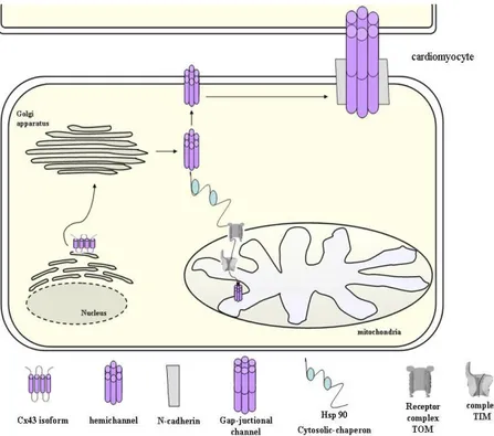
Figure 4: During biosynthesis, Cx43 is inserted into the endoplasmatic reticulum
where it correctly folds and oligomerize in the Golgi/trans-Golgi network. After assembly, Cx43 hemichannel is carried on the cell surface through the cytoskeleton. Once inserted into the cell membrane, Cx43 spreads in the region where there are GJ claque by a microtubule/dynein/β-catenin/N-cadherin-dependent pathway. In addition,
after assembly in Golgi, Cx43 may translocate from cytosol to mitochondria with a mechanism that involves Hsp90 and translocase of the outer membrane. This system involves binding of the target protein to Hsp90, presentation to specific parts of TOM complex, and release into the inner mitochondrial membrane [Pecoraro et al., 2015 a].
Myocyte apoptosis is now recognized to mediate cell death in a variety of acute and chronic heart diseases [Goubaeva et al., 2007], and many studies confirmed the mCx43 as a novel regulator of mitochondrial function where inhibition results in
the release of cytochrome C and myocyte apoptosis.
Futhermore, mCx43 is involved in a signal transduction pathway that can prevent mitochondrial permeability transition pore (mPTP) formation [Ruiz-Meana et al., 2008]. The mPTP is a large non-specific conductance channel that forms at the inner mitochondrial membrane under conditions of calcium
12
overload/oxidative stress [Baines et al., 2007]. Once formed the mPTP results in mitochondrial swelling, rupture of the outer mitochondrial membrane, release of apoptogenic mitochondrial contents to the cytosol, and cell death. Signals preventing mPTP formation during repefusion after ischaemia promote cell survival and reduce myocardial damage [Martel et al., 2012]. Thus, Cx43 channels, both as a GJ and as a hemichannel, form large-conductance ion channel with chemical gating similar to the Bcl-2 channels. In addition, they are voltage gated perhaps allowing sensing of the mitochondrial membrane potential in addition to the chemical environment.
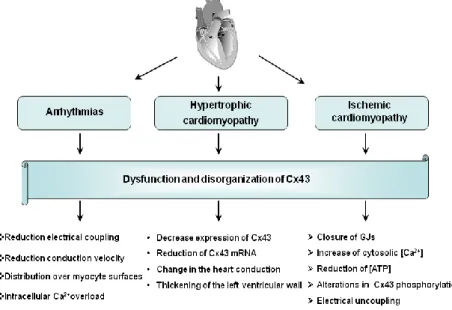
Changes in Cx43 expression and/or subcellular distribution in the heart have been associated with a wide variety of pathologic conditions and diseases, including the ventricular myocardium and cardiac arrhythmia [Due t al., 2017; Prevedel et al., 2017]. Moreover, myocardial hypoxia or ischaemia and conditions such as hypertrophy are associated with reduced cardiac action
potential conduction, raised intracellular [Ca2+] and GJ
uncoupling [Jabr et al., 2016].
These alterations contribute to abnormal impulse propagation and arrhythmogenic substrates leading to sudden cardiac death [Gutstein et al., 2001]. Arrhythmias are common complications of myocardial ischemia and infarction in humans. Reduced electrical coupling can increase the propensity for arrhythmias
13
rendering the ventricle more susceptible to re-entry. This condition seems to be due to dysfunction and disorganization of Cx43. Indeed, reduction of about 90% in Cx43 expression results in about 50% decrease in the conduction velocity. While 50% reduction in Cx43 may lead to some conduction slowing, high levels of electrical uncoupling are needed to increase arrhythmogeneity [Pecoraro et al., 2015 a].
Therefore, a large number of studies in recent years demonstrated a decrease in expression and/or lateralization and heterogeneous distribution of Cx43 in the myocardium of patients with hypertrophic cardiomyopathies (HCM), dilated cardiomyopathies (DCM), ischemic cardiomyopathies, as well as clinical congestive heart failure [Basheer et al., 2016].
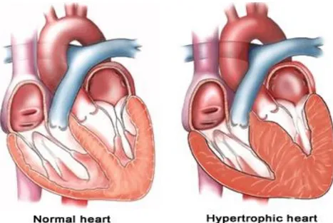
Figure 5: Hypertrophic Cardiomyopathy and Heart Failure. Hypertrophy are
associated with reduced cardiac action potential conduction, raised intracellular [Ca2+] and GJ uncoupling [Columbia University]
14
In general, in these cardiomyopathies, Cx43 expression appears to be unaltered or up-regulated during the initial and compensatory phase of hypertrophy, but redistributed along the cardiomyocyte surface and reduced when the hypertrophy becomes prolonged and putatively maladaptive in its progression to heart failure [Birgit et al., 2004].
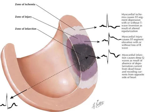
The most common myocardial dysfunction is ischemic injury, which is the principal cause of death in the world [Pecoraro et al., 2017 a].
Figure 6: Ischemic injury, the principal cause of death in the world. [Netter F et al.,
15
Cardiac ischemia is manifested by accumulation of metabolites in the extracellular compartment in combination with reduced oxygen supply. Furthermore ischemia induces closure of GJs,
due to increased cytosolic Ca2+ concentration, reduced ATP
concentration, changes in phosphorylation of Cx43 and acidification [Johanses et al., 2011]. Increased levels of
intracellular Ca2+ and H+ and accumulation of amphipathic lipid
metabolites during ischemia promote electrical uncoupling, mediated by alterations in phosphorylation of Cx43. In fact, acute ischemia may activate or inhibit protein kinases and phosphatases [Beardslee et al., 2000]. Cx43 plays a crucial role in ischemic preconditioning cardioprotection by improving myocardial function [Wang et al., 2015].
Figure 7: Schematic diagram that shows the effects of Cx43 dysfunction and
17
AIM OF WORK
Based on the data recently reported in the literature [Pecoraro et al., 2015 b; Severs et al., 2004; Du et al., 2017], we hypothesized that GJs, particularly Cx43, are largely involved in mechanisms that affect different cardiac pathologies. The aim of this doctoral project is therefore to investigate the involvement of Cx43, and in particular of the mCx43, in in vitro and in vivo cardiomyopaties models. In addition, we will focus on specific drugs and on their effects on the modulation of Cx43 expression/activity.
-As the first we investigated the involvement of mCx43 in the apoptotic pathway in an in vitro model of chemical hypoxia.
Hypoxia was induced by adding Cobalt Chloride (CoCl2) on
H9c2 cardiomyoblast cell line, both in absence and in presence of Radicicol, an Hsp 90 inhibitor that blocks Cx43 translocation to the mitochondria.
-As the second we investigated the involvement of Cx43 in a mouse model of cardiotoxicity. To this end we used a short-term mouse model of cardiotoxicity induced by Doxorubicin.
-As the third we investigated if the pretreatment with Diazoxide
(DZX), an opener of mitochondrial KATP-channels, attenuates
DOXO-induced cardiotoxicity and affect Cx43 expression
19
Chapter I
Role of mitochondrial Connexin 43 in a chimical
model of hypoxia
1.1 Introduction
Ischemic injury is the most common myocardial dysfunction, which is the principal cause of death in the world [Ruiz-Meana et al., 2001]. It’s irreversible and widespread loss of myocardial cells and subsequent ventricular remodeling induced by acute myocardial infarction are the main elements resulting in chronic heart failure and permanent loss of labor force [Roger et al., 2012].
This cardiomyopathy is manifested by accumulation of metabolites in the extracellular compartment in combination with reduced oxygen supply [Pecoraro et al., 2015 a].
Ischemia triggers a decrease in GJ communication as a result of
acidosis, increased intracellular Ca2+ concentration ([Ca2+]i) and
altered phosphorylation and nitrosylation levels [Schulz et al., 2015]. Revascularization could improve the prognosis of the patients with acute myocardial infarction [Roger et al., 2012], however, reperfusion could induce additional injury, which is sometimes extremely severe or fatal. Basic studies [Jiang et al., 2013; Wei et al., 2013] and small-sample clinical data [Crimi et
20
al., 2013] have verified that ischemic postconditioning (IPOC) could attenuate injury induced by ischemia/reperfusion (I/R). Other studies [Shimizu et al., 2014; Mykytenko et al., 2008] report that the Cx43 plays an important role in IPOC cardioprotection against I/R injury. In fact, many studies attribute the effect of cardioprotection to mitochondria, where Cx43 translocates throught Hsp90/TOM 20 machinery system [Goubeva et al., 2007].
So, the first step of this phD project was to study the role of mCx43 in an in vitro model of hypoxia.
21
1.2 Materials and Methods
1.2.1 Experimental Protocols
The hypoxia model was established by using the
hypoxia-inducing agent CoCl2 [Shi Yun et al., 2017]. CoCl2 is a well
known chemical hypoxia mimetic agent, which can mimic hypoxia/hischemic conditions by causing inactivation of hydroxylase enzymes and stabilization of hypoxia-inducible factor HIF-1α [Liu et al., 2014]. H9c2 cardiomyoblasts were
treated with CoCl2 at 50-100-150 µM for 3 or 6 h in DMEM 10
% FBS. In order to verify the role of mCx43 in CoCl2-treated
cells, we used Radicicol, a specific inhibitor of Hsp90 [Schulte et al., 1998], since the translocation of mCx43 on the mitochondrial membrane provides the binding of this protein to Hsp90 [Ruiz-Meana et al., 2008]. In the experiments that included the use of Radicicol (1 µM), an Hsp90 inhibitor, it has
been administered 30 min before CoCl2 treatment and left in the
incubation medium for all experimental time.
1.2.2 Cell Culture
The cell line used is rat cardiomyocytes H9c2 that demonstrate many similarities to primary cardiomyocytes, including membrane morphology, G-protein signaling expression, electrophysiological properties and constitutive expression of
22
Cx43 [Wu et al., 2013]. H9c2 was purchased from the American Tissue Culture Collection (Manassas, VA, USA).
H9c2 cells were subcultured weekly in 100-mm Corning dishes containing 10 ml Dulbecco’s modified Eagle’s Medium (DMEM; Gibco) with 10 % fetal bovine serum (FBS; Gibco) and antibiotics (25 U/ml penicillin and 25 U/ml streptomycin).
1.2.3 MTT assays
Cell viability was evaluated by means of MTT ([3-(4,5-dimethylthiazol-2-yl)-2,5-diphenyltetrazolium bromide]). H9c2
(3,5x10³ cells/well) were plated in 96-well tissue culture plates
and allowed to adhere for 24 h. Thereafter, the medium was
replaced with fresh medium alone or containing CoCl2
(50-100-150µM) and incubation was performed for 3 or 6 h at 37°C in an
atmosphere containing 5% CO2. Where indicated, Radicicol (1
µM) was added 30 min before CoCl2. Cell mortality was
assessed by means of MTT assay. Briefly, 25 µl of MTT (5 mg/ml) were added and cells were incubated for an additional 3h to allow the formation of formazan precipitate, which was solubilised with 100 µl of a solution containing 50% (v/v) N,N-dimethylformamide, 20% (w/v) SDS with an adjusted pH of 4.5. The optical density (OD) of each well was measured with a microplate spectrophotometer (Titertek Multiskan MCC/340-DASIT; Milan, Italy) equipped with a 620nm filter. H9c2
23
mortality in response to treatment with CoCl2 both in absence
and in presence of Radicicol was calculated as % mortality = 100-[100x(OD treated/OD control)].
1.2.4 Mitochondrial protein extraction and Western blot analysis for mitochondrial Cx43
H9c2 (1x10⁶ cells/well) were seeded into Petri plates and
allowed to adhere for 24 h. Thereafter, the medium was replaced with fresh medium and cells were treated as described. Mitochondrial protein extraction was carried out from cells in
lysis buffer A (sucrose 250 mM, K+ Hepes pH 7.5 20 mM, KCl
10 mM, MgCl2 1,5 mM, EDTA 1 mM, EGTA 1 mM, protease
inhibitors, NaF 50 mM, Na3VO4 0,2 mM, PMSF 1 mM, DTT 1
mM, digitonin 0,025%). Then the cells were centrifugated at 16000 g for 2 min at 4°C. The supernatant was discarded and the pellet was resuspended in lysis buffer B (NaCl 150mM, Triton X 1%, NaDeOH 0,5%, SDS 1% and Tris HCl 50mM pH 7.4) to obtain mitochondrial protein. Protein concentrations were determined with the Bio-Rad protein assay (BIO-RAD, Milan Italy). Equal amounts of protein (50 µg) were loaded into an acrylamide gel and separated by SDS-PAGE under denaturating conditions. Blots were incubated with primary antibody anti-Cx43 (BD transduction laboratories, 1:8000) or anti-TOM20 (1:250, used as loading control) over night. After incubation
24
with the primary antibodies and washing in PBS/0.1% Tween, the appropriate secondary antibody, anti-rabbit or anti-mouse (each diluted 1:4000) was added for 1h at room temperature.
Immunoreactive protein bands were detected by
chemiluminescence using enhanced chemiluminescence
reagents (ECL) in LAS 4000 (GE Healthcare). The images were analysed for densiometry using ImageJ Software.
In order to verify the purity of mitochondrial protein extraction, a Western blot analysis was performed to evaluate the presence of proteins expressed only in the mitochondria (ox-Phos Complex II, Abcam, 1:7000) and the absence of proteins
expressed in other cellular compartments (Na+/K+ ATPase,
Abcam, 1:3000). The purification assay was evaluated as the enrichment in mitochondrial protein (anti-Ox-Phos Complex II) as well as the elimination of the other cellular constituent
(anti-Na+/K+ ATPase) by means of Western blot analysis as
previously reported [Boengler et al., 2005].
1.2.5 Total protein extraction and Western blot analysis for procaspase 3 and caspase 9
H9c2 (7x10⁵ cells/well) were seeded in 6-well tissue culture
plates and allowed to adhere for 24 h. Thereafter, the medium was replaced with fresh medium and cells were treated as described. Total proteins were extracted from cells by freeze/
25
thawing in lysis buffer (containing Tris–HCl 50 mM pH 7.4, 50 mM NaF, 150 mM NaCl, 1 % Nonidet P40, 1 mM phenylmethylsulfonylfluoride, 0.2 mM sodium orthovanadate, 1mM EDTA and protease inhibitors). Protein concentrations were determined with the Bio-Rad protein assay (BIO-RAD, Milan Italy), and 50 µg protein/lane was loaded onto an acrylamide gel and separated by SDS-PAGE in denaturating conditions. Blots were incubated, over night, with primary antibody anti-procaspase 3 and anti-caspase 9 (each diluted
1:200; from Santa Cruz Biotechnology, DBA Italy). GAPDH
(1:1000) was used as loading control. After incubation with the primary antibodies and washing in TBS/0.1 % Tween, the appropriate secondary antibody, either anti-rabbit (diluted
1:5.000), was added for 1h at room temperature.
Immunoreactive protein bands were detected by
chemiluminescence using enhanced chemiluminescence
reagents (ECL) in LAS 4000 (GE Healthcare). Western blot data were quantified by using ImageJ Software.
1.2.6 Immunofluorescence Analysis with Confocal Microscopy
For immunofluorescence assay, H9c2 cells were seeded on
coverslips in 12-well plate (104 cells/well) and allow to grow
for 24 h; thereafter, the medium was replaced with fresh
26
3 or 6 h, with or without Radicicol (1µM). Then, cells were fixed with 4 % paraformaldehyde in PBS for 15 min and permeabilized with 0.1 % triton X in PBS for 15 min. After blocking with BSA and PBS for 1 h, cells were incubated with rabbit anti- HIF-1α antibody (TEMA Ricerca-Origene), mouse
anti-Cx43 antibody (Santa Cruz Biotechnologies, 1:250) and
rabbit anti-TOM20 (1:250) for 2 h at room temperature. The slides were then washed with PBS for three times and fluorescein-conjugated secondary antibody (FITC) or Texas red-conjugated secondary antibody were added for 1 h, DAPI was used for counterstaining of nuclei. Coverslips were finally mounted in mounting medium and fluorescent images were taken under the laser confocal microscope (Leica TCS SP5).
1.2.7 Measurement of Intracellular calcium Signaling
Intracellular calcium concentrations were measured using the fluorescent indicator dye Fura 2-AM, the membrane- permeant
acetoxymethyl ester form of Fura 2. Briefly, H9c2 (3x10⁴
cells/well) were seeded in 6-well tissue culture plates and allowed to adhere for 24 h. Thereafter, the medium was replaced with fresh medium and cells were treated as described. After incubation period (3 or 6 h), cells were washed in phosphate buffered saline (PBS), re-suspended in 1 ml of Hank’s balanced salt solution (HBSS) containing 5 µM Fura 2-AM for 45 min.
27
Thereafter, cells were washed with the same buffer to remove excess Fura 2-AM and incubated in calcium-free HBSS/0.5 mM EGTA buffer for 15 min to allow hydrolysis of Fura 2-AM into its active-dye form, Fura 2. H9c2 cells then were transferred to the spectrofluorimeter (Perkin-Elmer LS-55). Treatment with Ionomycin (1 µM final concentration), or with carbonyl cyanide p-trifluoromethoxy-pyhenylhydrazone (FCCP, 50 nM final concentration) was carried out by adding the appropriate concentrations of each substance into the cuvette in calcium-free HBSS/0.5 mM EGTA buffer. The excitation wavelength was alternated between 340 and 380 nm, and emission fluorescence was recorded at 515 nm. The ratio of fluorescence intensity of 340/380 nm (F340/F380) is strictly related to intracellular free calcium [Popolo et al., 2011]. Results are indicated as delta increase in fluorescence ratio (F340/F380 nm) induced by ionomycin-basal fluorescence ratio (F340/F380 nm) or FCCP-basal fluorescence ratio (F340/F380 nm).
1.2.8 Measurement of Mitochondrial Superoxide Evaluation with MitoSOX Red
Mitochondrial superoxide formation was evaluated by MitoSOX
Red. Briefly, H9c2 (4,0x10⁵ cells/well) were plated in 6-well
tissue culture plates and allowed to adhere for 24 h. Thereafter, the medium was replaced with fresh medium and cells were treated as described. After incubation period, MitoSOX Red (2.5
28
µM) was added for 15 min at 37°C before fluorescence evaluation by means of flow cytofluorometry. This indicator is a fluorogenic dye for highly selective detection of superoxide in the mitochondria of live cells and, once targeted to the mitochondria, it is oxidized by superoxide and exhibits red fluorescence. MitoSOX is readily oxidized by superoxide but not by other ROS-generating systems. Cells fluorescence was evaluated using a fluorescence-activated cell sorting and analyzed with Cell Quest software.
1.2.9 Measurement Mitochondrial Membrane Depolarization with TMRE
Mitochondrial permeability transition pore (mPTP) opening was measured with FACS scan by means of the fluorescent dye, tetramethylrhodamine methyl ester (TMRE). Due to its positive charge, TMRE readily accumulates in active mitochondria in inverse proportion to Δψm according to the Nernst equation. For
these experiments H9c2 (4,0x10⁵ cells/well) were seeded in
6-well tissue culture plates and allowed to adhere for 24 h. Thereafter, the medium was replaced with fresh medium and cells were treated as described. Cells were then collected, washed twice with phosphate buffer saline (PBS) and then incubated in PBS containing TMRE (5 nM) at 37°C. After 30 min, cells fluorescence was evaluated using a fluorescence-activated cell sorting and analyzed with Cell Quest software.
29
1.2.10 Cytochrome c release detection by cytofluorometry
Cytochrome c was checked by fluorescence-activated cell sorting (FACSscan; Becton–Dickinson). H9c2 cells were
cultured in a 6-well plate (4,5x10⁵ cells/well) and allow to grow
for 24 h; thereafter, the medium was replaced with fresh medium and cells were treated as described. After incubation period, cells were harvested with scraper and treated with permeabilization buffer (containing 2 % FBS and PBS in the presence of sodium azide 0.1 %, 4 % formaldehyde and Triton X 0.1 %) after the treatment with fixing buffer (containing 2 % FBS and PBS in the presence of sodium azide 0.1 %, 4 % formaldehyde) for 20 min. The permeabilization of cells was performed for 30 min and then anti-cytochrome c antibody was added. Anti-rabbit FITC antibody was used as a secondary antibody (eBioscience, CA, USA). Cells were then washed twice with fixing buffer and then analyzed by means of FACS. Data obtained were analyzed by means of Cell Quest software. Results were shown as percentage of positive cells.
1.2.11 Analysis of apoptosis
Hypodiploid nuclei were analysed using propidium iodide (PI) staining by means of FACs. Briefly, cells were cultured in a
6-well plate (4,5x10⁵ cells/well) and allow to grow for 24 h;
30
cells were treated as described. After incubation period, cells were washed in phosphate buffered saline (PBS) and resuspended in 500 μL of a solution containing 0.1% sodium citrate, 0.1% Triton X-100 and 50 μg/mL PI. After incubation at 4°C for 30 min in the dark, cell nuclei were analysed by means of FACS using CellQuest software. Cellular debris were excluded from the analysis by raising the forward scatter threshold, and the DNA content of the nuclei was registered on a logarithmic scale. Data are expressed as the percentage of cells in the hypodiploid region.
1.2.12 Statistical Analysis
Statistical analysis was performed with the aid of commercially available software GraphPad Prism4 (GraphPad Software Inc., San Diego CA, USA). Results are expressed as mean ± S.E.M. of at least three independent experiments, each performed in duplicate. Statistical analysis was performed by Student’s t test. A value of p<0.05 was considered as statistically significant.
31
1.3 Results
1.3.1 CoCl2-induced chemical hypoxia
CoCl2 mimics the hypoxic/ischaemic condition and is a simple
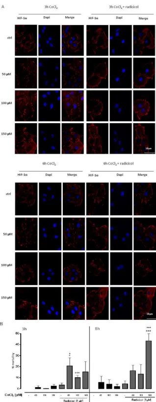
and validated in vitro tool to study the molecular mechanisms driven by hypoxia [Goldberg et al., 1988]. Cells respond to hypoxia by activation of the hypoxia-inducible factor HIF-1α, a transcription factor that modulates the expression of genes involved in angiogenesis, survival, metabolism and cell migration [Pennacchietti et al., 2003]. Immunofluorescence analisys confirm the induction of hypoxia in cells treated with
CoCl2 at 50-100-150µM for 3 or 6h. In fact, a marked increas of
HIF-1α levels were observed, both in absence and in presence of Radicicol (1 µM) (Figure 8A). Furthermore, by means of MTT we observed that in our experimental model cells mortality was lower than 50% (Figure 8B).
32
Figure 8: CoCl2 induces hypoxic state. CoCl2 (50-100-150µM) was administered for 3 and 6 h and H9c2 cells were stained with HIF-1α (red) and nucleus with DAPI (blue) and were determined by Immunofluorescence analysis. Where indicated, Radicicol (1µM) was administered 30 minutes before CoCl2. Scale bar, 10 µm. A representative of three experiments was shown. The panel B shows the cytotoxicity
measuring the cell mortality after treatment with MTT assay. Cell viability = absorbance of treated sample/absorbance of control. Cells mortality was calculated as:
% mortality = 100-[100x(OD treated/OD control)]. Results were analyzed by Student’s t test. *p<0.05 and ***p<0.001 vs non-treated;°p<0.05 and °°°p<0.001 vs
33
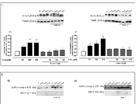
1.3.2 CoCl2 increased mitochondrial Cx43 expression
Western blot analysis showed a significant (p<0.05) increase of
mCx43 expression in CoCl2-treated H9c2 cells already evident
at 3h. Pre-treatment with Radicicol, significantly (p<0.05)
reduced CoCl2-induced mCx43 over-expression at either
experimental times (Figure 9A).
Figure 9: CoCl2 increases mitochondrial Cx43 expression. CoCl2 (50-100-150µM) was administered for 3 and 6 h in H9c2 cells and mCx43 expressions were detected by Western blot analysis. TOM20 protein expression was used as loading control (A).
Results are expressed as mean ± S.E.M. from at least three independent experiments each performed in duplicate. Data were analyzed by Student's t test. *p<0.05,
**p<0.005 vs non-treated; °p<0.05 vs cells treated with Radicicol alone. Representative Western blots of Na+/K+ATPase and Ox-Phos Complex II were used as markers, respectively, to demonstrate the purity of the mitochondrial extracts (B).
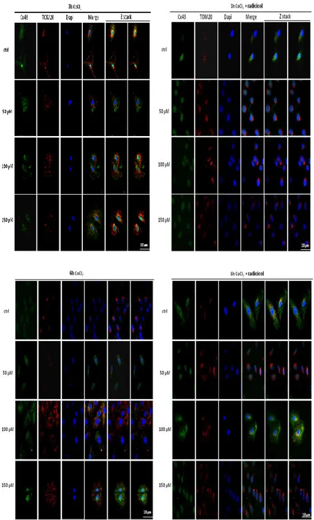
Data were confirmed by immunofluorescence analysis which
revealed that CoCl2 administration induced an increase in
mCx43 localization while Radicicol reduced this effect (Figure 10).
34
Figure 10: CoCl2 increases mitochondrial Cx43 localization. CoCl2 (50-100-150µM) was administered for 3 and 6 h and H9c2 cells and mitochondrial localization
of Cx43 was detected using immunofluorescence assay at confocal microscopy. Where indicated, Radicicol (1 µM) was administered 30 minutes before CoCl2. The
images were obtained throught software-generated reconstructions continuously acquiring overlapping (Z stack) images to get the whole cell image. Scale bar, 10 µm.
35
1.3.3 The inhibition of Cx43 translocation on mitochondria increased Mitochondrial Superoxide Production
The mitochondrial superoxide production was checked by
MitoSOX red. Flow cytometry analysis showed that the CoCl2
(50-100-150μM) treatment significantly (p<0.05) increased mitochondrial superoxide production at both experimental times.
Radicicol (1µM) pre-treatment, increases CoCl2-induced
superoxide production. This effect was significant (p<0.05) only at 6h, even if the amount of mitochondrial superoxide production in H9c2 pre-treated with Radicicol at 3h was
comparable to those of cells treated for 6h with CoCl2 alone
(Figure 11).
Figure 11: Radicicol increases mitochondrial ROS production in CoCl2 treated
H9c2 cells. Superoxide production by mitochondria was evaluated by means of the
probe MitoSOX Red in H9c2 cells by flow cytometry analysis. CoCl2 (50-100-150µM) was administered for 3 and 6 h. Where indicated, Radicicol (1 µM) was administered 30 minutes before CoCl2. Mitochondrial superoxide production was expressed as mean ± S.E.M. of percentage of MitoSOX positive cells of at least three
independent experiments each performed in duplicate. Data were analyzed by Student’s t test. *p<0.05 vs non-treated, °p<0.05 and p<0.005 vs cells treated with
36
1.3.4 CoCl2 induces calcium homeostasis alteration
Intracellular calcium concentrations was evaluated by means of
FURA 2-AM in Ca2+-free incubation medium (containing 0.5
mM EGTA). CoCl2-treated cells showed alteration in
intracellular Ca2+ levels. In fact, delta increase in intracellular
Ca2+ induced by Ionomycin in CoCl2-treated cells was
significantly (p<0.05) higher than control cells at all
experimental time points, indicating higher levels of Ca2+ in
cellular stores. Pharmacological inhibition of Cx43 translocation on mitochondria by Radicicol significantly affected intracellular
Ca2+ homeostasis. In fact, in Radicicol pre-treated cells, delta
increase in intracellular Ca2+ was significantly higher (p<0.05)
than cells treated with CoCl2 alone (Figure 12A). In order to
analyze the involvement of mitochondrial Ca2+ content, cells
were incubated with CoCl2 alone or pre-treated with Radicicol
and then the mitochondrial calcium depletory, carbonyl cyanide p-trifluoromethoxypyhenylhydrazone FCCP (50 nM), was
added. As reported in Figure 12B, in CoCl2-treated H9c2 cells
delta increase in intracellular Ca2+ induced by FCCP was higher
than control cells. In Radicicol pre-treated cells, delta increase in
intracellular Ca2+ was significantly (p<0.05) higher than cells
treated with CoCl2 alone, indicating higher levels of Ca2+ in
37
Figure 12: Radicicol increases mitochondrial calcium concentrations in CoCl2
treated H9c2 cells. CoCl2 (50-100-150µM) was administered for 3 and 6 h. Where
indicated, Radicicol (1 µM) was administered 30 minutes before CoCl2 . Intracellular
calcium content was evaluated on H9c2 cells in calcium -free medium by means of Ionomycin (1μM) (panel A). Effect of CoCl2 on mitochondrial calcium pool was
evaluated on H9c2 cells in calcium -free medium in presence of FCCP (50 nM) (panel B). Results are expressed as mean ± S.E.M. of delta (δ) increase of FURA 2 ratio
fluorescence (340/380nm) from at least three independent experiments each performed in duplicate. Data were analyzed by Student’s t test. *p<0.05 and **p<0.005 vs non-treated; °°p<0.005 and °°°p<0.001 vs cells treated with Radicicol
38
1.3.5 The inhibition of Cx43 Translocation Enhanced Mitochondrial Membrane Depolarization
CoCl2 (50-100-150μM) administration induced the opening of
the mitochondrial transition pore, as demonstrated by means of
TMRE. In fact, after 3 h of CoCl2 treatment, we observed an
increase of mitochondrial membrane depolarization. But it is interesting to note that inhibition of mCx43 translocation accelerates mitochondrial membrane depolarization. In fact, as reported in Figure 13, in Radicicol pre-treated cells at 3 h mitochondrial membrane depolarization values were higher than
those of cells treated for 3h with CoCl2 alone and comparable to
those observed in H9c2 treated with CoCl2 alone for 6h.
Figure 13: Radicicol increases CoCl2-induced mitochondrial membrane potential
collapse in H9c2 cells. CoCl2 (50-100-150µM) was administered for 3 and 6 h.
Where indicated, Radicicol (1 µM) was administered 30 minutes before CoCl2. The
mitochondrial membrane potential was evaluated by flow cytometry analysis with tetramethylrhodamina ethyl ester (TMRE), a cationic dye that gives a strong fluorescence signal. Results are expressed as mean ± S.E.M. of fluorescence intensity
of at least three independent experiments each performed in duplicate. Data were analyzed by Student’s t test. °°p<0.005 vs cells treated with Radicicol alone.
39
1.3.6 Inhibition of Cx43 translocation to mitochondria enhances CoCl2 -induced apoptotic response
Jung and co-workers [2004] report that one of the causes of cell death could be mitochondrial membrane permeabilization, therefore we analysed the apoptotic response in our
experimental conditions. Treatment with CoCl2 leads to an
increase of the cytocrome c release (Figure 14A) and in the number of aplodyploid nuclei (Figure 14B), as compared with untreated cells. In agreement with the activation of the mitochondrial apoptosis canonical way, Western blot analysis showed a significant and dose-dependent reduction of
procaspase 3 levels and an increase caspase 9 levels in CoCl2–
treated cells (Figure 14C). Radicicol pretreatment significantly (p<0.05) reduced cytocrome c release and the number of
hyplodyploid nuclei induced by CoCl2 at both experimental
40
Figure 14: Radicicol increased the trigger of the apoptotic pathway induced by CoCl2 in H9c2 cells. CoCl2 (50-100-150µM) was administered for 3 and 6 h, where indicated, Radicicol (1 µM) was administered 30 minutes before CoCl2.
Cytochrome c level into the cytosol was checked by flow cytometry analysis. Results are expressed as mean ± S.E.M. of percentage of cytochrome c positive
cells from at least three independent experiments each performed in duplicate (panel A). H9c2 were stained by propidium iodide and fluorescence of individual nuclei was measured by flow cytometry. Results are expressed as mean ± S.E.M. of percentage of hyplodiploid nuclei from at least three independent experiments each performed in duplicate (panel B). Caspase 9 and procaspase 3 expressions were detected by Western blot analysis; GAPDH protein expression was used as loading
control. Results are expressed as mean ± S.E.M from at least three independent experiments each performed in duplicate (panel C). Data were analyzed by Student’s t test. *p<0.05 and **p<0.005 vs non-treated; °p<0.05 vs cells treated
41
1.4 Discussion
GJ plaques are typically observed in the heart at theintercalated
disks of adjacent cardiomyocytes [Revel & Karnovsky, 1967; Severs, 1990] where they facilitate electrical current flow that coordinates cardiomyocyte contraction to sustain its pump
function [Severs et al., 2004]. In particular, Cx43 forms
hemichannels, which are predominantly closed in healthy myocardium [Goodenough et al, 2004; Krysko et al., 2005]. However, they can open in response to electrical and chemical triggers, most notably during ischemia and inflammatory conditions [Saez et al., 2005; Saez & Leybaert, 2014; Wang et al., 2012a; Wang et al., 2013a; Wang et al., 2013b]. A prolonged opening of Cx43 hemichannels during ischemia may lead to loss
of ionic gradients, excessive Ca2+ entry, cell swelling and
cellular damage. Apart from being present at the sarcolemma, Cx43 has been identified in mitochondria of cardiomyocytes, where it plays a pivotal role in mediating the cardioprotective effect of ischemic preconditioning [Boengler et al., 2005]. Rodriguez-Sinovas and co-workers [2006] demonstrated that ischemic preconditioning induces Cx43 translocation from cytosol to mitochondria with a mechanism that involves Hsp90 and TOM20. MCx43 is important in maintaining cellular mitochondrial homeostasis via regulating energy balance and calcium homeostasis [Wu et al., 2009], eliminating ROS [Jezek
42
et al., 2014], and regulating cellular apoptosis [Dando et al., 2013], thereby providing cellular protective effects.
Here, we evaluated the influence of mCx43 in in vitro hypoxia model. Hypoxia was induced by means of Cobalt Chloride
(CoCl2), a well-known hypoxia mimetic agent [Shi-Yun et al.,
2017], that is able to mimic the hypoxic response in many cellular process such as oxidative stress, dissipation of mitochondrial membrane potential, dysregulation of calcium homeostasis and consequent triggering of apoptosis [Wang et
al., 2013]. In our experimental model, CoCl2 is able to induce
hypoxia, like demonstrated by HIF-1α increased expression, without affecting cell viability in a drastic manner. In fact, we
chose to use CoCl2 doses and incubation times that could induce
hypoxia but did not give an excessive mortality because our goal was to observe early responses to induced damage.
As demonstrated by Western blot analysis, and then confirmed
by Immunofluorescence, in our experimental conditions CoCl2
induces an increase of mCx43 expression to mitochondria with a mechanism that involves the well designed Hsp90/TOM20 machinery system. In fact, blocking Cx43 interaction with
Hsp90, through Radicicol, significantly reduced the
mitochondrial import of Cx43. It is well known that the CoCl2
-induced hypoxia is characterized by dysregulation of calcium homeostasis, increase of reactive oxygen species, cell necrosis
43
and induction of pro-apoptotic signaling pathways [Wang et al., 2013]. MCx43 impacts on respiratory function [Boengler et al., 2012] and modulates the calcium overload, mitochondrial permeability transition and cell death [Srisakuldee et al., 2014].
Our data demonstrate that CoCl2 induces an increase in
intracellular Ca2+ levels in H9c2 cells, as revealed by means of
FURA 2-AM, instead inhibition of Cx43 translocation to
mitochondria significantly rises the intracellular levels of [Ca2+],
resulting in an increase of mitochondrial Ca2+ accumulation
induced by CoCl2.The role of mCx43 in calcium homeostasis in
hypoxic conditions is controversial. Gadicherla and co-workers indicate that mCx43 directly contribute to mitochondrial calcium entry/overload thus triggering cell injury/death pathways [2017]. On the other hand, other authors indicate that mCx43 exerts its cardioprotective effects by mitigating calcium overload, mitochondrial permeability transition and cell death [Srisakuldee et al., 2014]. This cardioprotective effect seems to be related to the increased mitochondrial calcium uptake and storage capacity during ischemia that helps to delay a rise in cytosolic calcium levels [Garcia-Dorado et al., 2012]. Our data
support this hypothesis. Indeed, Jung and co-workers [2001]
demonstrated that calcium levels are closely associated in mitochondrial ROS production and this suggests that chronic hypoxia-induced cell damages are related to mitochondrial
44
dysfunction. Here, we observed that CoCl2 induces an increase
in mitochondrial oxygen radical species, as demonstrated by means of MitoSOX. The inhibition of Cx43 translocation to mitochondria by Radicicol, significantly increases the effect of
CoCl2. It is well known that both mitochondrial calcium
overload and ROS production induce mitochondrial membrane depolarization [Gadicherla et., 2017]. In fact, data obtained by means of TMRE showed that in our experimental conditions
CoCl2-induced mitochondrial membrane depolarization was
more evident and faster in presence of the pharmacological inhibitor of Cx43 translocation on mitochondria. Hypoxia of cardiomyocytes cause cardiac dysfunction due to its triggering cell injury and apoptosis. In fact, as demonstrated by our data,
CoCl2 administration induces cytochrome c release, causing a
significant increase of apoptotic process, as shown by FACS
analysis with the use of propidium iodide. Furthermore, CoCl2
induces an increase of caspase 9 levels and a concomitant reduction of procaspase 3 levels. Use of Radicicol significantly
increases CoCl2-induced apoptotic signaling.
In conclusion, our data obtained suggest that mCx43 is essential for the cytoprotection stultify the apoptotic process induced by
chemical hypoxia and suggest thatthe presence of mCx43 is of
utmost importance for cardioprotective pathways being functional.
45
Chapter II
Involvement of Connexin 43 in Calcium
Impairment induced by Doxorubicin in a
short-term mouse model
2.1 Introduction
Anthracyclines, used alone or in combination with other chemotherapeutic agents, are a class of antitumour drugs widely used for the treatment of a variety of cancers, such as breast cancer, lymphoma and melanoma [Hrdinaet al., 2000]. Anthracycline agents are antibiotics isolated from soil microbe Streptomyces peucetius [Jain et al., 2017] and the commonly used include Doxorubicin (DOXO), Daunorubicin, and Epirubicin [Gewirtz et al., 1999]. DOXO is one of the most widely used and successful prescribed broad-spectrum chemotherapeutic [Smuder et al., 2013], but, unfortunately, its
usage is limited by the cumulative, dose-dependent
cardiomyopathy. In fact, one major side effect of this class of chemotherapeutic drugs is cardiotoxicity [Octavia et al., 2012], leading to dilated cardiomyopathy and heart failure [Wong et al., 2013]. The mechanisms of DOXO-induced cardiotoxicity are complicated and it is associated with the production of ROS and oxidation of lipids, DNA and proteins [Nordgren et al., 2014],
46
mitochondrial dysfunction, myofibril degeneration [Kavazis et al., 2016; Ghigo et al., 2016] and altered calcium handling by sarcoplasmic reticulum [Zhang et al., 2012]. Generation of ROS has been reported as the main mechanism that explains the pathophysiology of DOXO-induced cardiomyopathy [Fouad et al., 2011]. Similarly, other mediators such as Angiotensin II have also been found to play a critical role in the development of DOXO-induced cardiomyopathy [Toko et al., 2002]. The mechanisms of DOXO has been found to involve the activation of DOXO molecule into a more reactive semiquinone by mitochondrial complex I, resulting in increased oxidative stress [Carvalho et al., 2014]. However, Ren and co-workers [2012] reported that DOXO induces activation of both extrinsic and intrinsic apoptotic signaling pathways. Cardiac toxicity of DOXO was recently reported on electrocardiogram with a significant increase in heart rate, elevation of the ST segment, prolongation of the QT interval and an increase in T wave amplitude [Ammar et al., 2013]. Indeed, its disturbances in cardiac rhythm [Ferrans et al., 1997], changes in blood pressure [Medrano et al., 2001], reduction of ejection fraction and contractile function [Chatterjee et al., 2010] and cardiac dilation [Takemura et al., 2007], are often not clinically evident until the late stages.
47
Anthracycline cardiotoxicity is classified as acute and chronic. Acute cardiotoxicity occurs during or soon after initiation of therapy. This is usually transient and self-limiting with a myopericarditis like picture, non-specific repolarization changes on ECG, dysrhythmias, troponin elevation, and transient LV
dysfunction [Dazzi et al., 2001]. Chronic cardiotoxicity is
arbitrarily classified as type 1 or early onset (typically detected within one year of completion of chemotherapy) and type 2 or late-onset (usually detected after the first year with an unlimited time frame of up to decades after completion of chemotherapy) [Lipshultz et al., 1991; Lipshultz et al., 2005]. Majority of the patients develop chronic cardiotoxicity within the first year of completing therapy [Cardinale et al., 2015]. The total lifetime cumulative dose of anthracycline is the most important determinant of anthracycline cardiotoxicity [Von Hoff et al., 1979]. DOXO-induced cardiotoxicity is mainly related to accumulation of the repetitive doses required by the treatments,
in fact total DOXO concentration cannot exceed 500 mg/m2.
Recent evidence indicates that the damage caused by anthracyclines on cardiomyocytes is an early event, already evident after a single administration [Menna et al., 2008]. But until now most of the studies published report only the effects of long-term DOXO-administration [Zhang et al., 2014; O’ Connell et al., 2017].
48
DOXO-induced cardiomyopathy is characterized by abnormal calcium homeostasis. Recently it has been demonstrated that DOXO administration is able to induce calcium dysregulation and Cx43 re-arrangement in a rat cardiomyoblast cell line already evident after a short-term administration [Pecoraro et al., 2015 b]. Cx43 is functionally associated with calcium and Cx43 remodelling might be responsible for intracellular calcium overloading induced by DOXO, thus resulting in ischemic arrhythmia [Kristia’n et al., 1998]. On the basis of these data, we analyzed the effect of DOXO-administration on Cx43 expression and localization in a short-time mouse model of DOXO-induced cardiotoxicity.
49
2.2 Materials and Methods
2.2.1 Materials
DOXO was purchased from Baxter manufacturing S.p.a. (Officina di Sesto Fiorentino, Florence, Italy). Where not indicated otherwise, antibodies used were purchased from Santa Cruz Biotechnology (DBA, Milan, Italy), and all other products were purchased from Sigma (Milan, Italy).
2.2.2 Animals
Six week old female C57BL/6j (weighting 20–22 g) were purchased from Charles River (Lecco, Italy). All experimental procedures that involve animals have been conducted in agreement with the Italian and European Community Council for Animal Care (DL. no. 26/2014 protocol number of Ministerial approvation DGSAF 13788-A 02/07/2015) and in accordance with the guidelines of the Guide for the Care and Use of Laboratory Animals of the National Institutes of Health.
2.2.3 Experimental Protocols
C57BL/6j mice were randomly divided in three groups (n = 6 for each experimental group). The doses used were 2 and 10 mg/kg [Xujie et al., 2012; Wided et al., 2014] and they corresponded to 6.142 and 30.8 mg/m2 respectively [Freireich et al., 1997].
50
First Group: received one DOXO administration (2 or 10 mg/kg i.p.) and were sacrificed 24 h after the treatment;
Second Group: received two DOXO administrations (2 or 10 mg/kg i.p.) once a day for two days and were sacrificed three days after the first administration;
Third Group: received three DOXO administrations (2 or 10 mg/kg i.p.) once a day for three days and were sacrificed seven days after the first administration.
Mice that received saline were used as control group. Cardiac function was monitored echocardiographically at baseline and before sacrifice. At the end of each experimental time, heart samples were collected and prepared for molecular biological analyses.
Figure 15: Timetable. C57BL/6j female mice were randomly divided into three groups The 1st group received a single administration of DOXO (2 mg/Kg and 10 mg/Kg; i.p.) and was sacrificed after 24h; the 2nd group received two administrations
of DOXO (2 mg/Kg and 10 mg/Kg; i.p.) once every other day and was sacrificed 3 days after the first injection; the 3rd group received three administration of DOXO (2
mg/Kg and 10 mg/Kg; i.p.) once every other day and was sacrificed 7 days after the first injection. Control group received saline solution. At the end of experimental time, mice were sacrificed and the heart was removed and frozen for biochemical
51
2.2.4 Echocardiogram
Mice were lightly anesthetized with 1–1.5% of isoflurane in oxygen until the heart rate stabilized to 400–500 beats per minute. Echocardiography was performed using VEVO (VisualSonic, Toronto, (Canada) instrument. Ejection fraction (EF), Fractional shortening (FS), Left Ventricular Diastolic-Diameter (LVEDD), and Left Ventricular End-Systolic Diameter (LVESD) were calculated using the VEVO analysis software (Toronto, Canada).
2.2.5 Histological staining
For histological analyses, hearts were embedded in OCT medium (Bio-Optica, Milan, Italy). Sections (7 μm) were fixed with acetone and stained either hematoxylin and eosin (H&E staining) to evaluate the cardiac structure following the administration of DOXO. Coverslips were finally mounted in mounting medium and all the photomicrographs were taken at 20× magnification under the FluorescenceMicroscope (Axio Vision software).
2.2.6 Protein Extraction and Western Blot Analysis
Total proteins were extracted by homogenization of hearts with a dounce potter in lysis buffer (TRIS-HCl 50 mM, NaCl 500 mM, protease inhibitors, PMSF 0.25 μM, NaF 50 mM, Na3VO4
52
0.2 mM). Protein concentrations were determined with the Bio-Rad protein assay (BIO-RAD, Milan, Italy). Equal amounts of protein (50 µg) were loaded into an acrylamide gel and separated by SDS-PAGE under denaturating conditions. Blots were incubated with primary antibody anti-Cx43 (Sigma,
1:8000), anti-pCx43 phosphorylate on Ser368 (Santa Cruz,
1:250), anti-sarco/endoplasmic reticulum Ca2+-ATPase
(SERCAII; Santa Cruz, 1:250), anti-phospholamban (PLB;
Santa Cruz, 1:200), or anti-glyceraldehyde 3-phosphate
dehydrogenase (GAPDH; Santa Cruz, 1:1000, used as loading
control) overnight at 4 °C. After incubation period with the primary antibodies, blots were washed in PBS 0.1% Tween. The appropriate secondary antibody rabbit, mouse, or anti-goat (each diluted 1:4000) was added and allowed in incubation for 1 h at room temperature. Immunoreactive protein bands were
detected by chemiluminescence using enhanced
chemiluminescence reagents (ECL) in LAS 4000 (GE Healthcare, Björkgatan, Sweden).
2.2.7 Mitochondrial Protein Extraction and Western Blot Analysis for Mitochondrial Cx43 and pCx43
Mitochondrial proteins were extracted from homogenized hearts
with a dounce potter in lysis buffer (sucrose 250 mM, K+ Hepes
pH 7.5 20 mM, KCl 10 mM, MgCl2 1.5 mM, EDTA 0.1 mM,
53
mM, PMSF 100 μM, DTT 1 mM, digitonin 0.025%) as previously reported [Pecoraro et al., 2015 b; Sardão et al., 2009]. The protein yield was quantified with the Bio-Rad protein assay (BIO-RAD, Milan, Italy). Western blot analysis for Cx43 or pCx43 was performed as described above. Primary antibody anti-TOM20 (Santa Cruz) was used as loading control. In order to verify the purity of mitochondrial protein extraction, a Western blot analysis was performed to evaluate the presence of proteins expressed only in the mitochondria (ox-Phos Complex II, Abcam, 1:7000) and the absence of proteins expressed in
other cellular compartments (Na+/K+ ATPase, Abcam, 1:3000)
[Boengler et al., 2005].
2.2.8 Primary Cardiomyocytes Isolation and Measurement of Intracellular Ca2+ Signaling
Intracellular Ca2+ concentrations were measured in primary
cardiomyocytes, isolated from hearts of DOXO-treated mice and
control mice. Hearts were washed with HBSS 0.1 mM Ca2+ (140
mM NaCl, 5.4 mM KCl, 0.44 mM KH2PO4, 0.42 mM Na2H
PO4, 4.17 mM NaHCO3, 26 mM CaNa-EDTA, 0.10 mM
CaCl2·H2O, 5.0 mM HEPES and 5.5 mM dextrose). Next, hearts
were cut in 1–2 mm sections and incubated at 37 °C in HBSS
0.1 mM Ca2+ containing albumin 10 mg/mL, tryspin inhibitor 1
mg/mL, taurine 5 mM, dithiothreitol 0.4 mg/mL, collagenase II 0.6 mg/mL, and papain 0.6 mg/mL for 75 min. After the
![Figure 1: The image shows a GJ and its main element.[LadyofHats et al., 2006]](https://thumb-eu.123doks.com/thumbv2/123dokorg/7205172.76016/12.892.218.676.508.863/figure-image-shows-gj-main-element-ladyofhats-et.webp)