UNIVERSITÀ DEGLI STUDI DI SALERNO
Dipartimento di Farmacia
Dottorato di ricerca
in
Scienze Farmaceutiche
Ciclo XIV—Anno di discussione 2016
Coordinatore: Chiar.mo Prof. Gianluca Sbardella
INTEGRATED METABOLOMICS
APPROACHES FOR BERRY FRUIT USED IN
NUTRACEUTICAL FORMULATIONS
settore scientifico disciplinare di afferenza
:
BIO/15
Dottorando Tutore
Preface
My PhD in Pharmaceutical Science at the Department of Pharmacy of Salerno University was started in January 2013 under the supervision of Prof. Paola Montoro.
Targeted and untargeted metabolomics approaches were applied to investigate
the effect of environment, genotype, or both on variation of many metabolites in some berry fruits.
In particular the species under investigation during these three years of PhD course were: Fragaria ananassa, Fragaria vesca, Morus alba, Morus nigra and Myrtus communis. All these species are characterized by the production of small fruit, and all of them are plant species that can be used for the formulation of plant food supplements, in fact they are reported into the official list of Italian legislation (DM July 9, 2012-G.U. 21-7-2012, serie generale n. 169, and update on March 27, 2014). Some of them are recognized as Italian traditional food products, like Fragaria vesca, typical of Campania region and Myrtus communis, endemic of Sardinia region.
Furthermore, to improve my knowledge on metabolomics software, I spent a period of research activity at the Department of Bioscience of Wageningen University, Netherlands, under the supervision of Dr. Ric de Vos, where I focused my reaserch on Morus alba and Morus nigra.
During the three years course of PhD, liquid chromatography coupled to high resolution mass spectrometry and in conjunction with Multivariate Data Analysis were applied to investigate the metabolic composition of different berry species; with these approaches we could confirm that metabolomics represent a useful tool to evaluate the contribution of environmental and genetic factors to the differences in metabolite composition or content of berry fruit. Moreover, with the characterization of polar compounds we could also
Chapter 1: Metabolomics and “Berry” fruit
1 Introduction ... 1
1.2 Common Berry found in Italian regions ... 2
1.3 Foodomics ... 4
1.4 Metabolomics ... 4
1.4.1 Setup of metabolomics experiments ... 5
1.4.2 Metabolite analysis ... 8
1.4.3 Preprocessing and statistical analysis of plant metabolomics data 14 1.4.4 Statistical analysis: Principal Component Analysis and Partial Least Square ... 15
1.5 References ... 17
Chapter 2: Metabolomics approaches used for discrimination of different cultural system 2.1 Integrated Mass Spectrometric and Multivariate Data Analysis approach for the discrimination of organic and conventional strawberry (Fragaria ananassa Duch.) Crops 2.1 Introduction ... 25
2.2 Results and discussion ... 28
2.3 Conclusion ... 40
2.4 Materials and methods ... 42
2.5 References ... 46
Chapter 3: Metabolomics approaches used for typization of Berry fruit 3.1 Characterisation of Fragaria vesca fruit from Italy following a metabolomics approach through integrated Mass Spectrometry techniques 3.1.1 Introduction ... 55
3.1.2Results and discussion ... 57
3.1.3 Conclusion ... 71
3.1.4 Materials and Methods ... 72
3.1.5 References ... 77
3.2 Targeted and Untargeted Mass Spectrometric approaches in discrimination between Myrtus communis cultivars from Sardinia region 3.2.1 Introduction ... 81
3.2.2Results and discussion ... 83
3.2.3 Conclusion ... 95
3.3 Metabolic Profiling of different parts of myrtle's berry from Sardinia
3.3.1 Introduction ... 107
3.3.2Results and discussion ... 108
3.3.3 Conclusion ... 122
3.3.4 Materials and Methods ... 123
3.3.5 References ... 126
Chapter 4: Metabolomics approaches used for identification of bioactive metabolites 4.1 Combined use of α- glucosidase inhibitory activity and UHPLC-High Resolution mass spectrometry for the identification of active metabolites in Morus alba and Morus nigra extracts 4.1 Introduction ... 133
4.2 Results and discussion ... 135
4.3 Conclusion ... 150
4.4 Materials and Methods ... 151
4.5 References ... 156
1
Metabolomics and “Berry” fruit 1 Introduction
The term "berry fruit" generally refers to any small fruit that can be eaten whole. Botanically, a berry is the most common type of fleshy fruit in which the entire ovary wall ripens into an edible pericarp. They may have one or more carpels with a thin covering and fleshy interiors, in which the seeds are usually embedded. Many fruits, commonly known as berries, are not actual berries by the scientific definition, but fall into one of these categories, for example blackberry, mulberry and strawberry1. Berries are widely recognized for their several health-promoting properties including a reduced risk of cancer, cardiovascular disease, and other chronic disease. These properties may be due to the presence of polyphenols, particularly anthocyanins, which are a class of flavonoids that produce the bright red, purple, and blue pigments seen in many berries and are among the most consumed flavonoids in the american diet2,3.Polyphenols are a class of secondary metabolites found throughout the plant kingdom and can be categorized into a variety of families, for example phenolic acids, flavonoids, anthocyanins, stilbenes, coumarins, and tannins. In plants, phenols are essential to plant pigment, growth, reproduction, resistance to pathogens, and mediation of solar radiation and photosynthesis byproducts3.Moreover, when phenolic compounds are introduced to biological systems, they show antiallergic, antianxiety, anticarcinogenic, anti-inflammatory, antioxidant, antiproliferative, antitumorigenic, and antiviral properties3. Howewer systemic bioactivities ascribed to these compounds seems to be largely mediated by their metabolites, in fact, a large proportion of potentially protective berry
2
polyphenols are unable to enter the circulation and influence cellular interaction4. Therefore most polyphenols from berries remain in the gastrointestinal tract and pass through to the large intestine where they are subject to biotransformation by colonic microbiota5. Those compounds may also modulate nutrient availability through the inhibition of digestive enzymes involved in lipid and starch breakdown, which could influence obesity and blood glucose control5, and due to their high concentration and qualitative diversity in berry fruit, these fruit are increasingly often referred to as natural functional food.
Due to climatic conditions, fresh berries are generally available several months a year while some of the harvested fruits are processed to juice, fruit beverages, frozen product wine, jam and jelly. Studies report that constitutional and sensory qualities of berries are affected by many factors such as cultivars, geographic region, storage conditions, ripeness, climate and others may affect the concentration of phenolic compounds and in the antioxidant capacity6,7.
Furthermore, there has been a growing trend in the use of berry extracts as ingredients in functional food and dietary supplements, and omics ―approaches‖ such as genomics, proteomics and metabolomics, are increasing the possibility of carrying out better strategies to improve crops (including berry) for fruit quality enriched in bioactive compounds with nutraceutical properties8.
1.2 Common Berry found in Italian regions
Several plant species characterized by the production of berry fruit are cultivated or spontaneous in Italy. Most of these species are present in the list
3
of plant species that can be used for the formulation of plant food supplements (DM July 9, 2012)9.
They are: blackberry (Rubus ulmifolius L.), highbush blueberry (Vaccinium
corymbosum L.), black currant (Ribes nigrum L.), white currant (Ribes rubrum
L.), gooseberry (Ribes grossularia L.) black mulberry (Morus nigra L.), white mulberry (Morus alba L.), raspberry (Rubus idaeus L.), strawberry (Fragaria
× ananassa Duch.), wild strawberry (Fragaria vesca) and myrtle berry
(Myrtus communis).
Some of these species are endemic of Italian regions like Fragaria vesca, endemic and typical of Campania region in particular from "Alburni" and "Alto Sele" areas. In this area, the wild strawberry fruit are recognized as a traditional food product under the name ―Fragolina degli Alburni‖. The fruits are used for the production of liqueur and sweets. In medicine, fruits and leaves of this specie are used in the formulation of food supplements for their antioxidant activities. Another endemic species is Myrtus communis, that is a pleasant annual shrub endemic of Sardinia, commonly used for the production of myrtle liqueur. Fruit and leaves of Myrtus communis are also used in the preparation of food supplements used for fluidity of bronchial secretions and for balsamic effect. White mulberry (Morus alba) and black mulberry (Morus
nigra) are deciduous tree originating from China and Japan, spread in America
and Europe for the silkworm breeding. In south Italy trees of Morus alba and
Morus nigra are common spread and are used for the production of juice or
jam. Fruit and leaves are used in the preparation of food supplements for their several properties like: antioxidant, antihyperglicemic and fluidity of bronchial secretions.
4 1.3 Foodomics
Foodomics is defined as a discipline that studies the food and nutrition domains through the application of -omics technologies to characterize and demonstrate the beneficial effects on human health of food ingredients. Foodomics includes nutritional genomics sub-reaserch areas, such as nutrigenomics, nutrigenetics, proteomics and metabolomics 10.
In the last two decades, large investments were made to develop analytical approaches to analyze the different cell products, such as those from gene expression (transcripts), proteins, and metabolites. All of these so-called ‘omics approaches, including genomics, transcriptomics, proteomics, and metabolomics, are considered important tools to be applied and utilized to understand the biology of an organism and its response to environmental stimuli or genetic perturbation10.
1.4 Metabolomics
Metabolomics as follow-on from transcriptomics and proteomics was a term coined at the end of the 1990s11. the concept entailed the analyses of the metabolite composition of biological material, aimed to be fully complementarily with the others potentially unbiased or non targeted "-omics" approaches12.
Metabolome can be defined as the full set of endogenous or exogenous low molecular weight metabolic entities of approximately <1,000 Da (metabolites).
Metabolites are, in general, the final downstream products of the genome, and reflect most closely the operation of the biological system, its phenotype. One of the main challenges in metabolomics is to face the complexity of any metabolome, usually composed by a huge number of compounds of very
5
diverse chemical and physical properties (sugar, amines, aminoacids, peptides, organic acid nucleic acid or steroids). Sample preparation is especially important in metabolomics, because the procedure used for metabolite extraction has to be robust and highly reproducible. Sample preparation will depend on the sample type and the targeted metabolites of interest (fingerprint or profiling approach)11.
Metabolomics has proven to be a valuable tool for the comprehensive profiling of plant derived samples for the study of plant systems and natural products research, especially when combined with chemometric data analysis approaches13.
From the methodological point of view, there are basically only two different approaches used in any metabolomics study: ―targeted analysis‖ and ―metabolite profiling‖. Targeted analysis is restricted to quantitative analysis of a class of compounds that are related to a specific pathway or to intersecting pathways. Targeted analysis is very useful for the study of the primary effect of a genetic alteration, and the analytical procedures must include identification and absolute quantification of the selected metabolites in the sample. Metabolite profiling (or, sometimes, metabolic profiling), on the other hand, involves rapid analysis, often not quantitative, of a large number of different metabolites with the objective to identify a specific metabolite profile that characterizes a given sample14.
1.4.1 Setup of metabolomics experiments
The analysis of data obtained from metabolomics experiments is complex. Different platforms and experimental designs produce data in various format and units, making the study of metabolomics data difficult. Providing full information regarding the experimental design a sample preparation route for
6
the development of an analytical methodology and an approach to data analysis that includes all such information and is simultaneously easy to handle is challenging task15. However, a universally applicable standard procedure has not yet been established, and single publications only include suggestions that will have to be refined in the future16.
Sample preparation
The sample preparation is an important step in metabolomics analysis; in fact it can direct influence the results of the analysis. Basically it can be divided in the following four steps: 1) harvesting of the plant materials, 2) processing before extraction (for example drying), 3) extraction and 4) pre-analytical sample preparation. The execution of all these steps may not be necessary and can be avoided in some cases depending on the aim of research and the nature and properties of the analytes of interest15.
Harvesting
Metabolites are influenced by environmental factor. Seasonality, developmental stage and age, circadian rhythm, temperature, water availability, UV radiation, altitude, atmospheric composition, soil nutrients, and tissue damage may induce qualitative and or quantitative variations in the metabolites composition17. Moreover a plant metabolome may vary depending on the time or place of harvest. The influence of environmental conditions is extremely relevant for plant material collected in the natural habitat of plants; however, it may be considered minor for plants cultivated in a greenhouse or in cell culture plants16,18. Accurate timing is an important factor that should be considered because the levels of metabolites (primary as well as secondary metabolites) vary throughout the day, due to the different light exposition of
7
the plant19,20. Moreover, the developmental stage of the plant at the time of harvest affects the metabolite profile16,21. Additionally, different plant organs also show significantly different metabolite profiles. For this reason the separate analysis of different plant organs with separation by tissue age is fundamental to obtaining reliable results15.
Processing before extraction
Changes in the plant‘s metabolism occur from seconds up to a few minutes; for this reason, harvesting must be performed as quickly as possible and metabolism should be stopped immediately after harvesting by freezing of plant material in liquid nitrogen16,21. An appropriate technique would be freeze-drying, which is based on lyophilization and cryodesiccation. In this way, water is removed and the enzymatic activity is reduced. In addition to freezing or freeze-drying, the samples may also be subjected to a drying process by an oven, air or by microwave heating, which destroy enzyme activities 22. The selection of a drying method should always consider the molecules to be targeted for analysis because some methods are incompatible with certain metabolites, such as volatile and thermo-unstable compounds, among others. Further processing before extraction can include the pulverization of the plant material into small particles to improve the extraction process15.
Extraction
Extraction methods should be as simple and as fast as possible23. There is no current method for the extraction of all metabolites18,21. Plant tissue metabolites are highly diverse. Several critical factors must be considered during extraction, such as the ratio of solvent and plant material, solvent
8
characteristics, the time of extraction, the temperature and the choice of an appropriate method for the desired goals. The choice of a solvent is extremely important for the achievement of reliable results because it needs to be adequate for the metabolites targeted for extraction and for the analytical method15. Among solvent extraction methods, methanol and solvent mixtures that contain methanol are employed the most; furthermore acidified solution are common, and chloroform is often used for lipophilic compounds19,23,24. Several techniques have been applied to accelerate solvent extraction, such as ultrasonic extraction, microwave and pressurized solvent extraction 19,24. Ultrasonic extraction is also rapid and simple and is one of the most commonly used methods for solvent extraction at present26.
1.4.2 Metabolite analysis
Two analytical platforms are currently used for metabolomics analysis: Mass Spectrometry (MS) and Nuclear Magnetic Resonance (NMR). These techniques either stand alone or combined with separation techniques (typically, LC-NMR, GC-MS, LC-MS and CE-MS), can produce complementary analytical information to attain more extensive metabolome coverage27. MS and NMR-based technologies are both complementary and, therefore often used in parallel in metabolomics research. Compared to NMR, MS is a more sensitive technique; also MS coupled to GC, LC or CE allows higher resolution and sensitivity for low abundance metabolites. The use of high and ultra-high resolution analyzers (namely, TOF, FTMS, Orbitrap®) is essential to obtain accurate mass measurement for the determination of elemental compositions of metabolites and to carry out their tentative identification with database28. On the other hand, MS/MS or MSn experiments, especially when product ions are analyzed at high resolution (with Q-TOF,
9
TOF-TOF, or LTQ-Orbitrap®) provide additional structural information for the identification of metabolites29.
ESI source: ion generation and ion suppression
The ionization source that is generally used in metabolomics studies is electrospray ionization (ESI). Notwithstanding its widespread use, it is still a young technique and for this reason, fundamental studies about reaction in the ionization source, as well as fragmentation patterns (MS/MS or MSn) are still being investigated15.
In an initial step, the solution containing the analyte crosses a metallic capillary where an electric potential is applied (between 2 to 4.5 kV). This potential promotes a migration of charges to the interface of the capillary with the solution, which results in an electric double layer, and thus droplets with charged surfaces are formed. The process of solvent evaporation starts within a chamber with a slight reduction of pressure under a nebulizer gas. With the reduction of droplet sizes, an approximation of the charges occurs, and consequently, the electrostatic repulsive forces are increased, which leads to a decrease in the surface tension of the droplets until rupture, a process denoted as ―Coulomb explosion‖. This rupture results in the liberation of ions into the gas phase by a spray of charged particles, or in other words, an electrolytic current29. A positive ionization mode is used in a large majority of metabolomics studies and the addition of H+ and Na+ ions must be balanced. In metabolomics studies using the hyphenated system LC-ESI-MS, the co-elution of substances with a large difference in the ionisation potential can lead to the complete suppression of an ion, leading to an incorrect result indicating the non-appearance of a metabolite in question15.
10
Furthermore, it is also known that the use of hyphenated systems as well as the use of direct infusion mass spectrometry, still present several limitations, such as ion suppression, radical species formation and cross reactions15.
MALDI source: Ion generation and matrix effects
MALDI (matrix-assisted laser desorption/ionization) was introduced to the market with great success, principally in proteomic research. MALDI can be explained in the following three basic steps: 1) formation of a solid solution in which analytes are completely isolated from each other through dilution in an homogenous matrix for subsequent desorption; 2) excitation of the matrix, in this step a laser beam is applied to the matrix surface. Photons from the laser are absorbed by the chromophore parts of the matrix molecules, which causes rapid vibrational excitation followed by disintegration. Cluster of analyte molecules surrounded by the matrix and salt ions are ejected at the solution surface, producing the plume and subsequent evaporation of the matrix molecules to expose free analyte molecules to the gas phase; 3) ionization of the analyte, in the desorbed matrix, cloud ionisation (plume) occurs via proton or cation transference from the photo-activated matrix molecules, which leads to the formation of characteristic ions [M+X]+ (X= H, Na, K etc). In these steps, the process exhibits limitations for the analysis of organic micromolecules. This limitation was caused by the matrix, which is cationised or anionised after desorbition and extracted into the analyser. For this reason the mass range between 0 and 500 m/z may have shown numerous matrix signals, complicating any analysis of low molecular weight compounds31,32. MALDI is a promising techniques for metabolomics studies because of its high sensitivity, high analysis scan speed and lower contaminant influence on
11
ionisation15 but generally is used for proteomic analysis in which the molecular weight of the analytes of interest is higher.
MS/MS and MSn in metabolomics
The use of fragmentation spectra can help in the structural identification of metabolites. A series of databases has been established and with each day the information increases. An additional step is necessary for MS/MS analyses, namely, the isolation of an ion, which as an increased internal energy after receiving some type of activation that allows the beginning of the fragmentation process. Collision induced dissociation (CID) is without a doubt the most frequently used process33,34. Kinetic energy transference occurs through collision with the gas, and the ion is transformed by internal energy, inducing the fragmentation. MS/MS or MSn is extremely important for metabolomics studies, but for the interpretation of data, a detailed observation of the ions that show abnormal behavior is necessary because the reason might merely be the effects of the internal energy quantity of the analysed ion15.
Hyphenated mass spectrometry methods: GC/LC-MS
Prior to MS analysis, metabolites, are separated by a separation-based method, namely gas chromatography (GC), liquid chromatography (LC) or capillary electrophoresis (CE). In GC and LC, compounds are separated by exploiting different interaction of the substances with the stationary phase. However in CE, the separation is based on the size to charge ratio of the ionic molecules35. The choice of separation technique is made according to the type of molecules present in the target sample. GC is suitable for hydrophobic, low molecular weight compounds, that must be heat stable and volatile35. The ionization method that is most commonly used in combination with GC is electron
12
ionisation (EI), that is an hard ionization36; while in LC-MS soft ionization is most commonly used, such as electrospray ionization, where it is used to analyse a wide array of metabolites from sugar to fatty acids. The separation based MS methods are higly sensitive and the costs are relatively low compared to NMR methods20,35. For GC-MS new columns, such as narrow bore columns, were developed to reduce the analysis times to approximately 1 and 10 min, namely the Ultra Fast-GC and Fast-GC 37,38. In LC-MS, monolitich columns and fused core particle (2.7 µm) packing columns have been used to reduce the time and increase the resolution. The stationary phase of C18 (reverse phase) is most frequently used in plant metabolomics, despite new phases such as HILIC39,40,41.
Direct injection MS (DIMS)
Direct injection MS (DIMS) is an analysis performed by directly injecting a sample into the ionization souce. Soft ionization techniques are preferably applied in metabolomics studies because they cause less fragmentation than hard ionization techniques, resulting in a smaller number of signals and, consequently, less complex spectra35. The ions are generated in the ionization source, and they are subsequently separated in the analyser for the detection, so a mass spectrum is produced. The analysers can be of two types, namely, low or high resolution, and the latter enables high mass accuracy. Despite the high cost of the high resolution equipment, it is possible to obtain high repeatability, peak annotation and more precise data alignment15. Direct MS analysis of foods or food extracts has been demonstrated to be a useful approach in metabolic fingerprinting when rapid classification of food- sample types or rapid screening of food adulteration is wanted42.
13
The main disadvantages of the DIMS are the impossibility of distinguishing between chemical isomers and the lower quantity of chemical information that is obtained relative to hyphenated MS methods. A possible way to circumvent the chemical isomer problem is to perform MSn analyses. By performing MSn analyses, previous chromatographic separations can be avoided if information obtained by the fragmentation patterns can be used to differentiate the isomers. Furthermore, chemical information can also be increased by performing analyses in negative as well as in positive ion modes24. The type of mass analyser and the ionization method have a considerable influence on the resulting mass accuracy, resolution and detection limit, and advantages and disadvantages depend higly upon the chosen instrument type15.
Ambient MS
The main characteristic of ambient MS (AMS) is that it allows direct analysis of samples in open air with little or no sample preparation. For this reason MALDI and API techniques, such as ESI and APCI, are not considered to belong to this group. Since the usually still require sample preparation42. The development of AMS was initiated with the introduction of desorption electrospray ionization (DESI) by Cooks in 200443.
Almost 30 ambient sampling/ionization approaches were involved in MS analysis. Among them DESI and direct analysis in real time (DART) were the two most prevalent techniques. DESI shares the advantages of the matrix-free DIOS (laser desorption/ionization MS on porous silicon) and the advantageous production of multiply charged ions of ESI. DESI-MS has also be demonstrated to be promising tool in food safety control44. DESI was followed by DART in 200545. DART can be considered an API (Atmospheric Pressure Chemical Ionization)-related techniques based on the thermo-desorption of
14
condensed phase analytes by a (distal) plasma discharge in a heated gas stream. As other ambient ionization techniques DART is undergoing rapid development and it is beginning to deliver its potential in metabolomics45. Other techniques used for identification of metabolites are: Laser ablation electrospray ionisation (LAESI)47,48 and paperspray49.
1.4.3 Preprocessing and statistical analysis of plant metabolomics data Metabolomics experiments produce an enormous amount of data. Mathematical and statistical skills are essential for extracting as much information as possible from metabolomics data26,50. Data preprocessing is an important step in metabolomics data analysis, and it can influence data interpretation. Preprocessing includes noise filtering, normalization, peak detection, alignment and identification. Noise filtering is designed to separate compound signals from background signals originating from chemical or instrumental interference. Normalization is applied to correct the systematic variation and for direct comparison of different samples15. In different samples, slightly different m/z values may be obtained for the same compound. To compare different samples, alignment must be performed, and depending on whether individual metabolites are identified or not, finding out the chemical identity of a compound may also be necessary49. The metabolomics community is continuously growing, and new and exciting analytical strategies are steadily being developed to increase the amount of information extracted from mass spectrometric data sets. Such strategies include the development of software for the facilitation of data exchange52. The analysis of the large data sets generated by LC-MS requires data processing tools such as those based on multivariate data analysis. These techniques are robust to noise and missing data and enable one to deal with correlated variables;
15
different software such as the commercially available Markerlynx (Micromass Ltd., Manchester, U.K.) or freeware such as metAlign (Plant Reaserch International, Wageningen, The Netherlands) MzMine or XCMS perform the automatic extraction, alignment, and retention time correction of chromatographic peaks within individual mass to charge value using different algorithms.
These software generate a data table constituted by N observations (columns) and K variables (rows); the data table obtained must be analyzed in order to have information and to interpreted the results in a comprehensible way, for instance as a graph, and the MultiVariate Data Analysis (MVDA) is used for this purpose.53
1.4.4 Statistical analysis: Principal Component Analysis and Partial Least Square
Two are the multivariariate projection methods used for extracting information from a large or small table of data: Principal Component Analysis (PCA) and Partial Least Square (PLS).
Principal Component Analysis (PCA)
The PCA is an unsupervised method. The starting point for PCA is a matrix of data with N rows (observations) and K columns (variables). The observation can be analytical samples, chemical compounds or reactions, process time points of a continuous process, and so on. In order to characterize the properties of the observations, one measures variables. These variables may be of spectral origin (NIR, NMR, IR, UV, X-ray), chromatographic origin (HPLC, GC, TLC) or they may be measurements from sensors in a process (temperature, flows, pressures, curves, etc.)53.
16
The most important use of PCA is to represent the multivariate data table as a low-dimensional plane, usually consisting of 2 to 5 dimensions, such that an overview of the data is obtained. This overview may reveal groups of observation, trends, and outliers. This overview also uncovers the relationships between observations and variables, and among the variables themselves53. Principal component analysis summarizes the variation of a data matrix X, as product of two low-dimensional matrices, T and P, which can be easily overviewed and used. The data in the multi-dimensional space, defined by the measured variables, is modeled as a plane or hyperplane, the axes of which are called the principal components. Each principal component can be displayed graphically and may often be interpreted according to chemical, technical and/or biological knowledge53.
Partial Least Square (PLS)
PLS is a regression extension of PCA which is used to connect the information in two blocks of variables X and Y to each other. It derives its usefulness from its ability to analyze data with many noisy, collinear, and even incomplete variables in both X and Y. As in PCA, each observation can be represented graphically, however, the big difference in PLS is that each row of a data table corresponds to two points rather than one, one in the X-space and one in the Y-space.
PCA is a maximum variance least squares projection of X, whereas PLS is a maximum covariance model of the relationship between X and Y53.
17 1.5 References
1
Mikulic-Petkovsek M., Slatnar A., Stampar F., Veberic R. HPLC-MSn identification and quantification of flavonol glycosides in 28 wild and cultivated berry species. Food Chemistry 2012, 135, 2138-2146
2
Mattivi F., Vrhovsek U. Small berries with big nutritional benefits.
Mitteilungen Klosterneuburg 2010, 60, 442-448
3
Miller M. G. and Shukitt-Hale B. Berry Fruit Enhances Beneficial Signaling in the Brain.
Journal of Agricultural and Food Chemistry 2012, 60, 5709−5715
4
Brown E. M., McDougall G.J., Stewart D., Pereira-Caro G., Gonzales-Barrio R., Allsopp P., Magee P., Crozier A., Rowland I., Gill C. I. R. Persistence of anticancer activity in berry extracts after simulated gastrointestinal digestion and colonic fermentation. PLoS One 2012, 7 (11), e4974
5
Boat A. S., Grussu D., Stewart D. McDougall G. J. Berry Polyphenols inhibit digestive enzymes: a source of potential health benefits? Food Digestion 2012, 3, 1-7
6
Seeram N. P. Berry Fruits: compositional elements, biochemical activities, and the impact of their intake on human health, performance, and disease.
Journal of Agricultural and Food Chemistry 2008, 56, 627–629
7
Zhang Y., Seeram N. P., Lee R., Feng L., and Heber D. Isolation and identification of strawberry phenolics with antioxidant and human cancer cell antiproliferative properties. Journal of Agricultural and Food Chemistry 2008, 56, 670–675
8
Vattem D. A., Maitin V. Bioactivity, Bioavailability, and human health effects of berries' bioactive compounds. Functional Foods, Nutraceuticals and
Natural Products: Concepts and Applications 2015, chapter 22, 561-573
9Disciplina dell‘impiego negli integratori alimentari di sostanze e preparati vegetali. DM 9luglio 2012 (G.U. 21-7-2012 serie generale n. 169)
18 10
Puiggròss F., Solà R., Bladè C., Salvadò M-J., Arola L. Nutritional biomarkers and foodomic methodologies for qualitative and quantitative analysis of bioactive ingredients in dietary intervention studies. Journal of
Chromatography A 2011, 1218, , 7399-7414
11
Oliver S. G., Winson M.K., Kell D. B. et al. systematical functional analysis of the yeast genome. Trends in Biotechnology 1998, 16, 373-378
12
Hall R. D. Plant metabolomics in a nutshell: potential and future challenges.
Annual plant review 2011, 43, 1-24
13
Allwood J. W. and Goodacrea R. An introduction to liquid chromatography– mass spectrometry instrumentation applied in plant metabolomic analyses.
Phytochemical analysis 2010, 21, 33-47
14
Villas-Boˆas S. G., Mas S., Akesson M., Smedsgaard J.and J. Nielsen. Mass spectrometry in metabolome analysis. Mass Spectrometry Reviews 2005, 24, 613– 646
15
Ernst M., Silva D. B., Silva R.R., Vencio R. Z. N., Lopes N. P. Mass spectrometry in plant metabolomics strategies: from analytical platforms to data acquisition and processing Natural Product Reports 2014, 31,784-806 16
Bino R. J., Hall R. D., Fiehn O., Kopka J., Saito K., Draper J., Nikolau B. J., Mendes P., Roessner-Tunali U., Beale M. H., Trethewey R. N., Lange B. M., Wurtele E. S. and Summer L. W. Potential of metabolomics as a functional genomics tool. Trends in Plant Science 2004, 9, 418
17
Villas-Boas S. G., Koulman A., Lane G. A. Metabolomics. A powerful tool in system biology, ed. J.Nielsen, M.C. Jewett, Sprinfer-Verlag, Berlin Heidelberg, 2007, 11-52
18
Moritz T. Johansson A. I., in Metabolomics, Metabonomics and metabolite profiling, ed. W. J. Griffiths, RSC publishing, Cambrige 2008, 10, 254-272
19 19
Kim H. K. and Verpoorte R. Sample preparation for plant metabolomic.
Phytochemical Analysis 2010, 21, 4-13
20
Urbanczyk E., Baxter C., Kolbe A., Kopka J., Sweetlove L. J. and Fernie A. R. Profiling of diurnal patterns of metabolite and transcript abundance in potato (Solanum tuberosum) leaves. Planta 2005, 221,891-903
21
Verpoorte R., Choi Y.H., Mustafa N. R. and Kim H. K. Metabolomics: back to basic. Phytochemistry Review 2008, 7,525-537
22
Zhou H. Y. and Liu C. Z. Microwave assisted extraction of solanesol from tobacco leaves. Journal of Chromatography A 2006, 1129, 135-139
23
Morgenthal K. Wienkoop S., Wolschin F., Weckwert W., in Methods in
Molecular biology, vol 358: Metabolomics: methods and protocols, ed W.
Weckwerth, Totowa, 2007, 4, 57-75 24
Wolfender J. L., Rudaz S., Choi Y. H. and Kim H. Plant metabolomics: from holistic data to relevant biomarkers. Current Medicinal Chemistry 2013, 20, 1056-1090
25
Yuliana N. D., Jahangir M., Verpoorte R. and Choi Y. H. Comprehensive Extraction Method Integrated with NMR Metabolomics: A New Bioactivity Screening Method for Plants, Adenosine A1 Receptor Binding Compounds in
Orthosiphon stamineus Benth. Analytical Chemistry 2011, 83,6902-6906
26
Vinatoru M. An overview of the ultrasonically assisted extraction of bioactive principles from herbs. Ultrasonics Sonochemistry 2001,8,303-313
27
Shulaev V. Metabolomics technology and bioinformatics. Briefings in
Bioinformatics 2006, 7, 128-139
28
Brown S. C., Kruppa G. and Dasseux J.-L. Metabolomics applications of FT-ICR mass spectrometry. Mass Spectrometry Review 2005, 24, 223-231
20 29
Herrero M., Simò C., Garcia-Canas V., Ibànez E. and Cifuentes A. Foodomics: MS-based strategies in modern food science and nutrition. Mass
Spectrometry reviews 2012, 31, 49-69
30
Kebarle P., Verkerk U. H. Electrospray and MALDI mass spectrometry ed Cole R. B.,John Wiley &sons 2nd edn, 2010, 1,3-48
31
Karas M., Bahr U and Giessmann. Matrix-assisted laser desorption ionization mass spectrometry. Mass Spectrometry Review 1991,10, 335-57 32
Beavis R. C. Matrix-assisted ultraviolet laser desorption: evolution and principles. Organic Mass spectrometry 1992, 27,653
33
McLuckey S. A., Principles of collisional activation in analytical mass spectrometry. Journal of the American Society for Mass Spectrometry 1992, 3, 559-614
34
Baker M. and Garbyelski W. Int. Collision induced dissociation of deprotonated glycolic acid. Journal of Mass spectrometry 2007, 262,128-135 35
Hagel J. M. and Facchini P. J. Plant metabolomics: analytical platforms and integration with functional genomics. Phytochemical Review 2008, 7, 479 36
Kopka J., Fernie A., Weckwerth W., Gibon Y. and Sitt M. Metabolite profiling in plant biology: platforms and destinations. Genome Biology 2004, 5,109
37
Allwood J. W., Erban A., Koning S., Dunn S. K. B. Luedeman, Lommen A., kay L., Loscher R., Kopka J. and Goodacre R. Inter-laboratory reproducibility of fast gas chromatography-electron impact-time of flight mass spectrometry (GC-EI-TOF/MS) based plant metabolomics. Metabolomics 2009, 5,479-496 38
Bicchi C., Brunelli C., Cordero C., Rubiolo P. Galli M. and Sironi A. Direct resistively heated column gas chromatography (Ultrafast module-GC) for high-speed analysis of essential oils of differing complexities. Journal of
21 39
Kuehnbaum N. L. and Britz-McKibbin P. New advances in separation science for metabolomics: resolving chemical diversity in a post-genomic era.
Chemical Review 2013, 113,2437-68
40
Wang X., Sun H. , Zhang A., Wang P., Han Y. Ultra-performance liquid chromatography coupled to mass spectrometry as a sensitive and powerful technology for metabolomic studies. Journal Separation Science 2011, 34,3451
41
Spagou K., Tsoukali H., Raikos N., Gika H., Wilson I. D. and Theodoris G. Hydrophilic interaction chromatography coupled to MS for metabonomic /metabolomic studies. Journal Separation Science 2010,33, 716
42
Ibanez C., Garcia-Canas V., Valdes A., Simò C. Novel MS-based approaches and applications in food metabolomics. Trends in Analytical
chemistry 2013, 52, 100-111
43
Takats Z., Wiseman A. Gologan B., Cooks R. G. Mass spectrometry sampling under ambient condtions wit desorption electrospray ionization.
Science 2004, 306, 471-473
44
Chen H., Pan Z., Talaty N., Raftery D., Cooks R. G. Combining desorption electrospray ionization mass spectrometry and nuclear magnetic resonance for differencial metabolomics without sample preparation. Rapid Communication
in Mass Spectrometry 2006, 20,1577-1584
45
Cody R. B. Laramee J. A., Durst H. D. Versatile new ion source for the analysis of materials in open air under ambient conditions. Analytical
Chemistry 2005, 77, 2297-2302
46
Zhou M., McDonald J. F., Fernandez F. M. Optimization of a direct analysis in real time/time of flight mass spectrometry method for rapid serum metabolomics fingerprinting. Journal of the American Society for Mass
22 47
Deimler R. E., Razunguzwa T. T., Reschke B. R., Walsh C. M., Powell M. J. and Jackson G. P. Direct analysis of drugs in forensic applications using laser ablation electrospray ionization-tandem mass spectrometry (LAESI-MS/MS). Analytical Methods 2014, 6, 4810-4817
48
Vaikkinen A., Shrestha B., Koivisto J., R. Kostiainen, A. Vertes, T. J. Kauppila. Laser ablation atmospheric pressure photoionization mass spectrometry imaging of phytochemicals from sage leaves. Rapid
Communication in Mass Spectrometry 2014, 28, 2490-2496
49
Liu J., Wang H., Manicke N. E., Lin J.-M., Cooks R. G. and Z. Ouyang. Development, Characterization, and Application of Paper Spray Ionization.
Analytical Chemistry 2010, 82, 2463-2471
50
Trygg J., Holmes E. and Lundstedt T. Chemometrics in Metabonomics.
Journal of Proteome Research 2007, 6, 469-479
51
Castillo S., Gopalacharylu P., Yetukuri L. ans Oresi&ccaron M. Algorithms and tools for the preprocessing of LC-MS metabolomics data. Chemometrics
and Intelligent Laboratory Systems 2011,108, 23
52
Chambers M.C., Maclen B., Burke R., Amodei D., Rudermann D. L., et al. A cross-platform toolkit for mass spectrometry and proteomics. Nature
Biotechnology 2012,30,918.
53
Eriksson L., Byrne T., Johansson E., Trygg J. and Vikström C. Multi and Megavariate Data Analysis, Basic Principles and Apllications 3th revised edition 2013 Umetrics
23
Metabolomics approaches used for discrimination of different cultural system
Metabolomics represent a useful tool to evaluate the contribution of environmental factors to the differences in metabolite composition or content. Modern and sensitive analytical techniques coupled with Multivariate Data Analysis were developed for the discrimination of different cultural system, in particular the study was focused on organic and conventional strawberry crops.
Strawberries are one of the most widely consumed berries and are considered to be a functional food, with multiple health benefits, over and beyond nutritional needs, as demonstrated by extensive evidence regarding their antioxidant, anti-inflammatory, antihypertensive and antiproliferative properties1.
Organic products are becoming increasingly popular and previous research has reported that organic fruits and vegetables have higher levels of flavonoids and ascorbic acid.2
Moreover, a higher antiproliferative activity towards cancer cells was found in extracts from organically grown strawberries than conventionally grown3. During this study, metabolomic fingerprint approach was carried out on methanolic extracts of strawberries fruit to discriminate conventional and organic crops with the objective of apply these approaches for quality control, for authenticity assessment and for adulteration evaluation of food products.
25
2.1 Integrated Mass Spectrometric and Multivariate Data Analysis approach for the discrimination of organic and conventional strawberry
(Fragaria ananassa Duch.) crops 2.1 Introduction
The commonly known strawberry fruit [Fragaria ananassa Duch. (family Rosaceae)] is constituted by two botanical organs: the receptacle, which results from the enlargement of the flower receptacle upon pollination, and the achenes, the true botanical fruit, are attached to the surface of the receptacle through vascular bundles4. Strawberry is one of the most commonly consumed berry fruit. Together with other soft fruit, it is an important dietary source of fiber and bioactive compounds, both micronutrients and phytochemicals. In particular, strawberry fruits are a very rich source of phenolic compounds, which are well known for their antioxidant activities.
The main phenolic classes occurring in strawberries are anthocyanins, flavonoids, hydroxycinnamic acid derivatives, proanthocyanidins, ellagitannins, and ellagic acid derivates 5. Phenolic compounds possess an aromatic ring bearing one or more hydroxyl groups and their structures may range from that of a simple phenolic molecule to that of a complex
high-26
molecular weight polymer. The antioxidant activity of phenolic compounds depends on the structure, in particular the number and positions of the hydroxyl groups and the nature of substitutions on the aromatic rings6. In plants, phenols are constituents of plant pigments and are involved in plant growth and reproduction as well in the resistance of plants to pests and pathogens, and berry fruits are a rich source of phenolic compounds, particularly anthocyanins7. Due to these bioactive compounds, strawberry fruits are reported to have antioxidant, anticancer, inflammatory and anti-neurodegenerative biological properties8. Emerging research reports that consumption of berry fruits like strawberry and blueberry, has direct effects on the brain. Specifically, the ingestion of berries may help to prevent age-related neurodegeneration and resulting changes in cognitive and motor function, in fact berry fruits mediate signaling pathways involved in inflammation and cell survival in addition to enhancing neuroplasticity, neurotransmission, and calcium buffering, all of which lead to attenuation of age- and pathology-related deficits in behavior9.
Many methods so far used to identify strawberry fruit phenolics have been optimized for particular groups of compounds such as anthocyanins6 or ellagitanin-based compounds10,11. In addition many methods previously reported to identify strawberry fruit phenolics were based on spectrophotometry12 or high performance liquid chromatography coupled with UV detection (HPLC-UV) or diode array detection (HPLC-DAD)13,14. The use of HPLC coupled with mass spectrometry (HPLC-MS) detection provides useful structural information and allows the identification of tentative compounds when standard reference compounds are unavailable and when peaks have similar retention time (TR) and similar UV-absorption spectra. In addition, tandem mass spectrometric (MSn) techniques are useful for
27
distinguishing compounds with identical molecular weights10, 9, 15-17. Recently, Pavlovic et al. (2013)18 reported a UHPLC method coupled with a hybrid mass spectrometer, which combines a linear trap quadrupole (LTQ) and an Orbitrap mass analyzer, to investigate the main markers specific to each berry species. Numerous methods, both chemical and physical, may be used in laboratories for food research and control, mainly to evaluate the quality of the product, authenticity/adulteration, and traceability in the production and marketing chain. Besides demand for quality parameters, a very important issue is traceability of some chemical markers of food related to its origin and nutritional quality18. Metabolomic profiling techniques are highly relevant since they can reveal a comprehensive view on the relative levels of hundreds to thousands of metabolites present in the plant material19.
With regard to the metabolomic profiling aims, a full scan high resolution mass analysis appears to be most designated for such approaches since there is no need for compound tuning; therefore, the bioanalytical setup is simplified and information on compounds of interest, as well as untargeted components, is readily obtained20-24. Full scan mass analysis offers indeed the possibility to simultaneously analyze virtually unlimited number of compounds. Furthermore, the retrospective post-acquisition evaluation of data allows screening for analytes that were not selected a priori25. Metabolomic approaches have increasingly been used to gain insight into the metabolic composition of plant organs and to characterize the natural variance in metabolite content26. Metabolomics can provide a diagnostic tool for better understanding of a biological system and has now been successfully performed on a diverse array of plant species, including models such as Arabidopsis27, tobacco28 and potato29. In contrast to trascriptomics and proteomics, which rely to a great extent on genome information, metabolomics
28
is mainly metabolite dependent. Integrated metabolic profiling techniques are typically directed towards providing a wide-ranging estimation of the total metabolomes, including intermediate and end-product metabolites from multiple pathways. Wide-ranging metabolic profiling typically yields large data sets which can be mined for information using an array of statistical modeling techniques. The objective of the work was to verify if modern and sensitive analytical techniques are able to discriminate strawberry fruits grown under conventional and organic crops. In addition, with respect to the similar results obtained for the two different mass spectrometric approaches, the present work underlines as a fingerprint MS analysis could be used to classify two types of products obtained by different cultivations. By working on a selected number of samples used as models, it was possible to explore the application of Mass Spectrometry techniques coupled with Multivariate Data Analysis approaches to metabolomic studies on F. ananassa fruits.
2.2. Results and discussion
DI-ESI-MS untargeted analysis of strawberry crude extracts
Extracts from unripe (white) and ripe (red) strawberry fruits were analyzed by direct introduction mass spectrometry. Direct infusion electrospray ionization mass spectrometry (DI-ESI-MS) in the negative ion mode and in the positive ion mode were initially used to obtain fingerprints of the extracts of different fruit samples. Twenty extracts (samples) were used, obtained from: ripe (red) fruits (TR1, TR2, TR3, TR4, TR5) and unripe (white) fruits (TB1, TB2, TB3, TB4, TB5) from the conventional farm and ripe (red) fruits (BR1, BR2, BR3, BR4, BR5) and unripe (white) fruits (BB1, BB2, BB3, BB4, BB5) from the organic farm. The full spectra of each sample were recorded in triplicate with the aim to rapidly provide visual and statistical evaluations of similarities and
29
differences of secondary metabolites among them. The ESI-MS fingerprints of samples showed distinctive sets of markers for each sample, mainly flavonoids in the negative ion mode, and anthocyanins in the positive ion mode. ESI-MS fingerprint obtained in the negative ion mode [M-H]- evidenced the presence of ion peaks at m/z values of 289 (catechin) 463 (quercetin glucoside), 477 (quercetin glucuronide) 461 (kaempferol glucoside) and 577 and 865 corresponding to dimeric and trimeric procyanidins. Other peaks were present as major peaks in the ESI-MS positive fingerprint.
ESI-MS fingerprint obtained in the positive ion mode [M+H]+ evidenced the presence of ion peaks at m/z values of 433 (cyanidin glucoside), 579 (pelargonidin glucoside) 461 (pelargonidin rutinoside), together with other peaks characterizing the ESI-MS fingerprint in the positive ion mode. At this point, considering the large amount of data set obtained combining the positive and negative ion ESI-MS fingerprints of extracts under investigation, a chemometric approach as PCA was performed to differentiate the different fruits and to evaluate differences in terms of metabolites. The obtained data, in the positive and negative ion modes, were organized in a data matrix, using the
m/z values of observed peaks showing different intensities in the spectra as
variables, and the samples under investigation as observations. Data matrix was processed with Multivariate Data Analysis (MVDA): Principal Component Analysis (PCA), using SIMCA+ Software, was applied to the matrix. PCA is an unsupervised method and was used to reduce the dataset in order to obtain the maximum variation between the samples. PCA has been used widely in assessing the differences between plant varieties at metabolomic level30. Pareto scaling was chosen for scaling data. Fig.2.1 A shows the 2D projection plot of the samples. The first two principal components, having a values greater then unity, were selected. The first
30
component (R1X) explains the 70% of variance and the second (R2X) explains the 20%. Principal Component's choice was done on the basis of the fitting (R2X) and predictive (Q2X) values for the PCA model; in our case the second component gave the closest value to 1 for both of them. Variance was evaluated by significance level for Hotelling's T2. Fig.2.1 shows the PCA resulting to the total data analysis. PCA results showed a clear discrimination of unripe and ripe fruits, and in fact we can observe separated regions relative to these observations. Conventional and organic crop treatment is not directly separated. To better observe this separation, unripe and ripe fruits were considered separately by realizing two different matrices to submit to PCA analysis. Fig. 2.1 B shows the results of this second differentiated statistical evaluation. It is possible to observe that unripe samples are discriminated on the basis of the cultivation technique, while for ripe fruits there is a separation only on a diagonal axis. Based on these observations, a classification approach was applied to the samples.
31 A
B
Fig. 2.1: Principal Component Analysis (PCA) score plot of unripe and ripe fruits from
organic and conventional strawberry crops. A: PCA scatter plot total samples; B: PCA score scatter plot unripe and ripe fruits
The matrices were treated by a PLS-DA approach. PLS Discriminant Analysis (PLS-DA) is performed in order to sharpen the separation between groups of observations, by hopefully rotating PCA (Principal Components Analysis) components such that a maximum separation among classes is obtained, and to understand which variables carry the class separating information. PLS-DA consists in a classical PLS regression where the response variable is a categorical one (replaced by the set of dummy variables describing the categories) expressing the class membership of the statistical units.
The data sets were first analyzed by Principal Component Analysis, and specifically the data sets were obtained separately for the unripe and ripe fruits, defined homogeneous cluster of samples. These clusters were then used
32
as Y classes in Partial Least Squares Discriminant Analysis (PLS-DA). The data were thus modeled by Partial Least Squares Discriminant Analysis (PLS-DA) as a supervised approach in exploring clustering relationship. Fig. 2.2 shows the Score Scatter Plots obtained by matrix treatment with PLS-DA approach for unripe and ripe fruits. All the differences between fruits collected from the organic crops or conventional crops were underlined, both in the unripe fruit group and in the ripe fruit group. Fruit ripening is reflected by physiological changes associated with changes in gene expression, protein synthesis and metabolism; thus the ripe and unripe fruits were treated as different sets of data. However, changes in metabolism, generated from conventional organic cultivation, revealed by fingerprint MS analysis, gave classification of samples both in ripe and unripe fruits.
33
LC–ESI-MS and LC–ESI-MS/MS analyses of crude extracts
In order to obtain a metabolite profile of the crude extract from fruits, an analytical method based on MS and LC-ESI-Orbitrap-MS/MS was developed, both in negative and in the positive ion modes. The negative LC-MS profile highlighted the presence of a large group of compounds corresponding to the deprotonated molecular ions of different flavonoids. The positive LC-MS profile highlighted the presence of anthocyanins (Fig.2.3).
A
B
Fig. 2.3: LC-ESI-Orbitrap-MS profiles of methanolic extract of strawberry fruits (sample
34
Individual components were identified by comparison of their m/z values in the Total Ion Current (TIC) profile with those of the selected compounds described in literature (Table 2.1).
Table 2.1: Retention time, high resolution pseudomolecular ions of compounds occurring in
strawberry extracts by LC-ESI-Orbitrap-MS.
Name Retention time (min) Ion (+/-) [M-H]- / [M+H]+ References 1. Catechin 1.28 - 289.0718 31,32 2. Quercetin 2.04 - 301.0349 30 3. Kaempferol 2.26 - 285.0399 18 4. Ellagic acid 2.93 - 300.9985 31 5. Unknow* 3.01 - 383.0458 - 6. Unknow* 4.86 - 411.0766 - 7. Pelargonidin-glucoside 6.19 + 433.1129 31 8. Apigenin 7.14 - 270.0529 18 9. Unknow* 8.12 - 387.1136 - 10. Unknow* 10.10 - 369.1031 - 11. Myricetin-3-O-glucoside 14.23 - 479.0826 33 12. Kaempferol-glucuronide 19.89 - 461.0720 31 13. Unknow* 20.79 - 325.0924 - 14. Cyanidin-glucoside 22.20 + 449.1078 31 15. Chlorogenic acid 23.26 - 353.0873 18 16. Pelargonidin-diglucoside 28.20 + 595.1658 31 17. Quercetin-rutinoside 37.02 - 609.1456 31 18. Kaempferol-coumaroil-glucoside 38.57 - 593.1295 31 19. Quercetin-glucuronide 40.12 - 477.0669 31 20. Proanthocyanidin B2 41.15 - 577.1346 34 21. Quercetin-glucoside 45.33 - 463.0877 31 22. Galloyl-HHDP-glucoside 46.35 + 937.6532 31 23. Unknow* 48.53 - 489.1013 - 24. Methyl-Ellagic acid-pentose 49.03 - 447.0921 31 25. Pelargonidin-rutinoside 50.4 + 579.1708 31
* Compounds not identified neither by literature or standard compounds.
Additional LC-ESI-Orbitrap-MS/MS experiments were carried out in order to select and submit these ions to fragmentation experiments using the
35
parameters previously chosen by ESI-MS direct infusion experiments. By matching experimental MS/MS spectra with those reported in a public repository of mass spectral data called Mass Bank 35 compounds were identified (Table 2.2).
Table 2.2: Formula, exact mass and exact masses fragmentation of major compounds in
strawberry extract analysed by LC-ESI-Orbitrap-MS/MS.
Formula [ M-H]- [M]+ MS/MS Identification 7b C 21H21O10 433.1129 271.0606 (-C15H11O5) Pelargonidin- glucoside 12a C 21H18O12 461.0720 415.1605 (-C20H27O10) 283.8663 (-C 15H8O6) Kaempferol glucuronide 14b C 21H21O21 449.1078 287.0557 (-C15H11O6) Cyanidin-glucoside 17a C 27H30O16 609.1496 301.0348 (-C15H9O7) Quercetin- rutinoside 18a C 30H26O13 593.1295 327.0507 (-C17H11O7) 285.040 (-C 15H9O6) Kaempferol- coumaroil glucoside 19a C 21H18O13 477.0669 301.0351 (-C15H9O7) Quercetin- glucuronide 20a C 30H26O12 577.1346 289.0459 (-C15H13O6) Proanthocyanidin B2 21a C 21H20O12 463.0877 301.0350 (-C15H9O7) Quercetin- glucoside 25b C 27H31O14 579.1708 433.1139 (-C21H21O10) 271.0607 (-C 15H11O5) Pelargonidin-rutinoside
a: negative ion mode b: positive ion mode
Compounds 1, 2, 3, 4, 8, 11, 14, 15, 16, 22 and 24 were identified by the diagnostic [M-H]− or [M+H]+ ions showed in High Resolution (HR) ESI-MS
36
analysis, compared with standards and literature. Compounds 5, 6, 9, 10, 13 and 23 were not identified and are reported in the table as unknown.
The identities of compounds 12, 17, 18, 19, 20 and 21 were confirmed from the MS/MS data obtained by working in LC-ESI-Orbitrap-MS/MS in Product Ion Scan in the negative ion mode, and by comparing retention times and mass spectra of these compounds to those of commercial standards. Moreover, the identities of compounds 7, 14 and 15 were confirmed from the MS/MS data obtained by working in LC-ESI-Orbitrap-MS/MS in Product Ion Scan in the positive ion mode, and by comparing retention times and mass spectra of these compounds to those of commercial standards. Full negative HR-ESI-MS profile of compound 12 was in agreement with kaempferol glucuronide, showing the diagnostic [M-H]− molecular ion at 461.0721 m/z. The analysis of the ESI-MS/MS spectrum of compound 12 allowed one to determine the presence of a carboxihexose unit, with a prevalent fragment at 283.8663 uma. In literature the presence of kaempferol glucuronide for this species is reported, thus the compound was identified. Full negative HR-ESI-MS profile of compound 18 allowed the identification of kaempferol-coumaroyl-glucoside, showing the diagnostic [M-H]− molecular ion at 593.1295 m/z. The analysis of the ESI-MS/MS spectrum of compound 18 allowed to determine the presence of a hexose unit derivatized with a coumaroyl esterification, with prevalent fragments at 327.0507 and 285.0401 uma. In literature the presence of kaempferol-coumaroyl-glucoside for this species is reported, thus the compound was identified. The HR-ESI-MS profile of compound 19 was in agreement with quercetin glucuronide and, showing the diagnostic [M−H]− molecular ion at 477.0669 m/z. The analysis of the ESI-MS/MS spectrum of compound 19 allowed one to determine the presence of a carboxihexose unit, with prevalent fragment at 301.0351.
37
In literature the presence of quercetin glucuronide for this species is reported; thus the compound was identified. Full negative HR-ESI-MS profile of compound 20 assigned it to proanthocyanidin B2, showing the diagnostic [M-H]− molecular ion at 577.1346 m/z. The analysis of the ESI-MS/MS spectrum of compound 20 allowed one to identify a catechin dimer, with the major fragment ion at 289.0459. In literature the presence of proanthocyanidin B2 for this species is reported, thus the compound was identified.
Negative HR-ESI-MS profile of compound 21 was in agreement with quercetin glucoside and showing the diagnostic [M-H]− molecular ion at 463.0877 m/z. The analysis of the ESI-MS/MS spectrum of compound 21 allowed one to determine the presence of a hexose unit, with prevalent fragment at 301.0351. In literature the presence of quercetin glucoside for this species is reported, thus the compound was identified. The HR-ESI-MS of compound 7 suggested again an anthocyanin glucoside, showing a precursor ion at m/z 433.1129, corresponding to the [M]+ pseudomolecular ion of pelargonidin glucoside, previously found in the fruit of this species, and a major fragment ion at m/z 271.0606 corresponding to the loss of an hexose unit. Full positive HR-ESI-MS of compound 14 suggested that it was a glycoside of an anthocyanin, showing the major precursor ion at m/z 449.1078, corresponding to the [M]+ pseudomolecular ion of cyanidin glucoside and a major product ion at m/z 287.0557 corresponding to the loss of a hexose unit. The HR-ESI-MS of compound 25 suggested again an anthocyanin glucoside, showing the major pseudomolecular ion at m/z 579.1708, corresponding to the [M]+ pseudomolecular ion of pelargonidin rutinoside, previously found in the fruit of this species, and two major fragment ions at 433.1139 m/z 271.0607 corresponding to the sequential loses of two hexose units. All these compounds could be considered chemical markers of the species 14.
38
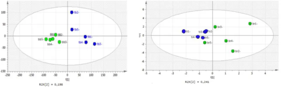
A new set of experiments was executed in LC-ESI-Orbitrap-MS in the negative ion mode working on 12 samples, with the aim of comparative qualitative analysis between traditional and organic ripe and unripe fruits. These analyses were run on a reduced number of samples based on the more time-consuming protocol. Samples included in the set were: Three samples of ripe fruits TR1, TR2, and TR3 and three samples of unripe fruits TB1, TB2, and TB3 from the conventional farm and three samples of ripe fruits BR1, BR2, and BR3 and three samples of unripe fruits BB1, BB2, and BB3 from the organic farm.
The negative ion mode at this stage was selected because of the major number of compounds detected in this operative mode.
A different approach was used for untargeted multivariate data analysis. For analysis of the acquired dataset with multivariate methods, LC-MS chromatograms were pre-processed using MZmine to compensate for variations in retention time and m/z value between the chromatographic runs. The pre-processed chromatograms were exported as a peak list table, with rows representing the individual samples, and columns representing the integrated and normalized peak areas. Moreover these data were used to confirm the MVDA (PCA and PLS-DA), through an approach of untargeted analysis. All the differences between fruits collected from the organic crops or conventional crops were underlined. Principal Component Analysis was performed by applying the peak areas of the total peaks present in the LC-MS dataset (excluding the noisy), thus a matrix was obtained by using these areas (variables), and the columns of the matrix were different analyzed samples. The data were also modeled by Partial Least Squares Discriminant Analysis (PLS-DA) as a supervised approach to exploring clustering relationship (Fig. 2.4).