Giulia Auriemma
Dottorato di Ricerca in Scienze Farmaceutiche XIII ciclo NS
2012-2015
DESIGN AND STRUCTURAL OPTIMIZATION OF NEW
MOLECULES AS POTENTIAL ANTIINFLAMMATORY
AND/OR ANTICANCER AGENTS
Dipartimento di Farmacia Via Giovanni Paolo II, 132 84084 Fisciano, Salerno M a ri a Stro cchi a D es ig n a n d s tr u ct u ra l o p ti m iz a ti o n o f n ew m o le cu le s a s potential antiinfla mmatory and/or anticancer agents Maria Strocchia Mari
Mari StrocchiaStrocchia Maria
UNIVERSITÀ DEGLI STUDI DI SALERNO
Dipartimento di Farmacia
Dottorato di ricerca
in Scienze Farmaceutiche
Ciclo XIII NS — Anno di discussione 2015
Coordinatore: Chiar.mo Prof. Gianluca Sbardella
Design and structural optimization of new
molecules as potential antiinflammatory
and/or anticancer agents
settore scientifico disciplinare di afferenza
: CHIM/06
Dottorando Tutore
Dott. Maria Strocchia Chiar.ma Prof. Ines Bruno
Preface
My PhD three years course in Pharmaceutical Sciences at the Department of Pharmacy of Salerno University was started in 2012 under the supervision of Prof. Ines Bruno.
My research project was mainly focused on the design and synthesis of small molecules as new modulators of emerging targets involved in inflammatory and cancer processes. Specifically, my research activity was addressed to the investigation of three major targets:
· the epigenetic family of readers, Bromodomain (BRD) containing proteins;
· the membrane enzyme, microsomal Prostaglandin E2 synthase-1 (mPGES-synthase-1);
· the molecular chaperone, Heat shock protein 90 (Hsp90). The entire work was carried out under the direct supervision of Prof. Ines Bruno and Dr. Stefania Terracciano.
Computational guided design of compounds was performed in collaboration with Prof. Giuseppe Bifulco’s research group.
Biological screenings were performed in collaboration with Dr. Panagis Filippakopoulos of the Structural Genomics Consortium (Oxford) in the case of BRDs, with Prof. Oliver Werz of Friedrich Schiller University (Germany) in the case of mPGES-1, and with Prof. Antonietta Leone and Fabrizio Dal Piaz of Salerno University in the case of Hsp90.
Furthermore, to improve my knowledge on mPGES-1, in 2013 I joined Prof. Hans Hebert’s research group at the Department of Biosciences and Nutrition of Karolinska Institutet (Sweden), where I spent seven months. During that period, my research was carried out under the supervision of Dr. Caroline Jegerschöld and was addressed to the heterologous expression and two-dimensional crystallization of human mPGES-1.
List of publications related to the scientific activity performed during the three years PhD course in Pharmaceutical Sciences
Papers:
Ø Lauro G., Strocchia M., Terracciano S., Bruno I., Fischer K., Pergola C., Werz O., Riccio R., Bifulco G. “Exploration of the dihydropyrimidine scaffold for the development of new potential anti-inflammatory agents blocking prostaglandin E2 synthase-1 enzyme (mPGES-1)”. Eur J Med Chem 2014, 80, 407-415.
Ø Terracciano S., Lauro G., Strocchia M., Fischer K., Werz O., Riccio R., Bruno I., Bifulco G. “Structural insights for the optimization of dihydropyrimidin-2(1H)-one based mPGES-1 inhibitors”. ACS Med
Chem Lett 2015, 6, 187–191.
Ø Strocchia M.,‡ Terracciano S.,‡ Chini M. G., Vassallo A., Vaccaro M. C., Dal Piaz F., Leone A., Riccio R., Bruno I., Bifulco G. “Targeting the Hsp90 C-terminal domain by the chemically accessible dihydropyrimidinone scaffold”. Chem Commun 2015, Article in press, DOI: 10.1039/C4CC10074C.
Ø Picaud S.,‡ Strocchia M.,‡ Terracciano S., Lauro G., Mendez J., Daniels D.L., Riccio R., Bifulco G., Bruno I., Filippakopoulos P. “The 9H-purine scaffold reveals induced-fit pocket plasticity of the BRD9 bromodomain”. J Med Chem. Accepted.
Conference proceedings:
Ø Terracciano S., Strocchia M., Chini M. G., Bruno I., Dal Piaz F., Bifulco G., Riccio R. “Structure-based approach for the discovery of potent inhibitors of the Hsp90 molecular chaperone bearing the triazole scaffold”. XXXIV National Meeting of Italian Chemical Society, Organic Chemistry Division, Pavia (Italy), September 10-14, 2012.
Ø Strocchia M., Terracciano S., Riccio R., Bruno I., Jegerschöld C. “Human microsomal prostaglandin E2 synthase-1 (mPGES-1) overexpression in LEMO21(DE3) E. Coli strain”. Giornate di Facoltà di Farmacia e Medicina, Salerno (Italy), May 22-23, 2014.
Ø Terracciano S., Strocchia M., Chini M. G., Vassallo A., Vaccaro M. C., Dal Piaz F., Leone A., Riccio R., Bifulco G., Bruno I. “3,4-dihydropyrimidin-2(1H)-one as a useful scaffold for Hsp90 C-terminal inhibition”. XXV National Meeting of Italian Chemical Society, Rende (Italy), September 7-12, 2014.
Ø Strocchia M., Terracciano S., Lauro G., Werz O., Riccio R., Bruno I., Bifulco G. “Identification of dihydropyrimidine derivatives as new mPGES-1 inhibitors”. XXV National Meeting of Italian Chemical Society, Rende (Italy), September 7-12, 2014.
Ø Strocchia M., Terracciano S., Lauro G., Werz O., Riccio R., Bruno I., Bifulco G. “New 3,4-dihydropyrimidin-2(1H)-one derivatives as efficient modulators of microsomal prostaglandin E2 synthase-1”. Ischia Advanced School of Organic Chemistry, Ischia (Italy), September 21-25, 2014.
Table of Contents
Abstract...I
Introduction
...1-34 Chapter 1...21.1 The role of organic chemistry in drug discovery 3-5 1.2 The crosstalk between cancer and inflammation 6-9 1.3 Epigenetic readers of acetylated lysines: bromdomains 9-15 1.4 Microsomal prostaglandin E2 synthase-1 (mPGES-1) 15-21
1.5 Heat shock protein 90 (Hsp90) 21-30
1.6 Workflow of the research project 30-34
Results and Discussion
...35-104 Chapter 2 Induced-fit pocket plasticity of the BRD9 bromodomain upon binding to 9H-purine inhibitors………...362.1 Background 37-38
2.2 9H-purines: new modulators of human bromodomains 39-51 2.3 Induced fit binding of 9H-purines to BRD9 52-56
2.4 In cell validation of 9H-purines 56-58
Chapter 3 Dihydropyrimidin-2(1H)-one: a new template for the modulation of microsomal Prostaglandin E2 Synthase-1 (mPGES-1).…...…59 3.1 Targeting mPGES-1: rationale from high-resolution X-ray crystal
structures 60-61
3.3 Investigation of DHPM-based compounds as mPGES-1 modulators: rationale from X-ray crystal structure 65-72 3.4 Structural optimization of compound 48, the promising DHPM-based
mPGES-1 inhibitor 72-78
Chapter 4 Discovery of new Hsp90 C-terminal modulators: synthesis and
biological evaluation of 3,4-dihydropyrimidin-2(1H)-one
derivatives……….79 4.1 Stressing the discovery of Hsp90 C-terminal inhibitors 80-82 4.2 Targeting Hsp90 C-terminal domain by DHPM-based derivatives 82-86 4.3 Antiproliferative assays, western blot analysis and effect on cell cycle
progression 86-88
4.4 Study of Hsp90!/54 interaction 89-92
Chapter 5 His-tagged human mPGES-1 overexpression in Lemo21(DE3) E.
coli strain and 2D-crystallization studies.…...…...93
5.1 Membrane protein overexpression in E. coli 94-97
5.2 Lemo21(DE3) E. coli strain 97-98
5.3 mPGES-1 overexpression in Lemo21(DE3) strain 98-104
Conclusions
...105-107Experimental Section
...108-170 Chapter 6 Synthesis of purine derivatives as new modulators of human bromodomains: Experimental procedures…..………1096.1 General synthetic methods 110
6.2.1 General procedure for the Suzuki-Miyaura cross-coupling of
free halo-purines 111-122
6.2.2 General procedure for TBAF-assisted N9-alkylation of purine
rings 122-125
6.2.3 General procedure for the synthesis of
2-hydroxy-6-arylpurines 125-127
6.2.4 THP-protection of 2-amino-6-bromo-9H-purine 127-128 6.2.5 Attempt of C-8 electrophilic fluorination reaction on the bis(THP)-purine 13a 127-128
Chapter 7 Synthesis of DHPM-based inhibitors of mPGES-1 and Hsp90: Experimental procedures ………130
7.1 General synthetic methods 131-132
7.2 Methods and materials 132-165
7.2.1 General procedure for microwave-assisted Biginelli
reaction 132-158
7.2.2 General procedure for microwave-assisted Liebeskind-Srogl cross coupling reaction 158-163 7.2.3 General procedure for reductive amination 163-165
Chapter 8 His-tagged human mPGES-1 overexpression in Lemo21(DE3) E.
coli strain and 2D-crystallization studies: Experimental procedures………...………166
8.1 Bacterial Overexpression of Human mPGES-1 167
8.2 Preparation and solubilization of whole cell extract 167-168
8.3 Purification of Human His6-mPGES-1 168-169
8.4 Gel Electrophoresis and Western Blotting 169
References...
...171-213I Abstract
Inflammation and cancer are two complex pathological processes, involving a variety of molecular actors. The deeply connection and crosstalk between cancer and inflammation is well-known and the modulation of these processes is one of the main goals of modern medicinal chemistry. The identification of new molecular entities able to interfere with biological targets placed at the crossroads of these two pathways is strongly needed, both for the development of new promising drug candidates and as chemical probes useful to further investigate less understood biological aspects. Three main targets, involved at different levels in inflammation and cancer, have been thoroughly investigated: bromodomain (BRD) containing proteins, microsomal Prostaglandin E2 Synthase-1 (mPGES-1) and Heat-shock protein 90 (Hsp90). The results obtained can be summarized in the three main sections, reported below according to the target of interest:
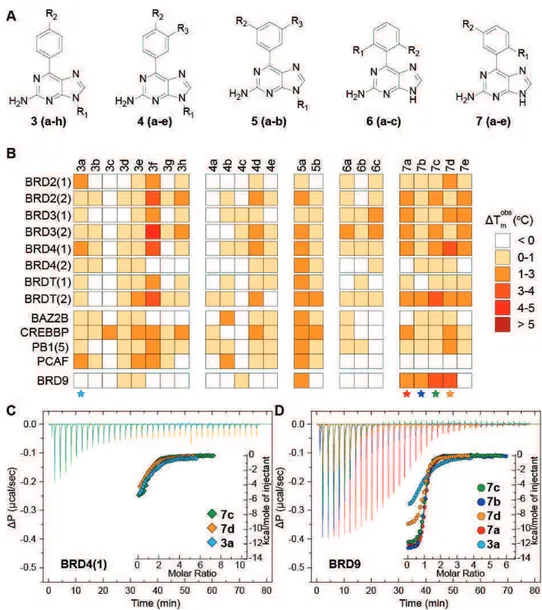
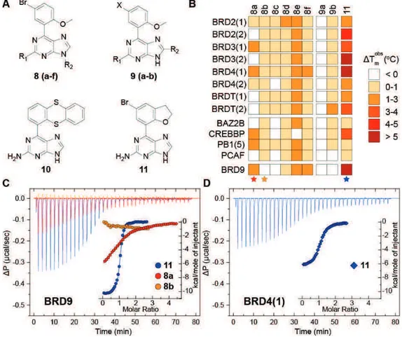
a) Discovery of new modulators of human bromodomains by structure-based and computer-aided combined approaches. BRDs are evolutionary conserved modules which act as readers of the histone code, by recognizing acetyl-lysine (Kac) residues on histone tails. The contribution of BRD containing proteins has recently emerged in a number of diseases, especially in cancer processes. With the aim of identifying a new Kac mimetic chemotype, a structure-guided approach was undertaken starting from small fragment-like 9H-purine scaffolds. One of the initial identified fragments (2a), that was shown to be a BRD binder, was systematically modified employing organic synthesis approaches in order to gather a structure activity relationships profile to be exploited in the next structural optimization process. These studies allowed to disclose potent nanomolar ligands for BRD9 (compounds 7d and 11), showing only residual micromolar affinity towards BRD4. Binding of 7d and 11 to BRD9 was investigated by crystallography and flexible docking
II
experiments and resulted in an unprecedented rearrangement of residues forming the Kac cavity, affecting plasticity of the protein in an induced-fit pocket. Finally, the compounds did not exhibit any cytotoxic effect in HEK293T cells and displaced the BRD9 bromodomain from chromatin in bioluminescence proximity assays without remarkably affecting the BRD4/histone complex.
b) Identification and structural optimization of DHPM-based mPGES-1 inhibitors. mPGES-mPGES-1 is a homotrimeric membrane protein involved in the arachidonic acid cascade, which acts as downstream synthase in the cyclooxygenase (COX) pathway by catalyzing the biosynthesis of Prostaglandin (PG) E2 from the PGH2 precursor. Inhibition of mPGES-1 can represent a valid therapeutic approach to interfere with inflammation-induced PGE2 formation without affecting the constitutively formed prostanoids. In order to find a new molecular platform for mPGES-1 modulation, a structure-based design approach was carried out on a focused collection of 3,4-dyhidropyrimidin-2(1H)-one (DHPM)-based molecules, docked in the first high resolution X-ray crystal structure of the enzyme in its active form (PDB code: 4AL0). The key interactions with the receptor counterpart were introduced as a qualitative filter for the selection of the most promising compounds to be synthesized. Biological results were consistent with the computational suggestions and disclosed two molecules (48 and 49) showing a promising in vitro mPGES-1 inhibitory activity. The most recently crystallized structure of mPGES-1 with the inhibitor LVJ (PDB code: 4BPM) was used to optimise compound 48 (IC50 = 4.16 ± 0.47 •M) to give compound 53, a 10-fold more potent mPGES-1 inhibitor (IC50 = 0.41 ± 0.02 •M).
In order to deeply investigate this complex enzyme, a heterologous expression of human His6-tagged mPGES-1 and two-dimensional crystallographic studies were also carried out.
III
c) The DHPM core as new chemotype for Hsp90 C-terminal modulation. Hsp90 is a molecular chaperone highly involved in the development, survival and proliferation of cancer cells. Traditional inhibitors of Hsp90 target its N-terminal domain. Nevertheless, this type of modulation produces scheduling and toxicity issues connected to the induction of the deleterious heat shock response. Although less explored, C-terminal inhibition of Hsp90 represents a very promising approach for developing new potential anti-cancer drugs as it is devoid of the negative effects triggered by the heat shock response. In an attempt to identify non-natural inspired modulators of Hsp90 C-terminus, a collection of DHPM derivatives was synthesized. The rationale for targeting Hsp90 C-terminal domain by DHPMs derives from the structural analogy between the DHPM core and uridine triphosphate (UTP), a nucleotide shown to selectively interact with the chaperone C-terminal site, but not with its N-terminus. Biological evaluation revealed that the privileged DHPM core can be considered as a new template for the modulation of Hsp90 chaperoning function, through the binding to its C-terminal region. In particular, compound 54 was identified as a novel promising antiproliferative agent against Hsp90 C-terminus.
- 1 -
- 2 -
1-- 3 -
1.1 The role of organic chemistry in drug discovery
The drug discovery process requires interdisciplinary approaches involving a multitude of scientific areas, e.g., biology, medicinal and synthetic organic chemistry, statistics, pharmacology, medicine, toxicology, structural biology, chembioinformatics, computational chemistry, genomics and proteomics.1 These disciplines work synergistically along the hard and complex journey toward the identification of a new drug.2 Indeed, the drug discovery and development process is one of the most challenging human endeavors, as the optimal balance between efficacy and safety of a drug must be ensured.3, 4 Moreover, the development of a new drug is a long, difficult, expansive and highly risky process, as the market access environment is very restrictive. Research and development for most of the available medicines has required 12–24 years for a single new medicine, from the beginning of the project to the launch of the drug.5
The process of drug discovery starts with the identification of a molecular target, whose modulation is expected to have positive therapeutic effect.6, 7 The selection of an appropriate target is a relevant issue: ideally, the biological target should be fully validated and its modulation should provide an unambiguous therapeutic response, with no susceptibility to the induction of resistance mechanisms.8 Appropriate assays, designed for the selected biological target are then needed to identify putative modulators.9 At this stage, the drug discovery process is focused on small organic molecules, as they are the main class of marketed drugs.10 Organic synthetic chemistry acts as the main player at this step, as its role is to identify and structurally optimize new active compounds both in potency and in their pharmacokinetic profile.11, 12 Actually, organic synthesis should be able to provide a more or less complex compound with high selectivity and efficiency.13 Many are the aims that organic chemistry is expected to achieve in the drug discovery process. Such examples include: to develop novel structural motifs with
- 4 -
improved pharmacological properties, to identify new synthetic methods and strategies, to expand applications of organic synthesis into the field of biology, to apply the information derived from structural biology studies on the selected target in order to synthesize molecules that can appropriately fit the receptor, to chemically modify an emerged lead structure in order to draw a structure–activity relationships (SARs) profile useful for its rapid optimization.14-18 Today, synthetic organic chemistry can rely on a wide range of tools for overcoming the several hurdles in the drug discovery process. These new tools include advances in synthetic, analytical and purification methods such as transition-metal-catalysed carbon–carbon couplings,19 multicomponent and domino reactions,20 microwave-assisted and flow chemistry,21, 22 high-field NMR23 and preparative high-performance liquid chromatography (HPLC),24 as well as computer-assisted approaches,25 combinatorial chemistry26 and high-throughput screening (HTS).27
An active compound, referred as a “hit” in the drug discovery process, may arise in many ways. It may be found in large or more focused compounds libraries, which have demonstrated prior reliability in drug discovery programs. If inhibitors or ligands of a given target are known, potential new binders may be selected on the basis of structural similarities through a “ligand-based design” approach.28 Alternatively, when the crystal structure of the target has been elucidated, structural complementarity to the binding site can be evaluated through “structure-based design” approaches.29 In this case, virtual screening studies can be also carried out, in order to select promising compounds belonging to a commercially available database.30 The resulting hits generally have modest activity, typically at micromolar concentrations, while the marketed drugs are commonly active at low nanomolar range.31 Development of the hit to a potent compound is the process of “lead” optimization.32 At this step, organic synthetic chemistry plays again a crucial role, as much synthetic effort is required to generate a large collection of
- 5 -
structurally related derivatives.33 In the course of this process, other problems related to drug pharmacokinetic profile must be taken into due account.34
Indeed, organic and medicinal chemistry should also work synergistically to overcome pharmacokinetic liabilities of test compounds. To this end, the prediction of “drug-like” properties has to be accomplished.32 Highly lipophilic compounds with high molecular weight are generally more potent in
vitro, but they tend to be usually not drug-like because of their poor
pharmacokinetics and oral bioavailability. As a predictor of drug-likeness, Lipinski et al.35 formulated the “rule of five” which consists of four important properties, each related to the number 5 (molecular mass <500 Da; calculated LogP <5; hydrogen-bond donors <5; and hydrogen-bond acceptors <10). However, this rule tries to predict oral bioavailability in a very basic manner, but drug discovery implies a very careful determination of the ADMET (absorption, distribution, metabolism, elimination, and toxicology) parameters of a drug, which implies more than Lipinski’s “rule of five”.36 An example is represented by antibiotics, cytostatic and many other drugs which suffer from the so-called “molecular obesity”,31 as they have higher molecular mass than the border of 500 Da, but possess elevated efficiency and bioavailability. Actually, the aim of lead optimization phase is to maintain favourable properties in lead compound, while improving any deficiencies in its structure. This represents a very complex issue and indeed, all the information gathered about the molecule at this stage will allow for the optimization of a target candidate profile which, together with toxicological and other control assays, will lay the basis first for preclinical candidate selection and finally for entering clinical trials.37-39
In the present PhD project, thanks to combined approaches of organic synthesis, computational chemistry and structural studies, new chemical entities with antiinflammatory or antitumor effects have been successfully identified.
- 6 -
1.2 The crosstalk between cancer and inflammation
It is beyond the scope of this thesis to discuss the complex mechanisms of cancer and inflammation, but the close relationship between these two pathological processes deserves some considerations. Although the involved pathways and the correlations between them have not been fully understood yet, the crosstalk between cancer cells and inflammatory mediators has been known for a long time.40
The first example of inflammatory processes related to cancer development in tissues was reported in the nineteenth century by the German physician Rudolf Virchow,who described leukocyte infiltrates within tumours.41 These leukocyte infiltrates were at first related to the immune surveillance and antitumor immune response, but it is now understood that they can act both as tumour-suppressors and as tumour-promoters.42-45
Cancers are composed of multiple cell types such as fibroblasts and epithelial cells, innate and adaptive immune cells, blood and lymphatic cells, as well as specialized cell types unique to each tissue.46, 47 Inflammation is a key component of the cancer microenvironment, also in tumours which are not related to an obvious inflammatory cause. Relevant aspects of cancer-related inflammation include the infiltration of white blood cells (mainly tumour-associated macrophages), the presence of inflammatory mediators (cytokines and chemokines) and the occurrence of tissue remodelling and angiogenesis.48
Both the intrinsic and the extrinsic inflammatory pathways have been related to cancer.44, 49, 50 The intrinsic one is activated by genetic events, mainly activation of oncogenes, resulting in the transformation of cells which trigger the expression of inflammation-related programs contributing to produce an inflammatory environment.51, 52 In the extrinsic pathway, chronic inflammatory conditions or infections increase the risk of developing cancer (e.g, prostatitis for prostate cancer, papillomavirus for cervical carcinoma).53-55 The two pathways converge in the activation of transcription factors, mainly
- 7 -
NF-•B and STAT3 which have emerged as key mediators in cancer development and progression.56, 57
In this context, it is not surprising that the traditional six hallmarks of cancer (self-sufficiency in proliferative signals, insensitivity to anti-growth signals, tissue evasion and metastasis, limitless replicative potential, sustained angiogenesis, resistance to cell death)58 have been joined by four additional emerging hallmarks (avoidance of immune destruction, induction of tumour-promoting inflammation, genome instability and mutation, and deregulation of cellular metabolism)59 which are all directly or indirectly related to the inflammatory process (Figure 1.1).
Figure 1.1 The ten hallmarks of cancer (adapted from ref. 59).
Both premalignant and malignant tissues have been found in an inflammatory state driven by cells of the immune system which ultimately disclose the tumour-promoting effect of the inflammatory response (Figure 1.2). Furthermore, inflammatory mediators contribute to genomic instability and to the occurrence of mutations associated with tumours, as many of them act as direct mutagens or as deregulators of DNA repair mechanisms and cell
- 8 -
cycle checkpoints, resulting in the acquired ability of cancer cells to proliferate, invade and escape from host defence, in particular from T and B lymphocytes, macrophages, and natural killer cells.60-63
Figure 1.2 Role of inflammation in cancer development.63
Whereas chronic innate immune inflammation in premalignant cells might promote cancer development, adaptive immune response to the tumour might result in abolition of the malignancy, a mechanism known as cancer immunosurveillance (Figure 1.2).64, 65 Adaptive immune cells can directly modulate cancer by inhibiting tumour growth through T-cell activity and cytokine-mediated lysis of malignant cells.66
These considerations highlight the dual opposite function of inflammatory reactions, which can result both in antitumour and in tumour-promoting effects.67, 68 Anyway, many evidences have been gathered supporting the improved therapeutic efficacy that can be achieved by blocking the two signalling networks and their pathways.69-73 In this respect, targeting inflammatory and neoplastic pathways can be accomplished at different levels
- 9 -
through, the modulation of specific proteins involved in key steps of these processes.
In the context of my PhD, three biological targets involved both in inflammation and cancer have been investigated: Bromdomain (BRD) containing proteins, microsomal Prostaglandin E2 synthase-1 (mPGES-1) and the chaperone Heat Shock Protein 90 (Hsp90). Although each of them is known for its major implication in inflammation (in the case of mPGES-1) or in tumour (in the case of BRDs and Hsp90), several evidences suggest their involvement in both processes, standing for an additional evidence of the crosstalk between cancer and inflammation.
1.3 Epigenetic readers of acetylated lysines: bromdomains
The array of post-translational modifications (PTMs) introduced on histone tails gives rise to the so-called “histone code”,74 a cellular language generated by proteins which introduce (writers) or remove (erasers) PTMs.75-77 Furthermore, this complex code involves also some evolutionarily conserved domains, found in structurally heterogeneous proteins, which act as readers of PTMs by recognizing covalent marks on histones.78-81
The combination of PTMs (acetylation, methylation, phosphorylation, ribosylation, biotinylation, citruillination, crotonylation and SUMOylation82-84 modulates chromatin plasticity and its functionality.85-89 For instance, -N-acetylation of lysine residues (Kac) is associated with neutralization of the positive charge of histone tails, resulting in an open chromatin structure (euchromatin) and transcriptional activation.90-94 Although lysine acetylation has been connected for a long time only to the histone code, this widespread PTM occurs throughout the entire proteome,95-99 and alterations in its levels have been associated to a large number of diseases, especially cancer.98, 100-103 In the context of epigenetics, the acetyl group is deposited on lysine residues by histone acetyl-transferases (HATs),104 removed by histone deacetylases
- 10 -
(HDACs)105 and recognized by conserved protein modules such as bromodomains,106 as well as the more recently discovered YEATS domains.107
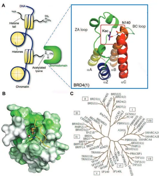
There are 61 bromodomains (BRDs) that have been identified in 46 different proteins in the human genome,108 which mainly act as transcriptional co-regulators and chromatin modifying enzymes, e.g., HATs and HAT associated proteins (PCAF, GCN5, BRD9),109-112 helicases (SMARCA),113 ATP-dependent chromatin-remodelling complexes (BAZ1B),114 SET domain containing methyl-transferases (MLL and ASH1L),115, 116 transcriptional co-activators (TAF1, TRIM/TIF1),117, 118 nuclear scaffolding proteins (polybromo PB1)119 and transcriptional regulators (BET family).120, 121 All BRDs share an architecturally conserved tertiary structure with an “atypical left-handed four-helix bundle” (!Z, !A, !B, !C) linked by two main loop regions (ZA and BC loops) (Figure 1.3A), a structural motif identified in the early 90s in the Drosophila melanogaster brahma gene.122 Despite the conserved BRD fold, the overall sequence similarity of the BRD family members is not high, as considerable variations have been found especially in ZA and BC loops.123 Nevertheless, the amino acids engaged in Kac recognition are among the most conserved in the hydrophobic Kac binding pocket and correspond to highly conserved asparagine and tyrosine residues (in BRD4(1): Asn140 and Tyr97).108 A peculiar feature of this module is also the presence of a network of water molecules, which form hydrogen bonds with carbonyl groups of the protein backbone at the base of the domain and are relatively conserved in most BRDs (Figure 1.3B).124-127 A large scale structure-based analysis of the human BRD family, using 34 high resolution crystal structures and 4 NMR models, as well as secondary structure prediction algorithms, grouped the 61 BRD modules into 8 distinct sub-families (Figure 1.3C).108 The BET subfamily of BRDs (group II) has attracted particular attention, as its members (BRD2, BRD3, BRD4 and BRDT) play a central role in cell cycle progression, cellular proliferation and apoptosis.128
- 11 -
Figure 1.3 Structure and classification of the bromodomain family.(A) The atypical
left-handed four-helix bundle structural motif in BRD4(1). Highlighted is the interaction with the conserved asparagine residue. (B) Molecular surface of the bromodomain of BRD4(1) showing conservation of Kac binding site. Green represents more conserved regions, and
white less conserved ones, as obtained from a multiple sequence alignment of all human BRDs. Conserved water molecules at the bottom of the Kac binding pockets are shown as
ball-and-stick models.(C) Phylogenetic tree of human BRDs.
BETs contain two N-terminal BRD modules that interact with acetylated histones,120 transcription factors129, 130 or other acetylated transcriptional regulators,131, 132 an extra terminal (ET) recruitment domain133 and a C-terminal motif responsible for the recruitment of the positive transcription elongation factor B (P-TEFb),121 in the case of BRD4 and BRDT.134 BET
- 12 -
BRDs have been successfully targeted by small molecule inhibitors, such as the triazolothienodiazepine (+)-JQ1134 (Figure 1.4A) and the triazolobenzodiazepine IBET762135 (Figure 1.4A) which were identified employing phenotypic screening 136 and have consolidated the emerging role of BRDs as viable therapeutic targets.137, 138 The discovery of these two compounds prompted in the last years a number of medicinal chemistry efforts, which resulted in a growing number of novel and structural diverse Kac mimetics targeting bromodomains, exhibiting excellent potency and selectivity, especially against the BETs (Figure 1.4A).139 More recently, a number of kinase inhibitors have also been identified as interacting with the Kac binding pocket of some bromodomains (Figure 1.4B).140, 141 Potent and selective molecules against non-BET proteins have also emerged, mainly targeting the bromodomain of CREBBP.142, 143 Finally, it was also possible to modulate more challenging BRDs such as BRPF1,144 ATAD2145, 146 and BAZ2B147 (Figure 1.4C), even though they had emerged as difficult to target from a druggability analysis carried out on all BRDs.148 In this context, fragment-based programs proved to be very reliable approaches to identify fragments interacting with these less druggable BRDs.145-147, 149-154
Potent and selective small molecules that inhibit the Kac•BRD interaction have been employed as chemical probes in elucidating the biology of several families of bromodomain-containing proteins, by shedding more light also on their role in pathological conditions. For instance, BET inhibition suppresses tumour growth in diverse mouse models of cancer, e.g., NUT midline carcinoma, acute myeloid and mixed lineage leukemia, multiple myeloma, glioblastoma, melanoma, Burkitt’s lymphoma, neuroblastoma and prostate cancer, leading to a number of clinical trials seeking to modulate BET function in diverse tumour settings.138
- 13 -
Figure 1.4 Bromodomain Inhibitors. (A) Representative BET inhibitors.134, 135, 155-159(B)
- 14 -
The first study to demonstrate the efficacy of a bromodomain inhibitor in a preclinical cancer model was carried out by Filippakopoulos et al.134 with the aim of evaluating the effect of (+)-JQ1 on mice bearing a NUT midline carcinoma (NMC) xenograft, a rare but aggressive form of cancer determined by the BRD4-NUT oncoprotein.162 Treatment with (+)-JQ1 induced a reduction of tumour volume and promoted survival with minimal toxicity against normal tissues.134 This outcome paved the way for some BET inhibitors to enter clinical trials in a range of malignancies, including NUT midline carcinoma (ClinicalTrials.gov identifiers: NCT01587703, NCT01987362), progressive lymphoma (ClinicalTrials.gov identifier: NCT01949883), solid tumours (ClinicalTrials.gov identifier: NCT02259114), glioblastoma (ClinicalTrials.gov identifier: NCT02296476), acute leukemia and other hematological malignancies (ClinicalTrials.gov identifiers: NCT01943851, NCT01713582).
The role of BETs in cancer is more than obvious, but these transcriptional factors have a relevant function also in inflammatory conditions, as emerged especially in the case of BRD4.163-169 The pan-BET inhibitor I-BET762 was shown to suppress inflammation by strongly attenuating the expression of LPS-induced pro-inflammatory genes during late macrophage activation.135 BET proteins have also emerged as an essential connection between chromatin signalling and IL-17-producing T helper cells differentiation and activation, which suggests their potential therapeutic role in autoimmune conditions.170 A very recent study has demonstrated the ability of (+)-JQ1 to interfere with the interaction between BRD4 and the transcription factor NF-•B.171 As described in the previous paragraph, NF-•B is the central mediator involved in the crosstalk between cancer and inflammation: its master function in modulating the immune response is regulated by the acetylation of Lys130 on its RelA subunit, which triggers transcriptional activation of NF-•B target genes and contributes to maintain its persistently active form in tumors.172, 173 This event
- 15 -
can be suppressed through depletion or inhibition of BRD4, as this BET member has been shown to bind to acetylated Lys310 of RelA and to regulate the transcriptional activity of NF-•B. As a consequence of Brd4 deletion or inhibition upon treatment with (+)-JQ1, NF-•B activation mediated by TNF-! is suppressed, as well as the expression of NF-•B-dependent target genes.171 Another BRD4 inhibitor, I-BET151, also exhibited anti-inflammatory properties, as it was shown to selectively regulate IL-6 production.174 In a chronic model of inflammation involving IL-6 (autoimmune encephalomyelitis used as a model of multiple sclerosis), treatment with I-BET151 resulted in a significant delay in the onset of clinical symptoms.174
Finally, BET bromodomains are involved also in heart failure,175, 176 adipogenesis177 and in viral transcription of HIV, herpesviruses, Merkel cell polyomavirus and murine leukaemia virus, suggesting potential therapeutic applications of BRD inhibitors also in these fields.178-184
1.4 Microsomal prostaglandin E2 synthase-1 (mPGES-1)
Prostaglandin E2 synthases (mPGES-1, mPGES-2 and cPGES) are downstream enzymes that specifically catalyze the biosynthesis of the crucial inflammatory mediator PGE2 from PGH2.185
PGE2 and all other eicosanoids are biologically active mediators, produced from the oxidation of long-chain 20 carbon atoms polyunsaturated fatty acids and obtained, either via the cyclooxygenases (COX-1 and COX-2) pathway, or
via the lipoxygenase (LO) one.186-191 The COXs pathway generates prostanoids, which include prostaglandins (PGs), prostacyclin and thromboxane (TXA), while the LO pathway results in the biosynthesis of leukotrienes (LTs).187 These inflammatory mediators are synthetized by most mammalian cells and tissues and their effect is mediated by the interaction with individual receptors, mainly G-protein coupled receptors (GPCR).192, 193 The biosynthesis of eicosanoids is initiated by release of arachidonic acid
- 16 -
(AA) from cell membrane by phospholipase A2 (PLA2),. in response to any inflammatory stimulus inducing an increase of intracellular Ca2+ levels.194, 195 In the case of prostanoids (Figure 1.5A), AA is converted to PGH2 by COX-1/2, in a process that requires two successive steps: firstly, AA is oxidized to generate endoperoxide PGG2 in the cyclooxygenase site of the COXs, and this AA-derived mediator is then reduced at the peroxidase site of COXs into PGH2.196 PGH2 is very unstable197 and is rapidly converted to PGD2, PGE2, PGF2•, PGI2 (prostacyclin) and TXA2 (thromboxane), depending on the expression of specific terminal enzymes of the biosynthetic pathway.198, 199
Among the three PGE2 synthases, cPGES and mPGES-2 are constitutively expressed, whereas mPGES-1 is an inducible isoform200, 201 specifically coupled with COX-2.202, 203 Low but constitutive expression of mPGES-1 is ubiquitous, but its level is up-regulated in response to various inflammatory stimuli and mediators, for example, cytokines (LPS, IL-1b and TNF-•).204-207 Identification of mPGES-1 was reported in 1999 by Jackobsson et al.,208 who recognized it as a member of the Membrane-Associated Proteins in Eicosanoid and Glutathione Metabolism (MAPEG) family,209 which includes five additional proteins (MGST1, MGST2, MGST3, FLAP, LTC4S).210, 211
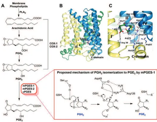
The first high resolution X-ray crystal structure of mPGES-1 in the active conformation was described by Sjögren et al.212 in 2013, who revealed that the protein is a membrane homotrimer with three active sites partially occupied by the cofactor (glutathione, GSH). The asymmetric monomer is characterized by four-helix, and each active site is oriented toward the cytoplasmic part of the protein, in particular between N-terminal parts of helix II and IV of a monomer and the C-terminal part of helix I and the cytoplasmic domain of the adjacent monomer (Figure 1.5B). This protein folding generates a pronounced deep active site occupied by GSH, and in the outer part, an extended groove between helix I of a monomer and helix IV of the adjacent monomer is observable (Figure 1.5C). Sjögren et al.212 also proposed a mechanism for
- 17 -
PGH2 isomerization to PGE2 mediated by the GSH cofactor (Figure 1.5A). According to this suggested mechanism, Ser127 activates the thiol function of GSH to form a thiolate anion that exerts a nucleophilic attack on the endoperoxide oxygen atom of PGH2, producing an unstable intermediate. Subsequently, Asp49 mediates the abstraction of the proton at C-9 followed by the cleavage of S-O bond, which results in the regeneration of GSH and in the formation of PGE2.
Figure 1.5 Biosynthetic pathway of PGE2 and structure of mPGES-1. (A) PGE2
biosynthesis and proposed mechanism of PGH2 isomerisation by mPGES-1, as reported by
Sjörgen et al.212 (B) Overall structure of mPGES-1. (C) Interaction of mPGES-1 with the
- 18 -
Figure 1.6 Some optimised scaffolds for mPGES-1 inhibition: (A) indole;213 (B)
phenanthrene imidazole;214, 215 (C)biaryl imidazole;216 (D) pirinixic acid;217-219 (E)
trisubstituted urea;220 (F) oxicam;221 (G) imidazoquinoline;222 (H) arylpyrrolizine;223 (I)
1,2,3-triazole;224, 225 (J) 1,2,4-triazine;226 (K) 1,2,4-triazole;226 (L) benzoxazole.227 HWB: human
- 19 -
Traditional treatment of inflammation is based on the use of NSAIDs, which inhibit PGs production by blocking both COX-1 and COX-2. However, their incapability to discriminate between the two COXs is responsible for their gastric side effects,228-230 mainly due to the massive inhibition of PGE2 synthesis, which is known to have a protective effect on the gastrointestinal mucosa. In order to circumvent this undesired effect, selective inhibitors of the inducible COX-2 (COXibs) were developed.231 However, they were shown to be associated with increased cardiovascular risk in patients after long-term treatments due to unbalanced levels of PGI2 and TXA2.232-234
In the light of the side effects connected to NSAIDs and COXibs, the development of inhibitory strategies, which specifically target the downstream PGs synthases, is the current goal of research in the modulation of AA inflammatory cascade. In particular, inhibitors of mPGES-1 are expected to manifest reduced adverse effects, by better maintaining the gastric mucosa integrity compared to traditional NSAIDs and by avoiding increased incidence of cardiovascular side effects related to COXibs. Modulation of mPGES-1 may not be associated with the perturbations in PGI2 and TXA2 metabolism, as indicated by Cheng at al.,235 who reported that mPGES-1 deletion does not result in hypertension or predisposition to thrombosis in normolipidemic mice, differently from deletion, disruption or inhibition of COX-2.
Inhibition of mPGES-1 offers a wide range of opportunities for therapeutic application. The potential use of mPGES-1 inhibitors is not limited to inflammatory condition, since mPGES-1 plays a crucial role in various phatological conditions such as pain,236, 237 fever,238 rheumatoid arthritis,239, 240 cardiovascular diseases,241 cancer.242-245 The impact of mPGES-1 in tumours is particularly relevant, as it results overexpressed in a number of neoplasias, including gastrointestinal cancers (esophageal, gastric, colorectal, liver and pancreatic cancer),246-251 brain cancers (glioma and medulloblastoma),252, 253 breast cancer,254 thyroid cancer255 and several cancers derived from epithelium
- 20 -
(head and neck, penis, lungs, larynx, cervix, endometrium and ovary).256-261 Elevated levels of mPGES-1 correlate with a worse prognosis in late stages of colorectal cancer,262 suggesting that this synthase may play a key role in cancer progression. Moreover, mPGES-1-derived PGE2, in cooperation with vascular endothelial cell growth factor (VEGF), seems to play a critical role in the development of inflammatory granulation and angiogenesis.263 Indeed, mPGES-1 deficiency has been well documented to be associated with reduced induction of VEGF in the granulation tissue.237
Despite the numerous potential applications in therapy and even though many companies and academic groups have worked to develop mPGES-1 inhibitors (Figure 1.6),264 since the discovery of this MAPEG member in 1999, no clinical trials have been reported yet. This can be ascribed to the poor in cell potency of many identified inhibitors, even though they showed very high and selective inhibitory potency on the recombinant human enzyme (Figure 1.6).265 An additional problem is sequence dissimilarity of mPGES-1 isoforms in the diverse species, as first described by Merck scientists.215 For example, potent inhibitors against the human enzyme may partially or completely lose potency against the rat isoform, mainly due to the variation between human and rat mPGES-1 in three individual amino acids located in transmembrane helix IV, which play a crucial role as gatekeepers for the active site of mPGES-1, regulating the access of an inhibitor in the enzyme. In the human enzyme, these residues are rather small (Thr131, Leu135 and Ala-138) but in the rat isoform they are bulkier or aromatic (Val131, Phe135 and Phe138), and thereby prevent the access to inhibitors for steric hindrance reasons.266 Similar bulky/aromatic residues are found also in mouse ortholog, but not in the guinea pig enzyme, suggesting the use of this specie as an animal model in pre-clinical studies.215
Although better results in terms of cellular activity have been obtained for some of the optimised templates (Figure 1.6) and, despite in few cases in vivo
- 21 -
studies displayed promising effects,219, 227 none of these compounds has entered clinical trials yet. Despite the challenging problems connected with the exploration of the biological target, selective inhibition of mPGES-1 might represent a promising approach for the design of effective anti-inflammatory drugs lacking the severe side effects related to the classic use of NSAIDs. However, whether mPGES-1 inhibitors are less afflicted with side effects and can achieve the same therapeutic efficiency of COX inhibition remains to be thoroughly investigated.267 In this context, the development of new selective mPGES-1 inhibitors is highly desirable in order to fully clarify this issue.
1.5 Heat shock protein 90 (Hsp90)
The key role of the molecular chaperone family of proteins is to prevent protein aggregation, to assist the maturation and folding of proteins and to generally maintain protein homeostasis (proteostasis).268-272 According to a general definition, a molecular chaperone is any protein that interacts, stabilizes and assists a client protein in the acquisition of its functional conformation. Heat shock proteins (HSPs) are highly conserved chaperones, classified according to their molecular weights (small HSPs (<40 kDa), Hsp40, Hsp60, Hsp70, Hsp90, and Hsp100) which can be localized in cytosol, mitochondria or in endoplasmic reticulum.273 Among them, Hsp90 is of particular interest as it is extremely conserved from bacteria to eukaryotes and is one of the most abundant proteins in the cell, thus confirming its key role in maintaining protein homeostasis.272, 274, 275 Hsp90 represents 1–2% of total cytosolic proteins in non-stressed eukaryotic cells, and its level can increase up to 4–6% in stressful conditions.276-278 Its expression is up-regulated as a consequence to external and cellular stress including infections, heat, drugs, fever, oxidative stress, inflammation, hormonal stimulation, and cancer.279-281 The two major Hsp90 isoforms are found in the cytoplasm and correspond to the inducible Hsp90 and the constitutive Hsp90•.282 In addition, two
non-- 22 non--
cytosolic forms are known, namely the Hsp75/tumor necrosis factor receptor associated protein 1 (Trap1) and the endoplasmic reticulum resident Hsp90 isoform, 94 -kDa gluclose-regulated protein (Grp94). The former resides in the mitochondrial matrix and is involved in oxidative cell death and in maintaining mitochondrial integrity,283 the latter assists the folding of both secreted and membrane proteins and plays an eminent role in embryonic development, immune response, Ca2+ balance, and cell adhesion.284 Hsp90 client proteins belong to different families and do not share any apparent functional or structural similarities.285, 286 A common feature may be their intrinsic instability and the conformational changes required in order to achieve their functional state. To date, more than 300 proteins are known whose maturation is regulated by Hsp90.287
Hsp90 offers important therapeutic opportunities. Its inhibition by cytotoxic agents induces the degradation of client proteins which are subsequently addressed to ubiquitinylation-mediated proteasomal degradation (Figure 1.7).288-290 Compounds that exhibit such effect have excellent therapeutic potential as anticancer drugs, as multiple signalling pathways involved in pathologies can be modulated.291, 292 On the other hand, non-toxic compounds inducing the expression of chaperone levels showed to reduce the accumulation of aggregated proteins, suggesting promising application against neuronal disorders (Figure 1.7).293-296
Hsp90 is overexpressed in many human cancers and plays a relevant role in the progression of malignancy, as its level in cancer cells can be increased up to 10-fold than in normal cells.297-300 Malignant cells are dependent on its chaperoning function, mainly due to the adverse microenvironment (hypoxia, low pH and poor nutritional status)300 which results in an altered state of cellular proteins, consequently requiring a higher production of Hsp90 for repairing degraded proteins280, 301. Hsp90 prevents aggregation and misfolding
- 23 -
of overexpressed and mutated client oncoproteins, e.g., ErbB2, Akt, p53, Bcr-Abl, Her-2, Cdk4, Cdk6, Raf-1, v-Src, MET, telomerase and survivin.302-304
Figure 1.7 Therapeutic opportunities for Hsp90 inhibitors
Hsp90 represents an exciting therapeutic target for the treatment of cancer and its inhibition allows for a combinatorial attack on transformed cells through the disruption of various signalling pathways.305, 306 Indeed, disruption of the Hsp90 protein folding machinery directly affects all hallmarks of cancer, by preventing maturation of proteins directly associated with each hallmark (Figure 1.8A).291, 307, 308 No other cellular protein has been ascribed to affect all cancer hallmarks, thus making Hsp90 one of the most promising targets for anti-tumour therapy at this time.309
In addition, Hsp90 is an investigated target also for neurodegenerative diseases, derived from cell death in the central nervous system such as Alzheimer’s, Huntington’s, and Parkinson’s disease.310 The reason for neuronal cell death in these pathologies can be ascribed to a variety of factors, but an important general aspect is the accumulation of misfolded proteins responsible for cytotoxicity. The rationale behind targeting Hsp90 in neurological disorders is based on the principle that non-cytotoxic small molecule inhibitors of this chaperone can up-regulate the expression of heat shock proteins through the induction of the heat shock response mechanism, which ultimately leads to solubilisation of protein aggregates and refolding of misfolded proteins.311
- 24 -
Particularly relevant is also the emerging role of Hsp90 in innate immunity, evidencing the deep connection between cellular stress and inflammation.312 The common player is again the transcription factor NF-•B, as Hsp90 is required for I•B kinase (IKK) biogenesis, homeostasis and activation.313-315 Inhibition of NF-•B pathway is observed upon treatment with the Hsp90 N-terminal inhibitor geldanamycin, suggesting the potential to prevent cancer development during chronic inflammation.316, 317 Moreover, the inhibitor SNX-7081 blocked nuclear translocation of NF-•B and strongly inhibited cytokines production in animal models of rheumatoid arthritis,318 modulation of Hsp90 function by radicicol attenuated intestinal inflammation,319 while 17-DMAG reduced inflammation in macrophages by suppressing Akt and NF-•B pathways320 and also attenuated inflammatory responses in atherosclerosis.321 The relationship between inflammation and chaperones is revealed also by Hsp90 involvement in endotoxin-induced uveitis,322 inflammatory myopathies,323 inflammatory bowel disease,324, 325 gastric inflammation and ulcer healing,326, 327colitis,328 liver injury,329 autoimmune encephalomyelitis,330 and inflammatory microenvironment associated with cancer prostate.331
Structurally, Hsp90 functions as a dimer, with each monomer consisting of an N-terminal ATP-binding domain, a middle domain, and a C-terminal dimerisation domain (Figure 1.8B).332 The N-terminal site triggers the conformational change of the protein through ATP hydrolysis, supplying the required energy for the chaperoning function;333 the middle domain regulates client protein interactions and interacts with the !-phosphate of ATP;334 the C-terminal contains a second nucleotide binding region,335-337 which does not exhibit ATPase activity, and is involved in the control of Hsp90 conformational rearrangement and in the binding of co-chaperones through a conserved pentapeptide sequence (MEEVD).338 In addition, a dimerisation motif, implicated in the functional switch between the open and closed protein conformation, is present at the C-terminus.339
- 25 -
Figure 1.8 Hsp90 client proteins and structure of the Hsp90 dimer. (A) Involvement of
Hsp90 client proteins in cancer hallmarks. (B) Hsp90 switch between open and closed conformation upon ATP binding.
In the absence of ATP, Hsp90 adopts an open conformation.340 Upon nucleotide binding, the N-terminal domain closes over the bound nucleotide and the two N-terminal domains of the dimer subsequently associate.341 A flexible loop of the middle domain interacts with the ATP-binding pocket of the N-terminal domain resulting in a twisted, closed conformation of Hsp90 and in ATP hydrolysis.342 In the final step of its chaperoning cycle, Hsp90 switches back to the open conformation and the hydrolyzed nucleotide is
- 26 -
released (Figure 1.8B). In this process, Hsp90 interacts with a number of co-chaperones which also mediate the maturation of client proteins.343, 344
Hsp90 contains several small molecule binding sites. The N-terminal ATP region has been the most extensively investigated, while less is known about the binding sites in the C-terminal and middle domains. The most common Hsp90 inhibitors bind competitively to the N-terminal domain and they include both natural products such as geldanamycin (GDA) and radicicol (RDC), and synthetic compounds such as GDA and RDC derivatives, purine-based molecules, benzamide- and resorcinol-containing inhibitors.345, 346
A number of clinical trials have been initiated from 1999 in order to evaluate the potential use of Hsp90 N-terminal inhibitors in cancer.347, 348 Although some N-terminal inhibitors are still under clinical investigation, 349-352
many trials have failed due to toxicity issues and to the occurrence of resistance against these agents,353-355 mainly associated with the induction of the deleterious heat shock response.356, 357 A strategy to circumvent this problem may be to target the less-explored Hsp90 C-terminal domain, as its modulation does not trigger the undesired heat shock response.358, 359 Potential Hsp90 C-terminal inhibitors, in fact, may maintain the anti-proliferative activity, without being associated with the side effects reported for N-terminal modulators and representing thus promising candidates for drug development.360, 361 However, only poor structural information on Hsp90 C-terminus are currently available representing a strong limitation for rational design of selective inhibitors. While the binding mode of Hsp90 N-terminal inhibitors has been well characterized by X-ray crystallography, there is no reported co-crystal structure of its C-terminal domain with any inhibitor.
The natural coumarin antibiotic novobiocin was identified as the first Hsp90 C-terminal inhibitor,362 followed by its analogues chlorobiocin and coumermycin A1 (Table 1). Novobiocin’s binding site is located at the C-terminal region of the chaperone containing amino acids 538-728.362, 363
- 27 -
Mechanistically, the binding of novobiocin to Hsp90 induces a conformational change of the protein that is dissimilar from that induced by N-terminal inhibitors.336, 364 For instance, novobiocin was shown to protect Hsp90• from cleavage with proteolytic enzymes in correspondence of two main sites at the C-terminus (Arg400 and Lys615/Arg620) and of a minor site at middle domain;364, 365 moreover, it prevents binding of TPR-containing co-chaperones to the C-terminal MEEVD motif.366 Given the weak interaction of novobiocin with Hsp90 C-terminus (IC50 = 700 µM in SKBr-3 breast cancer cells),362, 363 a number of structural analogues (novologues) have been synthesized and have exhibited a significant improved potency (Table 1).367-370 Other inhibitors of the Hsp90 C-terminal domain include epigallocatechin gallate (EGCG),371, 372 cisplatin,373 taxol374 and sansalvamide A derivatives375-377 (Table 1).
Further strategies to circumvent the liabilities of N-terminal inhibitors may be the development of isoform-selective inhibitors378-380 or modulators that work by alternative mechanisms, for example, co-chaperone disruptors.381-383 Even though more challenging, the modulation of Hsp90 through the inhibition of its C-terminal domain, together with the other alternative strategies, may allow to develop new potential effective anticancer drug candidates, that are expected to be free from side effects connected with the use of traditional N-terminal binders.
- 28 -
Table 1 Known Hsp90 C-terminal inhibitors and their optimised analogues.
Lead compound Optimised derivatives Ref.
-361, 374 - 361, 373 371, 372, 384
- 29 - 363, 367-370, 385-389
- 30 -
375-377, 390, 391
1.6 Workflow of the research project
The main goal of the present PhD research project has been the design, synthesis and biological evaluation of new inhibitors able to interfere with the activity of three relevant biological targets, involved both in cancer-related and inflammatory processes.
The general method employed in this study can be described through these main steps:
1. design of potential inhibitors of the target protein through fragment-based design, structure-fragment-based design, ligand-fragment-based design;
2. chemical synthesis of compounds selected by computational analysis or driven by structure-based approach;
- 31 -
3. biological evaluation and individuation of possible hits or lead compounds;
4. rationalisation of ligand/protein interaction by crystallographic or computational methods;
5. structural optimization of the identified lead compound in order to improve its biological profile.
Concerning step 1, fragment-based, structure-based and ligand-based approaches were used for the identification of a scaffold able to interfere with the target of interest. In more details, the 9H-purine and the 3,4-dihydropyrimidin-2(1H)-one (DHPM) cores have been disclosed to appropriately fit with the receptor counterparts. These chemical templates are considered “privileged scaffolds” in medicinal chemistry being endowed with relevant biological activities and, when appropriately decorated, they can selectively modulate diverse receptors, channels or enzymes responsible for a wide range of pharmacological effects.392, 393
With respect to step 2, suitable synthetic procedures have been employed and optimized in order to successfully obtain the desired compounds. For the synthesis of 6-aryl-9H-purine derivatives, a suitable strategy to overcome the necessity of a N9-protecting group394 in the Suzuki-Miyaura cross-coupling has been exploited. Indeed, the use of microwave irradiation and an appropriate aqueous solvent systems allowed to perform the Suzuki coupling by using boronic acids directly on the 6-halo-9H-purine precursors, at high yields and in short reaction times (Scheme 1.1).395 Concerning the synthesis of N9-alkylated purines, it is generally accomplished through the Mitsunobu reaction with alcohols396 or by strong basic conditions (NaH, K2CO3) with a variety of alkyl and benzyl halides.397 However, these reactions require long times (4–48 h), low temperatures for the Mitsunobu conditions or high temperatures for the basic conditions, and an inert atmosphere (Scheme 1.2).398 In our case, an alternative approach was employed, by using
- 32 -
tetrabutylammonium fluoride (TBAF) and alkyl halides at room temperature, a mild and efficient procedure that enabled to easily and rapidly accomplish the synthesis of N9-alkylpurines (Scheme 1.2).399
Scheme 1.1 Synthesis of 6-aryl-9H-purines by the Suzuki–Miyaura cross-coupling.
- 33 -
Regarding the DHPM core, it can be efficiently obtained by the well-known Biginelli reaction, a one-pot acid-catalyzed condensation of three components (urea, benzaldehyde and ethyl acetoacetate) that was first reported by the Italian chemist Pietro Biginelli in 1893 (Scheme 1.3). In the last decades, several procedures have been reported, replacing the traditional use of strong Brønsted acids400-402 with different Lewis acids such as FeCl3,403 LaCl3,404 Cu(OTf)2,405 SnCl2,406 InCl3,407 Yb(OTf)3,408 TMSCl.409 The use of phase-transfer catalyst,410 ionic liquids,411, 412 solvent-free conditions,413 polymer supported catalyst,414 solid-phase approaches,415 asymmetric synthesis416 have also been described. In addition, several high-speed microwave-assisted methods for the generation of diverse DHPM collections were developed in order to enhance product yield and reduce reaction time.417-421 In our case, DHPMs have been obtained through a protocol of the Biginelli reaction promoted by chlorotrimethylsilane (TMSCl)422 and microwave-irradiation.
Scheme 1.3 The Biginelli multicomponent reaction between benzaldehyde, urea and ethyl
acetoacetate, as reported by Pietro Biginelli.
Concerning step 3, biological evaluation of the synthesized compounds has been accomplished using suitable assays for each of the three investigated targets, e.g., thermal shift and isothermal titration calorimetry (ITC) assays in the case of BRDs, a cell-free assay using the microsomal fraction of interleukin-1•-stimulated human A549 cells to evaluate the effect of compounds on mPGES-1 activity, and finally Surface Plasmon Resonance (SPR), cytotoxicity and western blot assays in the case of Hsp90.
Regarding step 4, the rationalization of ligand/protein interaction has been performed using the support of X-ray crystallography and docking studies.
- 34 -
Lastly, the structure optimization step has required structure-based approaches that allowed to perform focused chemical modifications on the emerged lead molecule in order to improve its biological profile.
- 35 -
- 36 -
-CHAPTER
2-Induced-fit pocket plasticity of the BRD9 bromodomain
upon binding to 9H-purine inhibitors.
Based on: Picaud S., Strocchia M., Terracciano S., Lauro G., Mendez J., Daniels
- 37 -
2.1 Background
A number of medicinal chemistry studies have been addressed to target bromodomains, in particular BET proteins, with the aim of identifying novel scaffolds as mimetics of acetylated lysine (Kac), the natural substrate of these conserved protein modules. Phenotypic screening, fragment-based and molecular docking approaches were shown to be successful tools for the discovery of Kac-mimetics, as they enabled to find a number of new chemotypes, including 3,4-dimethylisoxazoles,423, 424 3-methyl-3,4-dihydroquinazolinones,425 indolizinethanones, phenylacetamides and N-acety-2-methyl-tetrahydroquinolines,149 triazolopyrimidines, methylquinoline and chloropyridones,426 thiazolidinones,152 4-acylpyrroles158 and triazolophtalazines427 (Figure 2.1).
Figure 2.1 Acetyl lysine (Kac) mimetic templates reported to bind to bromodomain
proteins. The Kac mimetic portion of each substructure is highlighted in colored circles.
Kac-mimetic fragments allowed to develop potent and selective BET inhibitors,151, 154 suggesting that it is possible to identify new BRD modulators
- 38 -
via initial fragment screening. In addition, fragment based approaches also
allowed the discovery of new scaffolds able to modulate BRDs outside the BET family, such as CREBBP/p300,142 ATAD2,145, 146 BAZ2B147 and BRPF1.144
Recent results disclosed some kinase inhibitors as interesting compounds endowed with high affinity and selective binding to the BET BRDs.140 Crystal structures with BRD4(1) revealed an acetyl-lysine mimetic binding of kinase inhibitors, without any significant distortion when compared to kinase complexes, indicating the possibility to develop dual inhibitors targeting both BRD and kinases at the same time. Interestingly, the cyclin-dependent kinase inhibitor dinaciclib was also identified as a binder of BRD4428 suggesting thus that other inhibitors classes might be good starting points for the discovery of new BRDs inhibitors.
In light of the successful fragment-based approaches and their reliability for the discovery of BRDs inhibitors, the purine scaffold was chosen to evaluate its putative Kac mimetic character. Purine is a privileged chemical core, as it is one of the most abundant N-based heterocycle in nature,429 and it is present in a number of currently approved drugs used for the treatment of cancer (6-mercaptopurine, 6-thioguanine), viral infections such as AIDS and Herpes (Carbovir, Abacavir, Acyclovir, Ganciclovir), hairy cell leukemia (Cladribine), and organ rejection (Azathioprine).430
Moreover, purine based compounds have emerged as reliable chemical-biology tools since they modulate a variety of biological targets involved in number of diseases. Some examples include their activity as microtubules (Myoseverin), 90-heat shock protein (PU3), sulfotransferase (NG38), adenosine receptor (KW-6002), and cyclin-dependent kinase (olomoucine, roscovitine) inhibitors.393, 431