SCUOLA DI DOTTORATO IN
SCIENZE BIOMOLECOLARI E BIOTECNOLOGICHE
INDIRIZZO MICROBIOLOGIA ED IMMUNOLOGIA
CICLO XXVIII
DIRETTORE: PROF. LEONARDO SECHI
Study of Salmonella pathogenicity mechanisms
in vitro and in vivo
Dottorando:
Dott.ssa Spiga Luisella
Tutor: Prof. Salvatore Rubino Co-tutor: Dott.ssa Daniela Chessa
Index Abstract Page 1. Summary 1 2. Introduction 2.1 Salmonella 9 2.2 Salmonella enterica 13 2.3 Host Specificity 13
2.3.1 Salmonella enterica Typhimurium 15
2.3.2 Salmonella enterica Typhi 17
2.3.3 Salmonella enterica Abortusovis 20
2.3.4 Salmonella enterica Abortusequi 23
2.4 Host cell Interaction 24
2.5 Toll-Like receptors 26
2.5.1 Toll-Like receptors 5 28
2.6.1 Flagella structure 31
2.6.2 Flagella energetics 34
2.6.3 Flagella rotation and motility 34
2.6.4 Flagella genes 35
2.6.5 Flagella phase variation 39
3. Hypothesis and aims 42
4. Materials and methods 4.1 Bacterial strains and growth condition 45
4.2 M9 Minimal media 48
4.3 Phage transduction 48
4.4 Motility assay in motility plates 52
4.5 Purification of bacterial flagella 52
4.6 Sodium Dodecyl Sulphate Polyacrylamide Electrophoresis 53
(SDS-PAGE) 4.7 Western Blotting 54
4.8 Flow Cytometry 55
4.9 Scanning Electron Microscopy (SEM) 57
4.10 T-84 cell line infection 58
4.11 RNA extraction 59
4.12 DNase treatment 60
4.13 RT-PCR 60
4.14 Real-Time PCR 61
5. Results 5.1 The right media is necessary to express Salmonella Abortusovis 63
flagella in vitro 5.2 Flagella expression in vitro 73
5.3 Quantification of flagella expression in Salmonella 76
5.4 Salmonella Abortusovis flagella: visualization of structures 79
5.5 Induction of TLR5 in vitro experiments 81
Study of Salmonella pathogenicity mechanisms
in vitro and in vivo
Abstract
Salmonella enterica subsp. enterica includes many pathogenic serovars. Models of the
study are S. Typhimurium and S. Abortusovis, a restricted serovar to sheep.
Bacterial pathogen’s transmission strategies are usually connected to pathogenesis
and limited information is available about the immune response of sheep to S.
Abortusovis and about flagellin interaction with S. Abortusovis. Knowledge about those
factors are mainly based on studies of S. Typhimurium.
Here we try to understand the role of flagella in S. Abortusovis. The idea is that S.
Abortusovis highly regulates expression of flagella within the host, and we performed
studies to clarify how S. Abortusovis evade activation of the immune system.
Mutations in S. Abortusovis flagella genes were generated and flagella from wild type
and mutant’s strains of the two different serovars were extracted using diverse media
to observe differences in flagellation and host interaction. Samples were analyzed by
Western Blot to determine expression of major flagellar protein. Surface expression of
flagella was verified by Flow Cytometry and by Scanning Electron Microscopy. To
colorectal carcinoma cell line (T84 cells) was treated with purified flagellin and the
transcriptional induction of a CXC chemokine IL-8 was measured.
We showed that S. Abortusovis flagella are expressed in specific media, and they
1. Summary
Pathogenic bacteria have various levels of host specificity. While many bacteria, such
as Salmonella Typhimurium and Pseudomonas aeruginosa can infect a wide range of
host, certain bacteria have strict host selectivity. For example, Neisseria gonorrhoeae
is host restricted to humans, Escherichia coli K-88 is specific for pig and Salmonella
Abortusovis is specific for sheep. This raises the question, which molecular
mechanisms underlie host restriction?
In this work Salmonella enterica was used as model organism since members of the
species Salmonella enterica are very closely related on a genetic level, but exhibit
differences in host specificity.
Models of this study are a broad host range Salmonella enterica subspecies enterica
serovar Typhimurium, and a highly host restricted serovar Salmonella enterica
subspecies enterica serovar Abortusovis. S. Abortusovis infection can seriously
damage a sheep-based economy, such as that of Sardinia. Salmonella Abortusovis is
in sheep. This is an important health problem in the Mediterranean area, where the
sheep industry has a significant economic impact1.
Previous work comparing adapted and non-adapted Salmonella serotypes suggests
that persistence of host-adapted serotypes such as Salmonella Abortusovis could be
based on circumvention of immune-based protective response2 with the ability to
persist at systemic sites3. Host adaptation of several serotype such as S. Typhi, S.
Gallinarum, S. Dublin and S. Pullorum appears to have coevolved with loss of the
intestinal lifestyle and the acquisition of the ability to cause systemic infection.
Bacterial pathogens’ transmission strategies are usually connected to pathogenesis,
and host responses are frequently typical for groups of pathogens, rather than being
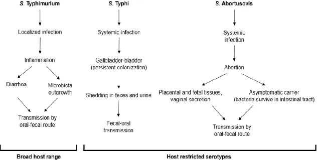
Figure 1. Different transmission strategies in different Salmonella serotypes.
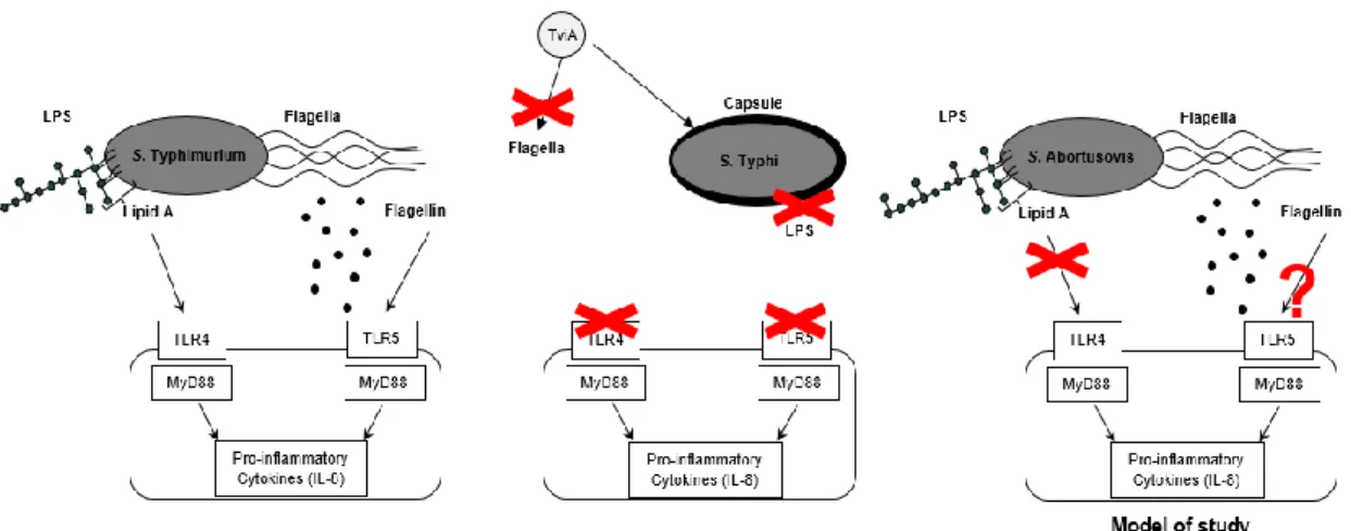
In Salmonella Typhimurium, bacterial flagellin and bacterial LPS are respectively
recognized by TLR5 and TLR4, causing the generation of CXC chemokines in a
process that has been implicated in neutrophil influx in the intestinal mucosa during
infection5. Recently, studies have shown that S. Typhi, in contrast to Salmonella
Typhiumurium, evades the recognition of TLR4 and TLR5. The regulatory protein TviA
represses the expression of flagella and the Vi capsular polysaccharide represses the
expression of the O-antigen. Through these mechanisms, S. Typhi evades receptor
recognition and the host responses6.
However, limited information is available about the immune response in sheep to S.
Abortusovis. S. Abortusovis, like S. Typhi, causes a systemic infection which the host
is not able to mount a prompt immune response to control infection before Salmonella
Abortusovis reaches the uterus and causes abortions7. Recently, it was shown that
LPS of Salmonella Abortusovis is not able to induce the expression of the CXC
chemokines, like IL-8, suggesting that this serotype is able to evade the TLR4
Very little is known about flagellin interaction with Salmonella Abortusovis. The
importance of flagella in host pathogen interaction is mainly based on studies of S.
Figure 2. Specific host responses for different Salmonella serotypes.
In this work we perform pioneering work to understand the role of flagella in S.
Abortusovis.
Our central hypothesis is to study how Salmonella Abortusovis evade activation of the
immune system. To this end, we compared differences between Salmonella
Typhimurium and Salmonella Abortusovis in recognition by TLR5 in vitro and in vivo.
Furthermore, we studied the major bacterial adaptive immune response as the
principal target for the humoral antibacterial response during the infection of the host.
The idea is that Salmonella Abortusovis highly regulates expression of flagella during
infection.
Previous work of studying Salmonella Abortusovis flagella in vitro had been difficult
since flagella are not readily expressed under standard laboratory conditions. In this
work we were able to identify a unique in vitro growth condition to express Salmonella
Abortusovis flagella in vitro for the first time.
Expression of flagella in vitro allowed us to study flagella expression in Salmonella
Abortusovis. To this end we generated defined mutations in Salmonella Abortusovis
Salmonella Typhimurium. We proceeded with the extraction of flagella from wild type
and mutant’s strains of the two different serovars using diverse media to observe
differences in flagellation and host interaction. We analyzed our samples with SDS
Page and Western Blot to determine expression of major flagellar protein. Also, we
verified surface expression of flagella by Flow Cytometry and by Scanning Electron
Microscopy (SEM). These different techniques enabled us to establish a media
suitable for inducing the expression of flagella in Salmonella Abortusovis, allowing us
to study Salmonella Abortusovis flagella in vitro for the first time.
To verify the capacity of S. Typhimurium and S. Abortusovis to stimulate TLR5, a
colorectal carcinoma cell line (T84 cells) was treated with purified flagellin and the
transcriptional induction of a CXC chemokine, IL-8, was measured in response to the
bacterial stimulation.
The fact that the expression of flagella only occurs in a particular media suggests that
Salmonella Abortusovis is able to control expression of flagellin to modulate activation
2. Introduction
2.1 Salmonella
Salmonella is an enteric pathogen of the Enterobacteriaceae family. Salmonella is
responsible for salmonellosis causing significant morbidity and mortality in humans
and animals with a socio-economic impact felt around the world.
The name Salmonella derived from Daniel Salmon’s name, a veterinary microbiologist
who first isolated Salmonella Choleraesuis from the intestine of a pig. Salmonella is a
Gram-negative facultative anaerobic bacterium. Motility is mediated by the presence of
peritrichous flagella.
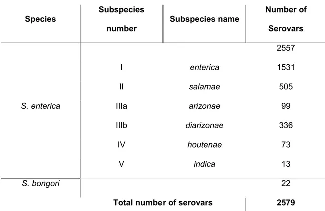
The genus Salmonella consists of 2 species, Salmonella enterica and Salmonella
bongori. S. enterica represents a group of 6 subspecies with multiple serovars9 (Table
Species Subspecies number Subspecies name Number of Serovars 2557 I enterica 1531 II salamae 505
S. enterica IIIa arizonae 99
IIIb diarizonae 336
IV houtenae 73
V indica 13
S. bongori 22
Total number of serovars 2579
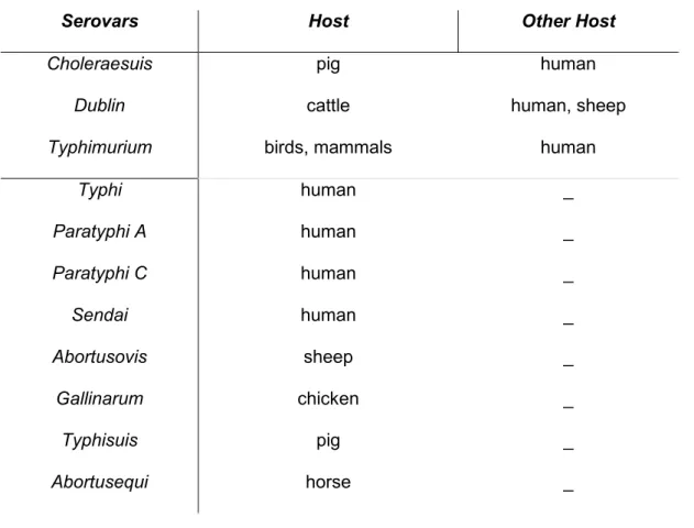
The different serotypes of Salmonella can be divided into two groups, host-specific
and non-host specific. The former are those that can infect a single animal species;
Serovars Host Other Host Choleraesuis Dublin Typhimurium pig cattle birds, mammals human human, sheep human Typhi Paratyphi A Paratyphi C Sendai Abortusovis Gallinarum Typhisuis Abortusequi human human human human sheep chicken pig horse _ _ _ _ _ _ _ _
2.2 Samonella enterica
Salmonella enterica is divided in 6 subspecies: enterica, salamae, arizonae, diarizonae, indica and houtenae.
Salmonella enterica subspecies enterica causes disease in vertebrates and is
transmitted through ingestion of contaminated food and water10.
2.3 Host specificity
Salmonella can be divided into two groups, host-specific and broad host range
serovars. Curiously, host specific serovars cause systemic disease in their respective
host species. For example, S. Gallinarum causes fowl typhoid in chicken, S. Dublin
causes a systemic diseases in cattle, S. Choleraesius and S. Typhisuis cause
septicemic and enteric salmonellosis in pigs and S. Abortusovis causes abortion in
sheep.
In human, specific serovar can cause severe systemic forms like typhoid in the case of
S. Typhi and paratyphoid with S. Paratyphi A, B and C11. In contrast, Salmonella broad
and reptiles, causing infections of varying severity depending on the serotype, the
mode of infection, and host characteristics12.
Salmonella Dublin and Salmonella Choleraesuis are non-specific serovars which
cause serious systemic diseases in cattle and pigs, but can also cause disease in
other mammalian hosts including humans13,14.
Serotypes like S. Dublin and S. Choleraesuis represent those microorganisms that are
prevalent in a particular species, but they can also cause disease in other host.
So it is important underscore the difference between host-specifc and host-restricted
serovars. The first group includes serovars which cause severe systemic infection in
their preferred host, but are usually excreted without any clinical symptoms in
accidental hosts.
In the second group are serovars which are restricted to one specific host.
Furthermore they exclusively cause systemic infection, which often proves to be fatal
The two serotypes commonly associated with human salmonellosis are S. Enteritidis
and S. Typhimurium; these pathogens are able to cause disease in many mammals,
including man1.
2.3.1 Salmonella enterica Typhimurium
S. Typhimurium is an enteric pathogen which cause diarrhea and gut inflammation and
during inflammation neutrophils are found in fecal samples16.
Salmonella enterica subspecies enterica serovar Typhimurium (S. Typhimurium) is
one serovar most widely associated with cases of human infection. Disease symptoms
usually develop after between 12 and 72 hours after infection in humans and the
transmission is oral-fecal, through the ingestion of contamined food or water. Infected
individuals develop gastroenteritis, fever, abdominal cramps, and diarrhea. Infection
typically lasts between 4 and 7 days and the majority of people recover without the
need for medical intervention17.
Once ingested, the bacteria travel through the stomach to the mucosa of the intestine
system-1 (T3SS-1) and type III secretion system-2 (T3SS-2) are required for causing
inflammation. Employing the T3SS-1 Salmonella Typhimurium, invades the intestinal
epithelium, and it is able to survive in mucosal macrophages using T3SS-218.
Initially, following the invasion of intestinal mucosa, the predominant influx cells were
the polymorphonuclear leukocytes (PMN)19,20.
S. Typhimurium is able to induce the apoptosis of PMN21, causing the production of
pro inflammatory cells and damage in the intestinal mucosa22.
Recently, it has been better understood an important mechanism of invasive capacity
of Salmonella Typhiumurium. The activation of gut inflammation is an important activity
for Salmonella to enhance colonization of the gastrointestinal tract. In fact, Salmonella
can use oxidation products that become available in this environment for respiration.
During the growth of the microbiota in the gut there is the formation of hydrogen
sulfide, a toxic product that is converted by oxidation in thiosulfate by the enterocytes23,
Thiosulfate is able to interact with reactive oxygen species (ROS), generate in the
intestinal lumen during inflammation by neutrophils, oxidizing thiosulfate in
tetrathionate.
The genes that allow Salmonella to use tetrathionate as an electron acceptor, enable
this pathogen to outgrowth the normal microbiota. The outgrowth in the lumen on the
intestine permit the transmission of S. Typhimurium25 (Figure 1).
Under anaerobic conditions, tetrathionate respiring S. Typhimurium is able to use
fermentation end products, which results in a substantial selective advantage for this
pathogen.
Salmonella Typhimurium is then able to grow and proliferate in competitive habitats
such as the intestine, since it is able to exploit the environment and substances
produced by the host26.
2.3.2 Salmonella enterica Typhi
Salmonella enterica subspecies enterica serovar Typhi (S. Typhi) is the causes of
S. Typhi is a highly adapted human-specific pathogen, usually contracted by ingestion
of food or water contaminated by fecal or urinary carriers excreting S. Typhi28.
Unlike other Salmonella serovars, which typically cause gastroenteritis characterized
by a massive neutrophil influx in the intestinal mucosa, S. Typhi causes a systemic
disease in which neutrophils are scarce in intestinal mucosa29.
The bacterium is serologically positive for the polysaccharide capsular antigen Vi
which is largely restricted to S. Typhi, and Vi-negative strains of this serovar are less
infectious and less virulent than Vi-positive strains27.
Initial colonization and proliferation is not associated with other disease symptoms,
suggesting that S. Typhi evades induction of immune response during infection.
S. Typhi must survive the gastric acid barrier to reach the small intestine, and a low
gastric pH is an important defense mechanism, in fact expression of tviA, which is
located in the viaB locus of S. Typhi, is activated by low tissue osmolarity when the
pathogen enters the intestinal mucosa.
The viaB locus has recently been implicated in preventing the generation of host
The mechanism through which the viaB locus reduces IL-8 production in human
intestinal epithelial cells is through the modification of the expression of Vi-capsular
antigen of TLR ligands on the bacterial cell surface, in fact the regulatory protein TviA
activates expression of the virulence-associated (Vi) capsular polysaccharide and
represses the expression of flagella. Furthermore activation of complement by the S.
Typhi O-antigen is obstructed by expression of the Vi capsular polysaccharide, which
attenuates neutrophil recruitment6.
Through these mechanisms, S. Typhi prevents initial activation of several pattern
recognition receptors that contribute to host responses against non-typhoidal
Salmonella serovars.
Following entry into the small intestine, the bacteria cross the intestinal epithelial
barrier, and then are phagocytosed by macrophages and spread systemically,
producing acute disease.
The most common sites of infection are the ileum, liver, spleen, bone marrow and gall
bladder. The bacteria reach the gall bladder through the vasculature or the ducts that
S. Typhi persists in the gall bladder through invasion of gall bladder epithelial cells.
Invasive bacteria replicate intracellularly, and shedding could occur as a part of
epithelial regeneration. Gall bladder epithelial cells containing S. Typhi would be
extruded to the lumen, and released bacteria could infect new cells or be shed into the
intestine via bile32.
Human carriers shed Salmonella Typhi in their feces, which can then contaminate food
and beverage, continuing fecal-oral transmission (Figure 1).
2.3.3 Salmonella enterica Abortusovis
Salmonella enterica subspecies enterica serovar Abortusovis (S. Abosrtusovis) is a
host specific serovar. It is restricted to sheep and does not cause any infection in
humans. Salmonella Abortusovis causes abortion in sheep which is an important
health problem in European countries where sheep-farming is the base of the
economy. Salmonellosis caused by Salmonella Abortusovis is common in France,
Spain, Germany, Switzerland and Italy33. Salmonella Abortusovis is very common in
The damage, both direct and indirect, includes the loss of lambs due to abortions and
loss of milk34.
Salmonella Abortusovis is introduced into a flock by an asymptomatic carrier35. When
S. Abortusovis is introduced for the first time in a flock, abortion becomes epidemic; in
later years the infection becomes endemic with sporadic abortions that occur most
often in young sheep or new sheep introduced into the flock. In the areas of
endemicity, abortion occurs in 30 or 50% of sheep in a flock, generally during the first
pregnancy and mainly during the last 2 months of gestation (a total of 5 months),
through an unknown mechanism36. Furthermore, after the first infection sheep
develops a protective immunity against Salmonella Abortusovis37.
The infected animals do not display other symptoms of diseases, in fact the only
symptoms is the abortion that usually occurs in the second half of the gestation.
Abortion occur several weeks after infection without any other symptoms before, and
sometimes is lethal for the sheep due to placental retention. Furthermore, lambs may
appear to be healthy but die within 3 weeks; some of them have diarrhea or symptoms
of pulmonary infections33.
During necropsy of infected lambs it is common to observe edema, hemorrhagic sites
and necrosis of different organs34.
After the abortion bacteria can be isolated from placental and fetal tissues as well as
from liver, spleen, brain and stomach, suggesting that those are the principal site of
multiplication. After 2-4 weeks is possible isolate bacteria from the vaginal secretion of
sheep38.
After a short period of bacteremia, where some organs like liver, spleen and lungs are
colonized, bacteria can be expelled by the host or they can survive in the intestinal
tract or in the gut, as in the asymptomatic carrier (Figure 1).
It is generally assumed that transmission occurs directly from the aborted fetus, the
placenta and uterine excretion. In fact, the fetus and placenta represent the most
important sites for the replication of the bacteria.
Nothing is known about a possible indirect transmission through foods and other
Salmonella Abortusovis is a contagious disease; to prevent its spread there are two
different kind of prophylaxis: direct and indirect prophylaxis.
Direct prophylaxis entails the elimination of the contaminated products and the
disinfection of all materials and surfaces exposed to this environment.
Indirect prophylaxis is characterized by the administration of attenuated or active
vaccine, those are more efficient but they are not available in Europe.
At the moment, no vaccine is available to immunize sheep against Salmonella
Abortusovis, but there are several promising studies.
2.3.4 Salmonella enterica Abortusequi
Salmonella enterica subspecies enterica serovar Abortusequi (S. Abortusequi) is a
host-adapted serotype specific to horses and donkeys that has been isolated in
association with abortion and a range of clinical conditions in foals39.
S. Abortusequi is usually transmitted by direct or indirect contact with pasture, food or
water that is contaminated with uterine discharges from carriers. Stallions can also
Clinical signs include late abortion (7-8 months) and retention of placenta and metritis
in mares. In foals born from infected mother there are clinical signs like acute
septicemia in the first week of life and polyarthritis in the second week of life. In
stallions symptoms are fever, orchitis with swelling in the prepuce and scrotum and
arthritis40.
The most common symptom is abortion and there are usually no premonitory signs of
impending abortion or other clinical signs are observed.
Antibiotic treatments against S. Abortusequi are efficient and inactivated vaccines are
available.
2.4 Host cell interaction
Host defense against invading microbial pathogens consists of two components:
innate immunity and acquired immunity.
Innate immune recognition is based on the detection of constitutive and conserved
endogenous danger-associated molecular pattern molecules (DAMPs) produced by
damaged tissues41.
Pattern-recognition receptors (PRRs), which include NOD-like receptors (NLRs) and
Toll Like receptors (TLRs), comprise the early components of the immune system that
function to detect invading pathogens through PAMPs and DAMPs and signal to
recruit and activate phagocytic cells such as neutrophils and macrophages42.
Specifically TLRs recognize microbes on the cell surface and in endosomes, whereas
NLRs detect microbial components in the cytosol.
These receptors trigger an immune response and are key to establishing an important
network between the innate and adaptive immune systems. Flagella, and LPS are
examples of PAMPs, which activate TLR5 and TLR4, respectively, in the host43.
In contrast the induction of an adaptive immune response begins when a pathogen is
ingested by an immature dendritic cell in the infected tissue. The adaptive immune
system provides further protection in addition to an immunological memory, which
2.5 Toll-Like Receptors
The TLR system provides important protection against microbes and they are present
mainly in macrophages, in mast cells, dendritic cells and endothelial cells.
The immediate protection provided by these receptors is based on stimulation of
macrophages or mast cells through TLR, through the synthesis and secretion of
pro-inflammatory cytokines, and the initiation of the pro-inflammatory process.
TLRs comprise a family of type I transmembrane receptors, which are characterized
by an extracellular leucine-rich repeat (LRR) domain and a citoplasmatic domain
homologous to the interleukin -1 (IL-1) receptor (TIR) domain44,45,46,47.
In mammalian species there are at least ten TLRs, and each have a distinct function in
innate immune recognition.
The different TLRs can be grouped on the basis of the cellular localization. The
receptors that recognize surface components of bacteria are found on the outer
membrane and the TLRs that recognize microbial nucleic acids are located on
The TLR2 can form heterodimers between TLR2 and either TLR1 or TLR6. For this
reason the TLR2 seems to be involved in the recognition of cell wall fragments, such
as peptidoglycan from Gram-positive bacteria, bacterial lipoproteins and
lipoarabinomannan.
They are found on the cell surface of monocytes, macrophages, dendritic cells, and B
cells. It is interesting to note that both TLR1 and TLR6 are expressed constitutively on
many cell types, whereas expression of TLR2 is regulated and seems to be restricted
to antigen-presenting cells and endothelial cells48.
TLR4 is found on the cell surface of monocytes, macrophages, myeloid dendritic cells,
mast cells and the basolateral side of intestinal epithelium. They are able to recognize
lipopolysaccharide (LPS) of Gram-negative bacteria.
Recognition of LPS by TLR4 requires several accessory molecules. LPS is first bound
to a serum protein, LBP (LPS-binding protein), which functions by transferring LPS
monomers to CD14. Another component of the LPS receptor complex is MD-2 that is
required for LPS recognition by TLR4. MD-2 is a small protein expressed on the cell
TLR5 is located on the cell surface of monocytes, macrophages, dendritic cells, and
on the basolateral side of the intestinal epithelial cells. TLR5 recognizes conserved
domains of flagellin present on the bacteria.
Recently, it was shown that TLR11 expressed in mice recognizes bacterial flagellin
and triggers a protective immune response against Salmonella Typhi50.
Of the 11 types of toll-like receptors TLR3 and TLR4 use the interleukin-1 receptor
associated kinase 4 (IRAK-4) as a transmitter for interferon. The IRAK-4 is essential
for the early induction of pro-inflammatory cytokines, such as tumor necrosis factor α
(TNF-α), interleukin-1β and interleukin-6.
2.5.1 Toll-Like Receptor 5
The epithelium represents the first line of defense for invading pathogens. TLR5 is the
most prominently expressed TLR.
TLR5 specifically recognizes bacterial flagellin, the principal component of bacterial
flagella. The activation of this receptor mobilizes the nuclear factor NF-kappaB, which
Flagellin is the only known activator of TLR5, and until recently flagellin-induced
inflammation was believed to be fully dependent on TLR5 expression. TLR5 is
responsible for flagellin-induced responses in epithelial cells, endothelial cells,
macrophages, dendritic cells (DCs), and T cells51.
There is strong evidence that the TLR5-activating region of flagellin is located within
the conserved domains that are constrained by the need to provide motility.
The structure of flagellin in Salmonella Typhimurium consists of four domains, D0, D1,
D2 and D3. D0 and D1 are conserved between bacteria species and are located on
the outside of the flagellum, D3 and D4 are variable domains that form the inner core
of flagellum52,53,54.
Human TLR5 recognize the inner core of flagellum filament which are the conserved
domains.
After TLR5-expressing cells are stimulated with flagellin, there is a signaling cascade
that involves phosphorylation of interleukin-1 receptor-associated kinase 1, leading to
activation of MEK kinases and I-kB kinases, which ultimately activates inflammatory
flagellin, TLR5 triggers myeloid differentiation primary response gene 88
(MyD88)-dependent signaling pathway to induce pro-inflammatory cytokine, such as the
neutrophil attracting chemokine CXC interleukin-8 (IL-8).
Flagellin activation of TLR5 requires a complicated interaction that could involve
additional protein-protein or protein-lipid interactions that are not yet understood.
2.6 Flagella
Flagella are significant for bacterial pathogenesis for two reasons, they allow bacterial
cells to move and are essential for microbial survival and additionally they are PAMPs
recognized by the immune system.
In Salmonella, flagella originate from the bacterial surface and they are dispersed on
all the surface of the cell. They vary in number between 5 and 10 per cell.
The flagellar filaments have a consistent diameter of approximately 20 nm but vary in
2.6.1 Flagella Structure
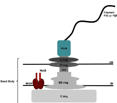
The flagella structure consists of three parts: the basal body, the hook and the filament
Figure 3. Flagella structure in Salmonella.
The basal body connects the flagella to the bacterial cell and consists of a rod and
several rings. Some of the rings make up the flagellar motor, which is divided into 2
major parts, the stator (MotA and MotB) that remains stationary, and rotor (C ring, MS
ring and rod). The remaining rings L and P located in the outer membrane are
stationary57. The MS ring is immersed in the cytoplasmic membrane and complexes
with proteins MOT (which allow the motion due to a proton-motive force) and protein
FLI (which reverse the rotation of the hook).
The hook is a single protein with the function of connecting the filament to the basal
body.
It is built in a similar way to the filament, and during the formation the hook-filament
junction proteins stay in place and the filament-capping protein (FliD) moves outward
as flagellin monomers polymerize.
The filament consist mainly of two proteins, FliC or FljB, and it is a cylindrical structure
made up of around 20,000 to 30,000 flagellin (FliC or FljB) subunits58, 59. The filament
can rotate on the left or on the right direction. The ‘normal’ form which is more stable is
2.6.2 Flagella Energetics
The energy for flagella is produced using a proton gradient, which produces proton
motive force61, 62. MotA and MotB proteins form a complex and are part of the stator
part of the motor and are involved in proton conduction.
MotA appears to be important for delivery of protons across the membrane and the
utilization of those protons in generating torque, MotB appears to be important for the
proton channel.
2.6.3 Flagella rotation and motility
The rotary movement is not random, a counter-clockwise movement creates motion
and a clockwise arrests motion. Maximum speed varies between 3 and 60 µm/s.
The motor contains a switch which allows the filament to be rotated in
counter-clockwise or counter-clockwise directions56, 63. When the left handed form is rotated in a
counter-clockwise direction the cell is pushed forward.
The motor can alternate through an interaction between a phosphorylated component
rotor: FliG that is most directly involved in rotation of the motor, FliN plays a part in
rotation and interacts with the stator protein MotA. The MS-ring proteins FliF and FliM
have a large role in switching between counter-clockwise and clockwise rotation by
binding to CheY phosphate.
2.6.4 Flagella genes
Regulation of flagellar assembly involves a combination of transcriptional, translational
and post-translational regulatory mechanisms.
The assembly of the flagellum can be divided into 7 stages: MS-ring formation,
assembly of the C-rings and secretion apparatus, rod formation, assembly of the P and
L rings, leading hook synthesis, hook completion, secretion substrate specificity switch
and filament elongation.
Assembly begins with the insertion of the MS-ring into the inner membrane. The
individual extra cytoplasmic flagellar subunits are secreted through the MS-ring after
In Salmonella the flagellar regulon consists of 17 operons, divided into classes 1, 2
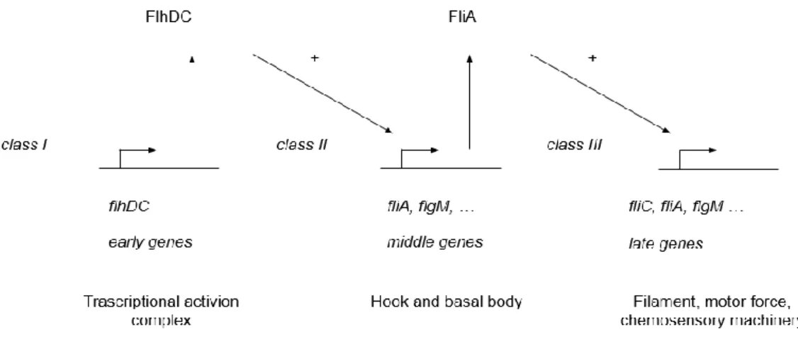
Figure 4. Trascriptional regulation of flagellar assembly in Salmonella.
The early genes belong to the class 1 promoters which control the expression of the
entire regulon. The middle gene possess class 2 promoters and are important for the
production of structural components of the hook and of the basal body. The late genes
belong to the class 3 promoters which products include the filament, the motor force
generators and chemosensory machinery.
During flagella formation, the first genes to act are the class 1 genes, flhDC.
FlhD and FlhC form FlhD4C2, an hexameric complex64, 65. This complex is a
transcriptional activator for σ70-dependent transcription of the class 2 promoters66.
The class 2 genes include fliA (σ28) which is required for transcription of many of the
class 3 genes, although some class 3 genes can be expressed independently of fliA67.
In addition, flgM and flgN genes can be expressed independently of fliA by read
through of the transcript from flgA, a class 2 promoter68,69,70,71.
flgM is the anti-fliA (σ28) factor that negatively regulates fliA by binding it. Upon
completion of the basal body-hook structure, the flagellar protein export apparatus
genes is then removed and transcription initiation by FliA dependent class 3 flagellar
genes can then proceed68.
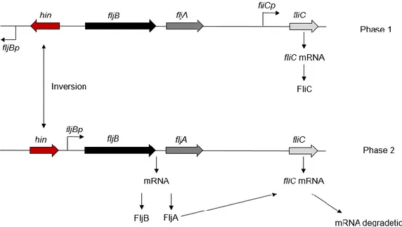
2.6.5 Flagellar phase variation
In S. Typhimurium there are two different flagellar filament proteins, FljB and FliC,
which are alternately expressed by a mechanism known as phase variation72. The
molecular mechanism mediating flagellar phase variation occurs by a site-specific
DNA inversion event in the chromosome by the stochastic inversion of a promoter, the
hin switch, which controls both FljB flagellin and an inhibitor (FljA) of FliC flagellin
formation. In one orientation the fljBA promoter directs transcription of the fljBA operon
and FljB flagellin is produced. The fljA gene is cotranscribed with fljB and encodes a
transcriptional inhibitor of the fliC gene. In the alternate orientation, the fljBA operon is
not expressed and transcription of the fliC gene ensues. So when the fljB-fljA operon is
expressed, only FljB flagellar filaments are produced; when the operon is not
transcribed, the gene for FliC flagellin (fliC) is released from inhibition and FliC flagellar
Figure 5. Flagellar phase variation in Salmonella Typhimurium.
The presence of a second phase type of flagella may help Salmonella to evade the
3. Hypothesis and aims
Limited information is available about the immune response of sheep to Salmonella
Abortusovis.
The objective of this application is to study the differences of innate immune
recognition and host responses to this pathogen compared with S. Typhimurium.
Our central hypothesis is that direct contact of Salmonella Abortusovis with host cells
does not trigger an initial innate immune response allowing the organism to reach
systemic sites.
Normally, epithelial cells initially herald the presence of invasive pathogens. After
penetration of the intestinal epithelium, bacteria encounter mononuclear cells, whose
stimulation through TLRs results in the release of cytokines (e.g. IL-8 and TNF-).
We hypothesized that Salmonella Abortusovis is able to escape the recognition by
TLR5 with an ‘on/off’ mechanism, in which initially flagellin is ‘off’, but later during the
In this work we want test our hypothesis that recognition of PAMPs, which normally
contributes to the initiation of inflammatory response in the intestinal mucosa, is
different during the infection of Salmonella Abortusovis in sheep.
To this end, we decided to study the initial interaction of Salmonella with the epithelium
using a colonic epithelial cancer cell line T84 (American Type CultureCollection) to
study if the serotypes Typhimurium and Abortusovis during infection of intestinal
epithelium are able to invade equally, and to understand if flagella in S. Abortusovis
are able to activate the TLR5.
The result of this analysis clarified the fundamental mechanisms leading to systemic
infection, which improved our understanding of systemic disease in general.
Our results provided first an important mechanistic insight into how S. Abortusovis is
able to modulate host responses that distinguish abortions from infections with S.
Typhimurium and we elucidated how serovar Abortusovis is able to not trigger the first
innate immune response, which is important in the early recognition of this pathogen.
This outcome was significant because it established key events and provided insights
thereby refining a paradigm for innate immune host-response to invasive enteric
pathogens.
The picture emerging in this study is that multiple factors are involved in initiating a
pro-inflammatory response in the intestinal mucosa during Salmonella Typhimurium
4. Materials and methods
4.1 Bacterial strains and growth conditions
The bacterial strains used in the present study are Salmonella Typhimurium,
Subspecies Strain Parent Genotype Source
S. Typhimurium SR11 SR11 wild type
Strain Collection S. Typhimurium TH1077 LT2 fliC5050::MudJ 74 S. Typhimurium TH714 LT2 fljB5001:: MudJ 75 S. Typhimurium TH4623 LT2 flgM5222::MudCm 76 S. Typhimurium TH4054 LT2 flhC5456:: MudJ 74
S. Abortusovis SS44 SS44 wild type 77
S. Abortusovis LS1 SS44 fliC5050:: MudJ This study
S. Abortusovis LS2 SS44 fljB5001:: MudJ This study
S. Abortusovis LS3 SS44 flgM5222::MudCm This study
S. Abortusovis LS4 SS44 flhC5456:: MudJ This study
S. Abortusequi wild type
Strain Collection
Strains (Table 3) were routinely grown at 37°C in LB broth. Undiluted serum of sheep,
young sheep and pregnant sheep were used to grow bacterial strains to test if, in
specific media, S. Abortusovis is able to express flagella in vitro. Strains were grown
over night in different media, and depending on the experiment directly used or diluted
1:50 in LB or in serum for 3 h.
Blood from sheep, young sheep and pregnant sheep was collected from a flock in the
Sardinian region, by a veterinarian that collaborated previously in this study.
Before blood collection, individual sheep were analyzed and no Salmonella infection
was detected as determined by rectal swab tests and plating on Salmonella-Shigella
agar plates (SS agar). Those plates contains lactose. If lactose fermentation occurs,
the medium will turn red due to the acidic pH. Salmonella, as non-lactose fermenters,
appears as transparent or translucent colorless colonies on SS agar. Samples were
also streaked on non-selective but differential medium such as MacConkey Agar.
Serological confirmation tests using polyvalent antisera for flagellar (H) and somatic
Blood was collected in heparin tubes, and it was treated in lab by centrifugation at
5,000 rpm for 30 min. Serum was collected and stored at -20°C.
4.2 M9 minimal medium
M9 minimal medium was used in this study to analyze the expression of flagella in
Salmonella Typhiumurium and in Salmonella Abortusovis.
This media was prepared using a final volume of 100 ml, 20 ml of M9 salts 5X
(Sigma-Aldrich), 2 ml of Glucose 20 % (Sigma-(Sigma-Aldrich), 200 µl of MgSO4 1M (Fisher
Scientific), 10 µl of CaCl2 1 M (Fisher Scientific) and 78 ml of H2O.
M9 minimal medium was filter sterilized and used to grow different serovars.
4.3 Phage transduction
Mutations of flagella genes were transduced in Salmonella Abortusovis to study
changes in motility using samples of Salmonella Typhimurium strains that had been
A P22 lysate of S. Typhimurium fljB5001::MudJ, S. Typhimurium fliC5050::MudJ, S.
Typhimurium flhC5456::MudJ, S. Typhimurium flgM5222::MudCm was used to
transduce in serotype Abortusovis flagella proteins mutations. The mutations were
Gene Sequence (5’ –3’) Source fliC AAGTCAGGTTGTTTACGGTGTTGC TGTCGCTGTTGACCCAGAATAAC This study flhC CAGCATCTCGGGAAAGTTTACG GCTTTATCTTGAGCAGTGTCCGC This study fljB GACAGATTGTTTACGGTATTGCCC GTATTACGCCGCAGATTACGATG This study flgM TGAGCGAGTCTGCTATTTTTCCC TGAGCATTGACCGTACCTCACC This study
Phage lysates were typically prepared by growing a 5 ml culture of the donor strain in
LB broth with the appropriate antibiotics and incubate at 37°C over night (O/N) with
aeration.
A volume of 4 ml of P22 broth were added to 1 ml of bacterial culture and incubate at
37°C for 16 h with aeration.
A volume of 1.4 ml of culture was centrifuged for 2 min at 12,000 g and the
supernatant was transferred to a cryovial, and 0.2 ml chloroform was added and
vortexed for at least 1 min.
For the transduction, the recipient strain was growth in LB broth with the appropriate
antibiotics and incubate at 37°C O/N with aeration.
Three 10-fold serial dilutions were prepared of the phage lysate in 0.1 ml PBS, and 0.1
ml of the recipient strain was added to each of the diluted phage lysate and incubate at
RT for 1 h.
Samples were centrifuged at 20,000 g for 1 min and the pellet was resuspended with
0.1 ml of LB and plated on LB plates containing the appropriate antibiotics and
Individual single colonies were streaked for single colonies on EBU plates, containg
25% (w/v) K2HPO4, 1% Evan’s Blue and 1% Sodium Fluoresceine, and incubated for
16 h at 37°C.
One colony from each streak was cross-streaked with 0.01 ml of P22 H5 on EBU
plates, incubated at 37°C for 8 h and the positive colonies were streak for single
colonies on LB plates containing the appropriate antibiotics.
4.4 Motility analysis in Motility plates
For motility assays, plates containing 10 g of tryptone/liter, 5 g of NaCl/liter, and 3 g/L
agar were inoculated with 10 µl of the indicated O/N cultures and incubated at 37°C 16
h.
4.5 Purification of bacterial flagella
The protocol followed for the flagella extraction is based on trichloroacetic acid
S. Typhimurium and S. Abortusovis were grown for 16 h at 37°C with aeration, diluted
1:50 in 6 ml of LB broth, and grown for 3 h at 37°C with aeration.
Flagella were sheared by treating the bacterial culture with a homogenizer (IKA) on ice
for 1 min. The suspension was centrifuged at 3,220 g for 10 min at 4°C, and the pellet
was washed with trichloroacetic acid to a final concentration of 10 %, followed by
centrifugation at 12,000 g for 30 min at 4°C. The pellet was washed two times with
ice-cold acetone and resuspended in 0.05 ml of water.
4.6 Sodium Dodecyl Sulphate Polyacrylamide Electrophoresis (SDS-PAGE)
The samples were boiled for 5 min with loading buffer (1x) containg 62.5 mM Tris-HCl
pH 6.8, 2.5 % SDS, 0.002 % Bromophenol Blue, 5 % β-mercaptoethanol, 10 %
glycerol.
A volume of 0.01 ml of sample was separated by sodium dodecyl sulfate 12 %
polyacrylamide gel electrophoresis. Gels were composed of 10.72 ml of H2O, 8 ml of
Tris-HCl 1.5 M (pH 8.8), 12.8 ml of acrylamide, and 320 µl of 10 % SDS. 0.05 %
The 4 % stacking gel was composed of 6.1 ml of H2O, 2.5 ml of Tris-HCl 0.5 M (pH
6.8), 1.3 ml of acrylamide, 10 % SDS and 10 % TEMED were added for the
polymerization.
Gel were visualized with Coomassie Blue 250 (Sigma), 0.2g of Coomassie Blue
G-250 were dissolved in 100 ml of H2O warmed to approximately 50°C. 100 ml of 2N
H2SO4 were added. The solution was filtered and 22.2 ml 10N KOH and 28.7g TCA
were added.
To stain the gel were immersed in the solution for 15 minutes, and stored in 7 %
HOAC.
4.7 Western Blotting
Protein extracted and separated by 12 % (v/v) SDS-PAGE gels were blotted onto
nitrocellulose membrane using a semi-dry method.
Blots were blocked in 3 % (w/v) skimmed milk powder (Sigma), 0.1 % (v/v) Tween20
Primary antibodies, anti-FliC monoclonal rabbit (Difco), were diluted in 5 % (w/v)
Bovine Serum Albumin (Sigma), 0.1 % (v/v) Tween20 in PBS and incubated with blots
for 2 h at 4°C, with gentle rocking.
Blots then were washed three times in 3 % milk solution at 5 min intervals with gentle
rocking at RT.
Horseradish peroxidase (HRP)-conjugated anti-rabbit IgG (BD) secondary antibodies
was diluted in 5 % (w/v) Bovine Serum Albumin, 0.1 % (v/v) Tween in PBS and were
incubated with blots for 1h at RT with gentle rocking.
The membrane was washed with 3 % milk solution at 5 min intervals three times and
three times with PBS at 5 min intervals.
Blots were developed using a chemi-luminescence reagent (EuroClone), and the
signal from the blots was captured using a specific software, Image Lab Software
(BioRad).
4.8 Flow Cytometry
The samples were centrifuged at 3,000 rpm for 10 min and resuspended in 0.1 ml of
PBS. 4 % paraformaldehyde was added and the samples incubated at RT for 20 min.
The samples were washed two times with PBS containing 0.02 % of gelatin,
resuspended in 0.5 ml 2 % Normal Goat Serum, and incubated for 30 min at RT.
1:250 dilution of the primary antibody was added directly and incubated at RT for 1
hour. After the incubation the samples were centrifuged at 3,000 rpm for 10 min, and
washed three times with PBS containing 0.02 % gelatin.
The samples were resuspended ml 0.5 ml 2 % Normal Goat Serum and 1:250 FITC
secondary antibody and 0.025 ml of Propidium Iodide Solution (1 µg/ml). They were
incubated for 1 hour in the dark at room temperature, and after the incubation washed
3 times with PBS containing 0.02 % gelatin.
After centrifugation at 3,000 rpm for 10 min, the final pellet was resuspended in 10 ml
of PBS.
BD Accuri C6 Flow Cytometer (BDBioScience) was used for this experiment and the
4.9 Scanning Electron Microscopy (SEM)
Processing and imaging of samples for scanning electron microscopy (SEM) was
carried out at the Electron Microscopy facility run by Salvatore Marceddu in the
University of Sassari.
The samples were prepared by growing S. Typhimurium and S. Abortusovis for 16 h at
37°C with aeration in different media, diluted 1:50 in 6 ml of media, and growth for 3 h
at 37°C with aeration.
Flagella were sheared by treating the bacterial culture with a homogenizer (IKA) on ice
for 1 min. The suspension was centrifuged at 3,220 g for 10 min at 4°C, and the pellet
was washed with trichloroacetic acid to a final concentration of 10 %, followed by
centrifugation at 12,000 g for 30 min at 4°C. The pellet was washed two times with
ice-cold acetone and air-dried. The samples were send to the microscopy facility for image
4.10 T-84 cell line infection
Human colorectal carcinoma cell line (T-84) (Invivogen) was cultured in DMEM/F12
media supplemented with Hepes buffer solution, Glutamax and 10 % FBS (Giboco).
Cells were split and a 24 wells plate was prepared for the experiment. Cells were used
at a density of 1*106 cells/well. After 24 h the media was changed in media without
serum.
After a total of 48 h flagella were added for 60 min (flagella samples 5 µl / PBS control
10 µl).
The flagella used for the T84 stimulation was extracted after growing the bacteria
culture O/N in LB or serum. A volume of 0.1 ml of overnight culture was inoculated in 5
ml of LB or serum, and grown at 37oC for 3 h. The samples were treated with a
homogenizer (IKA) on ice for 1 min and centrifuged at 3,220 g for 10 min at 4oC, and 5
4.11 RNA extraction
Total RNA was extracted with TRI Reagent (Molecular Research Center), according to
the recommendation of the manufacturer.
Briefly, 1 ml of TRI REAGENT was added to each wells and the cell lysate was passed
several times through a pipette.
The homogenate was stored for 5 min at room temperature to permit the complete
dissociation of nucleoprotein complexes, and the homogenate was supplemented with
0.2 ml chloroform and shaken vigorously for 15 seconds. The resulting mixture was
incubated for 15 min and centrifuged at 12,000 g for 15 min at 4oC.
Aqueous phase was transferred to a fresh tube and 0.5 ml of isopropanol was added
per 1 ml of TRI REAGENT used for the initial homogenization. The samples were
incubated at room temperature for 10 minutes and centrifuged at 12,000 g for 8 min at
4oC.
The RNA pellet was washed with 75% ethanol and centrifuged at 7,500 g for 5 min at
RNA pellet was air-dried for 15 min and dissolved in sterile water made RNase-free by
diethyl pyrocarbonate (DEPC) treatment.
For RT-PCR analysis, DNase treatment was necessary for optimal results.
4.12 DNase treatment
A volume of 20 µl of 10X DNase I Buffer and 1 µl rDNase I (Ambion) were added to
the RNA, and incubated at 37°C for 30 min. A volume of 1 µl of resuspended DNase
Inactivation Reagent was added and incubated 2 min at RT.
The samples were centrifuged at 10,000 g for 1 min, and the RNA was transferred to a
fresh tube.
The RNA obtained was used for the RT-PCR.
4.13 RT-PCR
Briefly, 2 µg of RNA was added to 1 µl of random primers (InvivoGen) and 1 µl to 3 µl
of H2O, for a final volume of 5 µl, and incubated at 70° C for 5 min followed by 10 min
A volume of 5 µl were added, for a final volume of 20 µl, to a mixture contained 4 µl of
H2O, 3 µl of MgCl2, 4 µl of buffer 5X, 2 µl RNasi inhibitor, 1 µl of nucleotides and 1 µl of
Taq (InvivoGen).
The samples were incubated for the following RT-PCR cycle: 5 min at 25° C, 60 min at
42° C and 15 min at 70°C.
The samples were used for the Real-Time PCR.
4.14 Real-Time PCR
Real-time PCR was performed using SYBR green (Promega) and the 7900HT Fast
real-time PCR system.
To determine transcription of the IL8 gene, the following primers for GAPDH gene and
for IL8 were used: GAPDH, 5’-3’ FW: CTGCTTTGATGTCAGTGCTGCTAC and RW:
CTGCCGTGTGAAGCCCACAATAAA; IL-8, 5’-3’ FW:
GCCAACACAGAAATTATTGTAAAGCTT and RW: CCTCTGCACCCAGTTTTCCTT.
The analysis was performed in triplicate using the following cycle: 95°C for 10 min
The fold change in mRNA levels was determined using the comparative threshold
cycle (CT) method (Bio-Rad).
For statistical analysis (fold increases in IL-8 expression), data were transformed
logarithmically to calculate geometric means. A parametric test (paired Student’s t-test)
was used to calculate differences in the increases in IL-8 expression. A two-tailed P
5. Results
5.1 The right media is necessary to express Salmonella Abortusovis flagella
in vitro
Our goal of studying the role of flagella in host-pathogen interaction was initially
hampered by the fact that flagella are not expressed by Salmonella Abortusovis under
standard laboratory conditions. Thus, we sought to experimentally identify a growth
condition suitable for the expression of flagella in vitro. Specifically, we varied carbon
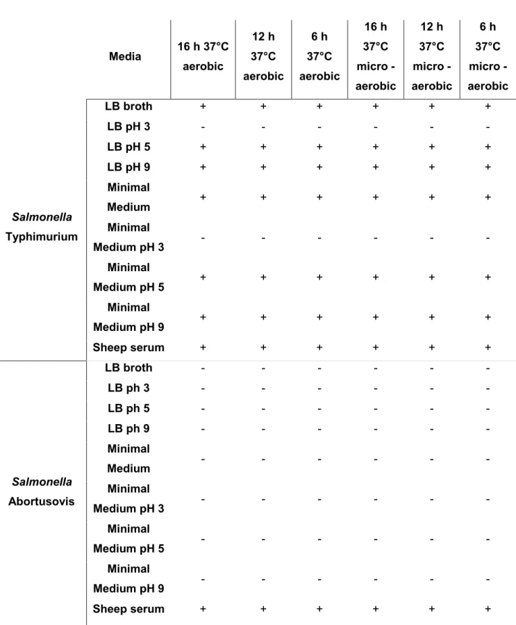
sources, pH, growth phase and temperature. Motility was used as a functional
Table 5. Flagella expression in different media. Media 16 h 37°C aerobic 12 h 37°C aerobic 6 h 37°C aerobic 16 h 37°C micro - aerobic 12 h 37°C micro -aerobic 6 h 37°C micro -aerobic Salmonella Typhimurium LB broth + + + + + + LB pH 3 - - - - LB pH 5 + + + + + + LB pH 9 + + + + + + Minimal Medium + + + + + + Minimal Medium pH 3 - - - - Minimal Medium pH 5 + + + + + + Minimal Medium pH 9 + + + + + + Sheep serum + + + + + + Salmonella Abortusovis LB broth - - - - LB ph 3 - - - - LB ph 5 - - - - LB ph 9 - - - - Minimal Medium - - - - Minimal Medium pH 3 - - - - Minimal Medium pH 5 - - - - Minimal Medium pH 9 - - - - Sheep serum + + + + + +
As shown in Table 5, Salmonella Typhimurium expresses flagella in almost all media
condition tested. In contrast, no expression of flagella was observed in S. Abortusovis
serotype. The same results were observed using the same media at 30°C.
We reasoned that the absence of a specific inducer may prevent flagella expression
and motility in vitro. Therefore we tested motility of Salmonella Abortusovis in a media
that better resembles in vivo growth condition, for example, the serum from sheep.
Interestingly, we observed that when sheep serum was used for growing bacteria
Salmonella Abortusovis was able to express flagella.
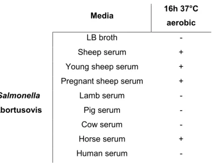
On the basis of those results different kind of sheep serum were tested. Additionally,
Media 16h 37°C aerobic
LB broth -
Sheep serum +
Young sheep serum +
Pregnant sheep serum +
Salmonella Lamb serum -
Abortusovis Pig serum -
Cow serum -
Horse serum +
Human serum -
As shown in Table 6, Salmonella Abortusovis expressed flagella in serum obtained
from sheep, but no expression was observed using other media as lamb serum, pig
serum, cow serum and human serum. Expression of flagella in this serotype was
observed using horse serum.
The status of the sheep and their pregnancy does not seems to play a role for the
expression of flagella and for the motility.
Those results were collected by studying the expression of flagella in motility agar
plates, and when flagella was expressed, a motility halo was observed. In addition to
the motility LB agar plates, in this study motility plates containing an equal mixture of
LB agar and each serum were used.
Salmonella Abortusovis is not able to move using LB media, in contrast to Salmonella
Typhimurium that showed motility in LB (Figure 6A). However, S. Abortusovis is able
to move in agar plates if serum of sheep is use to pre-grow this serotype. An
interesting result was also observed using horse serum, in this media Salmonella
Abortusovis showed a positive result, which means that flagella are expressed in this
Figure 6. Representative images of Salmonella Typhimurium (SR11) wild type (WT) and
Salmonella Abortusovis (SS44) WT in motility plates. A) SR11 WT and SS44 WT were grown 16 h in LB media at 37°C with aeration. A volume of 10 µl was inoculated on LB motility agar plates for 16 h at 37°C. This experiment was repeated three times. B) SS44 WT was grown 16 h in undiluted sheep serum, young sheep serum, pregnant sheep serum and horse serum, at 37°C with aeration. A volume of 10 µl was inoculated on motility agar plates containing an equal mixture of LB agar and each serum. This experiment was repeated three times.
Growth media that closely mimics the host is suitable to induce expression of flagellin
in a host-restricted serovar.
On the basis of this result Salmonella Abortusequi was growth using horse serum and
Figure 7. Representative images of Salmonella Abortusequi wild type (WT) in motility
plates. A) Salmonella Abortusequi WT was grown 16 h in LB media at 37°C with aeration. A volume of 10 µl was inoculated on LB motility agar plates for 16 h at 37°C. This experiment was repeated three times. B) Salmonella Abortusequi WT was grown 16 h in undiluted horse serum at 37°C with aeration. A volume of 10 µl was inoculated on motility agar plates containing an equal mixture of LB agar and horse serum. This experiment was repeated three times.
For this serotype it was observed that S. Abortusequi is not able to swim in motility
agar plate if grow in LB but motility was observed when the bacteria was grown in
horse serum.
As a control Salmonella Typhiumurium and Salmonella Abortusovis mutants were
tested on motility plates.
Salmonella Typhimurium non-flagellated mutants were not able to move (Figure 8A),
and the same was observed for Salmonella Abortusovis non-flagellated mutants using
Figure 8. Representative images of Salmonella Typhimurium (SR11) and Salmonella
Abortusovis (SS44) in motility plates. A) SR11 wild type (WT), S. Typhimurium fliC (TH1077), S. Typhimurium fljB (TH714), S. Typhimurium flgM (TH4623) and S. Typhimurium flhC (TH4054) were grown 16 h in LB media at 37°C with aeration. A volume of 10 µl was inoculated on LB motility agar plates for 16 h at 37°C. This experiment was repeated three times. B) SS44 WT, SS44 fliC (LS1), SS44 fljB (LS2), SS44 flgM (LS3) and SS44 flhC (LS4) were grown 16 h in sheep serum at 37°C with aeration. A volume of 10 µl was inoculated on motility agar plates containing an equal mixture of LB agar and sheep serum. This experiment was repeated three times.
So we were able to express Salmonella Abortusovis flagella in vitro using a media that
was as close as possible to the in vivo condition.
The next results will answer the question if now we can study this expression in vitro.
5.2 Flagellin expression in vitro
Although it is likely that the observed motility phenotype were due to flagella based
motility, other modes of motility have been reported in Salmonella, like Salmonella
Typhi has type IVB pili. To confirm that exposure to serum indeed resulted in the
upregulation of flagella proteins, we analyzed expression of the major subunit of phase
I flagella, the flagellin FliC by western blotting.
To understand if it is possible analyze flagella expression in Salmonella Abortusovis,
we decided to extract flagella from serovar Typhimurium and Abortusovis, both wild
type and mutant.
Following the trichloroacetic acid precipitation we were able to extract flagella, and
Our analysis confirmed our previous results obtained with motility agar plates.
Expression of flagella was detected in Salmonella Typhimurium growth in LB, and any
detection was observed for Salmonella Abortusovis growth in LB, but when S.
Abortusovis serotype was grown in different sheep serum we were able to observe the
expression of flagella (Figure 9A).
As a control, the same analysis was performed for the non-motile mutants in S.
Typhimurium and S. Abortusovis, and any flagellin detection was observed (Figure
9B).
On the basis of the results obtained with motility agar, we also analyzed the
expression of flagella in Salmonella Abortusovis growth in horse serum and of