Comments on triassiC pterosaurs with a Commentary on the “ontogenetiC stages” of Kellner (2015) and the validity of
Bergamodactylus wildi
Fabio M. Dalla Vecchia
Museo Friulano di Storia Naturale, Via Sabbadini 22-34, I-33100 Udine, Italy; Institut Català de Paleontologia Miquel Crusafont (ICP), Carrer de l’Escola Industrial 23, E-08201, Sabadell, Spain. E-mail: [email protected]
To cite this article: Dalla Vecchia F.M. (2018) - Comments on Triassic pterosaurs with a commentary on the “ontogenetic stages” of Kellner (2015) and the validity of Bergamodactylus wildi. Riv. It. Paleontol. Strat., 124(2): 317-341.
Abstract. Six stages (OS1-6) were identified by Kellner (2015) to establish the ontogeny of a given pterosaur fossil. These were used to support the erection of several new Triassic taxa including Bergamodactylus wildi, which is based on a single specimen (MPUM 6009) from the Norian of Lombardy, Italy. However, those ontogenetic stages are not valid because different pterosaur taxa had different tempos of skeletal development. Purported diagnostic characters of Bergamodactylus wildi are not autapomorphic or were incorrectly identified. Although minor differences do exist between MPUM 6009 and the holotype of Carniadactylus rosenfeldi, these do not warrant generic differentiation. Thus, MPUM 6009 is here retained within the taxon Carniadactylus rosenfeldi as proposed by Dalla Vecchia (2009a).
Received: February 19, 2018; accepted: April 17, 2018 Keywords: Pterosauria; ontogeny; Carniadactylus rosenfeldi; Triassic, Italy.
I
ntroductIonThe pterosaur specimen MPUM 6009 was discovered in 1971 or 1972 in the Norian (Upper Triassic) Calcare di Zorzino Formation at the Cene Quarry (Cene, Bergamo Province, Northwestern Italy) (Dalla Vecchia 2014). It was first identified as an immature individual of Eudimorphodon ran-zii Zambelli, 1973 by Wild (1979; it is reported as “Exemplar Milano”) and later retained as such in many following papers (e.g., Wild 1994; Dalla Vecchia 1998, 2003; Jenkins et al. 2001; Wellnhofer 2003). However, Kellner (1996, 2003), advanced doubts about the conspecificity of MPUM 6009 and the holotype of E. ranzii (MCSNB 2888). Dal-la Vecchia (2009a) demonstrated that MPUM 6009 differs from the holotype of Eudimorphodon ranzii and that differences cannot be considered as on-togenetic features as claimed by Wild (1979). Dalla Vecchia (2009a) listed several features that are uni-quely shared by MPUM 6009 and the holotype of Eudimorphodon rosenfeldi Dalla Vecchia, 1995 (spe-cimen MFSN 1797). Dalla Vecchia (2009a) consi-dered MPUM 6009 and MFSN 1797 to be con-specific and erected the new genus Carniadactylus
(monospecific, C. rosenfeldi) for them.Dalla Vecchia (2009a) also highlighted several differences betwe-en the two specimbetwe-ens, suggesting that they might alternatively belong to different (although closely related) species, but considered it more parsimo-nious to regard them as conspecific. Dalla Vecchia (2013, 2014) retained MPUM 6009 in Carniadactylus rosenfeldi and stated that MGUH VP 3393 (holotype and unique specimen of Eudimorphodon cromptonel-lus Jenkins, Shubin, Gatesy & Padian, 2001) should be referred to a distinct genus and that BSP 1994 I 51 (first referred to as Eudimorphodon cf. ranzii by Wellnhofer 2003) belongs to a new genus and spe-cies. Dalla Vecchia (2014) provided an emended diagnosis for ‘Eudimorphodon’ cromptonellus, but did not rename it; a list of potentially diagnostic fea-tures of BSP 1994 I 51 (reported as “Genus and species to be named”; Dalla Vecchia 2014: 82) was also published, but the formal naming of the new taxon was postponed to a later paper.
Kellner (2015) revised the taxonomy of MPUM 6009, MGUH VP 3393 and BSP 1994 I 51, erecting the new taxa Bergamodactylus wildi, Arc-ticodactylus cromptonellus and Austriadraco dallavecchiai, respectively. Kellner’s (2015) taxonomic statements on Triassic pterosaurs are substantially based on his hypothesis of the existence of “six
ontogene-tic states (OS)” (sic, p. 684) during pterosaur life history.
Here, the validity of Kellner’s six ontogenet-ic stages (OS) and of Bergamodactylus wildi are dis-cussed. The referral of MPUM 6009 to the genus Carniadactylus and provisionally to the species C. rosenfeldi, as proposed by Dalla Vecchia (2009a), is defended. The features characterizing each of the six ontogenetic stages is discussed and their pres-ence in Triassic pterosaurs examined in order to confirm or deny their purported order of appear-ance during pterosaur ontogeny. Specimen MPUM 6009 is described, with particular focus on the fea-tures used by Kellner (2015) to establish its onto-genetic stage and the erection of Bergamodactylus wildi. Finally, the characters supporting the validity of Bergamodactylus wildi are analyzed and critiqued.
M
aterIal,
Methods andterMInologyThe object of this work is pterosaur speci-men MPUM 6009, a partially articulated skeleton from the uppermost part of the Calcare di Zorzino Formation of Lombardy, Italy. The specimen was studied in detail using a Wild M3 binocular micro-scope with particular focus on those features used by Kellner (2015) to establish its ontogenetic stage and support for the erection of Bergamodactylus wildi.
The term “adult” is not considered here in the sense of sexual maturity, but of somatic ma-turity of an individual as it can be hypothesized through its osteological maturity. The degree of skeletal maturity is estimated by size-independent criteria such as the degree of ossification of the diaphyses and epiphyses of the long bones and the fusion or non-fusion of bones that normally fuse in the taxon of interest (Bennett 1993). The degree of skeletal maturity is also estimated by the his-tology of bone, which relates to bone growth rate and supposedly changes during ontogeny (e.g., de Ricqlès et al. 2000; Padian et al. 2004; Chinsamy et al. 2008). Macroscopic and histological features are interelated: the immature grain of the shaft surface due to the emergence of vascular canals is related to the relatively high degree of vascularization of the fast growing cortical bone and disappeared when the External Fundamental System (observ-able in histological thin sections of fossil bone) formed. However, although both macroscopic
and histological observations provide information on skeletal maturity, they can lead to different in-terpretations of the ontogenetic stage of a same specimen because of differing definitions of adult-ness (based on complete fusion of the composite skeletal elements and on the histological features suggesting a marked slowdown in growth, respec-tively; Bennett 2017). For example, Prondvai et al. (2012) observed that three specimens of the Late Jurassic non-pterodactyloid Rhamphorhynchus muen-steri belonging to the medium size-class of Bennett (1995) show different degrees of histological turity and that one of these specimens was as ma-ture histologically as the large size-class specimens. The medium size-class specimens were character-ized by a fused scapula and coracoid, fused ischium and pubis, but the ilium is unfused to the pubois-chiadic plate and the proximal tarsals may not be fused to the tibia, whereas all composite bones are fused in specimens belonging to the large size-class (Bennett 1995). Apparently, Prondvai et al. (2012) suggested that an individual of Rhamphorhynchus muensteri with a fused scapula and coracoid, fused ischium and pubis, but with the ilium that is un-fused to the puboischiadic plate and the proximal tarsals that may not be fused to the tibia, could be as histologically mature as one with all fused bones. Therefore, osteological maturity based on the fu-sion of the composite bones (i.e., the macroscopic criteria used by Kellner 2015) cannot be directly compared with histological maturity as defined by Prondvai et al. (2012).
The osteological structure termed as “suran-gular dorsal process” by Kellner (2015) has been reported previously as “coronoid process” (i.e., Wellnhofer 2003: 10). Recognizing that it is part of the surangular, Dalla Vecchia (2009a, 2013, and 2014) reported it as ‘coronoid’ process to avoid misunderstandings in the comparisons within old-er litold-erature. In genold-eral, the told-erm epiphysis is used to indicate the terminal portion of a long bone, which is composed also by the diaphysis (the shaft) and the metaphysis (the portion between the shaft and the epiphysis). Kellner (2015) used the term “epiphysis” to indicate the bones that are sepa-rated from the long bone extremity by a cartilagi-nous zone in immature individuals of species with determinate growth and fuse to it when an indi-vidual stops growing (Romer 1956). The cartilagi-nous zone is named “growth plate” or “epiphyseal
plate”; the latter term was used improperly as a synonym of epiphysis by Kellner (2015: 684-685). The term “secondary center of ossification” is used here instead of the “epiphysis” as employed by Kellner (2015). The orientation (anterior, pos-terior, dorsal, ventral etc) of forelimb elements is that of the flying animal with the forelimb extend-ed laterally. Measurements and long bones ratios of Triassic pterosaurs are from Dalla Vecchia (2014). Raeticodactylus filisurensis is probably congeneric with Caviramus schesaplanensis (see Dalla Vecchia 2009a), but the two taxa are here kept distinct, following Dalla Vecchia (2014), and pending a formal revi-sion based on new specimens, and ‘Raeticodactylus’ is cited in quotes.
InstItutIonalabbrevIatIons
Bnm, Bündner Naturmuseum, Chur, Switzerland; Bsp, Bayerische Staatssammlung für Paläontologie und Geologie, Munich (Germany); Cm, Carnegie Museum of Natural History, Pittsburgh (USA); gsm, British Geological Survey, Keyworth (UK); mCsnB, Museo Civico di Scienze Naturali di Bergamo “E. Caffi”, Bergamo (Italy); mfsn, Museo Friulano di Storia Naturale, Udine (Italy); mgC, Museo Geologico della Carnia, Ampezzo (Italy); mguh, Geologisk Museum - Statens Naturhistoriske Museum, Københavns Universitet, Copenhagen (Denmark); mh, Museum Hauff, Holzma-den (Germany); mnhu, Museum für Naturkunde Berlin, (former Humboldt Museum für Naturkunde), Berlin (Germany); mpum, Museo Paleontologico del Dipartimento di Scienze della Terra, Uni-versità di Milano (Italy); mt, Geologisch-paläontologisches Institut und Museum der Universität Tübingen (Germany); nhmuK, Na-tural History Museum, London (UK); nhmw, Naturhistorisches Museum Wien, Vienna (Austria); nsm-pv, National Science Mu-seum, Tokyo (Japan); smns, Staatliches Museum für Naturkunde Stuttgart (Germany); ulowen = University of Leuven, Leuven/ Louvain (Belgium); uupm, Palaeontological Museum, University of Uppsala (Sweden); uZurich, Palaeontological Institute and Mu-seum of the University, Zurich (Switzerland).
P
terosaurontogenyThe ontogeny of Triassic pterosaur specimens was discussed by Wild (1979, 1994) and Dalla Vec-chia (2003, 2009a, 2013 and 2014). The osteological features of immaturity in other basal pterosaurs have been analyzed in detail by Wellnhofer (1975a-c) and Bennett (1993, 1995, 1996), while those in pterodac-tyloids were reported by Wellnhofer (1970), Bennett (1993, 1996, 2017), Kellner & Tomida (2000) and Codorniù & Chiappe (2004). Here only the six onto-genetic stages of Kellner (2015) are examined with arguments against their universality presented.
the six ontogenetic stages of Kellner (2015) Kellner (2015: 684-685) identified six ontoge-netic stages for pterosaurs based on the progressive ossification of the skeleton, the fusion of compo-site elements (i.e., sacrum, scapulocoracoid, carpus, and pelvis) and the fusion of secondary centers of ossification to forelimb bones. The stages OS1 to OS6 are numbered in order of increasing maturity and are listed below. The diagnostic characters of each stage are numbered in order to score them for Triassic pterosaur specimens in Table 1 and are li-sted and discussed below.
OS1: “All bones or complex [sic] of bones are unfused” (character OS1-1); “ossification of some elements like the sternum” (character OS1-2) and “the articulations [sic] of some long bones (ulna, radius, wing phalanges, tibia) present, but not fully developed” (character OS1-3). This is considered to be the condition of hatchlings.
OS2: “All bones [are] ossified although the degree of ossification might vary” (character OS2-1); “all long bones have their articular ends molded [sic]” (character OS2-2). “The texture of the ex-ternal bone surface is immature, showing pits and small struts of bones” (character OS2-3). This is considered to be the condition of juveniles. Accord-ing to Kellner (2015), MGUH VP 3393, the small holotype of Arcticodactylus cromptonellus, belongs to stage OS2.
OS3: Sacral vertebrae are fused (character OS3-1), “followed by fusion of the carpal elements” (character OS3-2), “with the distal carpal elements fusing earlier than the ones of the proximal series” (character OS3-3). Characters OS3-2 and OS3-3 have been rephrased as fusion of the distal carpals (OS3-2) and fusion of the proximal carpals (OS3-3) in the scoring of the Table 1.
OS4: “Scapula and coracoid are fused” (char-acter OS4-1), “followed by the pubis and ischium that form a puboischiadic plate” (character OS4-2). “The ilium, however, is not fused with the remain-ing elements of the pelvic girdle” (character OS4-3). OS5: “The ilium [is] fused with the pubois-chiadic plate” (character OS5-1) and “the extensor tendon process [is] fused with the wing phalanx
1” (character OS5-2). According to Kellner (2015: 685), the holotypes of Austriadraco dallavecchiai and Bergamodactylus wildi “have... reached at least OS5”. As the holotype of Austriadraco dallavecchiai is report-ed as “adult” on Kellner (2015: 677), it is dreport-educreport-ed that individuals at OS5 are considered as adults by Kellner (2015).
OS6: “All bones or complex [sic] of bones are fused” (character OS6-1). “The epiphyses [= sec-ondary centers of ossification] of the humerus are among the last bones to fuse, with the larger dis-tal epiphysis fusing before the smaller ventrodisdis-tal epiphysis” (character OS6-2). Also “the epiphyseal plate [= secondary center of ossification] of the proximal articulation of the ulna is among the last bones to fuse” (character OS6-3). OS6 represents the ontogenetic maturity and specimens can be sidered full adults at this stage. Kellner (2015) con-sidered the holotype of E. ranzii (MCSNB 2888) to belong to this stage.
discussion
Characters OS1-1 to 3 are usually impossi-ble to be fully checked on fossils because pterosaur specimens are in most cases incompletely preser-ved. However, character OS1-1 can be scored as “absent” when at least the elements of one compo-site bone are fused. The only specimen that is ambi-guous in this sense is the holotype of Arcticodactylus cromptonellus, because it does not preserve or expose all composite bones, but all the bones that are visi-ble are unfused.
As for “ossification of ...the sternum ...pre-sent but not fully developed” (character OS1-2; p. 64), it can be supposed that Kellner meant that some parts of the sternum are not yet ossified, because a somewhat incomplete ossification of the whole sternal plate occurs also in large Triassic individuals that are usually considered as adults (e.g., MCSNB 2888, holotype of E. ranzii; see below). The small specimen MCSNB 8950 (its humerus is only 26 mm long) is the only Triassic specimen showing some unossified portions of the sternum (Wild 1994). Other features of immaturity found in MCSNB 8950 are the absence of ossified sternal ribs; unfu-sed sacral vertebrae; sacral ribs separated from their centra by visible sutures and unfused to the ilia; left scapula unfused to the coracoid (but the right sca-pula and coracoid could be fused); possible
presen-ce of secondary presen-centers of ossification at the end of humerus and/or the proximal extremity of ulna (reported as sesamoids by Wild 1994); proximal car-pals probably unfused in a syncarpus; pubis and the ilium separated by a suture; unfused tibia and fibula; and proximal tarsals unfused to tibia (Wild 1994). However, the phalanges of the pes are all ossified with well-formed ginglymi; the articular extremities of the long bones are well-ossified and have their final shape (which is plausibly the meaning of Kell-ner’s “articular ends molded” mentioned above); the distal end of humerus is well-ossified; the process for M. triceps brachii on the ulna is well-formed and is not located on a secondary center of ossification; there is a distinct crista metacarpi of the metacarpal IV; the sesamoids between the penultimate manual phalanx and the ungual are ossified; and the exten-sor tendon process of the wing phalanx 1 is fused with the phalanx (which is character OS5-2 and should occur much later in ontogeny) (Wild 1994). Thus, this immature specimen has one feature of Kellner’s earliest ontogenetic stage (OS1), but it also possesses features of later ontogenetic stages, up to the penultimate stage (OS5).
Character OS1-3 can be scored as “absent” when the condylar end of at least one ulna, radius, wing phalanges and tibia are fully developed, but note that this feature is often difficult to recogni-ze in small and crushed specimens (i.e., a majority of Triassic pterosaurs). In Triassic and at least in some Jurassic pterosaurs, the tibia fused with the proximal tarsals to form a distal condylar end of the tibiotarsus only later in ontogeny. Proximal tarsals are still unfused to the tibia in individuals having fused scapulocoracoids with obliterated suture (e.g. MCSNB 3359; Dalla Vecchia 2003). Thus, the tibia should be excluded from the bones characterizing Kellner’s stage OS1. In all Triassic pterosaurs, inclu-ding the holotype of Arcticodactylus cromptonellus, the distal end of at least one long bone appears to be fully developed. In the holotype of Preondactylus buf-farinii, this is established on the basis of the shape of the impressions of the bones.
The character “some bones are not ossified” should have been added to this first ontogenetic stage to account for specimens with totally unossi-fied skeletal elements as in, for example, specimens considered to belong to Pterodactylus where small phalanges of pedal digits III and IV are missing be-cause they were composed of cartilage when the
pterosaurs died (Wellnhofer 1970, 1991). All pha-langes of pedal digits III and IV are ossified in early juvenile individuals of the pterodactyloid Pterodau-stro guinazui (see Codorniù & Chiappe 2004).
The character OS2-1 is highly ambiguous. The degree of ossification clearly varies within ske-letal elements of specimens like MCSNB 2888 (see below), which Kellner (2015) refers to stage OS6. Furthermore, no Triassic pterosaur preserves all skeletal elements, thus this character has no prac-tical use. Note that MCSNB 8950 is scored ‘no’ in Table 1, because some portions of the sternum are not ossified at all, while the rest of the skeleton is fully ossified. Only nearly complete skeletons with preserved pedes (MCSNB 3359 and MFSN 1797) are scored ‘yes’ in Table 1, although all bones were ossified in all other specimens and with variable de-grees of ossification.
Character OS2-2 could be the most useful fe-ature to distinguish between individuals belonging to the stages OS1 (hatchlings) and OS2 (juveniles) as defined by Kellner (2015), if all long bones were preserved, which unfortunately is rarely the case for Triassic pterosaurs. In all specimens, except possibly the holotype of Arcticodactylus cromptonellus (MGUH VP 3393), the articular ends of the preserved long bones are well-ossified and have their final shape. MGUH VP 3393 has well-ossified articular ends of the long bones, but the distal articular end of the wing metacarpal has a shape unlike that of the same region in the wing metacarpals of other pterosaurs (Jenkins et al. 2001; Dalla Vecchia & Cau 2015). This could be a case of an articular end without its final (adult) shape (not molded, according Kellner’s terminology). As noted above, according to Kellner (2015) MGUH VP 3393 belongs to stage OS2 and is a juvenile. Jenkins et al. (2001) and Padian et al. (2004) had considered it a juvenile based on non-fusion of skeletal elements and also on histological analysis.
It is often impossible to assess the surface texture of the bones (character OS2-3) in the usual-ly small-sized and crushed individuals of Triassic and Jurassic pterosaurs that are preserved in do-lostone, recrystalized limestone and sandstone. In no Triassic specimens does the external surface of all skeletal elements show “pits and small struts of bone”. Such a texture is not reported even for the holotype of Arcticodactylus cromptonellus (see Jenkins et al. 2001). The bone surfaces of MPUM 6009 are
pitted, consequently Wild (1979) considered the pitting as evidence of immaturity. However, that pitting was caused by the diagenetic growth of a thin crust of small crystals over the bones (Dalla Vecchia 2009a; see below). Conversely, the grainy or ‘orange-peel-like’ appearance of the bone sug-gests the presence of local incomplete ossification in many specimens, including the large MCSNB 2888 (holotype of E. ranzii). I consider this feature as ‘inapplicable’ to Triassic pterosaurs.
According to Kellner (2015), fusion of the sacral vertebrae (character OS3-1) occurs before fu-sion of the scapula and coracoid (character OS4-1) and the ischium and pubis in a puboischiadic plate (character OS4-2). This is not the case for speci-mens MCSNB 3359 and MCSNB 3496 of Peteino-saurus zambellii (see Dalla Vecchia 2003, 2014). The scapulocoracoid is fused without suture and there is a puboischiadic plate in MCSNB 3359, but the sacral vertebrae remain unfused. MCSNB 3496 has puboischiadic plates, but the sacral vertebrae are unfused and a suture exists between the proximal tarsals and tibia. The sutures between the first th-ree sacral vertebrae are not obliterated even in the large MCSNB 2888 and the last sacral vertebra is unfused (Wild 1979; Dalla Vecchia 2014: fig. 4.1.87). However, fusion of sacral vertebrae very early in ontogeny is known for some pterosaur taxa. Two early juvenile specimens of the pterodactyloid Ptero-daustro guinazui have fused sacral vertebrae, although their transverse processes and neural arches remain unfused (Codorniù & Chiappe 2004). These P. gui-nazui specimens are quite immature having barely ossified condylar ends of long bones, cortical bone of long bones with a porous appearance, unfused neural arches in at least the proximal dorsals, unfu-sed scapula and coracoid, only two poorly ossified elements in the carpus, unfused extensor tendon process of wing phalanx 1 and proximal tarsals not fused to the tibia (Codorniù & Chiappe 2004).
According to Kellner (2015), fusion of the sa-cral vertebrae occurs just before fusion of the carpal elements. This is based on the condition observed in the holotype of the pterodactyloid Anhanguera santanae (see Kellner & Tomida 2000: 84). However, this is not the case for the holotype of A. piscator, in which the carpals are partially fused while the sacral vertebrae are not (Kellner & Tomida 2000: 34 and 84).
order of fusion of the carpals (first the distal, then the proximal carpals; characters OS3-2 and 3) as universal among pterosaurs. His statement is based only on the Cretaceous pterodactyloids Anhanguera piscator and A. santanae (see Kellner & Tomida 2000: 84). In some non-pterodactyloid pterosaurs, the proximal carpals are clearly fused into a syncarpal, while the distal carpals comprise a large element corresponding with the wing metacarpal and one or more, much smaller elements articulating with me-tacarpals I-III (e.g., Salée 1928, fig. 5; Wild 1975: figs 1 and 3-4; 1979: 209, pl. 2 and fig. 17; Wellnhofer 1975a: fig. 12a-b; Padian 2008a: figs. 3; Dalla Vec-chia 1998: fig. 2; 2009a: fig. 5; 2009b: fig. 4). In the small MCSNB 8950, Wild (1994: 105) mentions a large proximal carpal and two distal carpals, but he does not figure them. In the holotype of P. buffarinii (MFSN 1770), MGC 332466 (Austriadactylus crista-tus) and the holotypes of C. rosenfeldi and E. ran-zii, the pattern is that described above. In MCSNB 3359, the structure of the carpus is unclear, but the-re is a single large carpal articulating with the ulna, thus the proximal carpals could be unfused (Wild 1979: pl. 14; Dalla Vecchia 2014: fig. 4.1.65). The condition in MPUM 6009 is described below. The carpus of specimen CM 11424 of Campylognathoides liasicus (see Wellnhofer 1974) suggests that proximal carpals could remain unfused later in ontogeny: in CM 11424 they are still unfused, whereas the sacral vertebrae are fused (character OS3-1), the scapu-lae and coracoids are fused with obliterated sutu-res (character OS4-1), there is a puboischiadic plate (character OS4-2) that is fused with the ilium with
an obliterated suture (character OS5-1), the ilium is fused to the sacral ribs and the extensor tendon pro-cess is fused to wing phalanx I (character OS5-2).
Definitive evidence for a single distal syncar-pal in non-pterodactyloid pterosaurs (characters OS3-3) remains to be reported in literature; pro-bably, a single distal syncarpal never formed (Dal-la Vecchia 2009a). Even the (Dal-large MCSNB 2888 (holotype of E. ranzii) has two small distal carpals (Wild 1979: 209, pl. 2 and fig. 17). The fusion of the distal carpals into a syncarpal is probably a ptero-dactyloid feature or perhaps even of a part of the pterodactyloids only.
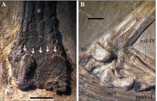
Fusion of the scapula and coracoid (character OS4-1) is seen in all Triassic pterosaurs were those bones are preserved and visible, excluding only the holotype of Arcticodactylus cromptonellus (see Jenkins et al. 2001) and possibly MCSNB 8950. The state in the holotype of Preondactylus buffarinii is uncertain (Dalla Vecchia 2014). Specimen MCSNB 3359 (Pe-teinosaurus zambellii) has a fused scapula and coracoid, fused ischium and pubis (character OS4-2) and the ilium is unfused to the ischiopubic plate (character OS4-3), but the sacral vertebrae are unfused (cha-racter OS3-1), proximal tarsals are unfused to tibia and the proximal carpals are possibly unfused, too (character OS3-3). The scapula and coracoid and all the three pelvic bones are fused (characters OS4-2 and OS5-1, respectively) in the holotype of Austria-draco dallavecchiai, but the suture between the tibia and distal tarsals can still be seen (Fig. 1A). In one of the smaller Dorygnathus banthensis specimens (BSP 1938 I 49; see Tab. 1 and Padian 2008a), the
scapu-Fig. 1 - Non-fusion of the distal tar-sals with the tibia. A) Right tibiotarsus of the holotype of Austriadraco dallavecchiai (BSP 1994 I 51) with the still visible suture; B) Right tibia, tarsus and proximal part of the pes of BSP 1938 I 49, Dorygnathus banthensis, with completely unfused proxi-mal tarsals and tibia. Abbre-viations: fi, fibula; lco, lateral condyle; lpt, lateral proximal tarsal (calcaneum); mtI-V, metatarsals I-V; pphV-1, pedal phalanx V-1; ti, tibia. The arrows in A point to the suture line between the tibia and proximal tarsals. The scale bars equal 2 mm.
lae and coracoids are co-ossified and all the pelvic bones are fused into a single plate, but the proximal tarsals are not fused to the tibia (Fig. 1B). The for-mation of the tibiotarsus seems to occur relatively late in ontogeny in non-pterodactyloid pterosaurs. It is an important ontogenetic feature that is not con-sidered in the “six ontogenetic stages” of Kellner (2015). Note that fusion of scapula and coracoid also appears to occur relatively early in ontogeny in the pterodactyloid Pteranodon (Bennett 1993).
In MCSNB 2886 and MCSNB 3359, the ilia are unfused with the puboischiadic plates (character OS4-3), but the extensor tendon processes are cle-arly fused with the wing phalanges (character OS5-2). In MCSNB 2886, the extensor tendon processes has a slightly different texture in the suture zone (Dalla Vecchia 2014: fig. 4.1.56), but the suture is obliterated. The extensor tendon process is fused with wing phalanx 1 in all Triassic pterosaurs with the possible exclusion of the holotype of Arctico-dactylus cromptonellus (however, Jenkins et al. 2001 did not report this non-fusion in their list of evi-dence of immaturity occurring in the specimen). In MCSNB 8950, the extensor tendon process is fused with the wing phalanx and the ilium is fused with a visible suture to the pubis (Wild 1994), but the specimen shows many features of osteological immaturity, which are listed above, and is probably a juvenile. The extensor tendon process also seems to be fused with wing phalanx 1 in some very small and immature Rhamphorhynchus specimens (Wellnho-fer 1975b considered these to belong to ‘R. longi-caudus’), like that figured in Wellnhofer (1975a: fig. 12a, exemplar 15; its skull is only 34 mm long) and BSP 1938 I 503 (Wellnhofer 1975b: fig. 22; skull 41 mm long). The process is fused to the phalanx in the Dorygnathus banthensis specimen BSP 1938 I 49, where all the pelvic bones are fused into a single plate, but the plate is not fused to the sacrum and the proximal tarsals are not fused to tibia. Thus, the fusion of the extensor tendon process occurred re-latively early in the ontogeny of non-pterodactyloid pterosaurs, although total obliteration of the sutu-re occursutu-red somewhat later. However, the process appears to be unfused until late in ontogeny in the pterodactyloid Pteranodon (Bennett 1993). Thus, the late ontogenetic fusion of the extensor tendon pro-cess is probably a feature of the pterodactyloids or only of some of them. All of Kellner’s hypotheses on the ontogenetic stage of the Triassic pterosaurs
based on the fusion of the extensor tendon process are biased by this incorrect assumption.
Character OS6-1 (all bones or complex of bones are fused) is usually impossible to be ful-ly established, because pterosaur specimens are in most cases incompletely preserved, thus the sta-te cannot be known for all of the bones. Kellner (2015: 685) noted that the dentaries are not fused in MCSNB 2888 (holotype of E. ranzii), despite it being considered an adult individual. However, Kellner viewed that non-fusion as a “phylogenetic signal”, meaning that non-fusion can be a phylo-genetic feature in “some non-pterodactyloids”, not an ontogenetic feature. Actually, the mandibular rami are also unfused at the symphysis in the ho-lotypes of Preondactylus buffarinii (MFSN 1770; see Dalla Vecchia 2014: fig. 4.1.3A-B), Austriadraco dalla-vecchiai, Arcticodactylus cromptonellus, Peteinosaurus zam-bellii, Carniadactylus rosenfeldi, Caviramus schesaplanensis and ‘Raeticodactylus’ filisurensis, specimens MPUM 6009 and MFSN 1922 of Carniadactylus rosenfeldi, specimen MFSN 21545 (Dalla Vecchia, 2014: fig. 4.1.164), and in a large and still unnamed pterosaur from the Triassic of the USA (Britt et al. 2015; pers. obs). All Triassic specimens where the relative di-splacement of the two rami can be checked show that the rami were unfused. This is also the case for specimens NHMUK R 1035 and NHMUK 43486 of the Early Jurassic Dimorphodon macronyx (see San-gster 2003). Thus, lack of fusion of the mandibular rami at the symphysis is typical within earliest ptero-saurs. In contrast, mandibular rami fused very early during the ontogeny of the pterodactyloids (Kellner & Tomida 2000).
Kellner (2015) apparently thought that full skeletal maturity was reached in pterosaurs when the humeral (character OS6-2) and ulnar (character OS6-3) secondary centers of ossification fused to their respective bones. This is based on his observa-tions on the large pterodactyloids from the Lower Cretaceous of Brazil (see Kellner & Tomida 2000: 84). Actually, those secondary centers of ossification are generally not reported in non-pterodactyloid pterosaurs (e.g., Wellnhofer 1975a-b; Wild 1979; Pa-dian 2008a, b; Dalla Vecchia 2014), possibly because they are difficult to identify due to their small size or cannot be correctly identified in disarticulated ske-letons, or because they fused very early during on-togeny, or because non-pterodactyloid pterosaurs did not have them. Possible secondary centers of
ossification are reported only in the immature spe-cimen MCSNB 8950 between the left humerus and ulna-radius and were identified as “sesamoids” by Wild (1994: 106). Note that the fusion of seconda-ry centers of ossification in the humerus appear to occur relatively early in the pterodactyloid Pteranodon (Bennett 1993).
Adult specimens of Pteranodon have a low process for the insertion of M. triceps brachii on the ulna (e.g., Bennett 2000: fig. 74). That process is located on a large and unfused proximal secondary center of ossification in the immature holotype of Anhanguera piscator (see Kellner & Tomida 2000: figs. 33-34). The proximal extremity of the ulna presents a comparatively well-developed process for the in-sertion of M. triceps brachii in Triassic pterosaurs; this process is never located on an unfused secon-dary center of ossification and no suture separates it from the ulna. This situation is also see in imma-ture individuals including MCSNB 3359 (Dalla Vec-chia 2014: fig. 4.1.64), MCSNB 3496 (Dalla VecVec-chia 2014: fig. 4.1.70) and MCSNB 8950 (Wild 1994: fig. 2).
The holotype of E. ranzii (MCSNB 2888) and the holotype of Austriadraco dallavecchiai (BSP 1994 I 51) are supposed to have reached ontogenetic stages OS6 and “at least O5” (Kellner, 2015: 685), respec-tively. However, this is unsupported following the ‘six ontogenetic stages’: secondary centers of ossi-fication are unreported in both specimens and both specimens are incompletely preserved, thus the complete fusion of all composite elements cannot be checked. Furthermore, the fusion of the ilium with the puboischiadic plate (character OS5-1) can-not be confirmed in MCSNB 2888 and the fusion of the extensor tendon process (character OS5-2, occurring in both specimens) is present also in some juveniles and subadults too, as shown above.
Specimen MCSMB 2888 shows other evi-dence of non-fusion and incomplete ossification of skeletal elements, most of which was previously listed by Dalla Vecchia (2014): the nasals are unfu-sed to the premaxillae, maxillae and frontals; the postorbital and supraorbital are unfused to adjacent elements; a suture is visible between the maxilla and the premaxilla and the maxilla and jugal (the ma-xilla and premama-xilla are rostrally fused very early in the ontogeny of pterodactyloids; Kellner & Tomida 2000); the last sacral vertebra is unfused to the pe-nultimate sacral vertebra; the sutures between the
other sacral vertebrae remain visible; the neurocen-tral sutures in the last dorsal vertebrae seem to be present; prepubes are not fused at the symphysis; tibia and fibula are not completely fused proximally (a groove separates them); the sternal plate, sternal ribs and prepubic blade are very thin and with a grainy, unfinished surface; and the sacral ribs also show a grainy, unfinished surface. The degree of fusion of the proximal tarsals to the tibia cannot be evaluated as the distal portion of both tibiae are missing.
The holotype of Austriadraco dallavecchiai also shows evidence of osteological immaturity (Dalla Vecchia 2014): the jugal is not fused with maxilla and the postorbital is unfused with adjacent ele-ments; sutures between mandibular elements are still visible; the ilium is unfused with the sacrum; fibula is probably unfused proximally with tibia; the suture between the proximal tarsals and the tibia is still visible (Fig. 1A); and some bones (e.g. scapula and ischium) have a grainy bone surface suggesting incomplete ossification (Dalla Vecchia 2014: figs 4.1.40 and 4.1.44).
In the large holotype of Anhanguera piscator (NSM-PV 19862), which has a 617 mm-long skull and an estimated wing span of about 5000 mm (Kellner & Tomida 2000), most of the skeletal ele-ments (including many skull bones, atlas-axis com-plex, neural arches of dorsals and some cervicals, the notarium, the sacral vertebrae, scapula and co-racoid, proximal and distal carpals, epiphyses of hu-merus and ulna, the extensor tendon process of the wing phalanx 1, pelvic elements, proximal tarsals and tibia) are unfused. According to the ontogene-tic stages of Kellner (2015), this large pterosaur was in a pre-OS3 when it died, thus it would have been an old juvenile at best. The histological features of this specimen should be investigated to test this conclusion.
The smallest Pteranodon specimen (1760 mm estimated wing span), which is considered a juve-nile by Bennett (2017), shows the same evidence of osteological immaturity in the distal radius and ulna, carpus, pteroid, wing metacarpal and wing phalanx 1 as the subadult Pteranodon individuals with wingspans over twice its size.
In Triassic pterosaurs, two options are pos-sible: 1) no fully mature individuals have ever been found, or 2) the full osteological maturity (fusion and complete ossification) of all skeletal elements
was never reached during their life history (i.e., their growth was indeterminate and they grew throu-ghout life, albeit at a slower rate in later years.
The order of fusion of skull elements could be species specific or even individually variable in early pterosaurs. For example, the suture between maxilla and jugal is still visible in the largest indivi-dual of Dimorphodon macronyx (see Sangster 2003), while it is obliterated in a close and still unnamed taxon (Britt et al. 2015) that is about 20% smaller in linear size and shows other evidence of somatic immaturity.
Nevertheless, there seems to be an appro-ximate, although not universal, order of fusion
of some skeletal elements in Triassic pterosaurs, which does not fully coincide with those of Kellner (2015). The extensor tendon process fused early to the wing phalanx 1, probably earlier than the co-ossification of scapula and coracoid (e.g. MCSNB 8950). Scapula-coracoid and ischium-pubis co-ossified before than ischiopubis-ilium and sacral vertebrae into a synsacrum. If distal carpals ever fused into a distal syncarpal, they did so after the formation of the proximal syncarpal. Proximal car-pals could form a syncarpal later than the fusion of scapula and coracoid and ischium and pubis. Possi-bly, proximal tarsals fused to tibia at this stage, but obliteration of the suture occurred later (later than
1 2 3 4 5 6 7 8 9 10 11 12 13 14 OS1 1 ? no no no no no no no no no no no no no 2 ? yes ?* no ? ? ? ? ? no† ?* no no ? 3 yes no* no no no ? no no ? no no no no no OS2
1 ? no yes ? ? ? yes yes ? ? yes yes ? ? 2 ? yes* yes yes yes ? yes yes ? yes yes yes yes yes 3 ? ? ?* ? ? ? ? ? ? ? ? ? ? ?
OS3
1 ? no ? no ? ? ? no no ? ? no†† ? ? 2 ? no+ no ? no no ? ? ? ? no no ? ? 3 ? yes+ no ? yes yes ? ? ? ? yes yes ? ?
OS4
1 no no* yes yes ? yes ? yes ? yes yes yes yes ? 2 ? ? ? ? ? ? yes yes yes yes yes ? ? ? 3 ? yes# ? ? yes ? yes yes yes no ? ? ? ?
OS5
1 ? no# ? ? no ? no no no yes ? ? ? ? 2 ?* yes yes ? yes yes yes yes ? yes yes yes ? yes
OS6
1 no no no no no ? no no no no ? no ? ? 2 ? no* ? ? ? ? ? ? ? ? ? ? ? ? 3 ? yes§ yes§ yes§ yes§ yes§ ? yes§ yes§ ? yes§ yes§ ? ?
Tab. 1 - The presence or absence of the features characterizing the six ontogenetic stages (OS1-6) of Kellner (2015) in the Triassic pterosaur specimens listed in order of increasing body size. See text for the description of the features. Legend: 1 = Arcticodactylus cromptonellus, holotype (MGUH VP 3393); 2 = MCSNB 8950; 3 = MPUM 6009; 4 = MCSNB 2887; 5 = Preondactylus buffarinii, holotype (MFSN 1770); 6 = Austriadactylus cristatus (MGC 332466); 7 = Peteinosaurus zambellii, holotype (MCSNB 2886); 8 = Peteinosaurus zambellii (MCSNB 3359); 9 = Peteinosaurus zambellii, (MCSNB 3496); 10 = Austriadraco dallavecchiai, holotype (BSP 1994 I 51); 11 = Carniadactylus rosenfeldi, holotype (MFSN 1797); 12 = Eudimorphodon ranzii, holotype (MCSNB 2888); 13 = Austriadactylus cristatus, holotype (SMNS 56342); 14 = ‘Raeticodac-tylus’ filisurensis, holotype (BNM 14524); no = absent; yes = present (it fits the character state); ? = unknown because missing, uncertain or impossible to establish. * = Alternative interpretation is discussed in the text; + = fide Wild (1994); # = a suture is visible between bones; § = if the process for M. triceps brachii of ulna is considered to have been on a secondary center of ossification in immature individuals; † = I provisionally assume that the bone identified as the sternum by Wellnhofer (2003) is the sternum, not the fused frontals as done by Kellner (2015, text; however, it is reported as a sternum in fig. 2e); †† sutures are visible (see text).
obliteration of sutures between the ilium and the puboischiadic plate). The sacrum fused with ilium later in ontogeny. Sutures between some mandibular elements disappeared relatively late in ontogeny too. Some skull bones and the mandibular rami are un-fused also in the largest known individuals. As noted above, this is not necessarily indicative of the imma-ture status of the individual. Suimma-tures between skull bones are not obliterated during the ontogeny of the living archosaur Alligator mississippiensis (see Bailleul et al. 2016). Adult reptiles still preserving open sutu-res in composite skeletal elements occur also in the fossil record. For example, neural arches and caudal ribs are always unfused in non-plesiosaur eusaurop-terygians (Rieppel 2000).
Probably the neurocentral fusion in the ver-tebral column followed a pattern in pterosaurs, as it does in other diapsids (Brochu 1996; Irmis 2007). This fusion pattern was mentioned by Kellner & To-mida (2000), but not by Kellner (2015) and needs to be further investigated.
Kellner’s six ontogenetic stages are an oversim-plification of a very complex process. Ontogenetic features of different taxa that probably had distinct growth patterns have been grouped together into a sequence that Kellner (2015) considered to be valid for all pterosaurs. Furthermore, those stages have been based mainly on observations from large Cre-taceous pterodactyloids, and then applied to Triassic specimens of basal pterosaur taxa. It is evident that the order of fusion of composite skeletal elements during ontogeny differs in pterodactyloids and early pterosaurs and there is no universal pattern that can be extrapolated to pterosaurs in general.
s
ysteMatIcP
alaeontologydiapsida Osborn, 1903 PterosaurIa Kaup, 1834
eudimorphodontidae Wellnhofer, 1978 (sensu Dalla Vecchia, 2014)
Carniadactylus Dalla Vecchia, 2009a carniadactylus rosenfeldi (Dalla Vecchia, 1995)
Specimen MPUM 6009 Figs 2, 3A and 4
1979 Eudimorphodon ranzii - Wild, pp. 179-180, 182, pls. 4-5, figs. 2, 5-7, 18, 24a, 26a, 27a, 28a, 29a and 42.
1994 Eudimorphodon ranzii - Wild, pp. 106, 112 and 115, fig. 13b, tabs. 1-2.
1998 Eudimorphodon ranzii - Dalla Vecchia, p. 357.
2001 Eudimorphodon ranzii - Jenkins, Shubin, Gatesy & Padian, pp. 151-152, 157 and 163, tabs. 1-2.
2002 Eudimorphodon ranzii - Dalla Vecchia, p. 46, fig. 10, tab. II. 2003 Eudimorphodon ranzii - Wellnhofer, pp. 8, 10-13, 15 and 17-19,
tab. 1.
2003 the Milano specimen, which differs from the holotype of Eu-dimorphodon ranzii - Kellner, p. 116.
2003 Eudimorphodon ranzii - Dalla Vecchia, pp. 24, 26, 27-28, 37 and 40, tabs. 1-2.
2004a Eudimorphodon - Dalla Vecchia, pp. 50, 62, 65-67 and 69, figs. 5B and 6D, tab. I.
2004b I retain it in Eudimorphodon, but I consider dubious [sic] its con-specificity with MCSNB 2888 - Dalla Vecchia, p. 13, fig. 2. 2006 Eudimorphodon - Fröbisch & Fröbisch, p. 1087.
2008 Eudimorphodon sp. - Stecher, pp. 194-197, fig. 10d, tabs. 2-3. 2009a Carniadactylus rosenfeldi Dalla Vecchia - pp. 159-160, 168,
172-179 and 181-183, figs. 3D, 4D, 6B and 11B, tabs. 2-3. 2010 Carniadactylus rosenfeldi - Ősi, pp. 138, 140-141, 143-144 and
146, figs. 1F and 2I, tab. 1.
2010 Eudimorphodon - Bonaparte, Schultz & Soares, p. 66.
2013 Carniadactylus rosenfeldi - Dalla Vecchia, pp. 127, 133, 141 and 145, tab. 1.
2014 Carniadactylus rosenfeldi - Dalla Vecchia, pp. 160-162, 191-203 and 306, figs. 4.1.98B, 4.99.B-C, 4.100B, 4.1.137-146, tabs. 4.1.1-2, 4.1.6 and 4.1.17.
2015 Bergamodactylus wildi Kellner, p. 677 - pp. 677-683, figs. 3-4 and 5b, tabs I-II.
description of mpum 6009
MPUM 6009 is preserved on a thin limestone slab and lies on its right side. Although the skeleton was originally complete, most of its posterior part split away or was weathered leaving only a faint im-pression, or no impression at all (see Dalla Vecchia 2014: fig. 4.1.137). The preserved part of the skel-eton is crushed and most of the bones as well as the teeth (see Dalla Vecchia 2014: fig. 4.1.140) are bro-ken into many fragments. The bones were originally covered by a thin layer of minute whitish crystals (probably calcite, Wild 1979: 180), which can still locally be observed on the slab surface. As noted above, crystal growth is probably responsible of the pitted aspect of the bone surface of this specimen; for the same reason, the surface of the slab is also pitted where the thin layer split away.
Crushing is particularly evident in the skull, which is flattened and exposes its left lateral view (Fig. 2). Crushing caused some bones to twist with respect to their original position. Some bones partly split away when the specimen was collected in the field or when it was prepared. Some skeletal ele-ments are partly overlapped by other eleele-ments. Fur-thermore, some palatal bones and possibly some elements of the right side are mixed with the lateral elements of the left side. The specimen was also covered with a lacquer that makes the bone surface shiny black like the enamel of the tooth crowns,
sometimes making it difficult to distinguish a bro-ken crown from a brobro-ken fragment of bone. This problematic state of preservation makes identifica-tion of single skull elements highly interpretative and sometimes prevents unambiguous interpreta-tion of their morphology.
Wild (1979) published an interpretative draw-ing of the entire specimen (Wild 1979: pl. 5), an interpretative drawing of the skull and the skull
re-construction (Wild 1979: fig. 2), and an interpreta-tive drawing of the left manus (Wild 1979: fig. 18). Dalla Vecchia (2014: figs 1.37B and 4.1.139B-C) published slightly modified versions of the inter-pretative drawings of the whole skeleton and skull by Wild (1979). Kellner (2015: fig. 4b-c) published his own interpretative drawing and reconstruction of the skull.
The following description supports the
dis-Fig. 2 - Skull and lower jaw of MPUM 6009. A) Photo; B) Drawing. Abbreviations: af, jugal antorbital fossa; aof, antorbital fenestra; bpt, ba-sipterygoid process; cop, ‘coronoid’ process (surangular dorsal process); d, dentary; en, external naris; fr, frontal; j, jugal; lac, lacrimal; mx, maxilla; n, nasal; pmx, premaxilla; or, orbital rim of the frontal; po, postorbital; q, quadrate; qj, quadratojugal; rap, retroarticular process of the mandible; rd, ridge; sq, squamosal. Elements in parentheses are from the right side. Teeth are in dark gray; manus pha-langes are in pale gray. The scale bar equals 10 mm.
cussion of the ontogenetic stage of the specimen and the validity of Bergamodactylus wildi. Further-more, mistakes in Kellner’s (2015) interpretation of the specimen are corrected.
Skull and lower jaw (Fig. 2). Authors dealing with MPUM 6009 followed the interpretation and reconstruction of the skull by Wild (1979), mainly in the details that are more difficult to understand on the actual specimen. Dalla Vecchia (2009a; 2014: fig. 4.1.139) and Kellner (2015: fig. 4b-c) proposed alternative interpretations, but only for some skull bones. Dalla Vecchia (2014) revised the postcra-nium, but did not attempt a thorough revision of the skull and lower jaw, because of the difficulty of interpretation caused by its poor state of preserva-tion. However, some points need to be clarified be-fore arguing against the diagnostic features reported by Kellner (2015) for Bergamodactylus wildi.
The degree of fusion of the skull elements cannot be reliably established because of the gen-eral crushing. Fractures are barely distinguishable from sutures and can easily be incorrectly interpret-ed as such. The skull length/height ratio in the skull as preserved is 2.48 (41.5/16.75 mm; height as the distance between the highest point of the skull roof and the base of the quadrate condyle measured per-pendicular to the main axis of the skull).
The external naris is relatively small (it is about 5.5 mm long and probably the smaller skull open-ing), rostroventrally to dorsoposteriorly elongated and probably with a sub-elliptical or sub-trapezoidal outline. The antorbital fenestra has a sub-triangular outline, but it is possibly distorted by crushingand its dorsal margin is not well defined. The orbit was sub-circular and by far the largest skull opening. The shape of the postorbital suggests that the up-per temporal fenestra had the outline of an inverted tear drop.
The tip of the snout of MPUM 6009 is over-lapped and partly covered by some phalanges of the right manus. The dorsal margin of the poste-rior two-thirds of the premaxillae split away, but the impression of the missing portion remains. The posterodorsal processes of the premaxillae rotated because of crushing and are both exposed in dor-sal view; in this view, they are very narrow and are probably unfused (the midline is visible). They ex-tend posteriorly to the level of the middle of the antorbital fenestra, but their posterior end is miss-ing because a 5 mm-long portion of the dorsal
mar-gin of the skull at mid-skull split away. The rotation of the posterodorsal processes of the premaxillae reduced the apparent extent of the external naris. The premaxillary body and the premaxillary process of the maxilla are separated by a straight fracture starting from the rostral apex of the external naris. That fracture becomes an irregular line rostrally that is swollen along its ventral (maxillary) side and ends just posterior to the most distal premaxillary tooth. Thus, that line is the premaxillary/maxillary suture and the entire structure (fracture plus line) plausibly corresponds to the premaxillary/maxillary bound-ary. Therefore, the suture between the two bones is not obliterated and the premaxilla does not have a significant maxillary process bordering the external naris ventrally. However, the part of the premaxilla bordering the rostral apex of the external naris is damaged (Fig. 2), thus the extent of the participa-tion of the premaxilla to the rostral margin of the external naris was estimated by Wild (1979: fig. 2b).
The outline of the triradiate maxilla is some-what interpretative because of its fracturing, the fracturing of adjacent bones and the sometimes in-distinct boundaries with them. The latter is the case of a shapeless element with the consistency of a film, which covers the central part of the external naris and partly overlaps the maxilla along its con-tribution to the ventral margin of the external naris (Fig. 2). The ascending process is plausibly that iden-tified by Wild (1979: fig. 2a), but it is not as angled as depicted and its dorsal margin is straight. The ascending process is proportionally much thicker in lateral view than that of MCSNB 2888 and is lance-like instead of regularly tapering apically. Its apical termination is not preserved. Kellner (2015: fig. 4) drew the ascending process as more slender than it actually is. It is unclear which is the actual maxilla edge along the rostral and rostroventral margin of the antorbital fenestra (i.e., which are the margins of the posterior side of the base of the ascending process and the dorsal side of the proximal part of the jugal process), because there are two distinct, similar and parallel structures there. I opt here for the ‘slender’ maxilla option (Fig. 2B), chosen also by Kellner (2015), but I am unable to understand which other skeletal element the other, parallel, one could be. It apparently mimics the right maxilla (Fig. 2B), but it cannot be the right maxilla, because it lies on the presumed palatal elements. The posterior extent of the jugal process of maxilla is also problematic.
For Wild (1979) and Kellner (2015), it reaches the level of the ‘coronoid’ process of the surangular. However, its actual posterior end is slightly rostral to the ‘coronoid’ process and about at the level of the mid-jugal and is abrupt, not tapering like that depicted by Kellner (2015). The upper and ventral margins of the jugal process are parallel for most of the length of the process. The jugal process is longer and shallower than the premaxillary process.
The nasal is a narrow strip of bone starting at the posterior end of the external naris. It is dorsally bordered by the posterodorsal process of the pre-maxilla and ventrally by the ascending process of the maxilla. Its rostral end, bordering the external naris, is damaged. It possibly has a small rostroven-tral process, but it is impossible to establish whether a rostrodorsal process like that of the nasal of MC-SNB 2888 existed or not (contra Kellner, 2015: 681) because that part of the bone is missing. Its absence in skull reconstructions by the various authors is just a graphic compromise, not an anatomical fea-ture. The posterior portion of the nasal is missing.
The jugal is probably tetraradiate and with a comparatively deep body like that of the jugals of Eudimorphodon ranzii and ‘Raeticodactylus’ filisuren-sis. The rostrodorsal (lacrimal) process is probably complete and is the longest process of the jugal, while the rostroventral (maxillary) process is very small, but it could be laterally overlapped by the ju-gal process of maxilla. A film-like, vertical strip of bone rostral to the antorbital margin of the jugal, which was identified as a lacrimal by Wild (1979) and ignored by Kellner (2015), may be the bottom of a jugal antorbital fossa (as noted by Witmer 1997),
like that occurring in the new and still undescribed Late Triassic pterosaur from USA (Britt et al. 2015; Britt et al. Submitted), although it is much broader in MPUM 6009. The antorbital margin (antorbital fossa excluded) is shallowly concave (i.e., nearly ver-tical). The orbital margin is broad and shallowly con-cave bordering ventrally the large orbit. The outline of the posterior termination of the jugal cannot be traced because of fracturing. The postorbital pro-cess is quite inclined posteriorly (the angle with the main axis of the skull is 152°); its distal extremity is not preserved and could be broken or still covered by rock. The quadratojugal process overlaps or is overlapped by the bone that, because of its position, was identified by Wild (1979) and Kellner (2015) as the left quadratojugal. The latter is a relatively large element (at least 6 mm long) with an expanded, flat and thin anterior extremity; it tapers to the poste-rior extremity, becoming rod-like and straight. It is impossible to establish the exact boundary between this bone and the left jugal. According to Dalla Vec-chia (2014), that bone could alternatively be the bro-ken and moved postorbital process of the jugal, but this appears to be improbable.
The Y-shaped postorbital (Fig. 3A) has slen-der squamosal and frontal rami forming an angle of about 70°. This indicates that the upper temporal fenestra had an unusually acute ventral margin. The jugal process is also slender and is slightly recurved (as is the frontal process). The distal ends of the squamosal and jugal processes are covered by other bones, while the distal end of the frontal process is broken. Therefore, the apparent length of these processes is not the true length. The left postorbital
Fig. 3 - Postorbital of MPUM 6009 compared to those of Aus-triadraco dallavecchiai and Campylognathoides liasicus. A) Left postorbital of MPUM 6009; B) Left postorbital in medial view or right postor-bital in lateral view of BSP 1994 I 51, holotype of Aus-triadraco dallavecchiai; and C) Right postorbital of Cam-pylognathoides liasicus (SMNS 11879). Abbreviations: fp, frontal process; jp, jugal pro-cess; scl, sclerotic bone; sqp, squamosal process. Lines of the abbreviations point to the exposed extremities of the processes. The scale bars equal 3 mm.
of MPUM 6009 is very similar to the postorbital of the holotype of Austriadactylus dallavecchiai (Fig. 3B), and moderately similar to that of the Early Jurassic Campylognathoides liasicus (Fig. 3C).
Kellner (2015) reported a relatively massive lacrimal along the orbital margin of the rostrodor-sal process of the jugal, while Wild (1979) drew the element much thinner and identified it as the pre-frontal. In both cases, it has the shape of an upside-down L. The bone has a thin shaft overlapping the thickened orbital margin of the jugal and a modest dorsorostral expansion that, as preserved, overlaps the apical part of the rostrodorsal process of the jugal. As preserved, the expansion is smaller than that figured by Wild (1979) and much smaller than that reconstructed by Kellner (2015). The shape of the shaft and the position of the bone are those of the lacrimals of the holotype of Eudimorphodon ran-zii and ‘Raeticodactylus’ filisurensis. Kellner (2015: fig. 4b) indicates the presence of a lacrimal in the draw-ing of the skull of MPUM 6009 with the abbrevia-tion “la”, but that abbreviaabbrevia-tion does not correspond with any bone in the figure. In that area, MPUM 6009 preserves a small, rod-like element that could be the right lacrimal, rotated at 90° counterclock-wise from its original position (see Fig. 2A-B), but this bone is not drawn in Kellner (2015: fig. 4b).
The frontals were rotated because of crush-ing and are exposed in dorsal view. They appear as a misshapen, broad plate of bone that is broken into many fragments, some of which split away. Only the orbital margins have a defined outline, because they are thicker. There are no ridges on the dorsal surface of the frontals (unlike MCSNB 2888) and there is no evidence of a suture separating the right from the left frontal (contra Kellner 2015: fig. 4b). The outline of the parietals is indistinguishable; there are no evident ridges for the origin of the ad-ductor musculature. Only the straight and thicker lateroposterior side of the left quadrate is exposed, with the distal lateral condyle that appears to be sub-spherical in lateral view. Possible traces of the medial lamella could be present too. The posterior slope of the left quadrate (135°) was somewhat ex-aggerated by the extreme crushing and flattening of the posterior part of the skull, which originally was transversely wide. The close rod-like bone that Wild (1979) identified as the right quadrate has a lower slope (about 120°). However, comparison with the holotype of ‘Raeticodactylus’ filisurensis (see Dalla
Vec-chia 2014: fig. 4.1.160) suggests that this bone could be the left basipterygoid process of the basisphe-noid.
At least six, apparently rod-like, and antero-posteriorly oriented bones cross the antorbital fe-nestra. At least some of them are plausibly palatal elements (Wild, 1979 identified the dorsal-most ele-ments as the vomers or palatines), while the others could be parts of the elements of the right cheek side of the skull.
The mandibular ramus is slender with a length/height ratio of 17 (length is 34 mm and height is only two mm at mid-ramus). Its dorsal margin is slightly concave, while the ventral margin is nearly straight. Its height is constant along most of den-tary length, but the ramus expands near the extrem-ities. In fact, the ramus slightly flares by mandibular tooth 5 and tapers rostrally to tooth 2; the ramus reaches it maximum depth at the apical part of the ‘coronoid’ process and tapers toward the retroar-ticular process. The left mandibular ramus is slightly displaced posteriorly with respect to the right one, therefore the two rami were unfused at the sym-physis. All mandibular elements appear to be fused, but possible sutures cannot be distinguished from cracks caused by crushing. The rostral tip of the ra-mus is sharply pointed, unlike the interpretation by Kellner (2015: fig. 4b-c), and the first fang-like tooth is close to the tip. An arched ridge that is bounded ventrally by a groove is present along the lateral side of the central part of the ramus (rd in Fig. 2B). It can also be observed in the holotype of Austriadraco dallavecchiai (BSP 1994 I 51), MFSN 21545 and also MFSN 1797 (as an impression). There is no exter-nal mandibular fenestra. The apex of the dorsal margin of the surangular (the ‘coronoid’ process) is overlapped and broken by another bone, probably the jugal (which overlapped the ‘coronoid’ process laterally in anatomical articulation). What appears to be the dorsal margin of the ‘coronoid’ process is a fracture line. Wild (1979: fig. 2b) recognized that the apical part of the ‘coronoid’ process is not ex-posed and traced it as a point. Wild’s reconstruction is plausible since the opposite sloping of the dorsal margins of the surangular anterior and posterior to the broken part. Instead, Kellner (2015: fig. 5b) de-picted the broken apical edge as if it were the ac-tual apex of the surangular. This mistake biased the choice of one of the purported features supporting MPUM 6009 as a taxon distinct from
Carniadacty-lus rosenfeldi (see below). Kellner (2015) incorrectly drew the retroarticular process in his figure 5b as if it were entire, but actually the figured profile is clearly that of the broken process. The complete process had a more rounded profile in lateral view.
No one has attempted another reconstruction of the dentition or proposed an alternative tooth count to that of Wild (1979), probably because the teeth are poorly preserved and some of them are difficult to identify, with both Dalla Vecchia (2009a) and Kellner (2015) accepting Wild’s interpretation. A more thorough analysis of the specimen shows that the most distal unambiguous mandibular tooth is the penultimate one in figure 2a of Wild (1979). Thus, the position of the last indisputable tooth is more distant from the apical part of the ‘coronoid’ process than previously supposed.The original in-correct interpretation biased the choice of one of
the purported features to distinguish MPUM 6009 from the holotype of C. rosenfeldi (see below). Twelve left mandibular teeth can be recognized in situ: three are mesial laniaries (the first two monocuspid and the third bicuspid) and nine are smaller multicusped teeth. However, three segments of the dentary tooth row are covered by displaced maxillary teeth. Based on the mesiodistal length of a mid-row crown, the assumption that there are no interdental spaces (which is the case of the exposed crowns) and the position of the last unambiguous dentary tooth, my estimated tooth count is 17-18 (uncertainty in the count is caused by the different sizes of the mul-ticusped crowns along the row, because the mesial and distal ones that are smaller than the others). Wild (1979) counted 17 mandibular teeth and Kell-ner (2015: fig. 4C) reported 16 teeth plus a probable empty alveolus distal to tooth 14; both authors
in-Fig. 4 - Left carpus of MPUM 6009 and adjacent bones in pos-terior (postaxial) view. A) Photo; B) Drawing. Abbre-viations: cm, crista meta-carpi; dc, distal carpal; etp, extensor tendon process of wing phalanx 1; ldc, large distal carpal; mcI-III, meta-carpals I-III, mc IV, wing metacarpal; pcr, proximal carpal (radiale); pcu, proxi-mal carpal (ulnare); ph, manual phalanges of digits I-III; pt, pteroid; ra, radius; u, ulna; wph1, wing phalanx 1. Elements in parentheses are from the right side. The carpals are in pale gray. The scale bar equals 5 mm.
cluded in the count the distalmost tooth that I am unable to find in the specimen.
Postcranium. The postcranium of MPUM 6009 was fully described by Wild (1979) and Dalla Vecchia (2014) and there is little need to replicate those de-scriptions here. Only details useful for discussing the validity of the characters used by Kellner (2015) to erect Bergamodactylus wildi and some additional details not previously described are reported here.
The vertebral column is articulated, but only the cervical segment is completely preserved. Eight cervical vertebrae are exposed, including the atlas-axis complex. The cervical-dorsal transition is cov-ered by the left scapulocoracoid. Cervical vertebra 8 has a large pneumatic foramen (Dalla Vecchia 2014: fig. 1.1.141). The tail was long; the few preserved mid-tail caudal vertebrae have been described by Dalla Vecchia (2002).
Scapula and coracoid are fused without a su-ture (Dalla Vecchia 2014: fig. 4.1.99B-C). The shaft of the better preserved left coracoid is broad, flat and with nearly parallel anterior and posterior mar-gins (i.e., it is not fan-shaped). Only the proximal portion of the left scapula is preserved. It is dor-soventrally low and with parallel dorsal and ventral margins (i.e. there is no evidence that the scapular blade was fan-shaped). The broad and flat sternal plate crops out from below the two humeri but its outline is unknown and its complete ossification cannot be checked, because it is mostly covered by appendicular bones.
The left humerus is complete; only the ante-rior margin of the ‘squared’ deltopectoral crest is slightly damaged (Dalla Vecchia 2014: fig. 4.1.142). Secondary centers of ossification cannot be iden-tified at the extremities of humerus and ulna. The process for the insertion of M. triceps brachii on the ulna is moderately developed; no suture is visible between the process and the rest of ulnar epiphysis (Dalla Vecchia 2014: fig. 4.1.143). The right carpus is poorly preserved and the various elements cannot be reliably identified. The left carpus (Fig. 4) is bet-ter preserved, although it is partly damaged. The two proximal carpals (ulnare and radiale) are separated: there is a clear proximodistal line of separation be-tween them (Fig. 4). This separation was noted by Wild (1979: fig. 18), but not by Dalla Vecchia (2009a, 2014). The distal carpus is composed of a large car-pal corresponding with the wing metacarcar-pal and one, or possibly two, much smaller carpals in
corre-spondence of metacarpals II and III (Wild 1979: fig 18 reported two small distal carpals). The remain-der of the carpus split away with the corresponding proximal portion of metacarpal I (Fig. 4). Although both carpi are articulated and also both pteroids are in situ, both preaxial carpals are missing. The pteroids have the same boomerang-like shape as those of MFSN 1797 (Dalla Vecchia 2009a: fig. 6; 2014: fig. 4.1.100). Their proximal ends contact the distal tubercle of their respective radii, which is the same position they have in the articulated holotypes of C. rosenfeldi and E. ranzii.
The left wing metacarpal has a prominent crista metacarpi. The extensor tendon process of wing phalanx 1 is fused with the phalanx (Dalla Vecchia 2014: fig. 4.1.144).
Only the preacetabular and postacetabular processes of the left ilium (Dalla Vecchia 2014: fig. 4.1.145) are preserved of the entire pelvis. Thus, the degree of fusion of the pubis and ischium in a single plate and fusion of the latter with the ilium cannot be checked.
Both femora are incomplete. The right femur lacks a small portion of the shaft and its distal ex-tremity is damaged. Its proximal part is covered by a film of dark substance, thus it cannot be seen and its extent must be interpreted. Therefore, the length of the femora is an estimate (as acknowledged by Wild 1979: tab 1) and could be slightly higher than the 19 mm reported by Wild (1979) and the 18.5 mm reported by Dalla Vecchia (2014), although probably not much higher (i.e., 19.5-20 mm). Very little is preserved of the tibiae. Their distal articular ends are not preserved even as an impression. Thus, the length of the bone cannot be reliably estimated and it is impossible to establish whether the proxi-mal tarsals were fused to the tibiae or not. Some phalanges of both pedes are preserved, including the ungual ones (Dalla Vecchia 2014: fig. 4.1.136). They are well ossified and the non-ungual phalan-ges have well-formed terminal ginglymi.
d
IscussIonofK
ellner’
s(2015)
PoInts foradIstIncttaxonBergamodactylus wildiThe diagnosis of Bergamodactylus wildi includes the following purported autapomorphies (Kellner 2015: 678 and 680):
A) Gracile postorbital with elongated frontal process;
B) Premaxilla not participating in the ventral margin of the external nares;
C) Wing metacarpal IV [sic] small, about 40% and 30% the length of the humerus and ulna, re-spectively;
D) Femur small, about half the length of the ulna and the first wing phalanx.
autapomorphy (a)
This feature cannot be used to separate MPUM 6009 from MFSN 1797 (holotype of Car-niadactylus rosenfeldi), because the postorbitals are not preserved or exposed in the latter (Dalla Vecchia 2009a), thus their shape is unknown. The holotype of Austriadraco dallavecchiai has a gracile postorbital, too (Fig. 3B). Wellnhofer (2003) did not mention it in his description of the specimen and Kellner (2015) followed the description by Wellnhofer (2003) without substantial improvements, thus he ignored this element. A similarly slender postorbit-al occurs postorbit-also in MFSN 21545 (pers. obs.), which represents a still unnamed taxon with multicusped
teeth from the Norian Dolomia di Forni Formation of NE Italy (Dalla Vecchia 2010, 2014) that is clear-ly distinct from MPUM 6009. As noted above, the processes of the postorbital of MPUM 6009 are all broken or covered by other bones, thus the relative elongation of the frontal process cannot be reliably established. In the postorbital of the holotype of Austriadraco dallavecchiai (Fig. 3B), all of the process-es seem to be broken distally and the prprocess-eserved part of the frontal process is as long as the squamosal process. In MFSN 21545, the frontal and squamo-sal processes are concealed distally by other bones, but the frontal process appears to be longer than the squamosal process. MPUM 6009 (Fig. 3A), the holotype of Austriadraco dallavecchiai (Fig. 3B) and MFSN 21545 share a slender and recurved jugal process. Also Campylognathoides liasicus has a gracile postorbital with an elongated frontal process (the frontal process is much longer than the squamosal process) as it can be observed in the complete right postorbital of SMNS 11879 (Fig. 3C; see also Pa-dian 2008b: fig. 6). Therefore, the gracile postorbital with elongated frontal process is not autapomor-phic of MPUM 6009.
Specimen ws mcIV h u wph1 f mcIV/h mcIV/u f/u f/wph1 MH nn 665 19 38-39 60 44-45 30 0.49 0.32 0.50 0.67 SMNS 18880 820 25 51 68 59/60 38? 0.49 0.37 - - SMNS 50702 870 26 50 79 63 39 0.52 0.33 0.49 0.62 BSP 1938 I 49 890 25 51 82 60 42 0.49 0.30 0.51 0.70 MT lost 935 28.5 57.5 92 ~75 48 0.50 0.31 0.52 0.64 SMNS 52999 960 29 51 - 65-72 42 0.57 - - 0.65-0.58 SMNS 55886 ~960 27 51 - 67 - 0.53 - - - MNHU 1905.15 970 32 65 105 79 53 0.49 0.30 0.50 0.67 SMNS 51827 975 28 52 89 70 45 0.54 0.31 0.51 0.64 SMNS 56255 ~990 29 56 - 71 - 0.52 - - - SMNS 18969 ~1000 29 57 94 78 47 0.51 0.31 0.50 0.60 SMNS 52998 ~1000 - 47 - - 37 - - - - U Lowen (st) 1025 28 60 93 72 48 0.47 0.30 0.52 0.67 UUPM R157 1030 29 61 98-100 71-73 50 0.475 0.29 0.50 0.69 U Zurich A/III493 ~1050 26-27 ~61 ~92 ~75 43-44 0.43 0.29 0.47 0.58* NHMW nn 1070 32 61 101 79 50 0.52 0.32 0.49 0.63 GPIT 1645/1 1085 30 60 101 80 48/49 0.50 0.30 0.48 0.61 SMNS 51826 ~1150 29 - - 76 - - - - - SMNS 51106 1150 28 57 - 74 - 0.49 - - - SMNS 50164 1150 33 75 105 74/76 55 0.44 0.31 0.52 0.73 Swedish Mus. nn 1150 34 65 103 81 - 0.52 0.33 - - NHMUK R10087 1285 36 - 112 90 57 - 0.32 0.51 0.63 MNHU 1920.16 1400 38 80 133 99 65 0.475 0.29 0.49 0.66 MNHU 1977.21 1690 42 84 142 110+? 70 0.50 0.30 0.49 -
Tab. 2 - Wing span (in increasing order), wing metacarpal, hu-merus, ulna, wing phalanx 1 and femur lengths of Dory-gnathus banthensis specimens and relative ratios. Measu-rements (in millimetres) are from Padian (2008a). The extreme values of each ra-tio are in bold. Anatomical abbreviations: f, femur; h, humerus; mc IV, wing meta-carpal; u, ulna; wph1, wing phalanx 1; ws, wing span. Other abbreviations: nn = without museum number, st = stolen. Symbols: -, no measurement/ratio; +? = this bone “ may not be quite complete” (Padian 2008: 19); * = based on approximate measurements.