Analysis of Complications After Surgical Repair
of Orbital Fractures
Matteo Brucoli, MD, Francesco Arcuri, MD, Roberta Cavenaghi, MD, and Arnaldo Benech, PhMD
Background: The term ‘‘orbital blow-out fracture’’ is referred to as the mechanism by which an impact to the eyeball is transposed as a mechanical energy to the orbital walls, causing them to fracture. Despite a proper surgical technique, a successful anatomic recon-struction of the orbit, and an accurate follow-up, 3 complications are still frequently observed at long-term follow-up: diplopia, enoph-thalmos, and hypesthesia of the infraorbital nerve territory. In this retrospective study, we analyze the incidence, the specific character-ization, and the potential risk factors of these 3 complications. Methods: The records of 75 patients who underwent surgical repair of isolated orbital blow-out fracture from January 2001 to December 2007 at the Maxillofacial Surgery Unit of the Novara Major Hospital were reviewed retrospectively. Patients who had other coexisting facial fractures or orbital rim involvement were excluded from this study.
The mean follow-up reached 39 months (range, 6Y81 months). Enophthalmos was measured by a Hertel exophthalmometer; diplo-pia was evaluated by an optometrist with cover test, red glass test, and Hess-Lancaster test; and hypesthesia of the infraorbital nerve territory was checked by clinical examination. The studied parameters included patient’s age and sex, time interval between trauma and sur-gery, location of the fracture, and implant material. TheW2test for nonparametric data was used, and a P value of less than 0.05 was considered statistically significant.
Results: Sex, location of the fracture, and implant material were not considered statistically significant (P9 0.05). The unique variable that influenced our data was the time interval between trauma and surgery (P9 0.05).
Discussion: Although the surgical technique was executed properly and the immediate postoperative recovery was uneventful, diplopia, enophthalmos, and infraorbital nerve dysfunction were the frequent complications. We stress the fact that orbital blow-out fracture is generally not considered a technically demanding procedure, but the outcome can be very disappointing; the surgical procedure must be managed very carefully by experienced surgeons to lower the high rates of these 3 common complications. However, we can report that the incidence of diplopia, enophthalmos, and infraorbital nerve
dysfunction are decreased by an immediate intervention and an early surgical repair of the orbital blow-out fracture. Patients who had surgery within 2 weeks of trauma have a lower risk to develop postoperative complications; this study supports an early surgical treatment of orbital blow-out fractures, when it is indicated. Key Words: Orbital blow-out fracture, diplopia, enophthalmos, infraorbital nerve hypesthesia
(J Craniofac Surg 2011;22: 1387Y1390)
T
he term ‘‘orbital blow-out fracture,’’ initially described in 1889 by Lang,1is referred to as the mechanism by which an impact to the eyeball is transposed as a mechanical energy to the orbital walls, causing them to fracture2; in 1957, Smith and Regan3reported the signs and the symptoms of an orbital wall fracture with intact orbital rim.Although this lesion can potentially involve the 4 walls of the orbit, it commonly entails the floor and/or the medial wall.4 This condition can be associated with intracranial, optic nerve, lacrimal system, eyelid, and globe injuries.5,6
Various surgical accesses are used to repair blow-out frac-tures such as (a) transorbital approach using a skin or conjunctival incision, (b) transantral approach, and (c) endoscopic endonasal approach.7
The management of orbital blow-out fractures is controver-sial; many authors advocate an early intervention (within 2 weeks),8 whereas other surgeons prefer to wait believing that the symptoms improve spontaneously.9
Surgery is currently indicated for selected cases such as (a) fractures involving one half or more of the orbital floor and/or the medial wall, (b) computed tomographic evidence of orbital soft-tissue entrapment, (c) diplopia and ocular motility limitation within 30 degrees of primary position, (d ) enophthalmos of more than 2 mm, and (e) hypesthesia of the infraorbital nerve territory.10
Despite a proper surgical technique, a successful anatomic re-construction of the orbit and an accurate follow-up, 3 complications are still frequently observed at long-term follow-up: diplopia, enoph-thalmos, and hypesthesia of the infraorbital nerve territory.11Y16
In this retrospective study, we analyze the incidence, the specific characterization, and the potential risk factors of these 3 complications.
MATERIALS AND METHODS
The records of 75 patients who underwent surgical repair of isolated orbital blow-out fracture (Fig. 1) from January 2001 to December 2007 at the Maxillofacial Surgery Unit of the Novara Major Hospital were reviewed retrospectively; patients who had other coexisting facial fractures or orbital rim involvement were excluded from this study.
Ten patients were affected by ocular injuries such as retro-bulbar hematoma (7 patients), hyphema (1 patient), superior orbital
C
LINICAL
S
TUDY
The Journal of Craniofacial Surgery
&
Volume 22, Number 4, July 20111387
From the Department of Maxillo-Facial Surgery, Azienda Ospedaliera Maggiore della Carita`, University of Piemonte Orientale ‘‘Amedeo Avogadro,’’ Novara, Italy.
Received November 23, 2010.
Accepted for publication December 5, 2010.
Address correspondence and reprint requests to Francesco Arcuri, MD, S.C.D.U. di Chirurgia Maxillo-Facciale, Ospedale Maggiore della Carita`, Corso Mazzini 18, 28100 Novara, Italy; E-mail: [email protected] The authors report no conflicts of interest.
Copyright* 2011 by Mutaz B. Habal, MD ISSN: 1049-2275
DOI: 10.1097/SCS.0b013e31821cc317
fissure syndrome (1 patient), and posttraumatic optic neuropathy (1 patient); consequently, they were excluded. We could not contact 25 patients; finally, 40 patients were included in this study group.
The mean age was 47.7 years (range, 30Y60 years); 29 were men, and 11 were women. Orbital fractures were the results of fall (20 patients), motor vehicle accident (10 patients), assault (5 patients), sports injury (4 patients), and domestic accident (1 patient).
The patients had injury to the right orbit in 22 patients and to the left orbit in 18 patients; 24 patients had fractures involving only the orbital floor, 2 patients had injury to the medial wall, and 14 patients had fractures involving both walls. There was no evi-dence of fractures involving the lateral wall or the orbital roof.
Surgery was performed under general anesthesia, and surgical repair was performed through a subciliary incision (27 patients) and transconjunctival incision (13 patients); dissection was carried down to the inferior orbital rim, and then, the periosteum was incised to expose the fractured bone. The herniated orbital tissue was released and pulled back to the orbit while preserving the infraorbital nerve as much as possible.
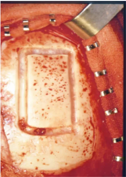
The orbits were reconstructed with Tutopatch sheet (26 patients; Tutogen Medical GmbH, Neunkirchen, Germany), titanium mesh (13 patients; Synthes GmbH, Oberdorf, Switzerland), and autologous calvarial bone graft (1 patient; Fig. 2); the forced duction test was performed to exclude tissue entrapment. The patients were then carefully observed until they attained maximum return of function or failed to keep a scheduled follow-up.
The mean follow-up reached 39 months (range, 6Y81 months). Enophthalmos was measured by a Hertel exophthalmometer (Richmond Products, Albuquerque, NM); diplopia was evaluated by an
op-tometrist with cover test, red glass test, and Hess-Lancaster test; and hypesthesia of the infraorbital nerve territory was checked by clin-ical examination.
The studied parameters included age, sex, time interval be-tween trauma and surgery, location of the fracture, and implant material. TheW2test for nonparametric data was used, and a P value of less than 0.05 was considered statistically significant.
RESULTS
Sex, location of the fracture, and implant material were not considered statistically significant (P9 0.05); the unique variable that influenced our data was the time interval between trauma and surgery (Table 1).
At a long-term follow up, postoperative diplopia was found in 17 patients of whom 13 patients were operated on before 2 weeks and 4 patients were treated after 2 weeks. The time of surgery was considered statistically significant (PG 0.05) to influence this con-dition (Fig. 3).
Eleven patients (Fig. 4) developed postoperative enophthal-mos (7 patients were treated before 14 days, and 4 patients were operated on after 14 days). The unique variable statistically signif-icant was the time of surgery (PG 0.05).
Finally, 22 patients complained of infraorbital nerve dysfunc-tion (Fig. 5) for hypesthesia and dysesthesia; 18 patients were treated before 2 weeks, and 4 patients were operated on after 2 weeks. Again, the time of surgery was considered statistically significant (PG 0.05).
DISCUSSION
In our retrospective study, diplopia, enophthalmos, and infra-orbital nerve dysfunction were the frequent complications, although the surgical technique was executed properly and the immediate postoperative recovery was uneventful. Our results showed that early repair of blow-out fracture leads to a less postoperative diplopia (PG 0.05). We believe that early surgery may minimize progressive fibrosis and contracture of the prolapsed tissues; scar tissue occurs afterward the extraocular muscles and orbital soft tissues are placed in their original anatomic position. Other authors report the same data and agree with our opinion.8,11,16
Conversely, other authors such as Jin et al17and Putterman et al9did not find any statistically significant correlation between postoperative diplopia and time of surgery (P9 0.05); they suggest an approach more conservative, believing that it is more convenient to ‘‘wait and see’’ for the resolution of the edema and the hemorrhage. FIGURE 1. Computed tomographic scan demonstrating
an isolated orbital blow-out fracture.
FIGURE 2. Intraoperative photograph showing an autologous calvarial bone graft.
TABLE 1. Time of Surgery After Injury
Time of Surgery e24 h e7 d 7Y14 d 914 d Total
No. patients 2 34 0 4 40
FIGURE 3. Correlation between postoperative diplopia and time of surgery.
Brucoli et al The Journal of Craniofacial Surgery
&
Volume 22, Number 4, July 20111388
* 2011 Mutaz B. Habal, MDIn our study, diplopia was present in 42.5% of the patients at the mean follow-up of 39 months. This condition did not develop or become worse in any patients after surgery. Literature is very dis-cordant about the incidence of postoperative diplopia after the sur-gical treatment of orbital blow-out fracture. Biesman et al18reported an incidence of 37%, whereas Greenwald et al19described 20% of patients with postoperative diplopia.
In our series, 27.5% of the patients had persistent enophthal-mos; other authors also report a high incidence of residual enoph-thalmos after surgery. However, this condition can be surgically corrected later with excellent results.11,19,20Prolapse of orbital tissues into the sinuses, enlarged orbital volume, atrophy of the orbital fat and loss of support of orbital walls can play a decisive role in the pathogenesis of enophthalmos. It may be masked in the first days after trauma because of the associated periorbital and intraorbital edema or hemorrhage.21
Infraorbital nerve dysfunction for hypesthesia and dysesthesia was the most frequent complication observed, and it was detected in 55% of the patients postoperative; these data agree with other studies.14,15,20Nevertheless, there is a limitation for this variable be-cause the examination was conducted by a standard neurologic visit with fine needle, bristle, and flocks; and the evaluation was not quantitative but subjective.
We stress the fact that orbital blow-out fracture is generally not considered a technically demanding procedure, but the outcome is disappointing; the surgical procedure must be managed carefully by experienced surgeons to lower the high rates of these 3 common complications.
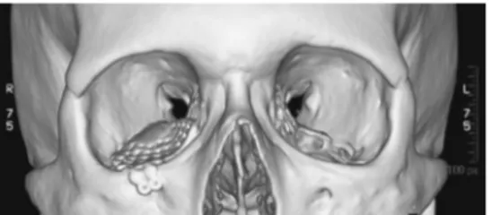
Actually, surgeons use various materials for orbital recon-struction; an ideal implant material should be chemically and biolog-ically inert with a minimal rate of inflammation, infection, extrusion, migration, and exposure. Titanium mesh was our preferred choice for the reconstruction of demanding fractures involving one half or more of the orbital floor and/or the medial wall with comminuted segments (Fig. 6).
We used Tutopatch sheet for less severe fractures; because of the high morbidity and the technical difficulty, we strictly reserve autologous calvarial bone grafts for high-energy impacts with loss of tissues. In this study, we did not observe any infections or extrusion of the different materials used to reconstruct the orbit.
CONCLUSIONS
Our retrospective study shows that it is very difficult to reach a successful result for a long-term surgical outcome free from com-plications, although the preoperative indication to surgery is carefully evaluated, the operative technique is conducted properly, and the postoperative recovery is uneventful.
However, we can report that the incidence of diplopia, enoph-thalmos, and infraorbital nerve dysfunction are decreased by an im-mediate intervention and an early surgical repair of the orbital blow-out fracture. Patients who had surgery within 2 weeks of trauma have a lower risk to develop postoperative complications; this study supports an early surgical treatment of orbital blow-out fractures, when it is indicated.
REFERENCES
1. Lang W. Traumatic enophthalmos with retention of perfect acuity vision. Trans Ophthalmol Soc UK 1889;9:41Y45
2. Fujino T. Mechanism of orbital blowout fracture. J Plast Surg 1974;17:427
3. Smith B, Regan WF. Blow-out fractures of the orbit. Am J Ophthalmol 1957;44:733Y739
4. Ellis E, Attar A, Moos KF. An analysis of 2067 cases of zygomatico-orbital fractures. J Oral Maxillofac Surg 1985;43:417Y428
5. Waterhouse N, Lyne J, Urdang M, et al. An investigation into the mechanism of orbital blow-out fractures. Br J Plast Surg 1999;52:607Y612
6. Brucoli M, Stecco A, Iaquinta C, et al. Diagnosis and treatment of orbit posttraumatic subperiosteal hemorrhage in a child, associated with a subdural intracranial hemorrhage. J Craniofac Surg
2005;16:407Y410
7. Bahr W, Bagambisa FB, Schlegel G, et al. Comparison of transcutaneous incisions used for exposure of infraorbital rim and orbital floor: a retrospective study. Plast Reconstr Surg 1992;90:585Y591
8. Matteini C, Renzi G, Becelli R, et al. Surgical timing in orbital fracture treatment: experience with 108 consecutive cases. J Craniofac Surg 2004;15:145Y150
9. Putterman AM, Stevens T, Urist MJ. Nonsurgical management of blow-out fractures of the orbital floor. Am J Ophthalmol 1974;77:232Y239
10. Gassner R, Tuli T, Hachl O, et al. Cranio-maxillofacial trauma: a 10 years review of 9543 cases with 21067 injuries. J Craniomaxillofac Surg 2003;31:51Y61
11. Hossal BM, Beatty RL. Diplopia and enophthalmos after surgical repair of blow-out fracture. Orbit 2002;21:27Y33
12. Sleep TJ, Evans BT, Webb AA. Resolution of diplopia after repair of the deep orbit. Br J Oral Maxillofac Surg 2007;45:190Y196
FIGURE 6. Three-dimensional computed tomographic scan showing the reconstruction with titanium mesh.
FIGURE 4. Correlation between postoperative enophthalmos and time of surgery.
FIGURE 5. Correlation between infraorbital nerve dysfunction and time of surgery.
The Journal of Craniofacial Surgery
&
Volume 22, Number 4, July 2011 Repair of Orbital Fractures* 2011 Mutaz B. Habal, MD
1389
13. Manson PN, Grivas A, Rosembaum A, et al. Studies on enophthalmos II. The measurement of orbital injuries and their treatment by quantitative computed tomography. Plast Reconstr Surg. 1986;77:203Y214
14. Freidel CK, Dumas P, Rougier J, et al. Ne´vralgie sous-orbitaire compliquant les fratctures de l’orbite. A propos de cinq observations. Rev Stomatol Chir Maxillofac 1970;71:357Y364
15. Vriens JP, van der Glas HW, Bosman F, et al. Information on infraorbital nerve damage from multitesting of sensory function. Int J Oral Maxillofac Surg 1998;27:20Y26
16. Jin HR, Lee HS, Yeon JY, et al. Residual diplopia after repair of pure blow-out fracture: the importance of extraocular muscle injury. Am J Rhinol 2007;21:276Y280
17. Jin HR, Shin SO, Choo MJ, et al. Relationship between the extent of fracture and the degree of enophthalmos in isolated blow-out
fractures of medial orbit wall. J Oral Maxillofac Surg 2000;58: 617Y620
18. Biesman BS, Hornblass A, Lisman R, et al. Diplopia after surgical repair of orbital floor fractures. Ophthal Plast Reconstr Surg 1996;12:9Y16
19. Greenwald HS, Keeney AH, Shannon GM. A review of 128 patients with orbital fractures. Am J Ophthalmol 1974;78:655Y664
20. Gas C, Sidjilani BM, Dodart L, et al. Fractures isole´es du plancher orbitaire: conclusions d’une etude retrospective portent sur 85 cas. Rev Stomatol Chir Maxillofac 1999;100:27Y33
21. Kolk A, Pautke C, Scott V, et al. Secondary post-traumatic enophthalmos: high-resolution magnetic resonance imaging compared with multislice computed tomography in postoperative orbital volume measurement. J Oral Maxillofac Surg
2007;65:1926Y1934
Brucoli et al The Journal of Craniofacial Surgery