Current Pharmaceutical Design, 2018, 24, 1-11 1
RESEARCH ARTICLE
1381-6128/18 $58.00+.00 © 2018 Bentham Science Publishers
Dietary Approaches to Attain Fish Health with Special Reference to their Immune
System
Thea Magrone
1,*, Matteo Antonio Russo
2and Emilio Jirillo
11
Department of Basic Medical Sciences, Neuroscience and Sensory Organs, University of Bari, School of Medicine, Bari, Italy;
2MEBIC Consortium, San Raffaele Open University of Rome and IRCCS San Raffaele Pisana of Rome, Rome, Italy
A R T I C L E H I S T O R Y Received: September 3, 2018 Accepted: December 28, 2018 DOI:
10.2174/1381612825666190104121544
Abstract: Fish despite their low collocation in the vertebrate phylum possess a complete immune system. In teleost fish both innate and adaptive immune responses have been described with melanomacrophage centers (MMCs) equivalent to mammalian germinal centers. Primary lymphoid organs are represented by the thymus and kidney, while spleen and mucosa-associated lymphoid tissues act as secondary lymphoid organs. Functions of either innate immune cells (e.g., macrophages and dendritic cells) or adaptive immune cells (T and B lympho-cytes) will be described in detail, even including their products, such as cytokines and antibodies. In spite of a robust immune arsenal, fish are very much exposed to infectious agents (marine bacteria, parasites, fungi, and viruses) and, consequentially, mortality is very much enhanced especially in farmed fish. In fact, in aquaculture stressful events (overcrowding), microbial infections very frequently lead to a high rate of mortality. With the aim to reduce mortality of farmed fish through the reinforcement of their immune status the current trend is to admin-ister natural products together with the conventional feed. Then, in the second part of the present review emphasis will be placed on a series of products, such as prebiotics, probiotics and synbiotics, β-glucans, vitamins, fatty acids and polyphenols all used to feed farmed fish. With special reference to polyphenols, results of our group using red grape extracts to feed farmed European sea bass will be illustrated. In particular, determination of cyto-kine production at intestinal and splenic levels, areas of MMCs and development of hepatopancreas will represent the main biomarkers considered. All together, our own data and those of current literature suggests that natural product administration to farmed fish for their beneficial effects may, in part, solve the problem of fish mortality in aquaculture, enhancing their immune responses.
Keywords: Aquaculture, cellular and molecular rehabilitation, fish, immunity, lymphocytes, melanomacrophage centers, natural products.
1. INTRODUCTION
Despite their low position in the vertebrate phylum, fish possess
quite robust immune machinery, which allows them to survive in a
very hostile environment. In analogy to mammals, fish immunity
can be divided into two major compartments, namely the innate and
the adaptive immune system. In fact, teleost fish possess both
pri-mary and secondary lymphoid tissues and, specifically, thymus and
head-kidney represent primary organs while the spleen and the
mucosa-associated lymphoid tissues (MALTs) are secondary
or-gans. In turn, MALTs can be divided into gut-associated lymphoid
tissue, gill-associated lymphoid tissue (GIALT), skin-associated
lymphoid tissue and nasopharinx-associated lymphoid tissue [1].
MALTs are covered by a protective layer of mucus containing
anti-bodies which interact with both commensal and invading
microor-ganisms [2-4]. They are not organized lymphoid structures as in the
case of mammal lymph nodes but fish myeloid and lymphoid cells
constitute a diffuse network, even if an interbranchial lymphoid
tissue has been identified in GIALT [5]. As later discussed in this
review, melanomacrophage centers (MMCs) are pigmented
phago-cytes distributed in kidney, spleen and liver of fish [6-8]. MMCs
belong to the innate immunity and are considered as precursors of
the mammal germinal centers (GCs) [9]. Furthermore, in fish,
in-nate immunity is based on the function of other immune cells, such
as granulocytes, macrophages and dendritic cells (DCs), but also
non-immune cells, such as intestinal M-like cells, red blood cells
and thrombocytes participate to immune defense [10-17].
*Address corrospondance to this author at the Department of Basic Medical Sciences, Neuroscience and Sensory Organs, School of Medicine, Univer-sity of Bari, School of Medicine, P.zza Giulio Cesare 11, 70124 Bari, Bari, Italy; Tal: +39 0805478492; E-mail: [email protected]
Fish innate-like lymphocytes are positioned between innate and
adaptive immunity, and, in particular, extrathymic γδ T cells have
been identified [18-20]. As discussed in the next section of this
review, fish intestine represents the major source of innate-like
lymphocytes in analogy to mammalian-like lymphocytes [21].
With special reference to fish adaptive immunity, both CD4
+and CD8
+cells are largely present in MALTs and their phenotypes
and functions will be discussed in a specific section of this review
[22-24]. B cells are also contained in MALTs and immunoglobulin
(Ig)T represents the equivalent of secretory IgA in mammals, thus,
protecting mucosal sites from invading pathogens [25]. Despite a
robust immune system, fish succumb because of severe infections
and, especially, in farmed fish mortality is very high due to invasion
by pathogens and stressful environmental conditions [26, 27].
Therefore, preserving fish health in aquaculture and prevention of
disease is a major focus of the current research in this field. This
achievement may result from the combination of different strategies
such as innovative feeds, low fish density and corroboration of the
immune response [28]. Particularly, administration to fish of dietary
supplements has been increasing in the last few years and, here, this
procedure will be elucidated in detail.
On these grounds, aims of the present review are the illustration
of piscine innate and adaptive immune responses but also of the
role played by natural products in the enhancement of fish
immu-nity against infectious challenges and stressful conditions.
2. INNATE IMMUNITY
2.1. Melanomacrophage Centers (MMCs)
MMCs are nodular accumulations of melanin-macrophages
mostly distributed in kidney, spleen and liver of fish. Pigments
(lipofuscin, melanin and hemosiderin) contained in MMCs can be
of both exogenous and endogenous origin and the latter is derived
from phagocytosed erythrocytes [29]. This last event suggests that
MMCs play a role in iron recycling similarly to hemosiderin-laden
splenic macrophages in mammals [30]. Besides erythrocytes, there
is evidence that MMCs phagocytose pathogens [31, 32]. In turbot,
the evidence that splenic MMCs are in close association with
ellip-soids (which are capillaries) may suggest their property to
phagocy-tose blood-borne pathogens [33]. Besides the above mentioned
functions, MMCs have been considered as primitive precursors of
mammalian GCs, even if such a contention needs further validation.
In fact, fish lack GCs and MMCs, which may also be due to the
adaptive immune responses [34, 35]. In this framework, fish
immu-nized with bacteria are able to retain antigens in and around splenic
MMCs, trapped within immune complexes (ICs) and such retention
can be enhanced by injection of preformed ICs [36-38]. More
spe-cifically, the above results support a similarity between MMCs and
mammalian follicular DCs in the context of GCs, thus, allowing
antigen presentation to T helper (h) 2 cells, and ultimately,
promot-ing B-cell antibody formation [39].
Conclusively, as recently reviewed by Steinel and Bolnick [40],
one can hypothesize that MMCs have evolved as “garbage dumps”
from a common precursor of vertebrates, and, then, after meeting
pathogens and antigens have acquired immunological functions,
finally, evolving into GCs.
2.2. Macrophages
Fish macrophages are distributed in mucosal and non-mucosal
tissues (spleen, kidney and liver) and in analogy to the mammal
counterpart can also be divided into subsets. In mammals, one can
distinguish M1 (classically activated macrophages) and M2 a, b and
c (alternatively activated macrophages) [41]. Functionally, M1 are
inflammatory, while M2 and, mostly, M2c are anti-inflammatory
cells, also secreting angiogenic mediators for the resolution of
inflammation. In particular, M1 cells exert phagocytosis of
patho-gens, produce free radicals and pro-inflammatory mediators
[42-44]. M2 fish macrophages have been analyzed for arginase gene
expression and activity, as well as, gene expression and function of
interleukin (IL)-10 and transforming growth factor-β. However, the
fine regulatory mechanisms involved in the polarization toward
M2a-c cells need further investigation [45-47].
Colony stimulating factor (CSF)-1 represents the major growth
factor for fish macrophages and in most fish CSF-1.1 and CSF-1.2
molecules have been identified [48]. CSF-1.1 promotes the
differ-entiation toward M1 macrophages in goldfish (Carassius auratus)
[49]. In support of the above finding, evidence has been provided
that a soluble CSF-1 receptor inhibits the pro-inflammatory
path-way in goldfish macrophages. Quite interestingly, in some fish
species, two distinct csf1r genes have been detected, thus,
suggest-ing that these animals have elaborated more complex mechanisms
of macrophage differentiation in comparison to mammals [50].
In order to cope with pathogens, fish macrophages bear both
extracellular and intracellular pattern recognition receptors (PRRs).
At least five types of PRRs have been identified, namely, Toll-like
receptors (TLRs), c-type lectins, nucleotide-binding
domain-leucine-rich repeat containing receptors (NLRs or NOD-like),
reti-noic acid inducible gene I-like receptors and absence in
melanoma-like receptors [51]. Besides macrophages, also neutrophils,
mono-cytes, DCs, epithelial and endothelial cells possess PRRs. Bony fish
display 17 TLRs with TLRs 20-23 present only on fish [52, 53]. Of
note, not all fish bear TLR-4, a specific receptor for Gram-negative
lipopolysaccharides (LPS) and, in particular, zebrafish TLR-4 does
not recognize LPS [54]. NLRs have been identified in fish and
seem to share conserved structural domains with mammalian NLRs.
In this respect, studies have been focused on gene expression and to
a lesser extent on signaling following NLR stimulation [55, 56].
Fish M1 macrophages exert a potent anti-microbial activity
which encompasses different phases. Following phagocytosis of
pathogens, generation of superoxide anions (respiratory burst) and
nitric oxide (NO) production are two independent
macrophage-mediated anti-microbial functions [57]. Another mechanism related
to the microbicidal activity of M1 macrophages is the indoleamine
2,3-dioxygenase (IDO)-mediated degradation of tryptophan [58].
However, IDO can control immune response via production of
kinurenins and, in turn, interferon (IFN)-γ-stimulated macrophages
produce IDO [59, 60]. Fish M2 macrophages play both
anti-inflammatory and pro-healing effects. The fish 4/13a and
IL-4/13b exert anti-inflammatory effects, dampening the gene
expres-sion of pro-inflammatory cytokines, such as tumor necrosis factor
(TNF)-α, IL-1β and IFN-γ [61, 62]. Quite interestingly, fish M2c
macrophages under influence of cortisol up-regulate the IL-10 gene
expression and in the gold fish recombinant IL-10 suppresses
reac-tive oxygen species production and inflammatory gene expression
[63]. Arginase production is a marker of M2 cell activity in fish
[64]. In fact, arginase converts L-arginine to L-ornithine which, in
turn, represents a precursor for polyamine and proline constituents
of collagen for tissue repair to occur [65]. Fish possess both
ar-ginase-1 and arginase-2 genes with M1 cells expressing more
in-ducible nitric oxide synthase gene (inflammatory profile) and M2
cells expressing more arginase genes (anti-inflammatory/tissue
repairing profile) [66].
Fish macrophages also serve as antigen presenting cells (APCs)
and interact with T lymphocytes via the major histocompatibility
complex (MHC)-1 expression, whose extent is different according
to species [67]. Of note, some fish species have lost the MHC-II,
even if they mount an effective immune response against pathogens
[68, 69]. Other fish species have expressed non-classical MHCs
whose lineages have separated before the emergence of tetrapods
[70]. In this framework, it is important to emphasize the role of fish
DCs which act as APCs. DCs express MHC-II and CD83 markers,
also exerting potent phagocytic properties and induction of CD4
+T
cell production [70-72].
In this context, the immune role exerted by fish erythrocytes is
worth mentioning. In vitro studies have demonstrated that trout
erythrocytes are able to engulf Candida (C.) albicans particles and
present them to surrounding head-kidney macrophages [13, 14].
Furthermore, mature fish erythrocytes which have engulfed C.
albicans have been shown to release in supernatants cytokine-like
factors able to potentiate macrophage function, while inhibiting
their migration (macrophage inhibition factor-like activity) [15].
Furthermore, evidence has been provided that C. albicans
phago-cyting trout erythrocytes form aggregates with macrophages (the
so-called rosettes), thus traveling via circulation to distant organs,
and, finally, contributing to the formation of MMCs [16]. Quite
interestingly, in infected trout with C. albicans, evidence of rosette
formation in blood has been provided, and, in addition, fish
throm-bocytes were present in close association with erythrocytes [17].
These data suggest that fish thrombocytes may also play an
immu-nological role in the clearance of C. albicans. Major innate immune
cells functions are summarized in Table 1.
3. FISH LYMPHOCYTES
In a recent review [73], the question has been poised on the
equivalence between fish lymphocytes and mammalian innate-like
T cells. In mammals and fish, innate-like T cells are mostly located
in the intestine as intestinal intraepithelial lymphocytes (IELs)
characterized by an archaic T cell receptor (TCR), the so called
γδTCR [74, 75]. γ
+δ
+cells can develop extrathymically in the
ab-sence of antigen encountering and exert an array of functions, such
as direct killing of infected cells, tumor immunosurveillance,
toxicity and release of both regulatory and pro-inflammatory
cyto-kines [76]. Quite interestingly, subsets of murine γδ cells have been
identified which are activated by innate stimuli rather than TCR
ligation and known as IL-17 producing (γδT-17) and IFN-γ
produc-ing (γδT-IFN-γ) cells [77].
Innate-like B cells in the mammals are the B1-B cells also
sub-divided into B1a and B1b-B cells [78]. B1 cells secrete natural
polyreactive antibodies in a T-independent fashion, being
phago-cytic, microbicidal and involved in autoimmune processes [79, 80].
In teleost fish, T cells are largely present in gills and intestine
and express the TCRαβ on their surface in the presence of either
CD4 coreceptor or CD8 coreceptor. Then, CD4
+Th cells, CD8
+T
cytotoxic cells, T regulatory (Treg) cells and Th17 cells have been
identified [81-83].
Fish intestine is the major site of immune responses as
de-scribed below. In the intestine, IEL in vivo undergo
recombinant-activating gene (RAG)-mediated somatic rearrangement of a given
V/C combination in the CDR3 junction length of
TCRβ-chain/TCRγ-chain [84, 85]. Conversely, gill T cells in vitro
prolif-erate when stimulated by lectins but do not express RAG [86]. In
the sea bass intestine, evidence has been provided that T cells
ex-press RAG-1, TCRα, TCRγ, CD8α and CD4, thus, suggesting that
gut is the main source of T cells in adult fish [87]. Conversely, gills
can be considered as a source of T cells committed as
effec-tor/helper cells [86]. In this context, a special issue is undoubtedly
represented by fish immune tolerance. Sugimoto and associates [88]
have clearly demonstrated that zebrafish lacking a functional FoxP3
ortholog exhibit a reduced survival with the development of a
pro-inflammatory profile. In fact, in analogy to mammals, Treg cells are
controlled by a master regulatory gene encoding FoxP3. The same
group has also demonstrated that zebrafish Treg cells mediate
or-gan-specific regeneration, while in the absence of FoxP3 deficient
Treg cells infiltrate damaged organs (e.g., spinal cord, heart and
retina) but do not express regenerative factors [89]. Furthermore,
the intrabranchial lymphoid tissue in gills promotes immune
toler-ance in view of its high expression of FoxP3 [90]. This homeostatic
mechanism seems to be very effective in the gills where continuous
exposure to a variety of antigens may lead to exaggerated
inflam-matory immune responses. Main functions of T lymphocytes are
expressed in Table 2.
Table 2. Subsets of fish T lymphocytes.
Cell types Functions
γ+δ+ T cells Direct killing of infected cells,
immunosurveil-lance, cytotoxicity and release of both regulatory and pro-inflammatory cytokines [76]. α+β+ T cells
(CD4+ Th cells,
CD8+ T cytotoxic
cells, Treg cells and Th17 cells)
Gill and intestinal T cells are the major sites of these lymphocytes and their anti-inflammatory
activity is prevalent [89, 90].
The origin of fish B cells is still uncertain. In zebrafish, the
pancreas has been indicated as the primary source of B cells [91]. In
sea bass, the presence of IgM producing cells in kidney may
sug-gest that this organ is the primary site of B cell production [92].
Fish B cells express µ,
θ, δ genes which lead to three different Ig
classes, namely, IgM, IgT and IgD, as recently reviewed by Flajink
[93]. IgM are tetrameric and their concentration is very abundant in
biological fluids [94]. IgT (for immunoglobulin teleost) is a
mu-cosal antibody isotype produced as a monomer [95]. However, in
trout mucus a non-covalent polymeric IgM association has been
reported [96]. Finally, IgD is expressed in monomeric form and in
analogy to humans it can be involved in inflammatory responses
binding to a still unknown receptor on the surface of basophils [97].
In Table 3, fish antibody classes and functions are outlined.
Table 3. Antibody classes in fish.
Class Functions
IgM They are tetrameric and very abundant in biologic fluids of non-immunized fish [93].
IgT This is a mucosal antibody equivalent to mammalian IgA and produced as a monomer [95].
IgD They are monomeric, being involved in inflammatory activi-ties via binding to basophils [97, 98].
Conclusively, according to Scapigliati and associates [73] fish
B cells have certain properties, such as elevated concentration of
IgM in non-immunized fish, low increase of IgM affinity following
secondary immunization, possible expression of TLRs and
patho-gen phagocytosis, kidney B1-B cells similar to the splenic murine
counterpart and proliferating B cells in peritoneum. Quite
interest-ingly, the first teleost CD5 molecule has recently been identified
[98]. This finding represents an additional evidence of phenotypical
and functional similarities between fish IgM
+B cells and
mammal-ian B1 cells.
There are experimental evidences in favor of immunological
memory in fish which are mostly based on adoptive transfer of
peripheral leukocytes from vaccinated donors to naïve
histocom-patible recipients [99]. Cell transfer experiments afford long term
protection in recipients but if the mechanisms triggered by these
cells may be considered “memory” needs further clarification.
As indicated in the previous paragraphs, fish lymphocytes are
able to secrete an array of cytokines. In this regard, the best studied
fish T lymphocyte cytokine is IFN-γ as a product of Th1 cells
[100]. Two different types of IFN-γ have been identified in fish,
namely IFN-γ and IFN-γ rel [101]. A functional dichotomy has
been observed in these two types of fish IFN-γ, mostly, in terms of
their effects on macrophages and monocytes, as extensively
re-ported by Grayfer and associates [102, 103]. It would be very
inter-esting to decipher whether there are differences between these two
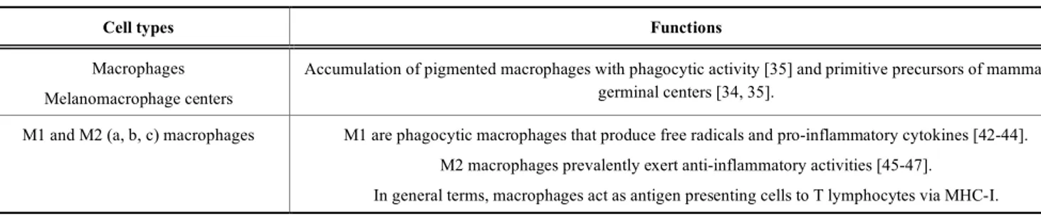
Table 1. Main innate immune cells in fish.
Cell types Functions
Macrophages Melanomacrophage centers
Accumulation of pigmented macrophages with phagocytic activity [35] and primitive precursors of mammalian germinal centers [34, 35].
M1 and M2 (a, b, c) macrophages M1 are phagocytic macrophages that produce free radicals and pro-inflammatory cytokines [42-44]. M2 macrophages prevalently exert anti-inflammatory activities [45-47].
IFNs in exerting antimicrobial activities against various fish
patho-gens.
At the end of this section, one has to emphasize that teleost fish
possess a commensal microbiota able to shape the immune
re-sponse, as recently reviewed [104]. Studies on the interaction
be-tween fish microbiota and immune system will contribute to solve
problems in aquaculture system, preventing development of
infec-tions.
4. NATURAL PRODUCTS AS MODULATORS OF FISH
IMMUNE RESPONSE
Fish disease (viral, bacterial, fungal and parasitic infections)
have been increasing, thus, causing noticeable economical losses in
the aquaculture industry [105-107]. Particularly, juvenile marine
fish undergo elevated mortality rate during the larvae culture phase
due to chronic stress and bacterial infections [108, 109]. In
aquacul-tures, high population density and manipulation seem to favor
growth of opportunistic bacteria with reduction of beneficial
micro-organisms, thus, leading to mortality [110, 111]. In order to
over-come the problem of fish mortality three main strategies have been
adopted, namely: i. the maintenance of microbiota equilibrium in
aquaculture; ii. the improvement of water quality and, finally, iii.
the enhancement of the immune response by larvae [112]. With
regard to immune enhancement in teleost fish, immune stimulation
strategy is based on the application of compounds which potentiate
the function of phagocytic cells, thus, increasing their bactericidal
and fungicidal functions [113]. The observed high mortality in
lar-vae may depend on their immature adaptive immune system, and,
therefore, immune stimulation may be very effective on their
natu-ral arm of immunity [114]. In synthesis, larval stage is a very
criti-cal phase in aquaculture where application of immunostimulant
strategies appears to be more effective. On these grounds, in the
next paragraphs several compounds of natural origin will be
illus-trated for their capacity to regulate/potentiate fish immune response
in aquaculture.
4.1. Prebiotics, Probiotics, Synbiotics
According to Bindels and associates [115], “prebiotic concept
typically refers to non digestible food ingredients or substances that
pass undigested through the upper part of the gastrointestinal tract
and stimulate the growth and/or activity of health-promoting
bacte-ria that colonize the large bowel.” Major prebiotics are fructose
oligosaccharides (FOS), galactose oligosaccharides (GOS), xylose
oligosaccharides (XOS), inulin, polydextrose, dietary fibers and
mannan-oligosaccharides [116]. Prebiotics administered to fish as
food or feed ingredients beneficially impact on the fish intestinal
immune system [117]. Common carp fed FOS, GOS and inulin
underwent a significant increase in skin mucus lysozyme activity,
especially in GOS-fed group [118]. Administration of GOS to
Cas-pian white fish increased immunoglobulin levels [119]. In
zebraf-ish, administration of sodium propionate increased appetite and
mucus gene expression [120]. In a series of reports, evidence has
been provided that administration of prebiotics to fish has led to
increased anti-inflammatory cytokine expression and regulation of
appetite gene expression also via production of short chain fatty
acids (SCFAs) [121-126]. Other lines of evidence have documented
that prebiotics are able to increase intestinal digestion and
absorp-tion in juvenile golden pompano, augmenting villus length, villus
width and their numbers as well muscle thickness at all gut
seg-ments [127]. In zebrafish, administration of chitosan silver
nano-composites reduced the number of Proteobacteria (a phylum of
Gram-negative bacteria enriched in pathogens) [128]. Conclusively,
as recently reviewed [129], prebiotics in addition to a direct effect
on the fish gut immune system are digested by intestinal microbiota
for the production of SCFAs which, in turn, promote the killing of
pathogens.
Probiotics are defined as “microorganisms that when consumed
(as in food or a dietary supplement) maintain or restore beneficial
bacteria to digestive tract” [130]. There are a few evidences on the
beneficial effects of probiotic supplementation to fish. Bioflocs are
rich in various probiotics and bioactive compounds and their effects
on juvenile sea cucumber Apostichopus japonicus have been
ana-lyzed [131]. From an immunological point of view, biofloc
in-creased phagocytosis and respiratory burst in comparison to
un-treated fish, also upregulating immune-related genes such as heat
shock proteins [90, 70, 105], rel, nitric oxidase and lysozyme. In
treated fish challenged with Vibrio splendidus, mortality
signifi-cantly decreased in comparison to controls. Lactobacillus (L.)
delbrueckii administration to Cyprinus carpio huanghe var has
re-cently been investigated to evaluate resistance against Aeromonas
(A.) hydrophila [132]. Results have demonstrated that L.
delbrueckii supplementation upregulated lysozyme, acid
phospha-tase and myeloperoxidase activities and antioxidant enzymes
[su-peroxide dismutase (SOD), catalase, gluthatione peroxidase)], while
downregulating mRNA levels of IL-1β, IL-8, TNF-α and NF-κB
p65. In view of these effects growth performance and survival rate
improved. In another study, two recombinant L. casei
(surface-displayed or secretory) expressing the OmpAI of A. veronii were
administered to Cyprinus carpio challenged with A. veronii [133].
In comparison to untreated fish after A. veronii infection, fish fed
with recombinant L. casei exhibited higher titers of serum and skin
mucus antibodies, lysozymes and gene expression of IL-1β, IFN-γ,
IL-10 and TNF-α. Immune modifications positively correlated with
higher survival in treated fish in comparison to controls.
In view of the beneficial effects exerted by pre- and probiotics
when independently administered to fish, their association, the
so-called synbiotics, has also been experimented in fish. As
exten-sively reviewed by Huynh and associates [134], synbiotics
admini-stration to fish increased nutritional absorptive capacities, on the
one hand, and, increased natural and adaptive immune responses on
the other hand. Besides that, synbiotics were able to modify
intesti-nal microbiota in Apostichopus japonicus during vibrio infection,
increasing proportions of Bacillus and Lactococcus in comparison
to controls [135].
4.2.
β-Glucans
β-glucans are natural polysaccharides largely produced by
bac-teria, fungi and yeast [136]. β-glucans act through a specific
recep-tor dectin-1 (not yet well defined in fish) and TLR-2 on leukocytes
[137]. Activation of dectin-1 triggers intracellular signaling, e.g.,
Syk/NF-κB pathways, thus, leading to increased expression of
im-mune genes and release of pro-inflammatory cytokines (IL-1, IL-8,
IL-12, TNF-α). β-glucans are profusely used in aquaculture for
their beneficial effects on the immune system and increased
resis-tance to stress. In cod larvae, β-glucans have been administered via
encapsulation by live food [138]. Mostly,
β-glucans extracted from
diatoms were very effective in survival and growth. In a recent
study [139], Sterigmatomyces halophilus β-glucans, when
adminis-tered to pacific red snapper, increased phagocytic activity,
respira-tory burst, NO production, SOD and catalase activities and immune
genes for IL-1β, IL-10 and IL-17. This led to an increased
resis-tance against A. hydrophila. Similar protective effects were
ob-served in Oreochromis niloticus challenged with Staphylococcus
iniae [140]. Increase in anti-oxidant, anti-inflammatory, anti-stress
and immune-related genes contributed to the protective effect
against bacterial infections. Furthermore, in an in vitro model,
fun-gal β-glucans increased in head-kidney leukocytes from pacific red
snapper NO production, myeloperoxidase, SOD and catalase
activi-ties as well as induction of pro-inflammatory cytokines [141].
4.3. Lipids
There is a large body of evidence that polyunsaturated fatty
acids (PUFAs), when ingested with food or as supplements exert
beneficial effects to the host, even including the switch-on of an
intestinal immune tolerogenic pathway and the improvement of gut
microbiota [142, 143]. In farmed fish, a few experimentations with
PUFAs have been conducted in order to promote growth of healthy
fish despite pollution, overcrowding and risk of infections. In a
recent report [144], rainbow trout juveniles were administered with
four PUFAs, namely,
α-linoleic (ALA), linoleic acid (LA),
eicos-apentaenoic acid (EPA) and docosahexaenoic acid (DHA). All four
PUFAs were combined with an environmentally-realistic
concen-tration of cadmium. Some fish were challenged with A. salmonicida
at the end of cadmium conditioning period. Despite the fact that
cadmium even at low doses could down-regulate the fish immune
response, PUFA administration improved growth (DHA), protected
against oxidative stress (ALA and EPA) and enhanced innate
im-munity. Hepatic steatosis frequently occurs in farmed fish,
ulti-mately leading to severe inflammation. In large yellow croaker,
administration of omega-3 PUFAs mitigated liver steatosis-induced
inflammation [145]. The protective mechanism exerted by omega-3
seems to rely on the sirtuin-1-mediated nuclear translocation of
NF-κB p65 subunit in hepatocytes.
Arachidonic acid supplemented to rabbit fish Siganus rivulatus
during an outbreak of S. iniae prolonged survival in comparison to
controls, while increasing lysozyme, complement and total serum
Ig levels [146]. Conclusively, this regimen could improve growth
survival and immune functions.
α-lipoic acid (α-LA) when supplemented to grass carp has been
demonstrated to prevent omega-3-induced lipid peroxidation [147].
In fact, in grass carp omega-3 dietary regimen could increase
pro-duction of malondialdehyde, thus, inducing lipid peroxidation in
liver and muscle. This α-LA-mediated effect was based on the
in-creased expression of NF-E2-related nuclear factor 2 and decrease
of kelch-like-ECH-associated protein 1 mRNA levels.
Sodium butyrate (SB) administration to young grass carp enhanced
intestinal immune function associated with NF-κB and p38 MAPK
signaling [148]. In general, SB ameliorated growth performance,
intestinal growth and function, increasing the number of
Lactoba-cilli in the gut microbiota. In particular, either natural immunity
(increase in C3 and C4, IgM and antimicrobial peptides) or adaptive
immunity (increase in anti-inflammatory cytokines) were
modu-lated by SB.
4.4. Vitamins
Vitamins are essential micronutrients endowed with multiple
immune functions in animals and humans. In an in vitro study,
us-ing the rainbow trout monocyte-macrophage cell line RT811 and
head-kidney leukocytes vitamin C increased production of
phago-cytic cell number [149]. Furthermore, vitamin C also enhanced the
transcription of several pro-inflammatory and antimicrobial genes
triggered by Escherichia coli. Of note, evidence has been provided
that vitamin C attenuates the toxic effects of
17α-methyltes-tosterone, reducing up-regulation of pro-inflammatory cytokines,
chemokines, TLR-7, IgM and apoptosis [150]. Finally, as recently
reviewed by Dawood and Koshio [151] vitamin C is essential for
growth health and stress resistance in fish.
In European sea bass administered with vitamin D3 an increase
in phagocytic capacities was observed [152]. In head-kidney and
gut, increase in fbl and rbl transcripts and hep gene, respectively,
were detected.
Vitamin E effects on fish have also been investigated. In turbot
fed high-fat diet vitamin E administration prevented induction of
hepatic oxidative stress [153]. Furthermore, increase in non-specific
immunity and modulation of inflammatory genes was documented.
In Tilapia nilotica, vitamin E supplementation attenuated the
ZnONPs-mediated oxidative stress [154]. In particular, ZnONPs
administration in the absence of vitamin E increased alanine
transaminase, aspartate transaminase and alkaline phosphatase
ac-tivities and generated oxidative stress inhibiting SOD and catalase
activities. In Chinese mitten crab fed the diet with fish oil and
vita-min E improved growth and anti-oxidant functions along with high
resistance to A. hydrophila were observed [155]. In Anguilla
japon-ica, dietary vitamin E led to higher hemoglobin concentration and
white blood cell numbers also increasing lysozyme and SOD
activ-ity [156].
4.5. Polyphenols
Polyphenols are natural products largely present in the vegetal
kingdom and, especially, in vegetables, fruits, cereals and
bever-ages such as wine, tea, chocolate and juices [157]. Structurally,
polyphenols can be divided into flavonoids and non-flavonoids
(e.g., resveratrol) compounds [158]. Numerous reports have firmly
confirmed the anti-oxidant and anti-inflammatory abilities of
poly-phenols. In the last decade, our own group has documented the
efficacy of polyphenols extracted from red wine or fermented grape
marc to exert anti-inflammatory activities, promoting a tolerogenic
pathway either in vitro or in vivo in experimental models and
hu-mans [155-168]. Also in farmed fish polyphenols have been used as
supplement to conventional feed. In gold fish, subjected to a
hyper-cholesterolemic diet, administration of polyphenols derived from
waste water from an olive mill reduced steatotic damage [169]. In
farmed turbot, resveratrol administration prevented the outcome of
Scutociliatosis, exerting potent anti-protozoal activity against the
Hystiophagous ciliate Philasterides dicentrarchi [170]. In this
framework, in juvenile GIFT tilapia administration of resveratrol at
higher doses (0.3 g/kg) induced hepatic damage and intestinal
de-formation in comparison to the lower doses (0.1 g/kg) [171]. In
zebrafish, caffeic acid and hydroxytyrosol prevented the obesogenic
effect generated by the peroxisome proliferator-activated receptor γ
agonist, rosiglitazone [172]. In the same test system, both
com-pounds prevented lipid accumulation in primary-cultured rainbow
trout adipocytes with hydroxytyrosol suppressing the mRNA levels
of the fasn gene in the adipose tissue of this fish. In another report,
farmed European sea bass juveniles have been administered with
polyphenols extracted from red grape (Canosina Nero di Troia,
Vitis vinifera) at two different concentrations (100 and 200 mg/kg)
[173]. Fish were evaluated for gut and splenic cytokine release and
MMCs areas and distribution. A decrease in intestinal IL-1β and
IL-6 release with an increase in MMCs areas was observed.
Con-clusively, following administration of polyphenols, reduction of
pro-inflammatory cytokines and increase in protective natural
im-munity and adaptive immune response (splenic IFN-γ) led to a
reduced fish mortality in comparison to controls. Using the same
dietary regimen, polyphenols led to an expansion of farmed sea
bass hepatopancreas, which may have contributed to reduced
mor-tality [174]. In Table 4* major interventions with natural products
in farmed fish are summarized.
CONCLUSION AND FUTURE PERSPECTIVES
As extensively discussed in the previous sections of this review,
fish mortality in aquaculture is still high due to stressful conditions
and mostly infectious agents. As recently reviewed by Mweemba
Munang’Andu and associates [175], genomic studies have been
applied to farmed fish for discovering new pathogens, genes linked
to disease resistance and innovative approaches to control and
pre-vent piscine disease. For instance, hepcidin-like antimicrobial
pep-tides (Th1-5, Th2-2, and Th2-3) have been identified in tilapia,
using phage hybridization [176]. Hepcidin has been shown to
ex-hibit antimicrobial activities against a wide range of extracellular
and intracellular bacteria and viruses in many fish either in vitro or
in vivo [177-180]. Recombinant IFNs and, especially, IFN-
α2 have
been shown to be protective against viruses in rainbow trout, even
if immunity had a short duration [181]. IFN encapsulated in
nanoparticles when administered to fish increased relative percent
survival against nervous necrosis virus infection [182]. However, in
Atlantic salmon infectious pancreatic necrosis virus could replicate
at higher rate despite the presence of type I IFN [183]. In synthesis,
according to Ooi and associates and Xu and associates [182, 184],
type I IFNs are able to induce the expression of antiviral genes, also
activating signaling pathways, which, in turn, generate other
antivi-ral genes in fish. Our group has recently contributed to the issue of
better health of farmed fish demonstrating that conventional feeds
enriched in polyphenols (extracted from seeds of red grapes)
im-prove the gut immune response also decreasing mortality in sea
bass (Dicentrarchus Labrax L.) [173]. Furthermore, in these fish
the same dietary regimen increased the size and morphology of
hepatopancreas, thus, further contributing to fish health [174].
Con-clusively, all the above cited data are representative of the current
studies aimed at preventing and controlling farmed fish disease.
LIST OF ABBREVIATIONS
α-LA
=
α-lipoic acid
ALA
=
α-linoleic
APCs
=
Antigen presenting cells
CSF
=
Colony stimulating factor
DCs
=
Dendritic cells
DHA
=
Docosahexaenoic acid
EPA
=
Eicosapentaenoic acid
FOS
=
Fructose oligosaccharides
GCs
=
Germinal centers
GIALT
=
Gill-associated lymphoid tissue
GOS
=
Galactose oligosaccharides
ICs
=
Immune complexes
IDO
=
Indoleamine 2,3-dioxygenase
IFN
=
Interferon
Ig
=
Immunoglobulin
IELs
=
Intraepithelial lymphocytes
IL
=
Interleukin
LA
=
Linoleic acid
LPS
=
Lipopolysaccharides
MALTs
=
Mucosa-associated lymphoid tissue
MHC
=
Major histocompatibility complex
MMCs
=
Melanomacrophage centers
NLRs
=
Nucleotide-binding domain-leucine-rich repeat
containing receptors
NO
=
Nitric oxide
PRRs
=
Pattern recognition receptors
PUFAs
=
Polyunsaturated fatty acids
RAG
=
Recombinant-activating gene
SB
=
Sodium butyrate
SCFAs
=
Short fatty acids
SOD
=
Superoxide dismutase
TCR
=
T cell receptor
Th
=
T helper
TLRs
=
Toll-like receptors
TNF
=
Tumor necrosis factor
Treg
=
T regulatory
XOS
=
Xylose oligosaccharides
CONFLICT OF INTEREST
The authors declare no conflict of interest, financial or
other-wise.
ACKNOWLEDGEMENTS
This paper was supported by Regione Puglia-project Vis Maris
(Bando FESR 2007-13-Azione 1.2.4 Bando “Aiuti a Sostegno dei
Partenariati Regionali per l’Innovazione”).
Table 4. Dietary products administered to farmed fish.
Products Main functions
Prebiotics (FOS, GOS, XOS), inu-lin, polydextrose, dietary fibers and
mannan oligosaccharides
Increase in immune performance [117-120].
Digestion by fish intestinal microbiota and production of short chain fatty acids [129].
Probiotics Increase in innate immunity, anti-inflammatory activities, enhanced resistance against microorganisms [131-133]. Synbiotics (pre+ probiotics) Increase in natural and adaptive immunity [134].
β-glucans Activation of the receptor dectin-1 and increased expression of immune genes and release of pro-inflammatory cytokines [137-140].
Lipids: 1. polyunsaturated fatty acids
Enhancement of innate immunity, protection against oxidative stress, mitigation of liver steatosis [144, 145].
2. α-lipoic acid Prevention of omega-3 induced lipid peroxidation [147]. 3. Sodium butyrate Increase in intestinal immune functions [148].
Vitamins (C, D, E) Vitamin C increases production of reactive oxygen species and phagocytic number [149]. Vitamin D increases phagocytic capacities [152].
Vitamin E prevents hepatic oxidative stress, increases non-specific immunity and modulates inflammatory genes [153, 154].
Polyphenols Prevention of steatotic damage and obesogenic effect mediated by rosiglitazone, reduction of intestinal pro-inflammatory cytokines, increase in splenic IFN-γ release and expansion of hepatopancreas [169, 172, 173].
Thea Magrone is a recipient of the grant “Intervento
cofi-nanziato dal Fondo di Sviluppo e Coesione 2007-2013-APQ
Ricerca Regione Puglia Programma regionale a sostegno della
spe-cializzazione intelligente e della sostenibilità sociale ed
ambientale-FutureInResearch”
REFERENCES
[1] Salinas I. The Mucosal Immune System of Teleost Fish. Biology (Basel) 2015; 4(3): 525-39.
[2] van der Marel M, Caspari N, Neuhaus H, Meyer W, Enss ML, Steinhagen D. Changes in skin mucus of common carp, Cyprinus carpio L., after exposure to water with a high bacterial load. J Fish Dis 2010; 33(5): 431-9.
[3] Estensoro I, Jung-Schroers V, Álvarez-Pellitero P, Steinhagen D, Sitjà-Bobadilla A. Effects of Enteromyxum leei (Myxozoa) infec-tion on gilthead sea bream (Sparus aurata) (Teleostei) intestinal mucus: glycoprotein profile and bacterial adhesion. Parasitol Res 2013; 112(2): 567-76.
[4] Jirillo F, Passantino G, Massaro MA, et al. In vitro elicitation of intestinal immune responses in teleost fish: evidence for a type IV hypersensitivity reaction in rainbow trout. Immunopharmacol Im-munotoxicol 2007; 29(1): 69-80.
[5] Haugarvoll E, Bjerkås I, Nowak BF, Hordvik I, Koppang EO. Iden-tification and characterization of a novel intraepithelial lymphoid tissue in the gills of Atlantic salmon. J Anat 2008; 13(2): 202-9. [6] Falk K, Press CM, Landsverk T, Dannevig BH. Spleen and kidney
of Atlantic salmon (Salmo salar L.) show histochemical changes early in the course of experimentally induced infectious salmon anaemia (ISA). Vet Immunol Immunopathol 1995; 49(1-2): 115-26. [7] Press McL C, Dannevig B.H., Landsverk T. Immune and enzyme histochemical phenotypes of lymphoid and nonlymphoid cells within the spleen and head kidney of Atlantic salmon (Salmo salar L.). Fish Shellfish Immunol 1994; 4(2): 79-93.
[8] Bermúdez R, Vigliano F, Marcaccini A, Sitjà-Bobadilla A, Quiroga MI, Nieto JM. Response of Ig-positive cells to Enteromyxum scophthalmi (Myxozoa) experimental infection in turbot, Scophthalmus maximus (L.): A histopathological and immunohis-tochemical study. Fish Shellfish Immunol 2006; 21(5): 501-12. [9] Agius C, Roberts RJ. Melano-macrophage centres and their role in
fish pathology. J Fish Dis 2003; 26(9): 499-509.
[10] Rieger AM, Hall BE, Barreda DR. Macrophage activation differen-tially modulates particle binding, phagocytosis and downstream an-timicrobial mechanisms. Dev Comp Immunol 2010; 34(11): 1144-59.
[11] Anderson HL, Brodsky IE, Mangalmurti NS. The Evolving Eryth-rocyte: Red Blood Cells as Modulators of Innate Immunity. J Im-munol 2018; 201(5): 1343-51.
[12] Fuglem B, Jirillo E, Bjerkås I, et al. Antigen-sampling cells in the salmonid intestinal epithelium. Dev Comp Immunol 2010; 34(7): 768-74.
[13] Passantino L, Altamura M, Cianciotta A, et al. Fish immunology. I. Binding and engulfment of Candida albicans by erythrocytes of rainbow trout (Salmo gairdneri Richardson). Immunopharmacol Immunotoxicol 2002; 24(4): 665-78.
[14] Passantino L, Tafaro A, Altamura M, Arena R, Passantino GF, Jirillo E. Fish immunology. II. Morphologycal and cytochemical characterization and phagocytic activities of head kidney macro-phages from rainbow trout (Salmo gairdneri Richardson). Immuno-pharmacol Immunotoxicol 2002; 24(4): 679-91.
[15] Passantino L, Altamura M, Cianciotta A, et al. Maturation of fish erythrocytes coincides with changes in their morphology, enhanced ability to interact with Candida albicans and release of cytokine-like factors active upon autologous macrophages. Immunopharmacol Immunotoxicol 2004; 26(4): 573-85.
[16] Passantino L, Cianciotta A, Jirillo F, et al. Lymphoreticular system in fish: erythrocyte-mediated immunomodulation of macrophages contributes to the formation of melanomacrophage centers. Im-munopharmacol Immunotoxicol 2005; 27(1): 147-61.
[17] Passantino L, Cianciotta A, Patruno R, Ribaud MR, Jirillo E, Pas-santino GF. Do fish thrombocytes play an immunological role? Their cytoenzymatic profiles and function during an accidental pi-scine candidiasis in aquarium. Immunopharmacol Immunotoxicol 2005; 27(2): 345-56.
[18] Matsunaga T, Rahman A. In search of the origin of the thymus: the thymus and GALT may be evolutionarily related. Scand J Immunol 2001; 53(1): 1-6.
[19] Flajink MF. A convergent immunological holy trinity of adaptive immunity in lampreys: Discovery of the variable lymphocytes re-ceptors. J Immunol 2018; 201(5): 1331-5.
[20] Peaudecerf L, Rocha B. Role of the gut as a primary lymphoid organ. Immunol Lett 2011; 140(1-2): 1-6.
[21] Xiao X, Cai J. Mucosal-Associated Invariant T Cells: New Insights into Antigen Recognition and Activation. Front Immunol 2017; 8: 1540.
[22] Takizawa F, Araki K, Kobayashi I, Moritomo T, Ototake M, Naka-nishi T. Molecular cloning and expression analysis of T-bet in gin-buna crucian carp (Carassius auratus langsdorfii). Mol Immunol 2008; 45(1): 127-36.
[23] Leal E, Granja AG, Zarza C, Tafalla C. Distribution of T Cells in Rainbow Trout (Oncorhynchus mykiss) Skin and Responsiveness to Viral Infection. PLoS ONE 2016; 11(1): e0147477.
[24] Sepahi A, Casadei E, Tacchi L, Muñoz P, LaPatra SE, Salinas I. Tissue Microenvironments in the Nasal Epithelium of Rainbow Trout (Oncorhynchus mykiss) Define Two Distinct CD8α+ Cell Populations and Establish Regional Immunity. J Immunol 2016; 197(11): 4453-63.
[25] Zhang YA, Salinas I, Li J, et al. IgT, a primitive immunoglobulin class specialized in mucosal immunity. Nat Immunol 2010; 11(9): 827-35.
[26] Pickering AD, Pottinger TG. Stress responses and disease resistance in salmonid fish: Effects of chronic elevation of plasma cortisol. Fish Physiol Biochem 1989; 7(1-6): 253-8.
[27] Parra D, Reyes-Lopez FE, Tort L. Mucosal Immunity and B Cells in Teleosts: Effect of Vaccination and Stress. Front Immunol 2015; 6: 354.
[28] Estévez RA, Mostazo MGC, Rodriguez E, et al. Inducers of salmon innate immunity: An in vitro and in vivo approach. Fish Shellfish Immunol 2018; 72: 247-58.
[29] Agius C. The role of melano-macrophage centres in iron storage in normal and diseased fish. J Fish Dis 1979; 2: 337-43.
[30] Klei TRL, Meinderts SM, van den Berg TK, van Bruggen R. From the cradle to the grave: the role of macrophages in erythropoiesis and erythrophagocytosis. Front Immunol 2017; 8: 73.
[31] Vogelbein WK, Fournie JW. Sequential development and morphol-ogy of experimentally induced hepatic melano-macrophage centres in Rivulus marmoratus. J Fish Biol 1987; 31: 145-53.
[32] Brattgjerd S, Evensen O. A sequential light microscopic and ultra-structural study on the uptake and handling of Vibrio salmonicida in phagocytes of the head kidney in experimentally infected Atlantic salmon (Salmo salar L.). Vet Pathol 1996; 33: 55-65.
[33] Ferguson HW. The relationship between ellipsoids and melano-macrophage centres in the spleen of turbot (Scophthalmus maxi-mus). J Comp Pathol 1976; 86: 377-80.
[34] Solem ST, Stenvik J. Antibody repertoire development in teleosts-a review with emphasis on salmonids and Gadus morhua L. Dev Comp Immunol 2006; 30(1-2): 57-76.
[35] Kaattari SL, Zhang HL, Khor IW, Kaattari IM, Shapiro DA. Affin-ity maturation in trout: clonal dominance of high affinAffin-ity antibodies late in the immune response. Dev Comp Immunol 2002; 26(2): 191-200.
[36] Lamers CH, De Haas MJ. Antigen localization in the lymphoid organs of carp (Cyprinus carpio). Cell Tissue Res 1985; 242(3): 491-8.
[37] Secombes CJ, Manning MJ. Comparative studies on the immune-system of fishes and amphibians-antigen localization in the carp Cyprinus carpio L. J Fish Dis 1980; 3: 399-412.
[38] Secombes CJ, Manning MJ, Ellis AE. Localization of immune complexes and heat-aggregated immunoglobulin in the carp Cyp-rinus carpio L. Immunology 1982; 47(1): 101-5.
[39] Victora GD, Nussenzweig MC. Germinal centers. Annu Rev Im-munol 2012; 30: 429-57.
[40] Steinel NC, Bolnick DI. Melanomacrophage Centers As a His-tological Indicator of Immune Function in Fish and Other Poikilotherms. Front Immunol 2017; 8: 827.
[41] Zhou D, Huang C, Lin Z, et al. Macrophage polarization and func-tion with emphasis on the evolving roles of coordinated regulafunc-tion of cellular signaling pathways. Cell Signal 2014; 26(2): 192-7. [42] Barreda DR, Neumann NF, Belosevic M. Flow cytometric analysis
of PKH26-labeled goldfish kidney-derived macrophages. Dev Comp Immunol 2000; 24(4): 395-406.
[43] Neumann NF, Stafford JL, Belosevic M. Biochemical and func-tional characterisation of macrophage stimulating factors secreted by mitogen-induced goldfish kidney leucocytes. Fish Shellfish Im-munol 2000; 10(2): 167-86.
[44] Mills CD, Ley K. M1 and M2 macrophages: the chicken and the egg of immunity. J Innate Immun 2014; 6(6): 716-26.
[45] Joerink M, Forlenza M, Ribeiro CM, de Vries BJ, Savelkoul HF, Wiegertjes GF. Differential macrophage polarisation during para-sitic infections in common carp (Cyprinus carpio L.). Fish Shellfish Immunol 2006; 21(5): 561-71.
[46] Grayfer L, Hodgkinson JW, Hitchen SJ, Belosevic M. Characterization and functional analysis of goldfish (Carassius auratus L.) interleukin-10. Mol Immunol 2011; 48(4): 563-71. [47] Castro R, Zou J, Secombes CJ, Martin SA. Cortisol modulates the
induction of inflammatory gene expression in a rainbow trout macrophage cell line. Fish Shellfish Immunol 2011; 30(1): 215-23. [48] Wang T, Hanington PC, Belosevic M, Secombes CJ. Two
macro-phage colony-stimulating factor genes exist in fish that differ in gene organization and are differentially expressed. J Immunol 2008; 181(5): 3310-22.
[49] Grayfer L, Hanington PC, Belosevic M. Macrophage colony-stimulating factor (CSF-1) induces pro-inflammatory gene expres-sion and enhances antimicrobial responses of goldfish (Carassius auratus L.) macrophages. Fish Shellfish Immunol 2009; 26(3): 406-13.
[50] Williams H, Brenner S, Venkatesh B. Identification and analysis of additional copies of the platelet-derived growth factor receptor and colony stimulating factor 1 receptor genes in fugu. Gene 2002; 295(2): 255-64.
[51] Hansen JD, Vojtech LN, Laing KJ. Sensing disease and danger: a survey of vertebrate PRRs and their origins. Dev Comp Immunol 2011; 35(9): 886-97.
[52] Roach JC, Glusman G, Rowen L, et al. The evolution of vertebrate Toll-like receptors. Proc Natl Acad Sci U S A 2005; 102(27): 9577-82.
[53] Oshiumi H, Matsuo A, Matsumoto M, Seya T. Pan-vertebrate toll-like receptors during evolution. Curr Genomics 2008; 9(7): 488-93. [54] Sepulcre MP, Alcaraz-Pérez F, López-Muñoz A, et al. Evolution of
lipopolysaccharide (LPS) recognition and signaling: fish TLR4 does not recognize LPS and negatively regulates NF-kappaB activa-tion. J Immunol 2009; 182(4): 1836-45.
[55] Xie J, Hodgkinson JW, Katzenback BA, Kovacevic N, Belosevic M. Characterization of three Nod-like receptors and their role in an-timicrobial responses of goldfish (Carassius auratus L.) macro-phages to Aeromonas salmonicida and Mycobacterium marinum. Dev Comp Immunol 2013; 39(3): 180-7.
[56] Angosto D, López-Castejón G, López-Muñoz A, et al. Evolution of inflammasome functions in vertebrates: Inflammasome and caspase-1 trigger fish macrophage cell death but are dispensable for the processing of IL-1β. Innate Immun 2012; 18(6): 815-24. [57] Iyengar R, Stuehr DJ, Marletta MA. Macrophage synthesis of
ni-trite, nitrate, and N-nitrosamines: precursors and role of the respira-tory burst. Proc Natl Acad Sci U S A 1987; 84(18): 6369-73. [58] Wang XF, Wang HS, Wang H, et al. The role of indoleamine
2,3-dioxygenase (IDO) in immune tolerance: focus on macrophage po-larization of THP-1 cells. Cell Immunol 2014; 289(1-2): 42-8. [59] Grohmann U, Bronte V. Control of immune response by amino acid
metabolism. Immunol Rev 2010; 236: 243-64.
[60] Yoshida R, Imanishi J, Oku T, Kishida T, Hayaishi O. Induction of pulmonary indoleamine 2,3-dioxygenase by interferon. Proc Natl Acad Sci U S A 1981; 78(1): 129-32.
[61] Takizawa F, Koppang EO, Ohtani M, et al. Constitutive high ex-pression of interleukin-4/13A and GATA-3 in gill and skin of sal-monid fishes suggests that these tissues form Th2-skewed immune environments. Mol Immunol 2011; 48(12-13): 1360-8.
[62] Wang T, Johansson P, Abós B, et al. First in-depth analysis of the novel Th2-type cytokines in salmonid fish reveals distinct patterns of expression and modulation but overlapping bioactivities. Onco-target 2016; 7(10): 10917-46.
[63] Grayfer L, Hodgkinson JW, Belosevic M. Analysis of the antimi-crobial responses of primary phagocytes of the goldfish (Carassius auratus L.) against Mycobacterium marinum. Dev Comp Immunol 2011; 35(11): 1146-58.
[64] Wright PA, Campbell A, Morgan RL, Rosenberger AG, Murray BW. Dogmas and controversies in the handling of nitrogenous wastes: expression of arginase Type I and II genes in rainbow trout: influence of fasting on liver enzyme activity and mRNA levels in juveniles. J Exp Biol 2004; 207(Pt 12): 2033-42.
[65] Mills CD. Macrophage arginine metabolism to ornithine/urea or nitric oxide/citrulline: a life or death issue. Crit Rev Immunol 2001; 21(5): 399-425.
[66] Joerink M, Ribeiro CM, Stet RJ, Hermsen T, Savelkoul HF, Wiegertjes GF. Head kidney-derived macrophages of common carp (Cyprinus carpio L.) show plasticity and functional polarization upon differential stimulation. J Immunol 2006; 177(1): 61-9. [67] Grimholt U. Whole genome duplications have provided teleosts
with many roads to peptide loaded MHC class I molecules. BMC Evol Biol 2018; 18(1): 25.
[68] Star B, Jentoft S. Why does the immune system of Atlantic cod lack MHC II? Bioessays 2012; 34(8): 648-51.
[69] Haase D, Roth O, Kalbe M, et al. Absence of major histocompati-bility complex class II mediated immunity in pipefish, Syngnathus typhle: evidence from deep transcriptome sequencing. Biol Lett 2013; 9(2): 20130044.
[70] Johansson P, Corripio-Miyar Y, Wang T, Collet B, Secombes CJ, Zou J. Characterisation and expression analysis of the rainbow trout (Oncorhynchus mykiss) homologue of the human dendritic cell marker CD208/lysosomal associated membrane protein 3. Dev Comp Immunol 2012; 37(3-4): 402-13.
[71] Soleto I, Fischer U, Tafalla C, Granja AG. Identification of a Poten-tial Common Ancestor for Mammalian Cross-Presenting Dendritic Cells in Teleost Respiratory Surfaces. Front Immunol 2018; 9: 59. [72] Shao T, Zhu LY, Nie L, et al. Characterization of surface
pheno-typic molecules of teleost dendritic cells. Dev Comp Immunol 2015; 49(1): 38-43.
[73] Scapigliati G, Fausto AM, Picchietti S. Fish Lymphocytes: An Evolutionary Equivalent of Mammalian Innate-Like Lymphocytes? Front Immunol 2018; 9: 971.
[74] Wu Y, Wu W, Wong WM, et al. Human gamma delta T cells: a lymphoid lineage cell capable of professional phagocytosis. J Im-munol 2009; 183(9): 5622-9.
[75] Wan F, Hu CB, Ma JX, Gao K, Xiang LX, Shao JZ. Characteriza-tion of γδ T Cells from Zebrafish Provides Insights into Their Im-portant Role in Adaptive Humoral Immunity. Front Immunol 2017; 7: 675.
[76] Born WK, Yin Z, Hahn YS, Sun D, O'Brien RL. Analysis of gamma delta T cell functions in the mouse. J Immunol 2010; 184(8): 4055-61.
[77] Corpuz TM, Stolp J, Kim HO, et al. Differential Responsiveness of Innate-like IL-17- and IFN-γ-Producing γδ T Cells to Homeostatic Cytokines. J Immunol 2016; 196(2): 645-54.
[78] Tung JW, Mrazek MD, Yang Y, Herzenberg LA, Herzenberg LA. Phenotypically distinct B cell development pathways map to the three B cell lineages in the mouse. Proc Natl Acad Sci U S A 2006; 103(16): 6293-8.
[79] Gao J, Ma X, Gu W, et al. Novel functions of murine B1 cells: active phagocytic and microbicidal abilities. Eur J Immunol 2012; 42(4): 982-92.
[80] Milner EC, Anolik J, Cappione A, Sanz I. Human innate B cells: a link between host defense and autoimmunity? Springer Semin Im-munopathol 2005; 26(4): 433-52.
[81] Nakanishi T, Toda H, Shibasaki Y, Somamoto T. Cytotoxic T cells in teleost fish. Dev Comp Immunol 2011; 35(12): 1317-23. [82] Takizawa F, Magadan S, Parra D, et al. Novel Teleost CD4-Bearing
Cell Populations Provide Insights into the Evolutionary Origins and Primordial Roles of CD4+ Lymphocytes and CD4+ Macrophages. J Immunol 2016; 196(11): 4522-35.
[83] Kasheta M, Painter CA, Moore FE, et al. Identification and charac-terization of T reg-like cells in zebrafish. J Exp Med 2017; 214(12): 3519-30.
[84] Buonocore F, Castro R, Randelli E, et al. Diversity, molecular characterization and expression of T cell receptor γ in a teleost fish, the sea bass (Dicentrarchus labrax, L). PLoS One 2012; 7(10): e47957.