DIPARTIMENTO DI SCIENZE BIOMOLECOLARI
CORSO DI DOTTORATO DI RICERCA IN
SCIENZE DELLA VITA, SALUTE E BIOTECNOLOGIE
Curriculum di Biologia della cellula e degli organismi
XXX CICLO
MAMMALIAN HIPPOCAMPAL NEURONAL
PLASTICITY UNDER NORMAL AND
PATHOLOGICAL CONDITIONS
Settore scientifico disciplinare BIO/09
Relatore
Dottorando
Patrizia Ambrogini
David Savelli
CONTENTS
Introduction ... 1
Plasticity of the central nervous system ... 3
Mechanisms supporting brain plasticity ... 4
Long Term Potentiation (LTP) and Long Term Depression (LTD) ... 4
The back-propagating action potential ... 7
Dendritic spine turnover ... 8
Adult Neurogenesis ... 9
Homeostatic Plasticity ... 9
PART I: Plasticity in healthy brain ... 13
Plasticity in hippocampus: adult neurogenesis ... 14
Neurogenesis in the dentate gyrus ... 15
Newborn granule cells’ “critical period” and their potential role in hippocampal functions ... 18
Adult-neurogenesis as substrate for experience-dependent change ... 20
Uncovering the effect of physical exercise on neurogenesis in the dentate gyrus . 24 Experimental procedures ... 25
Results ... 28
Discussion... 33
PART II: Plasticity in disease ... 36
Understanding and treating Major Depression ... 38
Etiopathology of depression ... 39
Treatments ... 41
Receptor-receptor interaction: discovery and their role in disease ... 43
FGFR1-5HT1A heteroreceptor complex as a novel target for the treatment of major depression ... 46
Experimental procedures ... 48
Results ... 52
Discussion... 57
Maladaptive plasticity in Temporal Lobe Epilepsy and its prevention ... 61
Epileptogenesis and aberrant circuit modifications in MTLE ... 62
MTLE and adult neurogenesis... 66
Role of inflammation in epilepsy ... 68
Oxidative stress and epilepsy ... 70
Animal models of epilepsy ... 73
Post-seizure -tocopherol treatment for preventing epileptogenesis ... 76
Experimental procedures ... 77 Results ... 85 Discussion... 92 Conclusions ... 98 Abbreviation list ... 99 References ... 101
1
INTRODUCTION
Neuroplasticity is a term that includes all the functional and structural changes within a neural circuit in response to external or internal events, changes at synaptic level, in the morphology, or in the number of cells. These changes are related with functional modifications and have great relevance under physiological conditions and in neuropathology.
The malleability of the nervous system has a central role in shaping the brain during the prenatal and early postnatal development, in the childhood, but also in the adulthood, supporting vital functions, such as learning and memory. Therefore, the first aim of my PhD itinerary was focused on the expansion of the knowledge about the mechanism of physiological plasticity in the hippocampus in relation with network activation induced by common every-day experiences, such as physical activity. Hippocampus, indeed, attracts great attention in the neuroscience research field because it takes part to certain types of learning and memory but also because of its extraordinary degree of neuronal plasticity. In this structure, much of the attention is mainly focused on neuronal plasticity phenomena, such as synaptic Long Term Potentiation (LTP) and adult neurogenesis: this last phenomenon represents a fascinating example of plasticity occurring in a specific hippocampal area called Dentate Gyrus (DG). Here, new granule cells are daily generated and incorporated in the existing network. In the hippocampus, stem/progenitor cell proliferation and newly-generated granule cell integration are affected by numerous stimulus both physiological and pathological. In keeping with this statement, physical exercise represents a pro-neurogenic activity. Our previous findings highlighted that a brief physical activity, and in particular voluntary running, produces short-term [1] effects in very immature newborn granule cells of adult DG. The attention is therefore shifted in the research for possible long-lasting effects of the voluntary running on newly-generated granule cells, evaluating morphological and possible functional implications related with this activity, with the purpose of removing part of the shadows upon the possible mechanism of cognitive enhancement widely reported in association with physical exercise.
Additionally, since abnormal plastic adaptation underlies many neural diseases, a second aim of my PhD project has considered two pathologies, depression and epilepsy, in order to uncover and highlight possible treatments able to influence, or prevent, the aberrant plastic support to these neuropathologies.
Depression, a chronic and recurrent disease linked to significant dysfunction of neural plasticity has been studied in collaboration with Prof. Kjell Fuxe’s group of the Karolinska Institutet in Stockholm. In particular, this project placed the focus on the study of Fibroblast Growth Factor Receptor 1 – 5-hydroxytryptamine 1A (FGFR1-5HT1A) heteroreceptor complex role in depression, which is a receptor-receptor (R-R) interaction of extreme interest since it represents the meeting point between two theories of depression, the serotoninergic and the neurotrophic factor hypotheses. The
2
FGFR1-5HT1A heteroreceptor complex is reported to exist in hippocampus [2] and midbrain raphe [3]. In addition, combined agonist treatment influences cellular throphism and morphology, suggesting that activation of FGFR1-5HT1A heteroreceptor complex might be related with antidepressant effect of serotonin in the brain and, combined activation of both receptors might result in more rapid and stronger antidepressant action than found with Selective Serotonin Reuptake Inhibitors (SSRIs). Indeed, an important clinical pursuit in the depression field is the research for fast-acting treatments or molecules able to speed up the effects of the canonical anti-depressive drugs, since commonly available treatments exert their therapeutic action after a delay that last from weeks to months [4]. Thus, this part of the PhD project has been focused on a first evaluation about the therapeutic potential of combined FGFR1 and 5HT1A agonists treatment, which has been firstly tested on Sprague Dawley (SD) rats, using electrophysiological, molecular and behavioural approaches. Afterward, to evaluate if disturbances of the FGFR1-5HT1A heteroreceptor complex might exist in depression and if the combined treatment with the agonists of the FRGR1 and 5-HT1A could exert antidepressant effects, the attention was moved on Flinders Sensitive Line Rats (FSL), a well-known model of depression [5]. Actually, the potential existence of disturbances in depression at FGFR1-5HT1A heteroreceptor complex level could represent an exciting finding since it might confirm these complexes as valid targets for future therapeutic treatments with possible fast-acting properties.
The other pathology concerning the second aim pursued in my PhD project is the mesial temporal lobe epilepsy (MTLE), the most common form of localization-related epilepsy, which is characterised by progressive plastic rearrangements that lead to the chronicization of the disease and the aberrant remodelling of the hippocampal network. Treatment able to counteract the chronicization of epilepsy represents an unmet clinical need. Previous findings from our laboratory of physiology suggested a potential and promising role of Vitamin E (as -tocopherol) as antiepileptogenic treatment [6, 7], which might act through different mechanisms than anti-oxidant one. To validate this assumption, using the kainate rat model of epilepsy, the excitability of hippocampus circuitry, the neuroinflammation markers, neuron cell death and microRNA (miRNAs) expression, have been investigated in adult rat after 15-days of -tocopherol treatment.
3
PLASTICITY OF THE CENTRAL NERVOUS SYSTEM
Plasticity is a term that has been adopted in neuroscience for over a century referring to the malleability of the nervous system, namely the capability to adapt through functional and structural modifications in response to events that organisms face during their life and to injury of its own integrity. Besides, it is firmly believed that plasticity is the substrate for learning and memory.
The term plasticity has been addressed for the first time by William James in his Principles of Psychology (1890) referring to possibility of changes in behavioural habits through modifications, after repeated use, in specific brain path [8]. He wrote: “Organic matter, especially nervous tissue, seems endowed with a very ordinary degree of plasticity […]: so that we may without hesitation lay down as our first proposition the following, that the phenomena of habit in living beings are due to the plasticity of the organic materials of which their bodies are composed.”[9]. Nevertheless, the first hypothesis that connect associative memories and practice-dependent motor skills with a localised facilitation of synaptic plasticity transmission was introduced by Eugenio Tanzi in 1893, and expanded by Ernesto Lugaro few years later through the relation of this plastic changes with the intuition about the chemical nature of synaptic transmission in the central nervous system (CNS) [8]. Concurrently, Ramón y Cajal completed Tanzi’s hypothesis with his own hypothesis of plasticity as the result of the formation of new connections between cortical neurons [10]. Indeed, an important contribution of Cajal, little known within the scientific community, is his application of the Neuron Doctrine to explain the relationship between brain plasticity and mental processes from a structural point of view, and his theories regarding the influence of the environment on brain development and function. Therefore, through Cajal's own work and his astute interpretation of the studies of others, the architecture of the cerebral cortex began to be considered as plastic and connections in this structure susceptible to change either in response to normal neuronal activity or to injury [10].
After the initial enthusiasm however, plasticity and the synaptic theory of learning came quickly under attack and some of the proposed connections between mental factors and neuronal activities were strongly criticized [8]. This trend was inverted in 1948 when Konorski attributed two fundamental properties to the central nervous system: reactivity and plasticity [11]. A year later, in 1949, Hebb published The Organization of Behaviour and the synaptic plasticity theory of learning was finally rehabilitated. He introduced the so called Hebbian plasticity, a form of synaptic plasticity that describes the increased synaptic strength that occurs if the presynaptic and postsynaptic element spike in a brief interval of time. In his words: “When an axon of cell A is near enough to excite a cell B and repeatedly or persistently takes part in firing it, some growth process or metabolic change takes place in one or both cells such that A’s efficiency, as one of the cells firing B, is increased” [12]. Hebb’s work returned to the topic of plasticity frequently during his carrier and researchers often refers to modifiable neuronal circuits
4
as “Hebbian” in honour of his theoretical contribute; moreover, synapses that change as a consequence of simultaneous firing are often referred to as “Hebbian Synapses” [8]. Nowadays the topic of plasticity is highly thriving and great interest is directed in the study of plasticity in healthy brain and, considering the impact of this topic on human health, in disease.
Mechanisms supporting brain plasticity
The mechanism behind neuronal plasticity could involve changes in synaptic strength, in the number of synapses or even in the number of neurons within circuits as pointed out by the more recent findings (1960s) about neurogenesis in the adult brain. Neuronal changes of plasticity are usually referred to as functional, as opposed to structural. It is nowadays clear that the dichotomy between functional and structural plasticity is arbitrary, as many of the changes that was previously been considered functional are accompanied by changes in number or shape of dendritic spines, or by the formation or apoptotic removal of neurons.
Long Term Potentiation (LTP) and Long Term Depression (LTD)
One of the most attractive cellular mechanisms sub-serving plasticity is synaptic plasticity, because it endows each neuron with the capacity to adapt dynamically the functional weight of specific inputs that it integrates. Long term changes in synaptic strength, such as LTP or LTD are believed to critical underline experience-induced neural adaptations in the brain [13]. These form of synaptic plasticity typically occur in the time scale of hours and can be expressed postsynaptically as a change in postsynaptic receptor number or function, or presynaptically as a change in neurotransmitter release.
Long term changes in synaptic strength was first discovered in the mammalian CNS by Bliss and Lømo in 1973, studying excitatory synapses response in hippocampal dentate gyrus after the application of a brief, 1-second bursts of high frequency stimulation (100Hz, called “tetanic”) [14]. Using this protocol, they were able to elicit a long-lasting increase in the strength of these synapses that could persist for many days. They also discovered an increased probability of the postsynaptic neurons to fire an action potential (AP) in response to a constant level of presynaptic stimulation. Taken together, they named this phenomenon LTP and Hebbian plasticity was documented to exist in the mammalian CNS. As a matter of facts, LTP undergoes the definition of Hebbian plasticity because it has the properties of “cooperativity” and “associativity”: a weak input, where only few excitatory synapses are tetanized, failed to induce LTP whereas a strong input able to activate many synapses, induces the potentiation (cooperativity); in addition, the simultaneous activation of two separate inputs, one of which is weak and fails to undergo LTP on its own, exhibits a robust LTP when tetanized together with
5
a strong input (associativity). It is nonetheless true that non-Hebbian LTP are documented to exist [15].
The complementary process of LTP is referred to as long-term depression and consist in a reduction of the efficacy of synaptic transmission. LTD was first discovered few years after LTP by Ito and Kano studying rabbit cerebellum and applying a low frequency stimulation of 4Hz for 30-120 seconds [16]. LTD is often observed after the induction of LTP, in which case has been referred to as “depotentiation”; in some cases however, LTD can be observed from baseline conditions and this has been termed “de novo LTD” [17].
To date, several forms of long-lasting synaptic plasticity have been observed in the mammalian central nervous system. Many, but not all, forms of LTP and LTD are dependent on the activation of glutamate receptors that characterise most excitatory synapses in the mammalian brain. In particular, glutamate activates the ionotropic -amino-3-hydroxy-5-methyl-4-isoxazole receptors (AMPARs) whose number could determine the efficiency of synaptic transmission. Although there is still debate over the mechanism involved in synaptic plasticity, one of the most common forms of LTP depends on postsynaptic activation of ionotropic N-methyl-D-aspartate receptors
(NMDARs). NMDA receptors are tetramers of various subunits (GluN subunits) and are cationic channels (allowing the passage of Na+, K+ and Ca2+) that open when their
blockage by Mg2+ ions is removed by depolarization of the postsynaptic cell as that
obtained after a strong activation of AMPARs. Calcium entering the postsynaptic neuron is a crucial signal triggering LTP or LTD. Indeed, LTP seems triggered by a fast and large increase in postsynaptic Ca2+, whereas LTD results from a slow, and less intense, influx
[18, 19]; besides, it should be considered that another source of Ca2+ necessary to trigger
LTP or LTD is via voltage-gated Ca2+ channels (VGCCs). A great flux of this second
messenger through NMDARs can activate kinases such as calcium/calmodulin-dependent protein kinase II (CaMKII), protein kinase C (PKC) and protein kinase A (PKA). These kinases then lead to LTP either by trafficking new AMPARs to activated synapses or by acting on the biophysical properties of postsynaptic membrane-localised AMPARs, via post-translational modifications. On the contrary, a smaller Ca2+ influx through
NMDARs triggered by weak synaptic activation will recruit phosphatases, as Phosphatase 2B (PP2B) and Protein Phosphatase 1 (PP1), that can lead to LTD via the opposite mechanism. Additional studies demonstrated that norepinephrine, dopamine, and acetylcholine-mimicking compounds, as well as the Brain Derived Neurotrophic Factor (BDNF), can all modulate the likelihood of induction of LTP at various central synapses, merging Hebbian plasticity with neuromodulation [20]. Considering synaptic potentiation more in detail, contemporary mechanistic models divide this phenomenon in short-term potentiation (STP) and long-term potentiation, divided again into at least two phases, “early” and “late”, based on additional studies probing the biochemistry of LTP. STP consist in the initially large potentiation of the evoked response after tetanic stimulation, fading after about 10 minutes in a more relaxed response that defines early LTP [21]. Such STD is synapse-specific and is largely dependent on NMDARs. The
6
mechanism of STP is not completely understood but considering that can develop within seconds after stimulation [22], it would seem likely that this potentiation might be produced by phosphorylation of AMPA receptors already held in the cellular membrane. Early LTP (E-LTP) is subserved by persistently activated protein kinases activated by Ca2+ entry through NMDARs, starts at around 30 minutes or less
post-tetanus, and is over after about 2-3 hours [20]. The potentiation observed in this phase is mainly due to the increased number of AMPARs within the synapse. Induction of late LTP (L-LTP) is dependent on changes in gene expression driven by mitogen-activated protein kinases (MAPKs) and lasts many hours. L-LTP is mechanistically different from E-LTP, and involves enlargement of the synapse itself [23], explaining the protein synthesis requirement for late LTP. During L-LTP not only post-synaptic density enlarges but also presynaptic bouton also enlarges [23]. Importantly, L-LTP it is now called neoHebbian because it involves not only pre- and postsynaptic terminals (Hebbian) but also a third element [24]. In CA1 hippocampal area, the third element is represented by dopamine [25]. The existence of this third factor makes the transition between E-LTP to L-LTP conditional on properties such as novelty, prominence, or reward value of the stimulus [26].
As aforementioned, also the presynaptic component may contribute to synaptic plasticity under some conditions [27]. The induction mechanisms of presynaptic plasticity are diverse and may involve repetitive activity of the presynaptic cell, a retrograde messenger released from the postsynaptic cell (nitric oxide, arachidonic acid or endocannabinoids), or some signal arising from adjacent synapses or astrocytes [28]. The entrance of Ca2+ ions via VGCCs or ligand-gated presynaptic receptors, triggers a
downstream cascade involving kinases and phosphatase activation. A well-known possible mechanism involves cAMP and PKA signalling. In particular, an increase in cAMP level could induce presynaptic LTP in many regions of the brain [28], while a presynaptic inhibition of the cAMP pathway via Gi/o – coupled receptors could result in
presynaptic LTD, which is also a widespread phenomenon [29]. Changes in the amount of neurotransmitter released have influence on the synaptic strength and support the expression of presynaptic LTP/LTD but the precise mechanism by which neurotransmitter release remains long-lastingly altered is largely unknown due to the difficulty of visualizing and manipulating axons and presynaptic terminals. Some possible mechanisms have been suggested, such as modification in Ca2+ influx through
VGCCs and changes of the release machinery [28]. Besides, presynaptic terminals undergo long-term experience and activity-dependent structural plasticity in the adult mammalian brain [30]. These structural changes could underline functional alterations of presynaptic strength, for example, via changes in the size or number of active zones, the number of vesicles recruited or docked, or through changes in the distance between synaptic vesicles and presynaptic VGCCs [31].
7
The back-propagating action potential
The active control of dendritic membrane potential by voltage-gated channels, such as sodium channels, was a paradigm shift from the previous assumption that active propagation of membrane depolarization was assumed to be limited to the axon. The discovery of active propagation of action potentials originating in the axon initial segment and soma into dendrites, a phenomenon called back-propagation of action potentials, opened new ways of thinking about dendrites and their role in neuronal information processing.
Back-propagating action potential plays a role in controlling depolarization envelope of the postsynaptic terminal regulating NMDA gating and influencing LTP and LTD [32]. Action potential back-propagation is sustained by dendritic voltage dependent Na+ and
activates Ca2+ channels. Dendritic K+ channels can modulate the amplitude and extent
of back-propagation. The extent of back-propagation is therefore dependent on the densities of Na+ and K+ voltage-gated ion channels in soma and dendrites. Johnston’s
group first reported that there was a high density of transient A-type potassium channels in dendrites of hippocampal CA1 pyramidal neurons. These channels prevent initiation of action potentials in the dendrites, limit back-propagation of action potentials into the dendritic area, and reduce excitatory synaptic events [33]. The A-type K+ channel, which
mediates IA current, is a low threshold, rapidly-inactivating potassium channel that opens
at subthreshold membrane potential (-50mV) and influences repolarization and propagation of action potentials. The unique rapidly activating and inactivating properties of transient A-type K+ channels and their distribution allow them to exert a
profound impact on the coordination of synaptic responses with neuronal activities and the regulation of synaptic plasticity through attenuation of action potential propagation. This group further reported that dendritic attenuation of action potentials was reduced by theta-like simulation (a typical sinusoidal oscillation of the hippocampal electroencephalography critical for mnemonic process) protocol in CA1 pyramidal neurons [34]. The decreased dendritic attenuation facilitated the back-propagation of action potentials and the induction of LTP. Besides, membrane potential is influenced by postsynaptic potentials (PSPs) which reflect the temporal and spatial summation of excitatory and inhibitory synaptic input. When an EPSP is produced at a dendritic spine by presynaptic release of glutamate and propagates to the axo-somatic AP initiation region, it will be attenuated along the dendrite due to electrotonic attenuation, inhibitory shunt, and activation of dendritic A-type K+ channels. Considering that the
intensity of IA is diminished significantly as a result of simultaneous induction of LTP, the
decrease of IA during the induction of LTP help to fulfil the function of synaptic and
intrinsic plasticity securing the induction of these plasticity by facilitating AP backpropagation, influx of Ca2+, and amplification of synaptic inputs, and finally,
reducing the induction threshold of LTP. A-type potassium channels could nevertheless be influenced by many neuromodulators such as dopamine, serotonin, acetylcholine and others, which indirectly modulate NMDA activity and could influence membrane
8
potential. These types of mechanisms have the capacity to confer the necessary molecular/biophysical mechanisms for multi-contingency precision timing of the induction of neuronal plasticity in triggering behavioural change.
Dendritic spine turnover
Accumulating evidence over the past decades indicates that the connectivity of the synaptic network is remodelled during life, through the mechanism of synapse formation, stabilization and elimination. Dendritic spines are primary sites for structural modifications during memory formation: spines are highly dynamic as they grow, shrink and change form during lifetime [35]. Indeed, the morphology and stability of excitatory and inhibitory synapses change over time and are constantly regulated by synaptic activity [23] and consequently strictly related to LTP and LTD processes [36]. This phenomenon is regulated by activity, and the size of spine heads correlates with synaptic strength, presynaptic properties, and the long term stability of the synapse. Electron microscopy studies indeed provide evidence that the induction of synaptic plasticity could affect the size and shape of dendritic spines [37]. In addition, two-photon glutamate uncaging and imaging experiments demonstrated a close association between increased synaptic strength and an enlargement of spine head [38], enlargement that could account for increased synaptic strength at many synapses. A small but significant fraction of synapses undergo a continuous turnover in the adult brain, probably allowing a constant adaptation of the neural circuit to experience [39]. Despite the magnitude of this process is decreased in the adult brain, a certain capacity of circuit remodelling is maintained and can be reactivated by lesions [40]. Activity and sensory experience are able to regulate synapse turnover, acting not only through the formation of new synapses but also destabilizing the existing ones [39]. An interesting feature of the activity-mediated spine turnover is that some evidence suggest that plasticity induction is facilitated in the vicinity of potentiated spines and that new spines preferentially form close to activated ones [36, 41]. Indeed, using repetitive motor learning, it has been shown that new spine formed during learning born clustered near spines formed in the training sessions for the same task, resulting also in a higher persistence over the time in comparison to non-clustered ones [42]. Depending on the protocol used, dendritic spine formation/loss could be shifted through an increase in the total number of spines or could result in a balanced change, without marked alteration in spine density [43, 44]. Taken together, all these observations suggest that dendritic spine turnover and rewiring of the network are important structural correlates of learning.
The molecular mechanisms that control spine turnover are not completely clear. Nonetheless, several mechanisms that can modify spine number and dynamics, have been reported. Firstly, the mechanisms that control long-term modifications of synapse (LTP/LTD), have influence on activity-mediated spine enlargement and stabilization [39], thus indicating a close relation linking induction and expression of plasticity, and
9
synapse stability. Phosphorylation processes induced by LTP involves CaMKII and PKC, which in turn have influence respectively on enlargement of spine heads and synapses stabilization [43, 45]. Moreover, BDNF has been reported to exert effects on spine formation and destabilization in the cortex and hippocampus [46, 47]: the effects on spinogenesis could be probably related to the activation of MAPK pathway and PI3K pathway, which interact with AKT and have functional links with mTOR signalling [48], thus influencing protein synthesis. An additional mechanism affecting spine growth is realized by the action and modulation of proteins implicated in cytoskeleton remodelling, such as Rho GTPases and their regulatory proteins [49]. The peculiarity of some of this factor is the possibility to diffuse in the cytoplasm. For instance, the protein RAS, which is activated during LTP induction, it is demonstrated to diffuse locally promoting plasticity in neighbouring spines [50].
Adult Neurogenesis
The adult mammalian brain continuously generates new neurons that become integrated in the pre-existing networks. Under physiological conditions adult neurogenesis is mainly restricted to the olfactory bulb and dentate gyrus of the hippocampus. These areas produce very specific subsets of new neurons. The olfactory bulb incorporates new granule cells and periglomerular cells, both of which are inhibitory interneurons and release c-aminobutyric acid (GABA) [51]. The dentate gyrus generates dentate granule cells (DGCs), principal neurons which release glutamate [52]. The contribution of adult-born DGCs to hippocampal function is a central question in the field of adult neurogenesis and brain plasticity. Different approaches developed over the past 20 years have contributed to the concept that adult neurogenesis is necessary for hippocampal function. Nonetheless, what makes adult-born neurons relevant to the dentate gyrus network remains a puzzle. The continuous addition of neurons represents a remarkable degree of circuit plasticity and will be treated more in detail below, in the section of “Part I: plasticity in healthy brain”.
Homeostatic Plasticity
In spite of the numerous changes that nervous system has to face during development and learning, such as modifications in synaptic strength or number, the brain has the capability to maintain the stability of its functions. Plasticity indeed, might be conceptualized as the balanced interplay of mechanisms promoting change and those promoting stability.
First Claude Bernard, and then Walter Cannon, suggest that complex physiological systems tend to promote stability, or “homeostasis”, making adjustments of physiological parameters to bring them at values near to the reference value, called
set-10
point. Nowadays it has become clear that neuronal activity is itself a parameter subject to homeostatic regulation.
Many of the changes that involve neural circuit work to destabilize its activity. Plasticity mechanisms such as LTP indeed, according with theoreticians’ opinion, generate a powerful destabilizing force because increase the probability that a neuron undergoes further LTPs, leading to unconstrained synaptic strengthening [53]. If synapses are able to be potentiated and the resultant increase in synaptic strength is very long-lasting, over time the total synapses of a neuron could be driven towards high levels of synaptic strength, potentially overwhelming the cellular metabolic capacity. Homeostatic plasticity is an additional non-Hebbian form of neuronal plasticity that is a critical regulator and stabilizer of behavioural change. To be considered homeostatic, a plasticity mechanism should regulate a key parameter around a set-point value. This concept implies that a neuron must sense the value of the parameter (firing rate for instance) and eventually generate an error signal when this deviates from the set point. Homeostatic forms of neuronal plasticity regulate all the synapses of a neuron in unison, in an orchestrated fashion, thus differing from Hebbian forms of plasticity such as LTP and LTD. Currently many phenomena have been described as form of homeostatic plasticity. These include: the activity dependent regulation of intrinsic neuronal firing [54]; pre- and post-synaptic forms of excitatory synaptic plasticity, as synaptic scaling, able to move all of a neuron excitatory synapses up or down, in order to stabilize firing [55]; the balancing of excitation and inhibition inside neural networks [56]; compensatory changes in synapse number [57]; mechanisms that regulate the probability to induce LTP or LTD [58]; homeostatic regulation of intrinsic excitability [59].
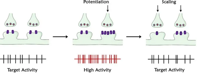
At present, the best understood form of homeostatic plasticity at the central excitatory synapses is synaptic scaling (Fig. 1). Synaptic scaling tends to modify synaptic strength in a compensatory way once brain network undergoes a perturbation of its activity, restoring average firing rates at baseline value [60]. Neurons of the CNS indeed, seem to be able to maintain average firing rates around a homeostatic set point [61]. Modulating network activity induces uniform increases or decreases of miniature excitatory postsynaptic currents (mEPSC) of a single neuron [60], so that the average firing rate is maintained but the relative strength of each single synapse is adapted. Perturbations in network activity could be sensed by individual neurons as changes in receptor activation, or changes in secreted factors, and induce modifications that are strongly suggested to be cell-related and resulting in a modification of neuron’s own firing. Thus, selectively blocking postsynaptic neuronal firing with Tetrodotoxin (TTX - a Na+ channel blocker), scales up synaptic strengths with an intensity degree comparable
to the blockade concentration [62]. Synaptic scaling involves postsynaptic changes in receptor accumulation. Blocking postsynaptic firing in neocortical neurons scales synapses up through the reduction of CaMKIV activation and transcription as a result of somatic calcium drop; this leads to AMPAR accumulation in postsynaptic membrane at all excitatory synapses [62]. Like scaling up, scaling down in response to elevated
11
network activity is dependent on calcium flux and in particular involves enhanced calcium influx, gene transcription, the CaMKK/CaMKIV signalling pathway, and targets the GluA2 subunit of AMPAR [63]. Scaling down is likewise realized through the activation of Plk2 (polo-like kinase 2) that after a CDK5-dependent recruitment to SPAR, induces its degradation activating a pathway necessary for the reduction in synaptic AMPAR accumulation triggered by elevated activity [64].
Figure 1. Illustration of synaptic scaling. When activity is perturbed (illustrated here as the potentiation of some inputs through Hebbian mechanisms) this triggers synaptic scaling, which produces a proportional reduction in strength at all synapses of the right magnitude to return firing to baseline levels. Note that, because this mechanism scales synaptic strength up or down proportionally, the relative difference in synaptic strengths induced by Hebbian mechanisms is preserved. Figure adapted from “Homeostatic Synaptic Plasticity: Local and Global Mechanisms for Stabilizing Neuronal Function” [61].
Homeostatic plasticity mechanisms could also exist at the network level operating through an activity-dependent release of secreted factors which modulate excitation/inhibition balance. In neocortical neurons, the activity-dependent release of BDNF seems able to mediate synaptic scaling inasmuch as blocking BDNF signalling mimics the effects of activity blockade on excitatory mEPSP while exogenous BDNF application does the opposite [65]. Another secreted factor suggested to contribute to homeostatic plasticity is the Tumour Necrosis Factor (TNF, a cytokine that is part of the inflammatory response to pathological states. Prolonged activity blockade with TTX stimulates glial release of TNF which in turn acts on neurons increasing AMPAR insertion and scaling up mEPSC amplitude [66]. It seems that TNF play an important role in the maintaining of scaling during prolonged (>24h) activity blockade, while it is not necessary for the induction of early scaling (4-6h) [66]. Considering that neural circuits are composed of excitatory and inhibitory synapses, it is expected that synaptic scaling would affect both type of synapses but, taking into account the homeostatic concept, in a different and maybe opposite manner. Indeed, it has been shown that inhibitory synapses onto pyramidal neurons are modulated in the opposite way in response to a drop in activity [67, 68]. Nevertheless, it has been shown that after activity blockade in hippocampus, under some conditions, inhibition and excitation change in
12
the same direction [69] and that interestingly, not all excitatory neurons express synaptic scaling since CA1 neurons scale synapses in response to activity blockade while CA3 neurons do not [70].
Synaptic scaling is induced in a global manner as a function of postsynaptic firing. Nevertheless, local or quasi-local changes in synaptic signalling can induce homeostatic modifications in synaptic strength. A truly local form of homeostatic plasticity would involve a single synapse in relation with changes in presynaptic transmitter release or postsynaptic receptor activation. Despite evidence for the existence of a synaptic-specific form of homeostatic compensation (realized through the accumulation of GluA1) at postsynaptic sites in response to reduced presynaptic neurotransmitter release [71], contrasting evidence make this topic still controversial [62]. Moreover, on a theoretical level, the meaning of truly local homeostatic plasticity is not clear considering that such a mechanism would counteract memory storage considering that potentiating a synapse through LTP would induce a homeostatic depotentiation (and the opposite after LTD). On the other hand, quasi-local forms of homeostatic plasticity act on groups of nearby synapses and would exert a useful normalization function without markedly affecting Hebbian plasticity [72, 73]. On the postsynaptic side, the global blockage of neuronal firing with TTX together with local glutamate receptor block with AP5 ((2R)-amino-5-phosphonovaleric acid) enhance GluA1–containing/GluA2–lacking AMPAR accumulation in the blocked synapses resulting in a substantial change in synaptic AMPA receptors composition [74]: this effect has recently been attributed to retinoic acid (RA) production and RA receptor (RAR) activation [75]. This is in contrast with the mechanism behind global synaptic scaling since blocking neuronal firing alone leads to the enhancement of mEPSC increasing the number of synaptic GluA2-containing AMPAR. On the presynaptic side, enhanced synaptic activity was observed to reduce the release probability (Pr) through a mechanism that was local to particular dendrites
[76]: considering that synapses that contact the same dendritic branch have the same Pr
and the less the number of synapses the more the Pr is, it is suggested that this regulation
happens in a quasi-local way in function of dendritic hyperpolarization. This form of homeostatic plasticity has been suggested to prevent synaptic saturation and excessive depolarization onto a dendrite [76] but the mechanism for the induction remains elusive. Interestingly, postsynaptic BDNF release has recently been suggested to locally regulate the Pr of neurotransmitters [77], highlighting the role of activity-dependent
13
PART I: PLASTICITY IN HEALTHY BRAIN
The adult brain has been often considered similar to a circuit board, and thus reliant on a fixed and precise connectivity. However, neural network undergoes an important and constant remodelling process throughout the lifetime. Brain plasticity is seen as a nature’s stratagem to adapt rapidly to a changing environment, thus overcoming genetic limitations, which has slower occurrence [78]. Plasticity represents an intrinsic property of the nervous system retained throughout life that enables modification of function and structure in response to environmental demands via the strengthening, weakening, pruning, or adding of synaptic connections and by promoting neurogenesis.
During the early childhood, there is a considerable capacity for cross-modal plasticity [79], i.e. the adaptive redeployment of neurons to integrate the function of different sensory systems. The observed brain changes in all conditions related to alterations of one of the sensory fields highlight the important physiological role of adaptative neuronal plasticity. For instance, tactile acuity is significantly superior in blind subjects compared to controls [80], speech processing and auditory localization activate the visual cortex in congenitally blind humans [81, 82] and the sensorimotor representation of the reading finger is expanded in blind Braille readers [83]. However, brain plasticity has a pivotal role also in more common and less extreme conditions. Indeed, an intriguing model for neuroplasticity studies are musicians. Exposure to musical training in early life shapes the brain. The anterior corpus callosum, consisting of nerve fibers connecting prefrontal regions crucial for coordination of bimanual motor activities and frontal motor-related regions, is larger in musicians that started their training before the age of 7 than in musicians without an early start, or controls [84]. Moreover, the cortical representation of the left hand fingers in string players is increased and is correlated with the age at which the person had begun to play [85].
Even though the developing brain is far more plastic than the adult brain, memory process involves brain plasticity and even in the adulthood the mature brain undergoes a continuous remodelling of the existing connections by experience of the everyday life and by performance of specific movements during motor and cognitive learning [86]. Plastic short-term modulations are important in the acquisition of new skills and can lead to structural changes in the cortical network as the skill become more established and automatic [87].
A fascinating example of plasticity in the adult brain regard London taxi drivers, who have a training period of 2 years before qualifying as drivers. The volume of grey matter in their right posterior hippocampus, a crucial region for spatial representation of the environment, is greater than controls subjects and increases together with the amount of time spent practicing as taxi driver [88].
Brain plasticity however has not the same efficiency in each subject: individual differences are likely quite large [78]. Important factors that contribute to differences in
14
mechanisms of plasticity include genetic and epigenetic mechanisms, such as polymorphisms or genetic expression, hormonal factors, such as gender or phases of menstrual cycle, impact of morbidities, such as diabetes or cancer, and lifetime experiences, such as brain injury stress, sleep deprivation, substance abuse or poor nutrition [78]. In keeping with this assertion, a number of genetic factors have been identified to regulate human brain plasticity [89]. The BDNF for example, has an important role in neuronal plasticity. A polymorphism of BDNF gene, which consists in a substitution of valine to methionine (Val66Met), leads to reduced levels of mature BDNF and differentially modulate human cortical plasticity and the response to training, brain stimulation and motor learning [90-92]. Another common mutation that highlights the influence of genetic factors on brain plasticity is the Apolipoprotein E (APOE) gene, and its allele, strictly linked with the risk of Alzheimer’s Disease [93], and able to influence brain network plasticity and the extent of plasticity throughout the lifespan [94]. Environmental factors contribute to genetic expression interacting with genes but also via epigenetic modifications, influencing plasticity mechanism across the lifespan [78]. These factors include educational experience, family upbringing and other social interactions, hormones, stress or physical activity.
Plasticity in hippocampus: adult neurogenesis
In recent years, neural plasticity has been a field which met an explosion in scientific research. It is now clear that environmental influences, including specific experiences, have a profound effect on adult brain structure and function [95]. In keeping with these evidence, much attention has focused on hippocampus, both because of its important role in certain types of learning and memory, in particular episodic and spatial memory, and its impressive degree of structural plasticity.
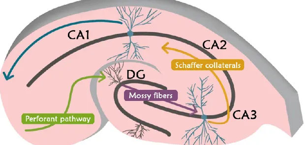
The hippocampus is localised in the medial temporal lobe (MTL), is considered a three-layer cortex and is made up by the Dentate Gyrus (DG) and Cornu Ammonis (CA), which is in turn divided in three main areas, CA1, CA2 and CA3 (Fig. 2). The information processing in the hippocampus follows a main pathway through its constituting areas forming the so-called “tri-synaptic circuit”: in particular, the information from the Entorhinal Cortex (EC) reaches the DG and then, in the order, CA3, CA2 and CA1, from which it returns to EC passing through the subiculum [96](Fig. 2).
In line with computational models, the hippocampus appears to rapidly learn associations between arbitrary events - one-shot learning -, form distinct representation from overlapping neocortical input - pattern separation - and, retrieve a complete representation in the presence of ambiguous or partial neocortical input at retrieval of memories - pattern completion - [97]. The hippocampal pattern separation function is thought to be primarily supported by DG, thanks to its large number of neurons (relative to its principal input, the EC) and sparse coding, which might lead to the production of distinct non-overlapping representations from similar overlapping input. Moreover,
15
despite many numerous questions still remain, vast literature suggests that the DG function is also strictly linked with a peculiar phenomenon which takes place in this hippocampal field: neurogenesis.
Neurogenesis in the dentate gyrus
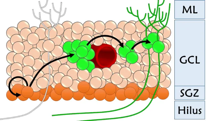
Adult neurogenesis in the DG has been demonstrated in a wide range of mammalian species, including humans [98, 99]. The dentate gyrus has a typical arrowhead shape (ventral and dorsal leaflets linked by the crest) (Fig. 2), and is considered a three-layer cortex. The molecular layer (ML), is the external one, is relatively poor in cells and contain the dendritic trees of granule cells and the fibers constituting the perforant pathway, which originate from neurons in the EC. The principal layer of the DG is the Granule Cell Layer (GCL) made up of 4-8 lines of granule cells which represent the principal neurons of DG; beneath the GCL is localised the Subgranular Zone (SGZ), where neural stem cells could be found and which therefore represents one of the only two widely acknowledged regions to date that retains neurogenesis under physiological conditions in adulthood [52, 100].
Figure 2. Hippocampus structure. The principal constituting areas are reported. DG: dentate gyrus; CA3:
Cornu Ammonis 3; CA2: Cornu Ammonis 2; CA1: Cornu Ammonis 1. The tri-synaptic circuit is also reported. In particular, the stellate cells located in the entorhinal cortex represent the principal input onto granule cells and their axons constitute the perforant pathway. Granule cell axons, the mossy fibers, make synapses onto CA3 pyramidal cells which, in turn, contact CA1 pyramidal cells. The information is subsequently brought to external areas.
Lastly, the third layer is the polymorphic one, which is placed between the dorsal and the ventral leaflet of GCL, constituting the hilus, where many different types of interneurons could be found and where the DGCs’ axons, the mossy fibers, pass until they reach and contact the CA3 pyramidal neurons.
16
In adult rodents, there are several thousand new neurons generated every day in the DG, modifying approximately 6% of the total DGC population per month [101]. Although most of these newborn DG cells (60–80%) undergo apoptosis within about one month following birth [101, 102], a remarkable number of new neurons survive and functionally integrate into the existing neural circuits [103] (Fig. 3). As new granules survive and integrate into the existing neural circuit, they form new connections with afferent projections and efferent targets within the neural circuit. Therefore, continuous addition of new DGCs in the DG introduces structural plasticity throughout the adulthood.
Figure 3. Hippocampal Neurogenesis. In the hippocampal dentate gyrus new granule cells are daily generated along the proliferative zone called subgranular zone. Progenitor cells (green cells - often localised around blood vessels) migrate along the granule cell layer, differentiate and integrate into the hippocampal network (green mature cells represented with dendrites and axons). ML: Molecular Layer; GCL: Granule Cell Layer; SGZ: Subgranular Zone.
Following what happens in the embryonic development, newborn DGCs in the adult brain follow a precise sequence of neuronal development and synaptic connectivity before they become fully mature [104-106]. During the first week, newborn DGCs have limited processes, crossing the granule cell layer toward molecular layer. All cellular properties resemble those of typical immature neurons of the developing brain, as they start to express neuronal sodium channels and fire immature action potentials [107]. After 2 weeks, new neurons have begun to migrate into the granule cell layer and to display typical granule cell morphology, with more numerous and elaborate dendrites traversing the molecular layer. However, no dendritic spines are observed at this stage [107]. Membrane properties become more mature but the characteristics of immature neurons still remain. At 4 weeks, newborn DGCs display the morphology of
17
mature granule neurons, including spiny dendrites that reach the outer border of the ML and axons that project to the CA3 region. Basic physiological properties mimic mature neurons at this stage, exhibiting mature action potentials and all known types of DGC synaptic connections. Nonetheless, the integration into functional circuit, the electrophysiological maturation and the plasticity seems to continue to evolve for at least three months [108]. During the development of newly-generated DGCs, GABA has been shown to play crucial roles [109]. Lacking synaptic inputs in the first week, newborn DGCs in the adult brain are tonically activated by ambient GABA. Functional GABAergic synapses that receive phasic GABAergic inputs from local interneurons start 8 days after birth [110]. Physiologically, these GABAergic inputs to adult-born DGCs share the same characteristics of those found on mature DGCs born in embryonic and early postnatal stages, and have similar functional properties [111]. GABA, as opposed to what happens in mature DGCs, has an excitatory action on immature granules owing to the high cytoplasmic chloride ion content of newborn DGCs in the first 2–3 weeks, and plays crucial role in regulating migration, development, and synaptic integration of newborn neurons [109]. Tonic GABA activation depolarizes newborn DGCs, and more importantly, it constitutes the majority of GABA-induced activation during the initial integration process when phasic GABA activation either does not exist or is weaker than tonic activation.
Following the formation of GABAergic synapses, glutamatergic inputs from the entorhinal cortex initiate synaptic connections onto the growing dendrites of adult-born DGCs 10 days after the birth [110]. By 4–8 weeks, adult-born DGCs display functional glutamatergic synaptic inputs similar to mature neurons [105]. Glutamatergic inputs also regulate neurogenesis in the adult hippocampus, presumably by modulating neuronal integration and survival during development. Some studies have shown that AMPA receptor potentiation increases adult neurogenesis [112], while loss of NMDA receptors activity decreases newborn neuron survival [113]. Seemingly, in contradiction, application of NMDA or AMPA receptors antagonists increase adult neurogenesis in the DG mainly by the regulation of cell proliferation [114-116]. These results suggest that the regulation of adult neurogenesis by glutamatergic activity is complex, possibly through different downstream signalling pathways, or sensitive to environment or behavioural changes following treatment.
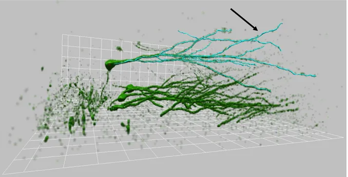
As newborn DGCs extend dendrites into the molecular layer, they extend axons rapidly toward the CA3 region. One week after birth, newborn axons pass through the hilus and reach the proximal CA3 region; from the second week, they begin to form en passant expansions [106, 117, 118]. Axons continue to grow along the CA3 within 3–4 weeks while their expansions grow into larger, mossy fiber buttons [117]. Newborn DGC axons do not extend beyond CA3, so they ultimately share the same trajectory as pre-existing mature mossy fibers. The earliest output synaptic contacts form on the dendritic shafts of target neurons starting from the second week. It takes 8–16 weeks for these new mossy fiber buttons to reach full maturity, with multiple invading dendritic spines and a stable number of synaptic contacts [117]. Recently, thanks to the optogenetic
18
approach, it has been found that mature adult-born DGCs establish functional synapses with hilar interneurons, mossy cells, and CA3 pyramidal cells and release glutamate as their main neurotransmitter, as mature DGCs [119]. However, the complete process of axonal integration and maturation remains unclear.
Newborn granule cells’ “critical period” and their potential role in hippocampal functions
Newborn neurons are continuously produced and incorporated into the existing hippocampal circuits throughout the adulthood. Therefore, a fundamental question is: do they contribute to hippocampal functions? Several studies using different approaches have shown the involvement of adult-born DGCs in hippocampal-dependent behaviours [120, 121]. The enhancement of neurogenesis, is usually associated with elevated synaptic plasticity in the DG and/or improved hippocampal-dependent learning and memory [122, 123]. Recent data also demonstrated that genetically increased DG neurogenesis through the specific inhibition of newborn cell death is able to improve hippocampal-dependent pattern separation [124]. In keeping with the hypothesis that neurogenesis exerts a positive effect on learning and memory, decreased neurogenesis in either transgenic mouse lines, such as Methyl-CpG binding protein 1 knockout (MBD1-/-) mice, or ablation of neurogenesis by irradiation results in decreased synaptic plasticity in the DG and/or deficits in some forms of hippocampal-dependent learning and memory [125, 126]. Besides, Drapeau and colleagues observed a direct connection between water maze performance and the number of newborn neurons in the hippocampus of aged animals, in which animals that retained spatial memory exhibited a higher level of cell proliferation and a higher number of new neurons in comparison to those with spatial memory impairments [127].
Additionally, the removal after learning of integrated, adult-born neurons, using a diphtheria toxin-based strategy without affecting ongoing neurogenesis, degraded existing hippocampal-dependent contextual fear and water maze memories, suggesting that adult-born neurons form a critical and enduring component of hippocampal memory traces [128]. Taken together, these studies suggest that adult-born DGCs are involved in hippocampal functions.
During their maturation, the functional properties of newborn granule cells face changes which give rise to periods of development possibly related with specific roles in hippocampal functions. Hubel and Wiesel established the term ‘‘critical period’’ to describe a particular time window in which neuronal properties are particularly susceptible to modification by experience, together with extensive anatomical changes that become irreversible after the closure of this period [129, 130]. This time window is characterised by enhanced morphological and synaptic plasticity, and is now considered a central mechanism for establishing fine-tuned neuronal circuits in the developing brain [131]. Thus, knowledge of neuroplasticity within critical periods emerged primarily from research on sensory systems such as the visual system. Nonetheless, even newborn
19
granules, during the integration in hippocampal circuit, when they start to receive experience-driven inputs from existing neural network, go through a phase of enhanced plasticity. Synaptic plasticity such as LTP has been thought to be the primary cellular basis of hippocampus-dependent learning and memory. As demonstrated by Ge et al., young adult-born DGCs display enhanced LTP with decreased induction threshold at the age of 4–6 weeks that rapidly drops by 8 weeks of age [132], indicating a critical period with enhanced synaptic plasticity. These data confirm previous findings [133, 134] and are consistent with other studies showing adult-born neurons display a high level of anatomical plasticity during this period which decreases thereafter, such as spine motility [106], suggesting that the newborn DGCs undergo a short period of fine-tuning while integrating into existing circuits. How are new DGCs more plastic during this period? Immature adult-born granules display distinct active and passive membrane properties such as high input resistance (IR) [105, 107]. Besides, in young neurons, high levels of T-type Ca2+ channels can generate isolated calcium spikes and enhance fast Na+
APs, contributing to the induction of synaptic plasticity [134]. Another key mediator of plasticity is the NMDA type of glutamate receptors. During adult neurogenesis, NMDARs are expressed early, starting from immature neuronal stages [107, 135, 136]. It is known that during early postnatal neuronal development, switching of NMDARs subtypes from NR2B to NR2A changes the direction and degree of synaptic plasticity [137-139]. NMDARs containing NR2B subunit are expressed early during postnatal development and appear to be associated with enhanced synaptic plasticity during the critical period [137, 140], while NMDARs containing NR2A, which are expressed and dominant later, mediate dramatically decreased LTP after the critical period [137]. Using field potential recordings, Snyder et al. revealed that LTP in the DG with intact GABAergic inhibition appears to be largely dependent on young adult-born neurons, and could be specifically blocked by NR2B antagonist ifenprodil [125]; in contrast, mature DGCs display much less LTP in the same condition [133], or have higher threshold for LTP induction [134]. Moreover, by specifically targeting adult-born DGCs, Ge et al. showed ifenprodil completely abolished LTP on DGCs of 4-weeks old, but not 8-weeks old or mature DGCs, providing a temporal correlation between synaptic expression of NR2B subtypes and critical period plasticity [132]. They also found that the plasticity of newborn DGCs within the critical period relies significantly more on NR2B-containing NMDARs than on pan-NMDARs, suggesting that NR2B, which is the major NMDARs subtype expressed during the critical period, plays an instructive role in the enhanced synaptic plasticity of adult-born DGCs within this time window [132]. These studies suggest that adult-born neurons in the critical period undergo molecular mechanisms similar to neurons in the early postnatal critical period.
Adult-born DGCs integrate into existing hippocampal circuits and express an improved plasticity during the critical period in relation to events other than mature neurons that have passed the critical period [132]. It is therefore natural to ask if they make unique contributions to hippocampal function. As suggested by numerous emerging evidence, adult-born DGCs might be preferentially recruited into hippocampal
20
circuits related to spatial information processing, contextual fear conditioning, novelty recognition and memory formation [103, 120, 141]. This preferential recruitment is consistent with the critical period of the adult-born DGCs and appears at 4–6 weeks after birth [120, 141]. In relation to their high excitability [105, 142] and the critical period of enhanced plasticity, adult-born DGCs of the critical period are more readily recruited into the hippocampal circuit for the encoding of novel information. As hypothesised by Aimone and his colleague, the special properties of young adult-born neurons are required for the formation of temporal clusters which associate individual elements of long-term episodic memories, a function called “pattern integration” [143]. Indeed, newborn neurons are preferentially activated in the critical period of enhanced plasticity and then, after the maturation, become less excitable but part of mnemonic traces: the progressive maturation of these granule cells create a temporal link between the different input that reach the DG. Overall, the available evidence strongly indicates that young adult-born neurons play an important role in participating in certain types of hippocampus-related behaviours, particularly learning and memory. However, the specific role of adult-born neurons which mature cells couldn’t achieve still remains under investigation.
Adult-neurogenesis as substrate for experience-dependent change
Adult neurogenesis is dynamic and highly dependent on the activity of the neural network. As DG receives various innervations from multiple brain regions, adult-born neuron development at distinct stages is regulated by numerous factors related to global and local neuronal activities. Actually, a growing body of literature indicates that adult neurogenesis is strongly influenced by the environment [95, 144]. Findings for overall literature on experimental modulation of adult neurogenesis seem to converge on a basic model: rewarding experiences, such as physical activity or mating, tend to increase the production of new neurons, whereas more aversive experiences, such as social defeat or predator smell exposure, tend to decrease the production of new neurons, [95, 144].
Hippocampal neurogenesis and physical activity
Voluntary physical exercise is one of the most studied activity able to positively influence adult neurogenesis. Neurogenesis improvement induced by physical activity is considered able to maintain the brain in fit: indeed, a large body of literature bring evidence about the cognitive performance improvement after physical and mental training. Physical activity is able to significantly increase the number of newly-born granule cells [123, 145]. The effect of physical exercise on precursor cell proliferation is related to many possible factors such as BDNF [146, 147], serotonin (5-Hdroxytriptamin – 5HT) [148, 149], Vascular Endothelial Growth Factor (VEGF) [150-153] or Insulin-like Growth Factor-1 (IGF-1).
21
BDNF seems to be the principal factor in mediating the effects of physical activity on neurogenesis, and its mRNA and protein levels increase in hippocampus after exercise [154]. Thus, the blockage of BDNF signalling lead to impairments in the learning improvement and neurogenesis promotion induced by physical activity [155]. Serotonin depletion in the brain of Tryptophan Hydroxylase 2 knockout mice (Trp2 -/-) affects the neurogenesis induced by physical activity, blocking the increase in granule number: it is therefore suggested that 5-HT play a role in stimulating neurogenesis after physical exercise [156]. Since Selective Serotonin Reuptake Inhibitors (SSRI) increase BDNF levels in the DG, the effect of serotonin might be linked to the BDNF action [157]. VEGF expression during exercise is also related to the increased neurogenesis but in a similar way to serotonin action, also VEGF probably act in concert with other factors, such as BDNF or IGF-1 [158]. Another factor which levels are found increased after exercise is IGF-1, which in turn increases angiogenesis and neurogenesis, in cooperation with VEGF action [159, 160]. Indeed, physical activity stimulates GH-IGF-1 axis, increasing IGF-1 blood level and the uptake of this factor not only in the muscle [161] but also in the brain [152]. Moreover, the increased levels of BDNF in the hippocampus after exercise seem to be triggered by the increasing in IGF-1 levels [152]. Physical activity is able not only to improve neurogenesis through enhanced proliferation but also to increase the number of surviving newly-generated granule cells, thus promoting survival [162].
Hippocampal neurogenesis and Enriched Environment
Donald Hebb, after the discovery of enhanced cognitive performance of pets over laboratory animals kept in cage condition, introduced for the first time the concept that an Enriched Environment (EE) could be able to improve the quality of life in animals [163]. After this first statement, Rosenzweig and colleagues developed the enriched environment model as we know it today [164]. Enriched environment consists in breading conditions able to offer a higher amount of sensory, motor and cognitive stimuli compared to standard laboratory conditions, which is considered a deprived environment [165, 166]. Often, the EE include social interaction since many animals are housed together. Many EE protocols consist in placing 8-12 rats in large cages containing many different objects, daily replaced: this condition provides novelty elements which assure cognitive stimuli, spatial representations and social interactions [167]. The items generally used in EE are characterised by different colours and shape, to provide visual stimuli, wood-items with different textures, to stimulate sensorial perception, tunnels with different shapes, for spatial navigation, and wheels for voluntary running: the number of items placed in an EE positively correlates with the number of immature granules in DG [168]. However, EE is able to affect neurogenesis only until the animal doesn’t familiarize with the environment; once the EE lost its novelty, neurogenesis in the DG is increased no more [169].
The effect of EE on neurogenesis consists in increasing cellular survival rather than enhancing neural proliferation [123, 169]; however, Kempermann & Gage [169]
22
revealed the ability of EE to increase proliferation as well. The effect of EE on proliferation seems to be mediated by GABA signalling, which could act on silent synapses characterised by only Mg2+-blocked NMDAR, promoting AMPAR
incorporation and the synaptic integration [170]. Since NMDAR is crucial for immature neuron survival and integration [113], the increase of synapses with activated NMDAR might promote neuronal survival.
Hippocampal neurogenesis and Learning
Learning is able to positively influence neurogenesis. Nonetheless, only certain types of learning can promote neurogenesis. For instance, a hippocampal-dependent learning such as the Morris Water Maze test, where the animal is tested for the detection of a hidden platform in a pool, is able to promote granule survival [171, 172], whereas learning to localise a visible platform (hippocampal-independent learning) has no effect [171].
Hippocampal-dependent task promotes neurogenesis when the task is quite tricky and the more the attempts to complete the test are, the more the effect on neurogenesis is high; in addition, the effect on neurogenesis is obtained only if the test is successfully ended [173]. These findings suggest that mental effort and network activity are crucial to rescue newly-born dentate granules, and suggests that a bigger mental effort induces better effects. In keeping with this assertion, comparing a demanding task such as Tool Use test and a less challenging task, such as the Radial Arm Maze test, Kumazawa-Manita and colleagues [174] clearly showed that Tool Use test produces a higher number of newborn granule cells than the Radial Arm Maze.
Spatial learning promotes neuronal survival and is influenced by the complexity of the task, the species employed in the test, sex differences, and the age of immature cells during learning [175]. Spatial learning promotes the survival of 6-10 days-old granule cells in rat, but seems to induce the opposite effects on older cells [107]. The survived cells, rescued by spatial learning, are reactivated when the animal is exposed to the test again, suggesting that these cells could be part of memory traces [176].
Hippocampus-dependent tasks make precocious the first GABAergic synaptic contact onto newborn granule cells, which are detectable 4-6 days after birth; this effect could represent the basis of increased survival induced by learning processes in newly-born granule [110]. It is therefore suggested that a higher number of GABAergic synapses could avoid an excessive activation of T-type Ca2+ channels, which might lead to
cytotoxic effect related to prolonged and elevated levels of this ion in the cytoplasm. Indeed, GABAAR activation tends to stabilize the membrane at the Cl- equilibrium
potential that, in immature neurons, is depolarized with respect to the RMP. On the other hand, fast changes of membrane potential, such as that observed in immature neurons [110], are more efficacious than a constantly depolarized potential in opening T-type Ca2+ channels, which are highly expressed in these cells [107]. Therefore, the