Group IV Phospholipase A
2
a Controls the Formation of
Inter-Cisternal Continuities Involved in Intra-Golgi
Transport
Enrica San Pietro1, Mariagrazia Capestrano1, Elena V. Polishchuk1, Alessio DiPentima1, Alvar Trucco1, Pasquale Zizza1, Stefania Mariggio`1, Teodoro Pulvirenti1, Michele Sallese1, Stefano Tete2, Alexander A. Mironov1, Christina C. Leslie3, Daniela Corda1, Alberto Luini1,4*, Roman S. Polishchuk1,4*
1 Department of Cell Biology and Oncology, Consorzio Mario Negri Sud, Chieti, Italy, 2 Department of Oral Sciences, University ‘‘G. D’Annunzio’’, Chieti, Italy, 3 Department of Pediatrics, National Jewish Medical and Research Center, Denver, Colorado, United States of America,4 Telethon Institute of Genetics and Medicine, Naples, Italy
Abstract
The organization of intra-Golgi trafficking and the nature of the transport intermediates involved (e.g., vesicles, tubules, or tubular continuities) remain incompletely understood. It was recently shown that successive cisternae in the Golgi stack are interconnected by membrane tubules that form during the arrival of transport carriers from the endoplasmic reticulum. Here, we examine the mechanisms of generation and the function of these tubules. In principle, tubule formation might depend on several protein- and/or lipid-based mechanisms. Among the latter, we have studied the phospholipase A2
(PLA2)-mediated generation of wedge-shaped lysolipids, with the resulting local positive membrane curvature. We show
that the arrival of cargo at the Golgi complex induces the recruitment of Group IVA Ca2+-dependent, cytosolic PLA2(cPLA2a)
onto the Golgi complex itself, and that this cPLA2a is required for the formation of the traffic-dependent intercisternal
tubules and for intra-Golgi transport. In contrast, silencing of cPLA2a has no inhibitory effects on peri-Golgi vesicles. These
findings identify cPLA2a as the first component of the machinery that is responsible for the formation of intercisternal
tubular continuities and support a role for these continuities in transport through the Golgi complex.
Citation: San Pietro E, Capestrano M, Polishchuk EV, DiPentima A, Trucco A, et al. (2009) Group IV Phospholipase A2a Controls the Formation of Inter-Cisternal
Continuities Involved in Intra-Golgi Transport. PLoS Biol 7(9): e1000194. doi:10.1371/journal.pbio.1000194 Academic Editor: Fred Hughson, Princeton University, United States of America
Received May 29, 2008; Accepted July 31, 2009; Published September 15, 2009
Copyright: ß 2009 San Pietro et al. This is an open-access article distributed under the terms of the Creative Commons Attribution License, which permits unrestricted use, distribution, and reproduction in any medium, provided the original author and source are credited.
Funding: The authors acknowledge financial support from the Associazione Italiana Ricerca Sul Cancro (AIRC) (Milan, Italy) and Telethon Italy (grants GGP05044 and GTF05007). AT was supported by an Alfredo Leonardi Fellowship (Mario Negri Institute, Milan, Italy), and Pasquale Zizza by a Fondazione Italiana per la Ricerca sul Cancro (FIRC) Fellowship (AIRC, Italy). The funders had no role in study design, data collection and analysis, decision to publish, or preparation of the manuscript.
Competing Interests: The authors have declared that no competing interests exist.
Abbreviations: AA, arachidonic-acid; Ab, Antibody; cPLA2a, cytosolic PLA2; EM, electron microscopy; ER, endoplasmic reticulum; GVIIIA, Group VIIIA; HFs, human
fibroblasts; IMLFs, immortalized lung fibroblasts; KDELR, KDEL receptor; KO, knock-out; NZ, nocodazol; PC-I, procollagen-I; PLA2, phospholipase A2; RNAi, RNA
interference; TGN, trans-Golgi network; VSV, vesicular somatitis virus; VSVG, vesicular somatitis virus glycoprotein. * E-mail: [email protected] (AL); [email protected] (RSP)
Introduction
After their synthesis in the endoplasmic reticulum (ER), cargo proteins move to the Golgi complex. This unique structure comprises numerous compact stacks of cisternae that are laterally interconnected into the Golgi ‘‘ribbon’’ through tubular-reticular networks (‘‘non-compact zones’’ [1]). Cargo proteins then traverse the Golgi cisternal subcompartments (where they are glycosylated), and at the trans-Golgi face they are sorted and delivered to their further destinations via large tubular/pleiomorphic carriers [2–6]. Despite significant advances in recent years, important aspects of the organization of intra-Golgi trafficking remain unclear. Three main intra-Golgi transport models have been traditionally considered for higher eukaryotes: trafficking by anterograde vesicles [7], trafficking by compartment maturation-progression [8–11], and trafficking by diffusion via tubular continuities joining different Golgi cisternae [12–18]. Among these, the cisternal progression model has recently gained broad consensus, although the nature of the intermediates involved in this mechanism (vesicles or intercisternal tubules) remains unclear. In addition,
more recently, a new model has been proposed by which cargo molecules mix rapidly throughout the stack (compatible with cargo diffusion via intercisternal continuities) and then partition into specialized export domains before leaving the Golgi complex [19]. Thus far, intra-Golgi transport models, the last, diffusion via tubular continuities, has received the least attention [12–17], and even the very existence of intercisternal tubules has long remained an issue of debate [20]. The main reasons for this uncertainty have probably been the technical difficulties of detecting the convoluted structures of the intercisternal tubules by traditional electron microscopy (EM) and the traffic-related dynamics of these tubules [18,21]. Over the last few years, however, tomography studies have indicated that tubules can be shown to join successive Golgi cisternae in animal cells, and that they form specifically under conditions of active trafficking [17,18,21]. Thus, although intercisternal tubules have still not been completely characterized (for instance, quantitative data on their frequency remain scarce [18,22]), they can be considered as potential players in intra-Golgi trafficking. It is therefore of interest to understand and manipulate their molecular mechanisms of formation.
The formation of an intercisternal tubule is likely to depend on several molecular events, including mechanical deformation by specialized proteins and changes in the distribution/geometry of the lipids within the membrane bilayer [23,24]. These latter can be induced in several ways, including the formation of local spontaneous positive membrane curvature via the generation of lysolipids and fatty acids by PLA2activity [24,25]. Indeed, a role
for PLA2in membrane shaping has been suggested in vitro [26], as
well as in a series of in vivo studies that have indicated that chemical blockers of PLA2 suppress tubule formation in several
endomembrane compartments and inhibit the related trafficking steps (see Brown et al. [27] for review).
Here, we show that transport through the Golgi complex coincides with the rapid recruitment of a specific molecular PLA2
isoform onto Golgi membranes: Group IVA, Ca2+-dependent cytosolic PLA2(cPLA2a). The activity of cPLA2a is required for
the formation of intercisternal connections in the Golgi. In addition, we show that treatments that inhibit cPLA2a and
suppress intercisternal tubule formation also block intra-Golgi trafficking of several cargo proteins. These data identify cPLA2a as
a component of the machinery underlying intercisternal tubular continuities and support a role for these continuities in intra-Golgi transport.
Results
As indicated above, PLA2 inhibitors have been reported to
suppress membranous tubules that extend from different cellular compartments, including the Golgi complex [28,29]. We thus sought to identify the Golgi-associated PLA2 isoform that might
serve as the molecular target of these inhibitors and might be involved in the regulation of Golgi-associated tubular structures. The superfamily of PLA2 enzymes consists of 15 groups
comprising secretory and cytosolic enzymes, with the latter divided into Ca2+-sensitive (cPLA2s, or Group IV PLA2s) and
Ca2+-insensitive (Group VI, VII, and VIII PLA2s) isoforms [30].
Among those that are Ca2+ sensitive, one of the Group IV isoforms, cPLA2a, that is normally cytosolic has been reported to
associate preferentially with the Golgi complex upon moderate increases in cytosolic Ca2+concentrations [31–34]. This affinity for Golgi membranes prompted us to examine whether cPLA2a
might itself be involved in intercisternal tubule formation and intra-Golgi trafficking.
Traffic Induces Binding of cPLA2a to the Golgi Complex
in a Ca2+-Dependent Fashion
Initially, we asked whether cPLA2a is recruited to the Golgi
complex during activation of transport through this organelle. We first generated a procollagen-I (PC-I) traffic pulse in human fibroblasts (HFs) using the 40–32uC transport synchronization protocol [8,35]. During the 40uC block, PC-I was diffusely distributed in the ER and cPLA2a was mostly cytosolic (Figure 1A).
After releasing the block, PC-I reached the Golgi complex, where its levels continued to increase for 30 min (Figure 1B). At the same time, remarkably, cPLA2a was partially recruited to the Golgi
complex (Figure 1B). This cPLA2a increase on the Golgi complex
lasted for at least 30 min. Similar experiments were carried out using the same 40–32uC transport synchronization protocol [35] in other cell types (HeLa and MDCK cells) with the temperature sensitive mutant of the vesicular somatitis virus glycoprotein (VSVG) as cargo, a well-characterized trafficking marker [35,36]. VSVG can be expressed by either transfecting or infecting cells with vesicular somatitis virus (VSV). In both cases, the generation of a traffic pulse induced the recruitment of cPLA2a to the Golgi
complex (Figure 1F and 1G), as seen with PC-I. We further examined the distribution of cPLA2a in the Golgi by immuno-EM.
We fixed VSV-infected cells expressing cPLA2a-GFP both during
the 40uC block and after releasing the block, and labelled the cells with an anti-GFP antibody (ab). Most of the cPLA2a-GFP was
dispersed in the cytosol during the 40uC traffic block, with very few gold particles on the ER and the Golgi complex. In contrast, the cells fixed 30 min after releasing the block (i.e., during a traffic wave) showed significant amounts of cPLA2a-GFP at the rims of
the Golgi cisternae and on rim-associated tubules (Figure 1H–1J). Some cPLA2a-GFP labelling was still seen in the cytosol and at
low levels in the ER, but not on other intracellular membranes. Notably, this distribution is consistent with an action of cPLA2a in
the formation of tubules from cisternal rims (see below). To verify that this cPLA2a recruitment to the Golgi complex
was due solely to changes in membrane transport rather than to the temperature changes required for inducing and releasing the trafficking block, we infected HeLa cells with VSV (as above) and then treated cells for 2 h with cycloheximide at 40uC (to deplete the cells of cargo and thus reduce transport [18]). Under these conditions, the temperature shift from 40uC to 32uC did not induce any significant changes in cPLA2a localization
(unpub-lished data).
We next investigated whether recruitment of cPLA2a to the
Golgi complex could be seen also under physiological steady-state trafficking conditions, rather than only during traffic waves. Indeed, HFs showed detectable levels of cPLA2a recruitment to
the Golgi complex also at steady-state (Figure 1C). Moreover, when PC-I transport was inhibited using the 40uC temperature block, cPLA2a lost its association with the Golgi complex, and
when trafficking resumed at 32uC, cPLA2a ‘‘rebounded’’ to high
levels on the Golgi complex (Figure 1A and 1B). Quantification of cPLA2a levels on the Golgi under these conditions is shown in
Figure 1E. Recruitment of cPLA2a to the Golgi at steady-state was
detected also in other cell types (HeLa, MDCK, and HepG2). In particular, in liver hepatoma HepG2 cells, which are professional secretor cells that release up to 20 different soluble serum proteins [37], the Golgi showed relatively high PLA2 levels (Figure 1D).
Thus, collectively, these data indicate that both ‘‘pulsed’’ and steady-state trafficking through the Golgi complex induces the association of cPLA2a with the Golgi membranes.
What is the mechanism of cPLA2a recruitment to the Golgi
complex? The binding of cPLA2a to membranes has been shown
to be due both to the Ca2+-binding C2 domain and to the catalytic domain of this protein. The C2 domain is required for initiating membrane association, in a process that depends exclusively on the intracellular Ca2+concentration ([Ca2+]i) and has thus been
proposed to function as a calcium sensor [32], while the catalytic domain prolongs this membrane binding of cPLA2a even after the
[Ca2+]ihas returned to lower levels [32]. We monitored the in vivo
dynamics of the calcium-sensor C2 domain of cPLA2a fused with
GFP (C2-GFP [32]) during the 40–32uC traffic pulse. Figure 1K shows that C2-GFP had a diffuse cytosolic pattern when most of the cargo protein (VSVG-YFP) was arrested in the ER. However, as soon as VSVG started to concentrate within the Golgi complex after the release of the traffic block (after 150–250 s), a significant portion of C2-GFP moved from the cytosol to the Golgi complex (Figure 1L and 1M; Video S1), and then gradually returned to the cytosol. Under the same conditions, the full-length cPLA2a protein
shifted to the Golgi complex and remained there for over 30 min (see above). This behaviour of C2-GFP and cPLA2a-GFP suggests
that the trafficking induces a transient increase in [Ca2+]i, which in
turn initiates cPLA2a recruitment to the Golgi complex [32], an
interaction that would then be prolonged by the catalytic portion cPLA2a-Dependent Tubules Drive Intra-Golgi Traffic
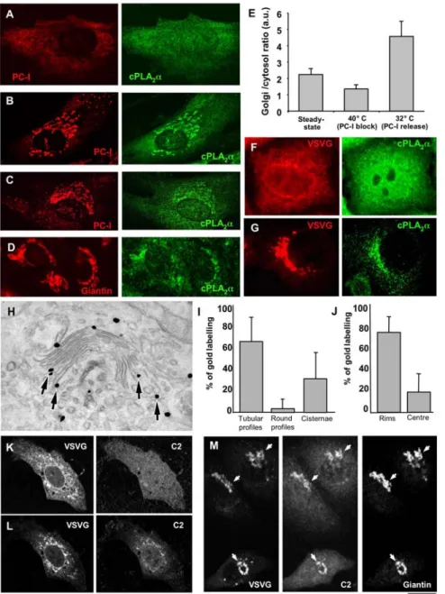
Figure 1. cPLA2ais recruited to Golgi membranes upon arrival of cargo from the ER. (A, B) HFs were incubated for 3 h at 40uC (A) and then
shifted to 32uC for 30 min in the presence of ascorbic acid (B), or HFs (C) and HepG2 cells (D) were grown under steady-state conditions. After fixing, the cells were double labelled with antibodies against PC-I and cPLA2a and examined under the confocal microscope. (A) When PC-I was trapped in
the ER after the 40uC block, cPLA2a enrichment was not detected in the Golgi area. (B) Arrival of PC-I at the Golgi complex after release of the
temperature block induced cPLA2a binding to the Golgi complex. (C, D) Under steady-state conditions, the cells were fixed, labelled with antibodies
against cPLA2a and either PC-I (C) or giantin (D). Confocal microscopy reveals that here some cPLA2a can be seen in the Golgi area together with PC-I
(C) and giantin (D). (E) Fluorescence intensity of cPLA2a in the Golgi region (defined as giantin-positive area) was quantified and normalized to cPLA2a
intensity in the cytosol of HFs. Plot shows that Golgi/cytosol ratio of cPLA2a (mean6SD; n = 20 cells) decreases in cells subjected to 40uC transport
block, but increases over the levels detected at steady-state conditions upon block release (at 32uC). (F, G) HeLa cells were infected with VSV and kept at 40uC for 3 h to accumulate VSVG in the ER. The cells were then fixed (F) or shifted to 32uC for 30 min to allow VSVG to exit from the ER (G), and prepared for confocal microscopy. Immunofluorescence labelling shows a diffuse pattern of cPLA2a when VSVG is blocked in the ER (F). In contrast,
cPLA2a undergoes recruitment to the Golgi membranes as soon as VSVG arrives at the Golgi complex from the ER (G). (H) HeLa cells were transfected
with cDNA encoding full length cPLA2a fused with GFP (cPLA2a-GFP), infected with VSVG, and subjected to the 40uC block. The cells were then fixed
30 min after the 40uC block release, to allow VSVG to reach the Golgi complex, and processed for immuno-gold EM with an anti-GFP ab, to reveal cPLA2a-GFP localization. After activation of VSVG transport, a cPLA2a-GFP signal was detected at the rims of the Golgi cisternae and flanking tubular
structures (arrows). (I, J) Quantification of gold labelling (mean6SD; n = 30 stacks) at the Golgi complex (see Materials and Methods) shows most of the cPLA2a-GFP is bound to the tubular profiles (I) and the rims of the cisternae (J). (K, L) HeLa cells were co-transfected with the cDNAs encoding
VSVG-YFP and the C2 domain of cPLA2a fused with GFP (C2-GFP), kept at 40uC for 3 h (K), and observed in vivo under the confocal microscope during
VSVG-YFP release from the ER. Images extracted from the time-lapse sequence show that C2-GFP moves from the cytosol (K) to the Golgi membranes (L) when VSVG appears within the Golgi area. (M) HeLa cells were transfected and incubated as above (K, L), and then observed under the confocal microscope and fixed during VSVG release when detectable amounts of C2-GFP started to appear in the perinuclear area. Further staining with anti-giantin antibodies reveals overlap of C2-GFP perinuclear signal with anti-giantin labelling (arrows). Scale bar, 7mm (A–D, F, G), 160 nm (H), 9 mm (K–M). doi:10.1371/journal.pbio.1000194.g001
of cPLA2a. While this increase remains to be further defined and
clarified, it could be related to the high lumenal Ca2+ concentrations in the Golgi complex [38] and the localization in the Golgi area of the signalling machinery that is involved in the release of Ca2+from intracellular stores [38,39]. Thus, the specific recruitment of cPLA2a to the Golgi complex can be explained by a
local traffic-induced Ca2+ release from the Golgi complex and probably, additionally, by an intrinsic affinity of cPLA2a for the
Golgi (possibly due to its affinity for lipids and proteins that are enriched in Golgi membranes [40–42]). Potentially related to this, Ca2+ released from the Golgi cisternae has been shown to be required for intra-Golgi transport [43].
In summary, traffic moving through the Golgi complex triggers the recruitment of cPLA2a to the rims of the Golgi cisternae,
possibly through a signalling mechanism [44] that might induce the local Ca2+ increase that is required for cPLA2a binding to
membranes [32].
Inhibiting cPLA2a Reduces Inter-Cisternal Tubules and
Suppresses Intra-Golgi Transport
We next examined whether cPLA2a is involved in the formation
of intercisternal tubules. Tubules can interconnect the stacked Golgi cisternae in at least two ways: tubular non-compact zones can join adjacent stacks ‘‘longitudinally,’’ to form the continuous Golgi ribbon [1], and tubules can link the Golgi cisternae in the cis-trans (‘‘vertical’’) direction within the same stack, as shown by EM tomography and stereoscopy [12,18,21]. To examine the role of cPLA2a here, we sought to inhibit/deplete cPLA2a by a variety
of approaches, while monitoring the presence/formation of Golgi tubules. HeLa cells were first exposed to siRNAs directed against cPLA2a. This resulted in a decrease in cPLA2a levels, as evaluated
by immunofluorescence (Figure 2A and 2B), western blotting (Figure 2C), and cPLA2a activity assays under basal and elevated
Ca2+conditions (Figure 2D). In these cells, growth was partially inhibited (50%–70%) in the last 24 h of exposure to the siRNAs; however, cell viability did not appear to be affected. In these cPLA2a-silenced cells, the Golgi ribbon was disassembled into
numerous fragments that remained perinuclear (Figure 2E, asterisks, 2F), as has been previously described upon application of PLA2 inhibitors [28,29]. EM showed that this was due to a
suppression of the longitudinal tubular elements (Figure 2G–2I) of the non-compact zones [1], which resulted in the breakdown of the Golgi ribbon into separate stacks (Figure 2H). We then investigated whether this cPLA2a deficit also affects vertical
intercisternal connections, which are presumably more relevant to cis-trans transport, using EM tomography (which is required to fully reconstruct these tubular structures [18,21]). This showed that tubules connecting different cisternae were present within individual Golgi stacks in these cells (Figure 3A and 3B, arrows; Video S2; see also below), as has been previously reported for other cell types [18], and that these tubules were almost completely suppressed by RNA interference (RNAi) of cPLA2a
(Figure 3C and 3D; Video S3). Other tools that specifically inhibit cPLA2a had similar effects: both microinjection of an ab against
the catalytic domain of cPLA2a (see below) and treatment with the
selective inhibitors of cPLA2a catalytic activity pyrrophenone and
pyrrolidine (not shown) [45,46] induced a significant fragmenta-tion of the Golgi ribbon corresponding to a reducfragmenta-tion in the tubular structures at the EM level (not shown). Of note, the tubules in the non-compact zones and the vertical intercisternal continuities always responded in the same way to a cPLA2a
deficit, suggesting that they both depend on the activity of cPLA2a.
Instead, other intracellular tubular structures (such as those of
endosomal origin, for example) were not affected by cPLA2a
depletion (not shown).
We also examined the effects of enhancing the levels of cPLA2a
by its overexpression. Remarkably, this treatment caused an overall increase in the Golgi tubular elements that was sometimes accompanied by a partial loss of the stack structure (Figure 2J), further supporting the concept that PLA2a promotes Golgi
tubulation.
A further point was whether the tubulating effects of cPLA2a
arise from the formation of lysolipids, which can create positive membrane curvature directly [24,27], or whether they are mediated by the formation of arachidonic-acid (AA) metabolites, perhaps via their signalling function. To test the latter possibility, we used chemical inhibitors to block the main metabolic enzymes of AA [47], the cyclooxygenases (using 50 mm indomethacin and 5 mm ibuprofen for 30 min) and the lipoxygenases (using 10 mm ketokenazol for 30 min). These agents had no influence on Golgi tubule formation (unpublished data), consistent with previously reported observations [28]. Moreover, the addition of AA to cPLA2a-siRNAs-treated cells did not counteract the
‘‘anti-tubular’’ effects of the cPLA2a deficit. Altogether, these data
suggest that cPLA2a is required to support tubulation and that its
effects are mediated via the formation of lysolipids [24,25,27,28]. We thus turned to investigate whether disassembly of inter-cisternal connections affects transport of cargo proteins across the Golgi complex. To test this, we first suppressed intercisternal connections by silencing cPLA2a, and then monitored the effects
of this treatment on intra-Golgi transport using the VSVG-synchronized transport assay. We thus infected cPLA2a-depleted
cells with VSV and accumulated VSVG in the ER at 40uC, and then we released the traffic block at 32uC. VSVG reached the Golgi complex apparently normally, but then it accumulated at the cis-Golgi pole (also inducing a moderate swelling of the cis cisternae) and did not proceed through the Golgi complex (Figure 4A–4E). Compatible results were obtained using biochem-ical transport assays (see Materials and Methods) (Figure 4F and 4G). Rescuing the cPLA2a activity in cPLA2a-siRNAs-treated cells
by microinjection of recombinant cPLA2a resulted in the
reactivation of VSVG trafficking (Figure 4H). Further along this line, an arrest of VSVG in the Golgi complex was seen also when the cells were transfected with a dominant-negative cPLA2a
mutant [48] (Figure 4I), and when cPLA2a was acutely inhibited
by the microinjection of antibodies against the catalytic portion of cPLA2a (Figure 5) or by specific inhibitors (not shown). Notably,
these inhibitory effects were marked but not complete (up to 60%– 80%; see Figure 4), possibly explaining the lack of visible toxicity (at least within our experimental time frame).
A possible concern here is that these experiments were carried out using a synchronization protocol that involves the sudden arrival of a large cargo load at the Golgi complex, raising the possibility that cPLA2a-dependent connections might have a role
only under conditions of Golgi overload. We therefore examined whether the inhibition of cPLA2a would cause similar effects
during more ‘‘physiological’’ non-synchronized trafficking. To this end, we infected cPLA2a-silenced cells with VSV and kept them at
32uC, to allow VSVG to continuously exit the ER. As was seen during the 40–32uC synchronized pulse, VSVG reached the Golgi complex but did not traverse it (see below), indicating that the role of cPLA2a in intra-Golgi trafficking is not limited to conditions of
cargo overload.
The transport of other cargo proteins was also examined using cPLA2a silencing. Ablation of cPLA2a in HFs resulted in a strong
delay of PC-I transport at the level of the Golgi complex (unpublished data). The use of cyclooxygenase and lipooxygenase cPLA2a-Dependent Tubules Drive Intra-Golgi Traffic
Figure 2. RNAi of cPLA2aaffects Golgi-associated tubular structures. (A, B) HeLa cells were fixed 72 h after transfection of control scrambled
(A) and cPLA2a-specific (B) siRNAs and stained for cPLA2a and giantin. Confocal microscopy shows a strong reduction in cPLA2a labelling in cells
treated with the specific siRNAs (B). (C) HeLa cells treated with siRNAs as in (A) and (B) and prepared for western blotting with antibodies against cPLA2a and actin. Expression of cPLA2a was strongly inhibited, while actin levels remained unaffected. (D) Quantification of cPLA2activity (mean6SD;
measured using [3H]-AA release; see Materials and Methods) revealed its reduction in cPLA2a-siRNAs-treated HeLa cells. (E, F) Control and cPLA2
-specific siRNAs-transfected HeLa cells. Labelling with an anti-giantin ab (E) and morphometric analysis (F) show extensive fragmentation of the Golgi complex in cells with low cPLA2a expression (E, asterisks). (G–I) Control (G, I) and cPLA2a-siRNAs-treated HeLa cells (H, I) were fixed and prepared for
EM analysis. Tubular structures were seen to connect the Golgi stacks to each other (G, arrows) in control cells but were lost upon cPLA2a
knock-down (H). Morphometric analysis indicates a reduction in surface area (mean6SD; n = 30 stacks) associated with tubular structures in the Golgi complex in cPLA2a-siRNAs-treated cells (I). (J) HeLa cells transfected with cPLA2a-GFP, fixed and processed for immono-gold EM with an anti-GFP ab.
Cells overexpressing cPLA2a-GFP show numerous long tubular profiles in the Golgi region (arrows) and partial loss of stack organization. Scale bar,
45mm (A, B), 22 mm (E), 300 nm (G, H, J). doi:10.1371/journal.pbio.1000194.g002
inhibitors had no effects on trafficking, and AA addition did not reverse the transport block in cPLA2a-siRNAs-treated cells, again
in parallel with the above effects on Golgi tubules. Thus, the induction of a cPLA2a deficit and the attendant disassembly of
intercisternal connections appear to be associated with the inhibition of intra-Golgi trafficking of different classes of cargo proteins.
A further question is which type of connection is required for trafficking. Our data indicate that the inhibition of cPLA2a
disrupts the ‘‘vertical’’ (cis-trans) and ‘‘longitudinal’’ (inter-stack) tubular connections equally (see Figures 2 and 3). Thus, although it may appear logical to assume that the vertical connections are those relevant for transport, a role for the longitudinal connections cannot be formally excluded by the above data. To resolve this issue, we used a Golgi system that contains vertical, but lacks horizontal, connections, and tested whether transport through such a system is sensitive to the ablation of cPLA2a. A Golgi
complex with only vertical connections can be generated by nocodazol (NZ) treatment, which results in the fragmentation of the Golgi ribbon into isolated (non-horizontally connected) stacks [18]. Any transport-relevant role of longitudinal connections can be excluded in this system. Control and cPLA2a-deficient cells
were subjected to 3 h of 30 mM NZ treatment and processed for EM tomography. As expected, vertical connections were revealed in stacks of control cells (Figure 6A; Video S4), while in silenced cells, these connections were almost absent (Figure 6B; Video S5). To test the efficiency of transport under these conditions, the cells were infected with VSVG and exposed to NZ during the 40uC block, and then shifted to 32uC to activate transport. Figure 6C shows that in control cells, VSVG moved to the plasma membrane first through GM130-positive and then through TGN46-positive compartments of the Golgi stacks, as has been previously reported [18]. In contrast, in cPLA2a-silenced cells, VSVG was retained in
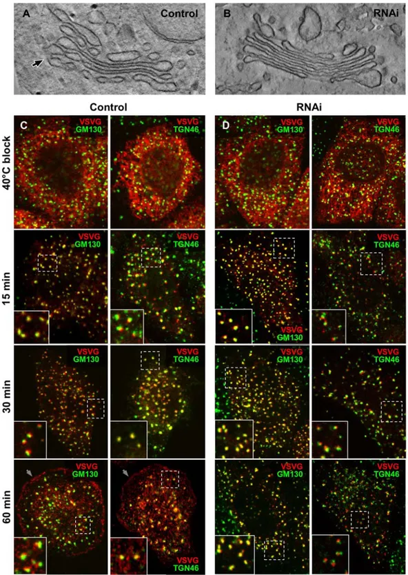
the Golgi stacks, where it showed strong overlap with the cis-Golgi Figure 3. RNAi of cPLA2aaffects intercisternal connections within the Golgi stack. Control (A, B) and cPLA2a-silenced (C) HeLa cells were
prepared for EM tomography after chemical fixation. (A, B) The digital slice (A) and zoom image (B; corresponding to the area outlined by black box in A) extracted from the tomogram (see Video S2) shows an intercisternal connection (arrow) bridging cisternae located at the different levels of the stack in control cells. (C) Digital image extracted from the tomogram reveals no bridges between cisternae within the same Golgi stack (see Video S3) as well as a lack of connection between neighbouring stacks (indicated by arrowheads). (D) Quantification of vertical connections per stack in sections (mean6s.e.; n = 10 stacks) in EM tomograms (see Materials and Methods), in control and cPLA2a-siRNAs-treated HeLa cells. Scale bar, 150 nm (A, C), 50 nm (B).
doi:10.1371/journal.pbio.1000194.g003
Figure 4. RNAi of cPLA2ainhibits intra-Golgi transport of VSVG. (A–E) Control (A, B, D) and cPLA2a-siRNAs-treated HeLa cells (A, C, E) were
infected with VSV and kept at 40uC for 3 h to accumulate VSVG within the ER. The cells were fixed directly at the end of the block (A) or after a 60-min (A) or 45-min (B–E) temperature shift to 32uC. (A) The cells were labelled with an anti-VSVG ab, showing that after accumulation within the ER, VSVG was efficiently exported to the plasma membrane in control cells and blocked in the Golgi complex in cPLA2a-siRNAs-treated cells. (B–E) The cells
were triple labelled with anti-VSVG, anti-cPLA2a, and anti-TGN46 antibodies (B, C) or prepared for immuno-EM using the nanogold protocol (D, E). In
control cells, VSVG showed good colocalization with TGN46 in the Golgi area (B, inset); this colocalization was poor in cPLA2a-siRNAs-treated cells (C,
inset). In control cells, EM showed little VSVG in the cis portion of the stack (7.8%), with most (61%) within trans-Golgi compartments (D, arrows), post-Golgi carriers (D, filled arrowhead), and at the plasma membrane (D, empty arrowhead). In cPLA2a-siRNAs-treated cells, most of the VSVG (62%)
remained within the swollen cis portion of the stack (E, arrows). (F, G) Control and cPLA2a-siRNAs-treated HeLa cells infected with VSV were
metabolically labelled with [35S]-methionine and chased at 32uC. At the indicated times, the cells were solubilized and digested with endoglycosidase H (Endo-H), which cleaves sugar chains built on the proteins early in the secretory pathway (i.e., before their processing by the medial Golgi enzyme mannosidase-II, which convert sugars into an Endo-H resistant form). The cell lysates were then separated by SDS-PAGE, and the gels scanned (F). The percentages of the Endo-H-resistant form of VSVG with respect to the total amounts of VSVG were quantified (G) using a FUJIFILM imager. The data indicate that VSVG processing to its Endo-H resistant form (which occurs in the medial Golgi) was reduced when cPLA2a was silenced. (H) cPLA2
a-siRNAs-treated cells were infected with VSV, microinjected with recombinant cPLA2a during the 40uC block, and fixed 45 min after the block release
at 32uC. The cells were then stained with anti-VSVG and anti-cPLA2a antibodies and observed under the confocal microscope. VSVG was delivered to
the plasma membrane after cPLA2a microinjection (arrows) but remained in the Golgi in noninjected cells (asterisks). (I) HeLa cells were transfected
with VSVG-GFP and the dominant-negative cPLA2a(1–522) isoform, subjected to a 40uC block, and fixed 45 min after the temperature shift to 32uC.
Confocal images reveal VSVG-GFP blocked in the Golgi complex in cPLA2a(1–522)-expressing cells (asterisks). Scale bar, 60mm (A), 6 mm (B, C),
270 nm (D), 200 nm (E), 15mm (H), 8.5 mm (I). doi:10.1371/journal.pbio.1000194.g004
marker (GM130) even 60 min after release from the ER (Figure 6D). Thus, the disruption of vertical intercisternal bridges by cPLA2a silencing inhibited the progression of cargo across
NZ-induced isolated stacks, which are devoid of horizontal connec-tions. This provides evidence that it is the intercisternal connections of the vertical type that are required for intra-Golgi transport (see Discussion).
The Catalytic Activity of cPLA2a Is Required to Support
Intra-Golgi Transport
Our experiments with specific cPLA2chemical inhibitors (see
above) taken together with already published observations [27] suggest that the changes in lipid geometry during Golgi tubulation require PLA2 catalytic activity. Nevertheless, given that the C2
domain of cPLA2a inserts deep into the membrane bilayer [49]
and could therefore be classified among the membrane-bending
protein modules [50], we wanted to determine whether it is indeed the catalytic activity of cPLA2a, rather than the insertion of this
enzyme into the Golgi membranes, that is responsible for the generation of tubules and transport across the Golgi.
We first tested whether cPLA2a maintains its ability to bind to
the Golgi complex in the presence of chemical inhibitors that suppress intra-Golgi transport. VSV-infected HeLa cells were treated with pyrrolidine, and the localization of cPLA2a was
monitored during a VSVG traffic pulse. cPLA2a translocated to
the Golgi complex to the same extent in control and inhibitor-treated cells, but only the latter showed VSVG retention at the Golgi (Figure 7A and 7B). Second, we examined the effects of two cPLA2a mutants, cPLA2a(1–522) and cPLA2aS228C, which lack
PLA2 catalytic activity and yet show normal binding to
membranes [48,51]. cPLA2a(1–522)is a deletion mutant that lacks
an amino acid (Asp549) essential for enzyme activity [52]. Of note, Figure 5. Microinjection of an anti-cPLA2aab inhibits intra-Golgi transport. (A–F) HFs were infected with VSV and kept at 40uC for 3 h to
accumulate VSVG in the ER. The cells were microinjected with an anti-cPLA2a ab (mixed with FITC dextran) during the block, then shifted to the permissive
temperature (32uC) and fixed at different times. (A, B) Staining of non-permeabilized cells with an ab against the ectodomain of VSVG 60 min after the 40uC block release shows reduced VSVG at the cell surface in microinjected cells (asterisks). (C, D) Microinjection with an anti-cPLA2a ab (asterisks) induces
fragmentation of the Golgi complex, prevents delivery of VSVG to the plasma membrane, and blocks VSVG within the early Golgi compartment without showing overlap with TGN46 (compare insets 1 and 2 in panel D, which correspond to dashed boxes in the control and injected cells). (E, F) Morphometric quantification shows poor colocalization of TGN46 with VSVG (E; mean6SD; n = 50 cells) and a reduction in the number of post-Golgi carriers (F; mean6SD; n = 50 cells) in anti-cPLA2a-ab-microinjected cells, compared to mock-injected cells. Scale bar, 30mm (A, B), 7.5 mm (C, D).
doi:10.1371/journal.pbio.1000194.g005
Figure 6. RNAi of cPLA2aaffects intercisternal connections and transport within NZ-induced Golgi ministacks. (A, B) Control (A) and
cPLA2a-silenced (B) HeLa cells were treated with 30mM NZ for 3 h, then fixed and prepared for EM tomography. The digital slices extracted from the
tomograms (see Videos S4 and S5) show an intercisternal connection (arrow) bridging cisternae located at the different levels of the stack in control cells (A) and reveals no bridges between cisternae within the Golgi ministack in silenced cells (B). (C, D) Control (C) and cPLA2a-siRNAs-treated HeLa
cells (D) were infected with VSV and kept at 40uC for 3 h in the presence of NZ to disassemble Golgi into ministacks and accumulate VSVG within the ER. The cells were fixed directly at the end of the block or at indicated times after the temperature shift to 32uC. The cells were double labelled with an anti-VSVG and either GM130 (cis-Golgi marker) or TGN46 (trans-Golgi marker) and investigated under confocal microscopy. In control cells (C), after release of 40uC block, VSVG moved from the ER to the Golgi ministacks where it first colocalized with GM130 and then with TGN46 (see insets) and finally arrived to the cell surface (arrows). In contrast, cPLA2a-silenced cells show VSVG within the GM130-positive compartment of ministacks (see
insets) even 60 min after release from the ER, with little or no VSVG detected both in the TGN46-positive compartment and at the plasma membrane. Scale bar, 150 nm (A, B), 6mm (C, D).
doi:10.1371/journal.pbio.1000194.g006
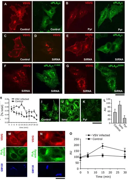
Figure 7. The catalytic activity of cPLA2ais required to support intra-Golgi transport. (A, B) HeLa cells were co-transfected with cPLA2
a-GFP and VSVG-Cherry and exposed to the 40uC block to accumulate VSVG-Cherry within the ER. Pyrrolidine (1 mM; Pyr) was added to the cells (B) 15 min before block release. Then cells were shifted to 32uC in the absence (A) or in the presence (B) of pyrrolidine for 45 min, fixed, and investigated under confocal microscopy. In control cells, VSVG-Cherry was detected at the cell surface (A) and its delivery to the PM was inhibited in pyrrolidine-treated cells (B) ,while cPLA2a-GFP recruitment to the Golgi membranes was not affected by inhibitor treatment (compare A and B). (C–G) Control (C)
and cPLA2a-silenced (D–G) mouse MC3T3 cells were transfected with VSVG-Cherry alone (C, D) or in combination with the following human cPLA2a
constructs: cPLA2a-GFP (E), cPLA2a (1–522)
(F), cPLA2a S228C
(G). The cells were subjected to 40uC block, then shifted to 32uC (to activate VSVG-Cherry exit from the ER) for 45 min, fixed, and stained with an anti-cPLA2a ab. Confocal microscopy revealed efficient delivery of VSVG-Cherry to the PM in
control cells (C), while in silenced cells, most of the VSVG-Cherry remained within the fragmented Golgi complex (D). Transfection of wild-type cPLA2a-GFP rescued VSVG-Cherry delivery to the cell surface in cPLA2a-silenced cells, while expression of the catalytically inactive mutants cPLA2a(1– 522)
(F) or cPLA2a S228C
(G) did not reactivate VSVG-Cherry transport. (H) PCCL3 cells were loaded with [3H]-AA (see Materials and Methods). Part of the loaded cells was infected with VSV. Then infected and noninfected (control) cells were exposed to the 40uC block, washed, and shifted to 32uC. Fresh medium was added to the cells for 3 min time intervals and then collected. The [3H]-AA released into the medium over each 3 min-long interval was measured (see Materials and Methods). Quantification of the AA release (mean6SD; n = 6 experiments) revealed increase of PLA2activity (with the
peak at 15 min) in VSV infected cells as compared to control. (I–L) HeLa cells were loaded with bis-BODIPY FL C11-PC (see Materials and Methods) and
fixed directly (I) or 15 min after incubation with either 5mM ionomycin (J) or 1 mM pyrrolidine (K). The fluorescent signal indicating PLA2activity was
detected by confocal microscopy both in the perinuclear region and the periphery of control cells (I). This signal increased after ionomycin treatment (J), but decreased after pyrrolidine addition (K), as also revealed by quantification (L) of the mean fluorescence per cell (mean6SD; n = 20 cells). (M, O) HeLa cells were infected with VSV, kept at 40uC for 3 h, and loaded with bis-BODIPY FL C11-PC at the end of 40uC block. The cells were then fixed
directly at the end of the 40uC incubation (M) or at different time intervals after temperature shift to 32uC (N, O). The cells were then stained with antibodies against VSVG and GM130 (Golgi marker). Confocal microscopy revealed only moderate PLA2activity overlapping with the Golgi when
VSVG resided within the ER (M) while activation of VSVG transport through the Golgi (15 min after release from the ER) induced an increase in the BODIPY signal in the Golgi area (N). Quantification of the mean BODIPY fluorescence per cell (mean6SD; n = 20 cells) indicates an increase in PLA2
activity in cells actively transporting VSVG, as compared to uninfected (control) cells (O). Scale bar, 8.5mm (A–G), 16 mm (I–K), 7 mm (M, N). doi:10.1371/journal.pbio.1000194.g007
cPLA2a(1–522)can be produced endogenously by caspase-mediated
cleavage at Asp522 during apoptosis [48], and it can act as a dominant-negative mutant of cPLA2a [48]. The cPLA2aS228C
mutant contains a single point mutation in the active site, again resulting in a complete loss of cPLA2a enzymatic activity [51].
Each mutant was transfected into cPLA2a-silenced cells and
compared to wild-type cPLA2a for its ability to rescue the
transport block induced by cPLA2a ablation and to translocate to
the Golgi complex. For the transport experiments, mouse cells were transfected with VSVG carrying a red fluorescent tag (VSVG-Cherry) and subjected to the transport-synchronization protocol. VSVG-Cherry was efficiently delivered to the surface in control cells (Figure 7C) but not in cPLA2a-silenced cells, as
expected (Figure 7D). The cPLA2a-silenced cells were then
transfected with either the mutants or the wild-type cPLA2a.
While the latter efficiently rescued VSVG-Cherry transport to the cell surface (Figure 7E), as expected, neither cPLA2a(1–522) nor
cPLA2aS228C modified the transport block (Figure 7F and 7G).
However, both cPLA2a mutants translocated to the Golgi complex
as efficiently as wild-type cPLA2a (Figure 7F and 7G). Therefore,
these collective results indicate that the catalytic activity of cPLA2a, rather than the ability of this enzyme to translocate to
Golgi membranes, is required to support transport across the Golgi complex.
Finally, we sought to directly monitor the increase in cPLA2a
activation that based on the above data should occur during cargo trafficking through the Golgi complex. First, we used a classical PLA2 activity assay based on the release of [3H]-AA from
AA-prelabelled cells [53]. A potential problem here is that during cargo trafficking, only a fraction of the total cellular cPLA2a is
bound to the Golgi complex (which represents, in turn, less than 5% of the cellular membranes). Thus, the increase in AA release over basal values might be very small. To overcome these problems, we used two approaches.
For the first, in addition to HeLa cells, we used a cell line that has been previously characterized in our laboratory to be an efficient AA releaser (PCCL3 cells) [46]. Both cell types were loaded with [3H]-AA, infected with VSV, and subjected to a 40– 32uC transport synchronization protocol. When they were shifted from 40uC to the permissive temperature of 32uC, the VSVG expressing PCCL3 cells showed a modest but statistically significant increase in AA release over that seen in control cells (Figure 7H). This increase coincided in time with VSVG transit through the Golgi complex. HeLa cells showed a trend in the same direction, which, however, was not statistically significant. To overcome this difficulty with HeLa cells, we used here a second approach based on the fluorogenic phosphatidylcholine analogue (bis-BODIPY FL C11-PC) as a sensor of local changes in PLA2
activity [54]. The hydrolysis of this lipid by PLA2enzymes results
in generation of fluorescent products by fluorescence dequenching [54]. After loading the bis-BODIPY FL C11-PC, HeLa cells at
steady-state showed a diffuse (ER-like) fluorescent signal in the cell periphery (indicating ongoing PLA2activity), and a clearer signal
in the perinuclear (Golgi) area (as assessed by confocal microscopy, Figure 7I). In control experiments, the application of the calcium ionophore ionomycin, which strongly stimulates cPLA2a [31],
markedly increased this signal both at the cell periphery and in the perinuclear region, while the cPLA2a inhibitor pyrrolidine
reduced overall BODIPY fluorescence (Figure 7J–7L), indicating that the probe functions as expected under our conditions. Then, the cells were exposed to the 40–32uC VSVG synchronized traffic pulse. During the 40uC block (Figure 7M) the cells did not show any significant concentrating of fluorescence signal in the Golgi area (consistent with the lack of transport through the Golgi and of
cPLA2a recruitment). When the traffic block was released, the
fluorescence signal increased selectively in the Golgi area, to nearly 2-fold the control (Figure 7N and 7O).
Thus, taken together, the AA release and the microscopy data suggest that the catalytic activity of cPLA2a increases during the
passage of cargo through the Golgi complex and that this activity is required for transport across the Golgi stack.
Suppression of cPLA2a Activity Does Not Inhibit Golgi
Vesicle Formation
A series of control experiments was then carried out. In the first, we asked whether the inhibition of cPLA2a activity might have an
effect on the Golgi COPI vesicles. We thus inhibited/depleted cells of cPLA2a and examined the features of the Golgi vesicles as well
as on the dynamics of the COPI machinery in these cells. cPLA2a
silencing affects neither the number nor the morphology of Golgi vesicles (Figure 2H and 2I). We also blocked vesicle fusion with their target membranes and monitored the kinetics of vesicle accumulation as an indicator of the rate of vesicle formation. This was achieved via inhibition of aSNAP (one of the main membrane fusion factors) by incubating permeabilized cells with an L294A aSNAP mutant that blocks fusion [55,56]. L294A aSNAP induced an accumulation of Golgi vesicles, as expected. This accumulation was the same in the absence and presence of the cPLA2a inhibitor
(Figure S1A–S1E). Also, the machinery responsible for COPI vesicle formation was not affected by the cPLA2a inhibitor, as
judged by the COPI and ARF1 dynamics of association with Golgi membranes in live cells (Figure S1F–S1K). Finally, we looked at the effects of cPLA2a silencing on a known
COPI-vesicle-dependent trafficking step: the recycling of the KDEL receptor (KDELR) from the Golgi complex to the ER. For this, we used a well-characterized assay based on a KDELR-VSVG chimera [57]. This assay indicated that the KDELR recycles from the Golgi complex to the ER equally well in control and cPLA2
a-siRNAs-treated cells (see below, Figure S2A–S2D), again indicating that the COPI machinery is not inhibited by a cPLA2a deficit.
Thus, treatments that block cPLA2a suppress the formation of
intercisternal tubules while having no apparent inhibitory effects on Golgi-associated COPI vesicles (which presumably rely mostly on coat proteins for their curvature; [23]).
Specificity of the Effects of cPLA2a Inhibition on Different
Trafficking Steps
We also examined the specificity of the effects of cPLA2a on
several transport steps. Since cPLA2a is recruited selectively to the
Golgi complex upon activation of transport and its inactivation selectively suppresses Golgi tubule formation, its effects on trafficking should be restricted to the Golgi complex. In contrast, some relatively nonspecific inhibitors of many PLA2isoforms used
previously, such as ONO [58], have been reported to block transport at multiple segments of the exocytic and endocytic transport pathways that rely on tubular transport intermediates (reviewed in Brown et al. [27]). To address this apparent discrepancy, we compared the effects of cPLA2a RNAi and of
pyrrophenone with those reported for ONO [28,29].
First, we examined the effects of silencing cPLA2a on several
transport steps. These included retrograde transport of the KDELR from the Golgi complex to the ER (as above; Figure S2A–S2D) plus endocytosis and recycling of transferrin to the plasma membrane (Figure S2E and S2F), and endocytosis of wheat-germ agglutinin lectin uptake and its transport to the trans-Golgi network (TGN) (Figure S2G and S2H). None of these steps was affected by cPLA2a silencing. We also examined the
plasma-membrane transport of VSVG after a 20uC transport block (at this temperature, VSVG is arrested and accumulates both in the TGN and in the medial-trans Golgi cisternae [4]). When the 20uC block was released in inhibitor-treated cells, a large fraction of VSVG reached the plasma membrane normally (presumably from the TGN), while the remaining fraction (presumably residing in the medial-trans cisternae) remained trapped in the Golgi complex (not shown), consistent with an effect of cPLA2a silencing on intra-Golgi trafficking (see above)
and with a lack of effect on TGN-to-plasma-membrane transport. Also, transport from the ER to the Golgi was not affected by cPLA2a silencing, as shown above (see Figure 4). Furthermore, the
labelling of different proteins that reside in the endocytic compartments (Figure S2I–S2L) or at the ER/Golgi interface (Figure S2M–S2P) showed no significant changes after this specific cPLA2a silencing. Thus, these data confirm the selectivity of the
cPLA2a role in intra-Golgi trafficking.
We then compared the above effects with those of ONO, a relatively nonspecific drug that inhibits several PLA2isoforms [27].
ONO inhibited the transport of VSVG (Figure S3A–S3F, S3K, and S3L) and PC-I (Figure S3G–S3J). Moreover, ONO induced the structural changes expected of PLA2 inhibition; namely,
fragmentation of the Golgi ribbon (Figure S4A and S4B) and suppression of Golgi-associated tubular elements (Figure S4C– S4E), including intercisternal connections (as revealed by EM tomography) (Figure S4F and S4G; Videos S6 and S7). These effects were reversible, as ONO wash-out resulted in a rapid reappearance of bridges connecting cisternae within the stack (Figure S4H–S4K; Video S8), which coincided with reactivation of transport through the Golgi complex (Figure S3M–S3O). These effects mimic those due to cPLA2a inhibition; however, in addition
to these, ONO had effects that were not seen in cPLA2a-silenced
cells, including the suppression of tubular structures in transferrin-containing early endosomes (not shown). Moreover, ONO has been shown by others to suppress the recycling of transferrin from the endosomes to the plasma-membrane [59], as well as retrograde transport from the Golgi complex back to the ER [60]. It is possible that as an inhibitor of many PLA2isoforms,
ONO has generalized effects on different membrane tubules because these depend on activities of different PLA2enzymes [27],
while cPLA2a is Golgi specific (see Discussion).
Other PLA2Enzymes Support Trafficking in Cells Derived
from cPLA2a Knock-Out (KO) Mice
An apparent difficulty in this study is that cPLA2a KO mice
show reduced fertility but, surprisingly, no other major phenotypes [61,62]. We examined whether cells obtained from these mice show transport abnormalities. To test this, immortalized lung fibroblasts (IMLFs) from cPLA2a KO mice (IMLFs2/2) or control
mice (IMLFs+/+) were infected with VSV and subjected to the 40– 32uC transport synchronization protocol (Figure S5). No signifi-cant differences in VSVG transport were detected between IMLFs+/+ and IMLFs2/2(Figure S5A, S5C, S5E, S5G), except that while intra-Golgi transport in IMLFs from control mice showed the expected inhibition in the presence of the specific cPLA2a inhibitor pyrrophenone, the IMLFs from KO mice were
insensitive (Figure S5B, S5D, S5F, and S5H). This suggests that the latter cells had developed an adaptive mechanism to compensate for the loss of cPLA2a (and, incidentally, confirms
the specificity of pyrrophenone). We considered the possibility that this mechanism might be based on other PLA2s. To verify this, we
used siRNAs to screen for the roles of all of the cytosolic PLA2
proteins from Groups IV, VI, VII, and VIII of the PLA2
superfamily [30] in VSVG transport in these cPLA2a KO cells
(Figure 8). The efficiency of siRNA delivery was checked with fluorescent siGLO. Among these siRNAs, only those silencing the Ca2+-independent Group VIIIA (GVIIIA)-PLA2inhibited VSVG
transport, resulting in accumulation of cargo within the Golgi complex (Figure 8L and 8P). Strikingly, this PLA2 isoform has
been detected by others at the Golgi membranes and appears to be important for maintenance of the tubular elements of the Golgi complex (W. Brown, personal communication). We also charac-terized the effects of the GVIIIA-PLA2ablation in IMLFs2/2and
saw clear similarities with the effects of cPLA2a silencing in
‘‘normal’’ cells. The Golgi remained perinuclear, but the ribbon underwent fragmentation (i.e., it exhibited numerous breaks; Figure 8P), presumably due to the loss of the tubular elements connecting cisternae. VSVG reached the Golgi complex normally, but to a large extent remained trapped in the Golgi complex, where it showed substantial overlap with the cis-Golgi marker GM130 (Figure 8P) (i.e., it remained in the cis-Golgi) and showed a marked delay of protein progression across the stack. It is thus likely that GVIIIA-PLA2is responsible for compensating for the
cPLA2a deficit in KO cells (or animals) and for supporting
transport through the Golgi complex in these cells.
We also asked whether and to what extend GVIIIA-PLA2is
involved in the regulation of transport in other cell lines with normal levels of cPLA2a by evaluating transport both in control
IMLFs+/+ and in HeLa cells depleted in GVIIIA-PLA2. The
silencing of GVIIIA-PLA2 inhibited VSVG transport less
markedly than that of cPLA2a, and the double knock-down of
these enzymes was slightly more effective than that of cPLA2a
depletion alone (Figure 9), indicating that GVIIIA-PLA2 has a
subsidiary role in normal cells.
Notably, these experiments were carried out under conditions of both synchronized (high load) and non-synchronized (low load) trafficking with similar results. Also notably, the traffic inhibition was marked (up to 80%), but not complete, even in double KD cells. This could be due either to incomplete silencing or to some further compensatory effects, or also to the presence of redundant transport mechanisms. As a consequence, silenced cells can survive, although their rate of growth was significantly decreased (by 60%–80%) in the last 24 h with the siRNAs, probably reflecting this inhibition of their secretory trafficking.
Altogether, these observations indicate that cPLA2a is the main
regulator of transport across the Golgi complex under different conditions of cargo load. GVIIIA-PLA2has a minor role in
intra-Golgi transport when cPLA2a is normally expressed, but appears
to be able to compensate for the lack of cPLA2a to support
trafficking in cPLA2a KO mice. Discussion
It has become clear in recent years that cells have at their disposal a vast repertoire of protein-based and lipid-based mechanisms for the bending of their membranes. The former, which have been more extensively characterized, include coat proteins such as clathrin, COPI and COPII complexes, as well as BAR proteins [23,24,50], while the lipids include substrates and products of phospholipases, acyltransferases, phospholipid transfer proteins, and flippases [24,50]. The main finding in this study is that in the Golgi complex, a specific PLA2isoform, namely Group
IV cPLA2a, is required for the formation of the intercisternal
tubules that appear to be involved in intra-Golgi trafficking. The simplest explanation for the role of cPLA2a in Golgi
tubulation is that this enzyme can induce the rapid accumulation of wedge-like lysolipids at the cisternal rims, resulting in a local increase in spontaneous positive membrane curvature, and hence cPLA2a-Dependent Tubules Drive Intra-Golgi Traffic
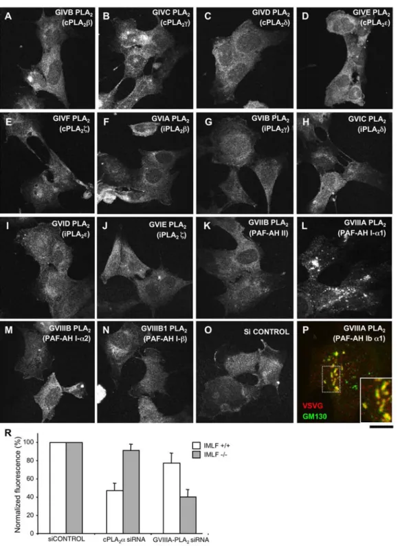
Figure 8. GVIIIA-PLA2inhibits transport of VSVG in immortalized murine lung fibroblasts from cPLA2a-KO mice. (A–P) IMLFs from
cPLA2a KO mice (IMLFs 2/2
) were incubated with siRNAs specific for the different cytosolic PLA2enzymes, and control siRNAs, for 72 h (A–P; as
indicated). The cells were then infected with VSV, kept at 40uC for 3 h to accumulate VSVG in the ER, and incubated in fresh medium at 32uC for 60 min before fixing. The fixed cells were either stained with only an anti-VSVG ab (A–O) or double labelled for VSVG and GM130 (P). VSVG was efficiently exported to the plasma membrane in cells incubated with either control (O) or most of the PLA2-isoform-specific siRNAs (A–K, M–O).
However, RNAi of GVIIIA-PLA2induced accumulation of VSVG within the Golgi complex of IMLFs (L). Notably, VSVG always overlapped strongly with
GM130 (see inset in P) in cells incubated with GVIIIA-PLA2-specific siRNAs, suggesting that VSVG is trapped within the cis-Golgi compartment. (R)
IMLF2/2and control lung fibroblasts from mice expressing cPLA2a (IMLFs+/+) were incubated with siRNAs specific for either cPLA2a or GVIIIA-PLA2
and control siRNAs for 72 h. The cells were then infected with VSV, subjected to the 40uC block with its further release for 60 min and fixed. VSVG was detected at the surface of IMLFs2/2and IMLFs+/+using an ab against its ectodomain. Afterwards, the cells were permeabilized and incubated again
with an anti-VSVG ab to reveal the total pool of VSVG within the cell. Then fluorescense intensities of surface and total VSVG were evaluated, expressed as a ratio, and normalized to the control. This quantification reveals that cPLA2a silencing significantly inhibits VSVG transport in IMLFs+/+
(but not in IMLFs2/2), while GVIIIA-PLA2 RNAi strongly affects VSVG transport in IMLFs2/2and only slightly in IMLFs+/+. Scale bar, 18mm (A–O), 7.2mm (P).
doi:10.1371/journal.pbio.1000194.g008
in tubulation (Figure 10). In addition to curvature, the generation of an intercisternal tubular continuity presumably requires the assembly of the fusion machinery at the tip of the budding tubule, for its connecting with a neighbouring cisterna. This, in turn, is likely to involve the formation of an ARF/COPI coat to recruit these fusion proteins into this bud [18,63,64]. Therefore, a simple model that fits our observations is that the cPLA2a-generated
lysolipids help to create and stabilize the curvature of the necks of COPI buds. This might prevent the fission of such buds into vesicles, allowing these buds to dock and fuse with the next cisternae, creating an intercisternal continuity. It is also possible that the cPLA2a-generated lysolipids favour the elongation of buds
into tubules, as suggested by the observation that overexpression of
cPLA2a induces tubulation of the stack structure (Figure 2J).
Notably, for these events to occur, diffusion of the lysolipids away from their site of synthesis should be limited by a diffusion barrier at the Golgi rims (perhaps similar to the molecular fences described at the plasma membrane [65]). This fence-like role could involve the COPI coat that resides at the rims of the cisternae. Future studies will elucidate these further components of the tubulation machinery. As noted, the inhibition of cPLA2a and
the attendant abrogation of the tubules are associated with the arrest of transit through the Golgi complex, indicating that the cPLA2a-dependent intra-Golgi tubules are involved in trafficking.
Interestingly, a similar association between intra-Golgi tubules and traffic has been reported for the effects of dicumarol. This drug is Figure 9. VSVG transport efficiency in HeLa cells after depletion of cPLA2aand/or GVIIIA-PLA2. HeLa cells were incubated with control
siRNAs or siRNAs specific for cPLA2a, GVIIIA-PLA2, or both for 72 h. (A) Cells exposed to these siRNAs were infected with VSV and kept for 6 h at 32uC
to allow continuous synthesis and transport of VSVG through the secretory pathway (‘‘steady-state’’ conditions). Then the cells were fixed and VSVG at the cell surface was stained with an ab against its ectodomain. Afterwards, the cells were permeabilized and incubated again with an anti-VSVG ab to reveal the total pool of VSVG within the cell. Scale bar, 10mm. (B) Cells were treated as in A or subjected to the 40uC block and then shifted to the permissive temperature of 32uC for 60 min (‘‘traffic wave’’ conditions), fixed, and labelled for surface and total VSVG as described above. Then the fluorescence intensities of surface and total VSVG were evaluated, expressed as a ratio, and normalized to the control. This quantification reveals that under both ‘‘steady-state’’ and ‘‘traffic-wave’’ conditions, cPLA2a silencing inhibits VSVG transport much more strongly than GVIIIA-PLA2depletion
(see also images in panel A). RNAi of both PLA2enzymes, in turn, shows a slightly stronger inhibitory effect on VSVG transport over cPLA2a silencing
alone. (C) Cells incubated with different siRNAs (as described above) were subjected to western blotting with antibodies against either cPLA2a or
GVIIIA-PLA2or GAPDH, as indicated.
doi:10.1371/journal.pbio.1000194.g009
an activator of the fission-inducing protein CtBP1/BARS, and it suppresses horizontal Golgi tubules (and thus, presumably, also the vertical intercisternal tubules, which, however, were not directly examined) and inhibits intra-Golgi trafficking [66]. Thus, suppressing the tubules appears to result in the arrest of trafficking independently of the molecular mechanisms that underlie tubule disruption.
How do cPLA2a-dependent tubules support intra-Golgi
traffick-ing, and which are the tubules—longitudinal or vertical—that are involved in intra-Golgi transport? The simplest hypothesis is that Golgi tubules allow the intercisternal diffusion of molecules crucial for trafficking. Both vertical and longitudinal tubular elements are cPLA2a-dependent. The former are much less abundant; however,
since traffic requires movement along the cis-trans (i.e., vertical) axis, they must be functionally crucial (as also supported by investigations in NZ-treated cells). Moreover, while relatively infrequent and difficult to detect [18,21,22], vertical tubules have the potential to be a very efficient means of intra-Golgi transit due to the great speed of diffusion over short distances (microns in the Golgi complex). Thus, very few vertical tubules per stack can be sufficient to achieve rapid diffusion between the cis and trans compartments of the Golgi complex (see Figure S6). This may be the case even when the connections are fewer than those required for complete intra-stack connectivity. For instance, in the Golgi ribbon, where stacks are connected to each other by horizontal membrane bridges, gaps in one stack might be compensated for by connections in neighbouring stacks (in this sense, also horizontal tubules may contribute to cis-trans diffusion; Figure S6). Connections might be cis-transient; here, again, very few connections need to be present in a stack at any given time to support intra-Golgi diffusion if they rapidly open and close between cisternae (Figure S6). Given the above, the question arises as to the role of intercisternal diffusion in intra-Golgi trafficking of cargo proteins that cross the Golgi by cisternal maturation/progression, such as PC-I and VSVG [35]. The simplest hypothesis is that these tubules allow retrograde movement of Golgi membranes and resident proteins (e.g., enzymes), which is required for maturation [18,21]. According to this scheme (also
discussed elsewhere [18]), during cisternal progression, the Golgi enzymes diffuse through the intercisternal continuities and explore the Golgi space, where they partition according to their physico-chemical properties into those cisternae that have their most favourable composition. This partitioning is driven by a physico-chemical gradient that is maintained across the stack at all times, possibly by the input of compositionally different intermediate compartment membrane into the cis cisternae and of endosomal membrane into the trans-Golgi. Thus, the arrival of intermediate compartment membranes at the cis pole (accompanied by consumption at the trans) promotes both enzyme backflow and cisternal progression (above), resulting in the synchronization of these two events and the maintenance of Golgi polarity. Clearly, this model requires more work to fully test it experimentally, but at this stage, it provides a logical explanation of the observations. At the same time, it should be noted that while our data point to a crucial role for tubules, complementary transport mechanisms cannot be excluded. For instance, if Golgi tubules indeed arise from the stabilization of COPI vesicles, as proposed above, it is possible that trafficking might switch between vesicular [67,68] and connection-mediated modes, with one or the other mechanism prevailing, depending on the cell type and the functional state.
How ‘‘general’’ is the requirement for cPLA2a-dependent tubules
in trafficking? Our findings show that the role of cPLA2a, is very
specific for intra-Golgi tubular structures and trafficking. However, other (non-Golgi) tubulation-dependent transport steps have been reported to be blocked by PLA2 inhibitors. For instance, ONO
(a rather nonspecific inhibitor of many PLA2 isoforms) has been
shown by us and others to also suppress non-Golgi tubules and to block non-Golgi transport steps that appear to be dependent on tubular intermediates, including endosome-to-plasma-membrane recycling of transferrin [59] and retrograde transport from the Golgi complex to the ER [60]. It is thus possible that these transport steps [59,60] are regulated by other PLA2isoforms that are located in the
different organelles. For instance, cPLA2b and cPLA2e have been
reported to be located to the early [69] and late [70] endocytic compartments, respectively, and may be involved in the regulation of Figure 10. Schematic illustration of cPLA2aaction at the Golgi complex. Inset 1: cPLA2a hydrolyses the fatty acids (FA) at the sn-2 position of
cylindrical phospholipids to form wedge-shaped lysophospholipids. Inset 2: This formation of wedge-shaped lysophospholipids favours generation of spontaneous membrane curvature and transformation of flat cisternae-like membranes into highly curved tubular membranes, which serve as intermediates for transport across the Golgi stack.
doi:10.1371/journal.pbio.1000194.g010
specific steps of endocytosis that are carried out via tubular carriers and that require PLA2activity [27,59]. If this is the case, then the
PLA2family in general (through different PLA2isoforms), rather than
cPLA2a itself, could underlie a membrane-bending mechanism based
on the induction of spontaneous membrane curvature [24] that is involved in tubulation and trafficking at the different levels of cellular membranes in mammals [27]. A further observation that is most probably related to these considerations is that mice knocked out for cPLA2a have reduced fertility, but do not show any other major
phenotypes [61,62], and that secretory transport and Golgi morphology in IMLFs obtained from KO mice [71] are normal. This appears to be because cPLA2a in KO cells is functionally
replaced by GVIIIA-PLA2(Figure 8). GVIIIA-PLA2also partially
localizes at the Golgi complex, where it appears to control tubulation processes (W. Brown, personal communication). Whether the mechanisms of action of this enzyme in trafficking are similar to those of cPLA2a is unclear at this time. At the mechanistic level, the
properties of GVIIIA-PLA2are not well defined in vivo. Although
GVIIIA-PLA2has been shown to have specificity in vitro towards
PAF-like lipids, its endogenous substrates remain unknown [72]. Thus, the precise metabolic reactions by which GVIIIA-PLA2
supports tubulation remain to be determined.
In conclusion, the activity of cPLA2a appears to be an
important mechanism for the formation of Golgi tubules in mammalian cells [24]. For other tubulation events (in other organelles or cell types), as noted, this role of cPLA2a might be
taken on by other PLA2 isoforms or even other phospholipases
(yeast). Nevertheless, the identification of cPLA2a as a player in
Golgi tubulation is a key finding, in that it reveals that generation of lysolipids is an important event in the formation of cellular tubules, and it should open the way towards the unravelling of further components of the tubulation machinery. It is now important to elucidate the mode of action of the intercisternal tubular connections and to define their underlying molecular machinery as well as the relationships of these tubules with other key players in intra-Golgi trafficking [73,74].
Materials and Methods Antibodies and Reagents
Ab sources: Ab against TGN46 from S. Ponnambalam (University of Dundee, Dundee, UK); Ab against PC-I from L.W. Fisher (NIH, Bethesda, MD, USA); Ab against giantin from H-P. Hauri (University of Basel, Basel, Switzerland); Ab against GM130 and ecto-domain of VSVG from M.A. De Matteis (Consorzio Mario Negri Sud, Santa Maria Imbaro, Italy). Ab against GFP and decorin from Abcam (Cambridge, UK); Abs against actin and VSVG from Sigma-Aldrich (Milan, Italy); Abs against cPLA2a from Santa Cruz Biotechnology (San Diego, CA,
USA) or were produced in our laboratory according to standard protocols. The Alexa 488, 546, and 633 IgG conjugates were from Molecular Probes Europe BV (Leiden, The Netherlands). The NANOGOLD gold-Ab conjugates and the GOLDENHANCE-EM kit were from Nanoprobes (Stony Brook, NY, USA). cDNA sources: cPLA2a-GFP from C. Leslie (National Jewish Medical
and Research Center, Denver, CO, USA) and T. Hirabayashi and T. Shimizu (University of Tokyo, Tokyo, Japan); cPLA2a(1–522)
from I. Kudo (Showa University, Tokyo, Japan); C2-GFP from R.L. Williams (MRC, Cambridge, UK); cPLA2aS228Cfrom B.P.
Kennedy (Department of Biochemistry and Molecular Biology, Merck Frosst Center for Therapeutic Research, P.O. Box 1005, Pointe Claire-DorVal, Canada); L294A aSNAP from R. Burgoyne (University of Liverpool, Liverpool, UK); YFP, VSVG-Cherry, VSVG-KDELR, and ARF1-GFP from J.
Lippincott-Schwartz (NIH, Bethesda, MD, USA); sialyltransferase-HRP from D.F. Cutler (University College London, London, UK). ONO-RS-082 was from Alexis (Lausen, Switzerland), pyrrophenone was from K. Seno (Shionogi Research Laboratories, Osaka, Japan), pyrrolidine was from Calbiochem (San Diego, CA, USA), bis-BODIPY FL C11-PC was from Molecular Probes (Eugene, OR, USA), [3H]-AA was from Amersham Pharmacia (Piscataway, NJ, USA).
Cell Culture, Transfection, RNAi, and Infection with Vesicular Stomatitis Virus
HeLa, MDCK, MC3T3, NRK and PCCL3 cells, HFs, and IMLFs from cPLA2a-KO mice were cultured in DMEM
(Invitrogen SRL, Milan, Italy) supplemented with 10% foetal calf serum and 1 mM L-glutamine. LipofectAMINE 2000 and Oliogofectamine (Invitrogen, Carlsbad, CA, USA) were used for the cDNA and cPLA2a-directed siRNA (SMART Pool,
Dharma-con , Chicago, IL, USA) transfections, respectively. Of note, both the mixture of siRNAs as well as two of the individual cPLA2
a-specific siRNAs from the SMART pool were effective in knocking down cPLA2a. The sense oligonucleotide sequences for human
cPLA2a are:
(#1) GGACAGUCGUUAAGAAGUA, (#2) GGAGAAACACUAAUUCAUA, (#3) GGAGAAGACUUUCAGACAA, (#4) GUACAAGGCUCCAGGUGUU.
The pool of three siRNA (QIAGEN Inc., Valencia, CA, USA) was used to silence mouse cPLA2a. The sense oligonucleotide
sequences for mouse cPLA2a are:
(#1) CCAGATGAATTTGAACGAATA, (#2) AAGCCTGAGGATTCTCATTTA, (#3) TAGGAGAAACACTAATTCAAA.
The efficiency of cPLA2a knock-down was evaluated by either
western blotting or immunofluorescense. Infection of cells with VSV was performed as described previously [18]. For VSVG rescue experiments, HeLa cells were incubated with oligonucle-otide #4 (directed against amino acids 649–655) and then transfected with mouse full length cPLA2a or the human
cPLA2a(1–522)mutant. Alternatively, mouse MC3T3 cells silenced
for cPLA2a with pool of three oligonucleotides were transfected
with full length human cPLA2a or human cPLA2a (1–522)
or cPLA2aS228Cmutants.
Cell Microinjection
VSV-infected HFs were microinjected with 4 mg/ml anti-cPLA2a ab in the presence of FITC or TRITC dextrans during
the course of the 40uC block, using an Eppendorf transjector 5246 (Eppendorf, Milan, Italy). They were then shifted to 32uC and processed for confocal microscopy. Similarly, VSV-infected cPLA2a-silenced HeLa cells were injected with 2 mg/ml
recom-binant cPLA2a protein in resque experiments. Treatment with the aSNAP Mutant
HeLa cells were permeabilized with streptolysin-O and incubated with the recombinant L294A aSNAP (4 mg/ml) protein in the presence of cytosol and an ATP-regenerating system, as described in Kweon et al. [56].
Immunofluorescence, Confocal Microscopy, and Live-Cell Imaging
For immunofluorescence analyses, the cells were fixed with 4% paraformaldehyde and permeabilised in 0.02% saponin, 0.5% BSA, and 50 mM ammonium chloride prior to their incubation with the cPLA2a-Dependent Tubules Drive Intra-Golgi Traffic