Journal Pre-proofs
Original articleA 2,3-diphenylpyrido[1,2-a] pyrimidin-4-one derivative inhibits specific an-giogenic factors induced by TNF-α
Serena Del Turco, Luca Quattrini, Rocchina Colucci, Melania Gaggini, Concettina La Motta, Giuseppina Basta
PII: S1319-0164(19)30127-6
DOI: https://doi.org/10.1016/j.jsps.2019.09.014
Reference: SPJ 949
To appear in: Saudi Pharmaceutical Journal Received Date: 7 February 2019
Revised Date: 3 September 2019 Accepted Date: 28 September 2019
Please cite this article as: Del Turco, S., Quattrini, L., Colucci, R., Gaggini, M., La Motta, C., Basta, G., A 2,3-diphenylpyrido[1,2-a] pyrimidin-4-one derivative inhibits specific angiogenic factors induced by TNF-α, Saudi Pharmaceutical Journal (2019), doi: https://doi.org/10.1016/j.jsps.2019.09.014
This is a PDF file of an article that has undergone enhancements after acceptance, such as the addition of a cover page and metadata, and formatting for readability, but it is not yet the definitive version of record. This version will undergo additional copyediting, typesetting and review before it is published in its final form, but we are providing this version to give early visibility of the article. Please note that, during the production process, errors may be discovered which could affect the content, and all legal disclaimers that apply to the journal pertain.
A 2,3-diphenylpyrido[1,2-a] pyrimidin-4-one derivative inhibits
specific angiogenic factors
induced by TNF-α
*Serena Del Turco, °Luca Quattrini, #Rocchina Colucci, *Melania Gaggini, °Concettina La Motta, *Giuseppina Basta
*CNR Institute of Clinical Physiology, Via G. Moruzzi, 1, 56124, Pisa, Italy; °Department of Pharmacy, University of Pisa, Via Bonanno, 6, 56126, Pisa, Italy.
#Department of Pharmaceutical and Pharmacological Sciences, University of Padova, Largo Meneghetti 2, 35131 Padova, Italy
Corresponding authors at:
National Research Council (CNR), Department of Biomedical Sciences, Institute of Clinical Physiology, Via Moruzzi, 1, 56124 Pisa, Italy
Phone 0039-050-315 2216 Fax 0039-050-315 2166
A 2,3-diphenylpyrido[1,2-a] pyrimidin-4-one derivative inhibits
specific angiogenic factors
induced by TNF-α
ABSTRACT
Low-grade chronic inflammation is a key process of angiogenesis in tumour progression. We investigated whether a synthetic analogue of apigenin, the 2-(3,4-dimethoxyphenyl)-3-phenyl-4H-pyrido[1,2-a] pyrimidin-4-one (called DB103), interfered with the mechanisms involved in the angiogenic process induced by the inflammatory cytokine tumour necrosis factor (TNFα). In endothelial cells, DB103 but not apigenin reduced the TNFα-induced oxidative stress. DB103 inhibited the activation of ERK1/2 but not JNK, p38 and Akt kinases, while apigenin was not so selective because it inhibited essentially all examined kinases. Similarly, apigenin inhibited the TNFα-induced transcription factors CREB, STAT3, STAT5 and NF-B, while DB103 acted only on NF-B. DB103 inhibited the induced-release of angiogenic factors such as monocyte chemotactic protein-1, interleukin-6 (IL-6) and angiopoietin-2 but not IL-8, while apigenin reduced the IL-6 and IL-8 release. DB103 revealed a better ability than apigenin to modulate proangiogenic responses induced by an inflammatory microenvironment.
1. Introduction
It is known that chronic inflammation potentiates or promotes cancer development, growth, and progression (Yao et al. 2016). Excessive production of inflammatory cytokines stimulates proliferation of endothelial cells (ECs) culminating in sustained tumour angiogenesis (Huang and Blobe 2016, Yao et al. 2016). The finding of high cytokine serum levels in cancer patients supported the opportunity to promote anti-inflammatory therapies in an attempt to counteract cancer progression. The tumour microenvironment is full of pro-inflammatory and proliferative factors including tumour necrosis factor (TNF)-α, interleukin (IL)-6, vascular endothelial growth factors (VEGF), metalloproteases, adhesion molecules and chemokines, whose expression is mainly regulated by the transcription nuclear factor-B (NF-B), which plays a key role both in the inflammatory process and in the cancer development (Pikarsky et al. 2004). TNF-α is angiogenic in vivo, while in vitro it does not directly induce EC migration and proliferation but promotes the angiogenic process only in the presence of growth factors and protease (Liu et al. 2016, Salvatore et al. 2017).
An increased intake of dietary flavonoids is associated with a decreased risk of cardiovascular diseases and cancer (Hussain et al. 2016, Lewandowska et al. 2016). Flavonoids have been found to inhibit angiogenesis through multiple mechanisms including inhibition of hypoxia-signalling cascades, down-regulation of the NF-B pathway, and direct interaction with intracellular signalling pathways of growth factor-receptors (Hussain et al. 2016, Lewandowska et al. 2016). There is to point out that most in vitro studies on flavonoid antiangiogenic properties generally exploited high concentrations of specific compounds until to 100 µmol/L (Zern and Fernandez 2005, Romagnolo and Selmin 2012). Hence apparent effects seen only at the limit of toxicity should be interpreted with caution. Thanks to their interesting functional properties, flavonoids have become a starting point for chemists to develop new synthetic analogues with improved functional properties than native or natural
changes that have been recently described (La Motta et al. 2007), we obtained a novel class of 2,3-diphenylpyrido[1,2-a]pyrimidin-4-ones (La Motta C. et al. 2013, Del Turco et al. 2015). We recently demonstrated (Del Turco et al. 2014). that, in our experimental conditions, flavonoid bioisosteres developed in our laboratory and inspired by the apigenin scaffold were well-tolerated even at 50 µmol/L, and for longer time, while the endothelial tolerability limit of apigenin for 72 hours of incubation was 10 µmol/L (Del Turco et al. 2014). In particular, we discovered that low concentrations of a synthetic derivative (2-(3,4-dimethoxyphenyl)-3-phenyl-4H-pyrido[1,2-a] pyrimidin-4-one), belonging to this class of bioisosteres and named DB103 (Chart 1), inhibited the smooth muscle cell proliferation but not the EC proliferation, and in both cell types it did not alter their vitality (Del Turco et al. 2014).
Chart 1. (A) 2-(3,4-dimethoxyphenyl)-3-phenyl-4H-pyrido[1,2-a] pyrimidin-4-one (DB103). (B) apigenin.
From this, it follows that its biocompatibility associated with a more specific functional activity than apigenin gives to DB103 improved functional properties. In continuity with what we have previously published, here we investigated whether DB103, compared with its natural analogue, modulated the inflammatory angiogenesis induced by TNF-α, by affecting proliferative factors and signalling pathways.
2. Material and methods
2.1. Materials
All reagents were purchased from Sigma-Aldrich (St. Louis, MO, USA) except where specified. Stock solutions of apigenin and DB103 were dissolved in sterile dimethyl sulfoxide (DMSO) and stored at -80 °C at the maximum solubility of 50 mmol/L. Since the final concentration of DMSO in the culture medium never exceeded 0.1%, DMSO (0.1%) alone served as a control. At this concentration, it did not show any effect on cell viability, cell proliferation, or related molecular mechanisms (data not shown).
2.2. Chemical synthesis of DB103
2-(3,4-dimethoxyphenyl)-3-phenyl-4H-pyrido[1,2-a]pyrimidin-4-one, (called DB103), was synthesized from the key intermediate 2-(3,4-dimethoxyphenyl)-4H-pyrido[1,2-a]pyrimidin-4-one, prepared as reported elsewhere (La Motta et al. 2007). Briefly, the treatment of the intermediate with N-bromosuccinimide, in refluxing chloroform, produced the corresponding 3-bromo derivative that after the reaction with phenylboronic acid in the presence of bis (triphenylphosphine)palladium(II) dichloride and Na2CO3, in refluxing toluene, led to the
target compound DB103.
2.3. Cell cultures and treatments
ECs were isolated from human umbilical vein endothelial cells, and characterized and maintained as described (Lazzerini et al. 2009). ECs were obtained from discarded umbilical vein and treated anonymously conforming to the principles outlined in the Declaration of Helsinki. Cells were used up to the fifth passage from primary culture.
If not otherwise indicated, experiments were performed on cells at pre-confluence, and cells were pre-treated for 5 minutes with 10 and 50 µmol/L of DB103, 10 µmol/L of apigenin or vehicle alone, before stimulation with TNF-α (10 ng/mL) for times that vary according to the experimental target. For each experimental condition, no less than 3 replicates had been performed. Cellular toxicity by apigenin and DB103 was checked at a range of concentration
from 1 µmol/L to 50 µmol/L (data not shown) through the phase contrast microscopy of cell morphology and WST-1 assay (Roche Diagnostics, Mannheim, Germany) using the manufacturers’ protocol as previously described (Cervelli et al. 2014).
2.4. Detection of intracellular oxidative stress
ECs were treated with TNF-α at 10 ng/mL in the presence/absence of DB103 or apigenin for 1 hour and the generation of intracellular ROS was measured with the fluorescent dye 6-carboxy-2′,7′ dichlorodihydrofluorescein diacetate bis(acetoxymethyl)-ester (C-DCF-DA) (Molecular Probes, Inc., Eugene, Oregon, USA) as described (Cervelli et al. 2014).
2.5. Cell signalling pathway analysis
ECs were plated in 100mm dishes, grown at sub-confluence, and subjected to partial fetal calf serum (2%) reduction for 18 hours before the addition of treatments and stimulation with TNF-α for 10 minutes. Cells were harvested in lysis buffer in the presence of protease inhibitor cocktail set III (Calbiochem, EMD Biosciences, San Diego, CA, USA). Each lysate (10 𝜇g of total protein) was analysed with the Milliplex® Map Multi-Pathway 9-plex Magnetic Bead Signalling kit (Merck Millipore, Billerica, MA, USA), to detect changes in phosphorylation of the kinases [ERK 1/2 (Thr185/Tyr187), JNK (Thr183/Tyr185), p38 (Thr180/Tyr182) and Akt (Ser473)] and transcription factors [NF-B p65 (Ser536), STAT3(Ser727), STAT5A/B (Tyr694/699) and CREB (Ser133)], according to the manufacturer’s instructions. Finally, the fluorescence intensity was read on an integrated multi-analyte detection platform (high-throughput technology MagPix system, Luminex® xMAP® technology, Merck Millipore).
2.6. ELISA analysis of the phospho-NF-B p65 protein
To confirm the results by the above-mentioned Luminex® xMAP® technology, the
quantification of phospho-NF-B p65 protein was determined with the PathScan® Phospho-NF-B p65 (Ser536) Sandwich ELISA Kit (Cell Signalling Technology, Danvers, MA, USA)
in the same cell lysates, according to manufacturer’s instructions. Optical density was read in a microplate reader at 450 nm.
2.7. 6, monocyte chemotactic protein 1 (MCP-1), VEGF-A, Angiopoietin-2(Ang-2) and IL-8 determination
After pre-treatment with the indicated compounds for 5 minutes and overnight stimulation with TNF-α at 10 ng/mL, culture supernatant samples were collected, frozen and thawed only once before the cytokine quantification. The amount of IL-6 and MCP-1 proteins were determined using the human IL-6 and MCP-1 Quantikine ELISA kits (R&D Systems, Minneapolis, MN, USA) according to the manufacturer’s instructions. The sensitivity of the IL-6 and MCP-1 was 0.7 and 1.7 pg/mL, respectively. The quantification of IL-8, VEGF-A and Ang-2 was performed with a custom-made Milliplex® Map Human Cytokine/Chemokine Magnetic Bead Panel (Merck Millipore). Cell culture supernatant samples were processed following the manufacturer’s recommended protocols and read on the Luminex® xMAP® platform (Merck Millipore). The sensitivity of VEGF-A, IL-8 and Ang-2 was 8.1, 0.2 and 3 pg/mL, respectively.
2.8. Statistical analysis
The results were expressed as mean ± SD, percentage or median. The percentage of inhibition was calculated by the formula (value of positive control – value of sample/ value of positive control − value of negative control) × 100. Multiple comparisons were performed by one-way ANOVA followed by Bonferroni's post-hoc tests. Values of P < 0.05 were considered statistically significant. Data were analysed with the use of statistical software SPSS 13.0 (SPSS Inc, Chicago, IL, USA).
3. Results and Discussion
3.1. DB103 inhibited the TNF-α-induced oxidative stress
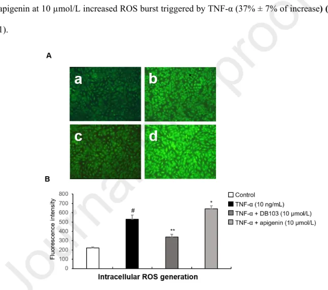
As shown in the Fig.1, ECs stimulated for one hour with TNF-α, produced intracellular reactive oxygen species (ROS) and pre-treatment with DB103 at 10 µmol/L strongly inhibited their production (61.3% ± 10% of inhibition). Pre-treatment with DB103 at 50 µmol/L inhibited in an almost identical manner (data not shown), indicating that the maximum inhibition of ROS production was reached at 10 µmol/L. Conversely, pre-treatment with apigenin at 10 µmol/L increased ROS burst triggered by TNF-α (37% ± 7% of increase) (Fig. 1).
Fig. 1. DB103, but not apigenin, inhibited the TNF-α-induced intracellular ROS generation (A) Representative photomicrographs (10x magnifications) of ROS produced in ECs incubated for 1 hour with vehicle (a) or with 10 ng/mL of TNF-α (b) in the continued presence of DB103 (c) or apigenin (d) at 10 µmol/L. (B) Quantitative analysis of ROS expressed as mean ± SD of arbitrary fluorescence units of 3 replicates. #P<0.001 vs. control;
*P<0.05 and **P<0.01 vs. TNF-α alone. Photomicrographs are representative of three experiments performed on different experimental days.
These results suggest that DB103 has antioxidant properties that are known to be related to anti-inflammatory properties (Griffiths et al. 2016). Instead, the ROS increase by apigenin is in agreement with that recently published by Souza et al.(Souza et al. 2017) and, as suggested by the authors themselves, could be associated to its pro-apoptotic and anti-proliferative properties (Sung et al. 2016, Souza et al. 2017).
3.2. Among the TNF-α-induced kinases, DB103 inhibited only the extracellular signal– regulated kinase (ERK) 1/2
We analysed the effects of DB103 on the main protein kinases activated by TNF-α, such as
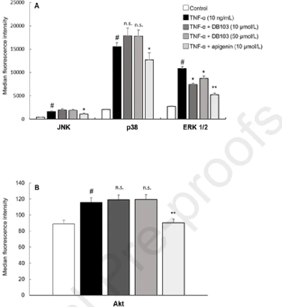
MAPKs and Akt kinase that regulate various processes including cell survival, growth and angiogenesis. After 10 minutes of stimulation, TNF-α activated the phosphorylation of c-Jun N-terminal kinases (JNK), p38, ERK 1/2 and Akt kinases (Fig. 2). DB103 inhibited the phosphorylation of ERK 1/2 kinase while it did not alter JNK, p38 and Akt (Fig. 2 A-B). At the opposite, apigenin inhibited all examined kinases (Fig. 2A-B). These different effects on cell signalling pathways highlight that DB103 is more specific than apigenin.
Fig. 2. Contrary to apigenin, DB103 inhibited only the TNF-α-induced ERK 1/2 kinase
ECs were incubated with vehicle or with TNF-α (10 ng/mL) in the continued presence of
DB103 or apigenin. Cell lysates were assayed with multiplex technology for the phosphorylation of JNK, p38, ERK 1/2 (A) and Akt kinases (B). Results are expressed as median fluorescence intensity. Each bar represents the mean of three independent experiments, each performed in n = 4 replicates. #P<0.001 vs. control; *P<0.05 and **P<0.01
3.3. Among the TNF-α-induced transcription factors, DB103 inhibited only NF-B
We evaluated the effect of both compounds on the main transcription factors induced by TNF-α. The efficacy of apigenin was remarkable on STAT3, STAT5, NF-B and CREB transcription factors (Fig. 3 A-B) while DB103 inhibited only NF-B phosphorylation ( Fig. 3A). The phospho-NF-B p65 induced by TNF-α was also analysed by the ELISA method, confirming the same inhibitory patterns of the two compounds on phospho-NF-B p65 (Fig. 3C). Once again, the performance of the two compounds was dissimilar.
Fig. 3. DB103 inhibited only NF-B while apigenin inhibited indiscriminately
ECs were incubated with vehicle or with 10 ng/mL of TNF-α in the continued presence of DB103 or apigenin. Cell lysates were assayed by multiplex technology for the phosphorylation of STAT3, STAT5, NF- B (A) and CREB (B). Results are expressed as median fluorescence intensity. Each bar represents the mean of three independent experiments, each performed in n = 4 replicates. The phospho-NF-κB p65 was also evaluated on cell lysates by an ELISA assay (C). The bars represent the mean ± SD from four replicates. °P<0.01 and #P<0.001 vs. control; *P<0.05 and **P<0.01 vs. TNF-α alone.
3.4. DB103 inhibited MCP-1, IL-6 and Ang-2 but not the IL-8 cell release
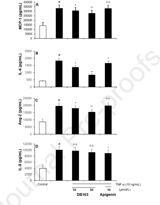
As shown in Fig. 4, upon overnight stimulation with TNF-α (10 ng/mL), ECs produced and released angiogenic cytokines, but not VEGF-A (data not shown). The absence of VEGF-A in the supernatant could be due to the fact that TNF-α-induced VEGF-A expression is not direct but it depends by other TNF-α-induced cytokines (Yoshida et al. 1997).. Consequently, since its detection requires a longer time of stimulation, the overnight treatment with TNF-α resulted insufficient for a sensible detection and a following estimate of the inhibitory treatments (data not shown). DB103 inhibited MCP-1 in a concentration-dependent manner, while apigenin did not inhibit it (Fig. 4A). MCP-1, which is best known for its role in recruiting monocytes/macrophages to the arterial wall, up-regulates the hypoxia-inducible factor 1 and consequently induces VEGF-A (Hong et al. 2005) and therefore may indirectly contribute to cell proliferation and angiogenesis (Hong et al. 2005, Gacche and Meshram 2013, Bianconi et al. 2018). Also, IL-6, which resulted to be inhibited by both DB103 and apigenin (Fig. 4B), plays a crucial role in the angiogenic process because it upregulates the VEGF promoter activity in tumour cells(Loeffler et al. 2005, Huang et al. 2016). DB103, contrary to apigenin, reduced in a concentration-dependent manner the TNF-α-induced release of Ang-2 (Fig. 4C), a cytokine promoting cell death that, nevertheless, combined with VEGF, can promote neo-vascularization (Hashizume et al. 2010). An opposite result was obtained about the interference of the two compounds on the TNF-α-induced release of IL-8
(Fig. 4D), a chemokine regulated by NF-B transcription factor which activates signal transducers and activators that in turn promote angiogenesis (Waugh and Wilson 2008, Zhang et al. 2012). Although DB103 inhibited strongly the p65 phosphorylation, it unreduced IL-8 release (Fig. 4D), suggesting that other post-transcriptional mechanisms can be affected by DB103 with consequent absence of a net effect on production and release of IL-8 (Hoffmann et al. 2002, Wang et al. 2012). On the other hand, we cannot exclude that DB103 can exert a stimulating action and favouring the IL-8 production through other cell-signalling pathways, which we have not investigated here.
Fig. 4. DB103 inhibited IL-6, MCP-1 and Ang-2 but not IL-8 cell release
ECs were incubated for 24 hours with vehicle or with 10 ng/mL of TNF-α in the continued presence of DB103 or apigenin. MCP-1 (A), IL-6 (B), Ang-2 (C) and IL-8 (D) expression were measured on cell supernatant by ELISA assay. Each bar represents the mean of three independent experiments, each performed in six replicates. ANOVA with Bonferroni’s post hoc comparison. #P<0.001 vs. control; *P<0.05 and **P<0.01 vs. TNF-α alone.
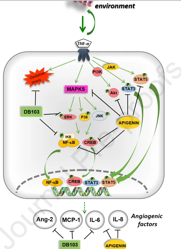
DB103 modulated the expression of several angiogenic factors induced by TNF-α as illustrated in the Fig. 5. These proliferating factors, including TNF-α, accumulate in the cancer-surrounding microenvironment and generate a low-grade, chronic inflammation, which is a crucial process involved in tumour progression (Candido and Hagemann 2013). DB103 revealed a superior ability than apigenin to modulate the angiogenic process induced by TNF-α.
In conclusion, although DB103 does not block the growth factors-induced physiological angiogenesis, as we previously published (Del Turco et al. 2014), it may hinder a tumour advancing by reducing factors and processes triggered by chronic and persistent inflammation of the tumour environment, characterized by the presence of cytokines and chemokines, all involved in the angiogenic development. Therefore, our compound could assist the action of current anti-proliferative drugs modulating the low-grade chronic inflammatory condition. Further studies in animal models will prove the robustness of this dietary supplement as a potential candidate for developing functional foods, utilisable to contrast the pathological angiogenic responses associated with the secretion of inflammatory cytokines in cancer.
Fig. 5. Effect of DB103 and apigenin on different TNF-α signalling pathways and their
Declaration of interest None.
ACKNOWLEDGMENTS
This work was supported by the Italian MIUR (Ministero dell'istruzione, dell'Università e della Ricerca) - CNR Flagship project Interomics (DSB.AD013.002) and the Micro-VAST project (Grant 153/09). The funders had no role in study design, data collection and analysis, decision to publish, or preparation of the manuscript.
AUTHOR CONTRIBUTIONS
G. B., S.D.T designed and supervised the project. L.Q. and C.L.M. conducted the chemical synthesis of the compound. S.D.T., M.G. and R.C. performed cellular and biochemical experiments. G. B., S.D.T. and C.L.M. wrote the paper. All authors analysed and discussed the achieved results and reviewed the manuscript.
REFERENCES
Bianconi, V., Sahebkar, A., Atkin, S. L. and Pirro, M., 2018. "The regulation and importance of monocyte chemoattractant protein-1." Curr Opin Hematol. 25, 44-51.
Candido, J. and Hagemann, T., 2013. "Cancer-related inflammation." J Clin Immunol. 33 Suppl 1, S79-84.
Cervelli, T., Panetta, D., Navarra, T., Andreassi, M. G., et al., 2014. "Effects of single and fractionated low-dose irradiation on vascular endothelial cells." Atherosclerosis. 235, 510-518.
Del Turco, S., Sartini, S., Cigni, G., Sentieri, C., et al., 2015. "Synthetic analogues of flavonoids with improved activity against platelet activation and aggregation as novel prototypes of food supplements." Food Chem. 175, 494-499.
Del Turco, S., Sartini, S., Sentieri, C., Saponaro, C., et al., 2014. "A novel 2,3-diphenyl-4H-pyrido[1,2-a]pyrimidin-4-one derivative inhibits endothelial cell dysfunction and smooth muscle cell proliferation/activation." Eur J Med Chem. 72, 102-109.
Gacche, R. N. and Meshram, R. J., 2013. "Targeting tumor micro-environment for design and development of novel anti-angiogenic agents arresting tumor growth." Prog Biophys Mol Biol. 113, 333-354.
Griffiths, K., Aggarwal, B. B., Singh, R. B., Buttar, H. S., Wilson, D. and De Meester, F., 2016. "Food Antioxidants and Their Anti-Inflammatory Properties: A Potential Role in Cardiovascular Diseases and Cancer Prevention." Diseases. 4.
Hashizume, H., Falcon, B. L., Kuroda, T., Baluk, P., et al., 2010. "Complementary actions of inhibitors of angiopoietin-2 and VEGF on tumor angiogenesis and growth." Cancer Res. 70, 2213-2223.
Hoffmann, E., Dittrich-Breiholz, O., Holtmann, H. and Kracht, M., 2002. "Multiple control of interleukin-8 gene expression." J Leukoc Biol. 72, 847-855.
Hong, K. H., Ryu, J. and Han, K. H., 2005. "Monocyte chemoattractant protein-1-induced angiogenesis is mediated by vascular endothelial growth factor-A." Blood. 105,
1405-Huang, J. J. and Blobe, G. C., 2016. "Dichotomous roles of TGF-beta in human cancer." Biochem Soc Trans. 44, 1441-1454.
Huang, Y. H., Yang, H. Y., Huang, S. W., Ou, G., Hsu, Y. F. and Hsu, M. J., 2016. "Interleukin-6 Induces Vascular Endothelial Growth Factor-C Expression via Src-FAK-STAT3 Signaling in Lymphatic Endothelial Cells." PLoS One. 11, e0158839.
Hussain, S. A., Sulaiman, A. A., Balch, C., Chauhan, H., Alhadidi, Q. M. and Tiwari, A. K., 2016. "Natural Polyphenols in Cancer Chemoresistance." Nutr Cancer. 68, 879-891.
La Motta, C., Sartini, S., Mugnaini, L., Simorini, F., et al., 2007. "Pyrido[1,2-a]pyrimidin-4-one derivatives as a novel class of selective aldose reductase inhibitors exhibiting antioxidant activity." J Med Chem. 50, 4917-4927.
La Motta C., Da Settimo F., Dario B., Sartini S., et al. (2013). A Therapeutic agent for treatment of blood vessels. PCT Int. Appl. : WO2013144860A2013144861.
Lazzerini, G., Del Turco, S., Basta, G., O'Loghlen, A., Zampolli, A. and De Caterina, R., 2009. "Prominent role of NF-kappaB in the induction of endothelial activation by endogenous nitric oxide inhibition." Nitric Oxide. 21, 184-191.
Lewandowska, H., Kalinowska, M., Lewandowski, W., Stepkowski, T. M. and Brzoska, K., 2016. "The role of natural polyphenols in cell signaling and cytoprotection against cancer development." J Nutr Biochem. 32, 1-19.
Liu, D., Wang, X. and Chen, Z., 2016. "Tumor Necrosis Factor-alpha, a Regulator and Therapeutic Agent on Breast Cancer." Curr Pharm Biotechnol. 17, 486-494.
Loeffler, S., Fayard, B., Weis, J. and Weissenberger, J., 2005. "Interleukin-6 induces transcriptional activation of vascular endothelial growth factor (VEGF) in astrocytes in vivo and regulates VEGF promoter activity in glioblastoma cells via direct interaction between STAT3 and Sp1." Int J Cancer. 115, 202-213.
Pikarsky, E., Porat, R. M., Stein, I., Abramovitch, R., et al., 2004. "NF-kappaB functions as a tumour promoter in inflammation-associated cancer." Nature. 431, 461-466.
Romagnolo, D. F. and Selmin, O. I., 2012. "Flavonoids and cancer prevention: a review of the evidence." J Nutr Gerontol Geriatr. 31, 206-238.
Salvatore, V., Teti, G., Focaroli, S., Mazzotti, M. C., Mazzotti, A. and Falconi, M., 2017. "The tumor microenvironment promotes cancer progression and cell migration." Oncotarget. 8, 9608-9616.
Souza, R. P., Bonfim-Mendonca, P. S., Gimenes, F., Ratti, B. A., et al., 2017. "Oxidative Stress Triggered by Apigenin Induces Apoptosis in a Comprehensive Panel of Human Cervical Cancer-Derived Cell Lines." Oxid Med Cell Longev. 2017, 1512745.
Sung, B., Chung, H. Y. and Kim, N. D., 2016. "Role of Apigenin in Cancer Prevention via the Induction of Apoptosis and Autophagy." J Cancer Prev. 21, 216-226.
Wang, Y., Wang, W., Wang, L., Wang, X. and Xia, J., 2012. "Regulatory mechanisms of interleukin-8 production induced by tumour necrosis factor-alpha in human hepatocellular carcinoma cells." J Cell Mol Med. 16, 496-506.
Waugh, D. J. and Wilson, C., 2008. "The interleukin-8 pathway in cancer." Clin Cancer Res. 14, 6735-6741.
Yao, M., Brummer, G., Acevedo, D. and Cheng, N., 2016. "Cytokine Regulation of Metastasis and Tumorigenicity." Adv Cancer Res. 132, 265-367.
Yoshida, S., Ono, M., Shono, T., Izumi, H., et al., 1997. "Involvement of interleukin-8, vascular endothelial growth factor, and basic fibroblast growth factor in tumor necrosis factor alpha-dependent angiogenesis." Molecular and Cellular Biology. 17, 4015-4023. Zern, T. L. and Fernandez, M. L., 2005. "Cardioprotective effects of dietary polyphenols." J
Nutr. 135, 2291-2294.
Zhang, Y., Wang, L., Zhang, M., Jin, M., Bai, C. and Wang, X., 2012. "Potential mechanism of interleukin-8 production from lung cancer cells: an involvement of EGF-EGFR-PI3K-Akt-Erk pathway." J Cell Physiol. 227, 35-43.