UNIVERSITA’ DEGLI STUDI DI VERONA
Dottorato di ricerca in
biotecnologie applicate
Ciclo XXII
S.S.D.: AGR/07
Study of Bacillus thuringiensis behaviour
in food environment by genome – wide
transcriptome analysis
* Per l’elenco dei Settori Scientifico-Disciplinari (SSD) si veda il D.M. del 4 Ottobre 2000, Allegato A
“Elenco dei Settori Scientifico –Disciplinari” reperibile sul sito del Ministero dell’Università e della
UNIVERSITA’ DEGLI STUDI DI VERONA
DIPARTIMENTO DI
BIOTECNOLOGIE
DOTTORATO DI RICERCA IN
BIOTECNOLOGIE APPLICATE
CICLO XXII
Study of Bacillus thuringiensis behaviour in food environment by
genome – wide transcriptome analysis
S.S.D. AGR/07
Coordinatore: Prof. Massimo Delledonne
Firma __________________________
Tutor: Prof. Massimo Delledonne
Firma __________________________
Co-Tutor: Prof. Pier Sandro Cocconcelli
Firma __________________________
Dottorando: Dott.ssa Francesca Colla
INDEX
CHAPTER 1
INTRODUCTION:
1. THE GENUS BACILLUS 2
1.1 Classification and Phylogeny 2
2. GENERAL PROPERTIES OF BACILLI 3
2.1 Structure surface of Bacillus 3
2.1.1 S-layers 4
2.1.2 Capsules 4
2.1.3 Cell wall 5
2.1.4 Flagella 6
2.2 Growth conditions and nutritional requirements 6
2.3 The Bacillus endospore 6
2.3.1 Sporulation 6
2.3.2 The structure of bacterial spores 8
2.3.3 Germination process 11
2.3.4 Bacilli receptors 13
3. OCCURRENCE OF BACILLUS SPP. IN THE ENVIRONMENT 14
3.1 The Bacillus cereus group 15
3.2 B. cereus as pathogenic organism 16
3.2.1 Emetic syndrome 16
3.2.2 Diarrhoeal syndrome 17
3.2.3 Mode of action of the enterotoxins 19
3.2.4 Regulation of enterotoxin expression 20
3.3 Presence of toxin in other Bacillus spp. 22
4. BACILLUS THURINGIENSIS 23
4.1 General characteristics 23
4.2 Ecology and serotyping 24
4.3 B. thuringiensis Cry proteins 25 4.4 Mode of action of B. thuringiensis Cry proteins 25
4.5 Transcriptional Mechanisms of cry gene 27
4.6 Development of B. thuringiensis biopesticides. 28
INDEX
CHAPTER 2DISTRIBUTION AND EXPRESSION PROFILES OF GENES CODING FOR BACILLUS CEREUS-LIKE ENTEROTOXINS IN BACILLUS
THURINGIENSIS STRAINS OF COMMERCIAL INTEREST
1. INTRODUCTION 41
2. MATERIALS AND METHODS 43
2.1 Bacterial strains isolation 43
2.2 DNA extraction for PCR and REP 44
2.3 Detection of crystal proteins 44
2.4 Repetitive extragenic palindromic DNA sequence (REP) 44
2.5 Detection of genes coding for enterotoxins 45
2.6 RNA preparation 45
2.7 Analysis of toxins expression by RT-PCR 46
2.8 Enterotoxin assay 46
2.9 Spores production 46
2.10 Food model preparation 47
2.1.1 B. thuringiensis spores germination assay in food model 47
2.1.2 Sample preparation for scanning electron microscopy (SEM) 48 2.1.3 X-ray microanalysis 48
3.RESULTS 48
3.1. Characterisation of the B. thuringiensis isolated strains 48 3.2. Detection and expression of enterotoxic genes in B. thuringiensis isolated strains 50 3.3 Enterotoxin production 51
3.4 Selection of sporification medium and spores production 51
3.5 Food model development 52
3.6 SEM observations of the germination process 54
3.6.1 Gold coating results 54
3.6.2. SEM X-ray analysis 58
4.DISCUSSION 58
INDEX
CHAPTER 3GENOME-WIDE TRANSCRIPTOME ANALYSIS OF
BACILLUS THURINGIENSIS SPORE GERMINATION OUTGROWTH
AND TOXIN PRODUCTION IN FOOD MODEL
1. INTRODUCTION 64
2 MATERIALS AND METHODS 67
2.1. Bacterial strain, and growth condition 67
2.2 Spore generation and germination conditions 67
2.6. Microarray construction 68
2.7. RNA isolation, cDNA synthesis, labelling and hybridization 70
2.8. Microarrays stripping for Re-hybridization 71
2.9. Microarray data analysis 71
2.10 Relative quantification of enterotoxic gene expression 72
2.11 Enterotoxin assay in food model 73
3. RESULTS AND DISCUSSION 73
3.1 Genome-wide gene expression analysis 74
3.1.1. Microarray validation 74
3.1.2. Transcriptional analysis 77
3.1.2. QT-clustering 78
3.1.3. Transcriptome analysis of spores 80
3.1.4. Functional analysis 82
3.2 Enterotoxin gene expression profiles 90
3.3 Enterotoxin assay in CPM model 92
BIBLIOGRAPHY 94
CHAPTER 4 RESEARCH AND INACTIVATION OF VIRULENCE REGULATING SYSTEMS IN BACILLUS SPP. 1. INTRODUCTION 99
INDEX
1.2 Site-specific chromosomal mutagenesis 100
2. MATERIALS AND METHODS 104
2.1 Bacterial strains and growth condition 104
2.2 DNA extraction and manipulation 104
2.3 Optimization of B. thuringiensis electroporation system 104
2.4 Detection of B.cereus transcriptional regulators homolog in B. thuringiensis UC10070 105 2.5 Construction of resE and fnr mutants with homologous recombination system 105 2.6 Detection of genes disruption 106
2.7 Construction of B. thuringiensis resE and fnr mutants with TargeTron system 106 2.8 Reverce transcription PCR to evaluate intron expression in B. thuringiensis UC10070 108 2.9 Confirmation of knockout by colonies PCR 108
3. RESULTS 109
4. DISCUSSION 117
BIBLIOGRAPHY 120
CHAPTER 5 GENERAL CONCLUSION 123
CHAPTER
1
INTRODUCTION:
THE GENUS BACILLUS
Chapter 1
1.The genus Bacillus
In 1872, Ferdinand Cohn, characterized the bacterium Bacillus subtilis. This Gram-positive organism, capable of growth in the presence of oxygen, and able to form a unique type of resistant cell called endospore, represented the first member of what was to become a large and diverse genus of bacteria named Bacillus, in the Family Bacillaceae . The ubiquity and diversity of these bacteria, the resistance of their endospores to chemical and physical agents, the developmental cycle of endospore formation, the ability to produce antibiotics, the toxicity of their spores and protein crystals for many insects, have attracted ongoing interest since their discoveries in the 1870s (Kennet Todar, 2009). 1.1 Classification and Phylogeny
The heterogeneity in ecology, physiology, and genetics of Bacillus species made difficult to categorize the genus Bacillus. The modern concept of the genus Bacillus can be ascribed largely to the work of Nathan R. Smith, Francis E. Clark, and Ruth E. Gordon; in the 1930’s these group of scientists, developed a definition of the genus Bacillus as comprising “ rod-shaped bacteria capable of aerobically forming refractile endospores that are more resistant than vegetative cells to heat, drying, and other destructive agencies”. First attempts to classify Bacillus species were based on two main characteristics: aerobic growth and endospore formation. This resulted in grouping of many bacteria possessing different physiology and occupying a variety of habitats. In Bergey's Manual of Systematic Bacteriology (1st ed. 1986), the G+C content of known species of Bacillus is reported to range from 32 to 69% (Holt, 1986), illustrating the genomic heterogeneity of the genus. There are variation from species to species, but sometimes it can be observed profound differences in G+C content within strains of the same species. Phylogenetic classification reported in the Bergey's Manual of Systematic Bacteriology (2nd ed. 2004) groups the two most prominent types of endospore-forming bacteria, clostridia and bacilli, in the two different Classes of Firmicutes: Clostridia and
Bacilli. The Phylogenetic evidence, mainly based upon RNA analysis of the small subunit
of ribosomes (16S rDNA) indicated that Bacillus species showed a kinship with several non spore-forming bacteria like Enterococcus, Lactobacillus, Listeria and
Staphylococcus. With the advent of ssRNA analysis, Bacillus genus, was divided into
several families of endospore-forming currently assigned to four genera in the family
Chapter 1
anaerobic Clostridium spp. for its ability to grow in the presence of air. Many Bacillus species can be allocate to one of six taxa that have distinguishable physiologies. This is generally consistent with the devision of the genus based on spore morphologies. The six groups are: B. polymyxa group (I), B. subtilis group (II), B. brevis group (III), B.
sphaericus group (IV), and thermophiles (V and VI). Group I includes species that are
facultative anaerobes and can grow strongly in the absence of oxygen. A variety of sugars are fermented to produce acid, and endospores are ellipsoidal. Species belonging to the B.
subtilis group, are phylogenetically and phenotipically consistent. All these bacteria
produce acids from a wide range of sugars and some strains, like B. cereus and B.
licheniformis, are facultative anaerobes. B. licheniformis can use glucose only under
anaerobic conditions but grows poorly anaerobically. Although B. subtilis is generally considered an aerobe, it can grow and sporulate slowly also in anaerobic conditions. When glucose, with nitrite is the terminal electron acceptor, it grows strongly anaerobically. These bacteria are therefore an intermediate stage between the true facultative anaerobes of the group I strains and the strict aerobes in groups III and IV. This is reflected in their production of acid from several sugars (Leuschner, Bacillus - Central Science Laboratory, York, UK. 2008). The oval endospores produced by these bacteria do not swell the mother cell and are generally located centrally or subterminally. Group III represents strict aerobes that generally do not produce acid from sugars. They produce ellipsoidal spores that swell the mother cell. In group IV all species produce spherical spores that may swell the mother cell and contain l-lysine or ornithine in the cell wall. All species are strictly aerobic, but some have a limited ability to produce acids from sugars. Thermophilic species of the Group V are heterogeneous physiologically and morphologically, and grow optimally at > 50° C. Most produce oval spores that swell the mother cell. In group VI are thermophilic and acidophilic species which membranes are characterized by the presence of omega-alicyclic fatty acid.
2. General properties of bacilli 2.1 Structure surface of Bacillus
Like many gram-positive bacteria, the properties of adhesion, resistance and tactical responses, making the surface of Bacillus species rather complex.
Chapter 1
Fig.1 Bacillus surface. C=capsule, S=S-layer, P=peptidoglycan (from Kenneth Todar PhD.2009
University of Wisconsin-Madison).
The surface of vegetative cells is a laminated structure consisting of a capsule, a proteinaceous S-layer, several multi-layers peptidoglycan, and proteins located on the outer surface of the plasma membrane.
2.1.1 S-layers
S-layers consist in crystalline surface layers of protein or glycoprotein subunits. They can be found in Bacillus, like in other bacteria but their function is not completely understood. Since it covers the entire cell surface, it seems likely that it can act as a molecular sieve, preventing large molecules from entering or leaving the cell. Other roles which have been ascribed to bacterial S-layers include protection of the cell from predation and provision of attachment sites for exoenzymes. It has recently been shown that in some Gram-positive bacteria S-layers can mask the negative charge of the peptidoglycan sheets and prevent agglutination processes.
2.1.2 Capsules
Bacillus species can produce different types of capsules: those of B. anthracis, B. subtilis, B. megaterium, and B. licheniformis, contain poly-D- or L-glutamic acid. B. circulans, B. megaterium, B. mycoides and B. pumilus, produce carbohydrate capsules, or with more
complex polysaccharides. Some polysaccharides produced by Bacillus may react with antisera of other genera of bacteria, including human pathogens: is the case of B.
mycoides with Streptococcus pneumoniae or B. pumilus with Neisseria meningitides. The
Chapter 1
by the closest B. cereus and B. Thuringiensis, can be used as a criterion for distinguishing between these species.
2.1.3 Cell wall
Bacillus genus does not present much variability in the structure of the cell wall as occur
in many Gram-positive bacteria. The wall of all Bacillus species consists of peptidoglycan of the mesodiaminopilmelic acid (DAP) (Weiss et al.1981). This type of polymer is the same type as the one universally found in Gram negative bacteria. DAP can be directly cross-linked to D-alanine, as in the Enterobacteriaceae; in other cases, like in most Gram-positive bacteria, two tetrapeptide side chains of peptidoglycan connect DAP and D-alanine, by an interpeptide bridge. The presence of teichoic acids bounded to muramic acid residues has been reported in large amount for all species. However, the type of teichoic acids varies widely between Bacillus species. As in many Gram-positive bacteria
Bacillus species present lipoteichoic acids associated with the cell membrane, which seem
to be involved in the synthesis of teichoic acids in the cell wall.
Fig.2 Schematic representation of muropeptide subunit of Bacillus peptidoglycan, without intrapeptide
Chapter 1
2.1.4 Flagella
Most of the spore-forming aerobic bacteria, are mobile and possess peritrichous flagella that cell use to move in the environment in response to external stimuli trough the chemotaxis mechanism; the composition of alkaliphile Bacillus species like B. firmus, present a low content in basic amino acids, thought to render cell more stable at pH value up to 11. Flagellar system and chemotaxis has been extensively studied in B. subtilis.
A B
Fig.3Flagellar strains: B. cereus (A) B. brevis (B) (from Kenneth Todar PhD.2009University of
Wisconsin-Madison).
2.2 Growth conditions and nutritional requirements
Spore-forming bacteria are generally chemoheterotrophs able to implement the process of respiration using a variety of simple organic compounds like sugars amino-acids and organic acids. In some cases they can ferment carbohydrates with reactions that produce glycerol and butanediol. Species such as B. megaterium need no organic factors for growth, while others require amino acids or vitamins. Most are mesophilic with an optimal of temperature growth between 30 and 45 degrees. Some species are thermophilic with optimal growth around 65 ° C. Psychrophiles species are few but are able to develop and sporify even at 0 ° C. Bacillus species can grow in a wide range of pH between 2 and 11. In laboratory environment, and optimal growing conditions, they present a regeneration time of about 25 minutes.
2.3 Bacillus endospore 2.3.1 Sporulation
Gram positive bacteria belonging to the Bacillus genus can undergo a complex developmental cell differentiation process what allows them to adapt to changing
Chapter 1
environmental conditions and lack of nutrients by producing highly resistant spores. This process, called sporulation, involves progression through different stages including initiation, chromosome segregation, sporulation-specific cell division (asymmetric in rod-shaped bacteria), differential gene expression and specific signal transduction mechanisms. The return pathway, leading to vegetative cell growth, involves spore germination followed by outgrowth of the germinated spore. All of these aspects of sporulation have been studied for many years in great detail and have had both a substantial impact on our understanding of many other basic cell processes and have started to fuel applied spore research with new ideas.
In general, factors that may affect sporulation the ability to sporulate are pH, oxygen, and temperature. Sporulation appears to be favoured by conditions which result in a decreased growth rate in the presence of adequate energy and carbon source reserves. When sufficient nutrients are present, the vegetative cell divides rapidly by cell division, but environmental triggers like nutrient depletion and/or population density do initiate the sporulation process, ultimately resulting in the bacterial spore (Barak and Wilkinson, 2005; Eichenberger et al, 2004; Errington 2003; Piggot en Hilbert, 2004; Wang et al, 2006). Spore development involves an unequal cell division, the smaller cell (forespore) being engulfed by the larger one so that the endospore develops inside the mother cell. In many species, the cell is distended by the spore. Spore formation, which takes several hours, is accompanied by morphological, physiological, and biochemical changes, and the resulting refractile spore is structurally very different from a vegetative cell. The formation of a spore is an expensive and complex process for the bacterial cell. Spores are only made under conditions where cell survival is threatened such as starvation for certain nutrients or accumulation of toxic wastes. Regulation of sporulation is tight and the first few steps are reversible. This helps the cell conserve energy and only sporulate when necessary. Initiation of spore formation is controlled by Spo0A, a transcriptional factor which modulates gene expression during the transition from the exponential to the stationary phase. Spo0A is the response regulator of a two-component, signal transduction regulatory system, and in growing cells exists predominantly in the dephosphorylated state. Under conditions in which sporulation is initiated, it is phosphorylated by a phosphorelay involving a number of kinases and is thereby able to activate or repress gene expression by binding specific DNA targets (“0A boxes”) found upstream of regulated genes. Subsequently, changes in gene expression are controlled by the synthesis
Chapter 1
and activation of alternative σ factors which associate with RNA polymerase and alter the promoter specificity of the enzyme. Five σ factors are known to be produced at various stages.
Sporulation is a seven step process during which cell will be dramatically reorganized. The first stages of sporulation are involved in forming a separate compartment for the spore in the mother cell. Activation of Spo0A and σH in the predivisional cell leads to asymmetric division. Once this occurs, sporulation is irreversible. The next stages involve laying down the various layers of the spore. Both the spore and the mother cell plays a role in this process. Within the two compartments, spore development is orchestrated by RNA polymerase σE regulated gene expression in the mother cell and σF regulated gene expression in the forespore. In the final stages, regulated by σK in the mother cell, and σG in the forespore (Wang et al, 2006), the spore dehydrates its cytoplasm while at this stage the mothercells lysis as a consequence of programmed cell death (Lewis, 2000), releasing the spore in the environment. The sporulation pathway is depicted in Figure 4.
Fig. 4The morphological stages of sporulation. Patrick Stragier. Annual Review of Genetics, 1996
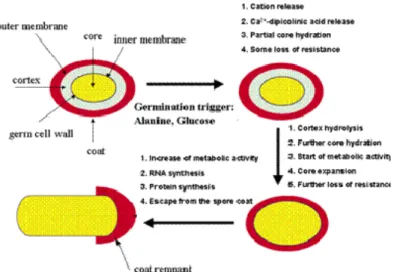
2.3.2 The structure of bacterial spores
The complex structure of the spore protects the cellular compartment from environmental challenge providing a formidable resistance against harsh conditions (Table 1). Another typical spore property, crucial for its longevity is the spore dormancy. The cross section
Chapter 1
of the spore in figure 5, reveals all the spore compartments including the core, the inner membrane, the cortex, the outer membrane and the coat layers followed by the
exosporium.
Table 1
Resistance capacity of growing cells and dormant spores of B. subtilis _______________________________________________________________
Treatments required to kill 90% ofthe population __________________________________
Growing Dormant Treatment cells spores ________________________________________________________________
UV radiation (254 nm)(KJ m-2) 36 330
Wet heat (90°C) (min) <0.1 18
Dry heat (120°C) (min) <0.01 18
H2O2 (15% at 23°C) (min) <0.2 50
Formaldehyde (25 g/l) (min) <0.1 22
Nitrous acid (100 mmol/l) (min) <0.2 100 Freeze dryings (number of cycles) < 1 >20
________________________________________________________________ Setlow, 2005
The outermost layer is the exosporium, which is a thin covering made of proteins. The exosporium is the primary site of contact with the environment, including host defences; it is not present on B. subtilis spores, but seems to be conserved among pathogenic bacilli on members of the B. cereus group. it is a loose-fitting, balloon-like structure composed of a paracrystalline basal layer and an external hair-like nap (Gerhardt, P., 1967). The filaments of the hair-like nap are apparently formed by a single collagen-like glycoprotein, whereas the basal layer is composed of a number of different proteins in tight and loose associations. The exosporium is the least understood part of the spore structure, but its presence on pathogenic Bacilli and, suggests a possible role in interactions with host organisms. Below there is the spore coat which is made up of highly cross-linked keratin and layers of spore-specific proteins. The role of many individual coat proteins remains unclear. The outer membrane is located between the cortex and inner coat layers. Its function is still not well elucidated, It is however an essential structure during formation of the spore (Piggot, 2004). The cortex consists of loosely cross-linked peptidoglycan. It is of important to maintain spore dormancy and
Chapter 1
heat resistance, and is thought to contribute to the dehydrated state of the core (Nicholson et al, 2000 and references therein). The cortex composition is similar between many spore-forming bacteria, including clostridia (Atrih and Foster, 2001). During spore germination, the cortex must be degraded quickly to allow the expanding spore core. During spore dormancy, spore cortex lytic enzymes are present in the dormant spore although in an inactive state. The two crucial cortex lytic enzymes in B. subtilis spore germination are CwlJ and SleB. CwlJ is synthesized in the mother cell during sporulation, and located in the outer layers of the spore, whereas SleB, synthesized in the forespore, is targeted to both the spore inner membrane and the outer spore layers (Bagyan and Setlow, 2002; Chirakkal et al., 2002). We know that the SleB protein of B. subtilis is a muramidase but we do not yet know how it is activated during germination. Not all sporeformers have the SleB, CwlJ pair of germination- specific cortex lytic enzymes The inner membrane, surrounds the spore core as selective permeability barrier. The germination receptors and gene products of the SpoVA operon, essential parts of the germination apparatus, (Vepachedu and Setlow, 2005) seems to be partly incorporated in this membrane. This suggested that the inner membrane plays an important role in the first stages of germination. Moreover, after the activation of the germination receptors, the inner membrane contributes in signal transduction directly to other parts of the spore as germination signal. Core contains the components of the vegetative bacterial cell (the cell wall, cytoplasmic membrane, cytoplasm, nucleoid, DNA, ribosomes, etc.) as well as significant quantities of dipicolinic acid and Ca2+ ions. The water content of endospores is only about 10-30% of the water content of vegetative cells; therefore, endospores are capable of surviving at levels of dehydration that would kill vegetative cells. The low water content also provides the endospore with chemical resistance (to chemicals such as hydrogen peroxide) and it causes the remaining enzymes of the spore cell to become inactive.
One chemical produced by endospores that is thought to lend to their high resistance is dipicolinic acid. This chemical has been found in the spore cell of all endospores examined. Dipicolinic acid interacts with calcium ions to form calcium dipicolinate (DPA), which is the main substance believed to lend endospores their resistance and represents about 10% of the dry weight of an endospore. The spore also contains small acid-soluble spore proteins (SASPs). These function to protect DNA from UV radiation,
Chapter 1
desiccation and dry heat, and they also serve as a carbon and energy source during the germination process (conversion back to a vegetative cell).
Fig.5Schematically representation of the spore structure,(from Department of Microbiology, Cornell
University. 2007)
2.3.3 Germination process
In addition to its intrinsic interest, spore germination has attracted applied interest, because it is through germination that spores ultimately cause food spoilage and poisoning (Setlow et al., 2003). In order to initiate germination and restore vegetative growth when conditions become favourable, bacterial spores must be able to monitor their external environment. Spore germination, as defined as those events that result in the loss of the spore-specific properties, is an essentially biophysical and degradative process (Moir and Smith, 1990). The spore’s inner membrane increases in fluidity (Stewart et al., 1979) and ion fluxes resume; monovalent cations, potassium and sodium, move across the spore membrane, and calcium ions and dipicolinate are excreted. The peptidoglycan of the spore cortex is degraded, and the coat layers are partially degraded (Atrih et al., 1998; Atrih et al., 1999). ATP synthesis and oxidative metabolism resume (Otani et al., 1986), DNA damage is repaired (Nicholson et al., 1997) and the DNA-complexing small acid-soluble proteins (SASPs) are degraded by a specific protease (Nessi et al., 1998), providing a source of amino acids for outgrowth. It occurs without any need for new
Chapter 1
macromolecular synthesis, so the apparatus required is already present in the mature dormant spore. Germination in response to specific chemical nutrients requires specific receptor proteins, located at the inner membrane of the spore. After penetrating the outer layers of spore coat and cortex, germinant interacts with its receptor: one early consequence of this binding is the movement of monovalent cations from the spore core, followed by Ca2+ and dipicolinic acid (DPA). Germinant molecules are able to activate these receptors, probably by allosteric interaction (Wolgamott and Durham, 1971). This initiates a cascade of processes that gradually degrade the protective structures of the spore and resume cellular processes and its metabolism, ultimately leading to the vegetative cell (Hornstra , 2007). In some species, an ion transport protein is also required for these early stages. Early events including loss of heat resistance, ion movements and partial rehydration of the spore core, can occur without cortex hydrolysis, although the latter is required for complete core rehydration and colony formation from a spore. In B.
subtilis two crucial cortex lytic enzymes have been identified: one is CwlJ, which is
DPA-responsive and is located at the cortex-coat junction. The second, SleB, is present both in outer layers and at the inner spore membrane, and is more resistant to wet heat than is CwlJ. Cortex hydrolysis leads to the complete rehydration of the spore core, and then enzyme activity within the spore protoplast resumes. We do not yet know what activates SleB activity in the spore, and neither do we have any information at all on how the spore coat is degraded (Moir, 2005).
Chapter 1
2.3.4 Bacilli receptors
Bacillus spores are equipped with a specific set of germination receptors that monitor the
environment for proper outgrowth conditions. As signalling molecules herein function germinants, often amino acids or ribosides, which are able to initiate germination when present in appropriate concentration and mixture in close proximity of the spore (Foerster and Foster, 1966; Gould, 1969). The process of germination involves interaction of chemical germinants with presumed specific receptors in the spore, and the transduction of this signal in some way. There is no evidence of bulk transport or metabolism of germinant (Scott and Ellar, 1978). The full molecular details of the signal transduction process in spore germination are not yet clear, but reasonable hypotheses can be constructed with the available information, most of which is derived from studies with B.
subtilis. One hypothesis to explain the germination-associated changes is that the earliest
events in germination would involve membrane changes that alter permeability properties, leading to a redistribution of ions and water in the spore, and thereby activate lytic enzymes (Keynan, 1978); evidence of inhibition of spore germination by ion channel blockers supports this (Mitchell, 1986), as does the likely membrane association of gerA gene products (Moir and Smith, 1990). The germinant has to first permeate the outer coat and cortex layers of the spore before coming in contact with the germinant receptors. The gerA operon in the genome of B. subtilis, encoding for the germination (Ger) receptor GerA, was the first germination operon described (Zuberi et al, 1987), and was shown to be involved in L-alanine initiated. germination. Later, gerB and gerK were described, both involved in a germination response on a mixture of L-asparagine, glucose, D-fructose and K+ (AGFK response) (Corfe et al, 1994).
Genomes of almost all sporeformers contain at least one, and usually several of these receptor operons, leading to the conclusion that sporeformers respond to different types of germinant via multiple receptors, encoded in gene clusters that have diverged from some common ancestor(s). Sometimes more than one receptor is involved in the response to single or multiple germinants (McCann et al., 1996; Barlass et al., 2002; Ireland and Hanna, 2002). The gerA operon, like most of its homologues, encodes three proteins, GerAA, GerAB and GerAC. These all have a potential association with the membrane – GerAA and AB are integral membrane proteins – GerAA has a predicted membrane-bound domain that would span the membrane at least five times, whereas GerAB is predicted to have 10 membrane spans, and is classified in evolutionary terms as a
Chapter 1
subfamily of single component membrane transporters (Jack et al., 2000)( it is the only one of the three proteins that has homology to any other known protein outside the spore-forming bacteria). The GerAC protein is a predicted lipoprotein. All are expressed in the developing spore compartment, and would therefore be targeted to the inner membrane of the spore. Experiments using antibodies against GerAA and GerAC proteins showed that they were present in the inner membrane, rather than the outer layers of the spore (Hudson et al., 2001) and experiments on GerBA showed that this too was present in the inner membrane (Paidhungat and Setlow, 2001). Evidence that receptor proteins directly bind germinant comes only, so far, from analysis of mutant phenotypes. A germinant may bind without mediating transport, but causing allosteric changes within the membrane protein(s). Therefore, we need a better understanding of these receptor proteins, which has been hindered by the failure of attempts so far to overexpress and characterize the membrane-associated components.
3. Occurrence of Bacillus spp. in the environment
Members of the genus Bacillus have a ubiquitous environmental distribution. The endospore production is basic for the dispersion of Bacillus spp..The reservoir of these bacteria is the soil. Strains have been isolated from the extremes of deserts and Antarctic samples.. The extreme spore resistance capacities have amazed many scientists and have been studied, trying to reveal the mechanisms behind spore resistance (Nicholson et al, 2000 and references therein; Setlow, 2005). They have been proved to be the most durable type of cells found in nature: thanks to their dormant state, they can survive for extremely long periods, even millions of years. Because of this incredible resistance, the presence of spores may causes several problems wherever hygienic and sterile conditions are a prerequisite, such as in the food industry and in medical environments. Spores are able to resist most of the preservation techniques currently applied and as a consequence are responsible for infections, serious food-borne illnesses and significant amount of food spoilage (Brul et al, 2006). Industry has developed preservation methods to reduce the microbial contamination on food products. As a result of these efforts our food can be regarded as safe. Unfortunately, currently used methods are not fully effective against spores as a consequence of their incredible resistance capacities (Oomes and Brul, 2004). The omnipresence of Bacillus spores in the environment inevitably results in the presence of spores in agricultural and dairy products. In recent years, consumers have shifted their
Chapter 1
preferences on “fresh-like” foods, since having better taste and texture characteristics, these products are expected to be also healthier. However, the use of milder food processing conditions, basal to accommodate these preferences, facilitate the presence of spores in food products, for frequently not completed inactivation of spore. Furthermore, the lack of microbial competition after the treatments, facilitates the rapid facilitates the rapid release of vegetative cells from germinating spores.
3.1 The Bacillus cereus group
The Bacillus cereus group comprises a highly homogeneous subdivision of the genus
Bacillus that exhibit highly divergent pathogenic properties.
B. cereus belongs, together with B. anthracis, B. thuringiensis, B. weihenstephanensis, B mycoides and B. pseudomycoides, to this group of closely related microorganism.
The reservoir of these bacteria is the soil, but they are widely distributed in the environment, or commensal inhabitants of the intestines of insects. Occasionally these species can cause food poisoning and soft tissue infections, particularly of the eye. B.
cereus is an opportunistic human pathogen most commonly associated with food
poisoning (Drobniewski et al. 1993). Other members of this group, currently classified as
B. thuringiensis, are primarily insect pathogens widely used as a biopesticide (Schnepf et
al 1998). A third pathogenic phenotype is exhibited by B. anthracis: it is the causal agent of anthrax, a zoonotic disease that can be lethal to humans. B. mycoides, B.
pseudomycoides and psychrotolerant B. weihenstephanensis are less well characterized.
Although the latter one, capable of growing efficiently at temperatures of 4°C, may form a hazard in food products stored at low temperatures (Hornstra 2007). The genome analysis of B. weihenstephanensis, revealed the presence of toxin genes (Stenfors et al, 2002), but has not yet been demonstrated its responsibility in food-borne disease.
Despite first studies on B. cereus group started in19th century, the relationships between some of these organisms have yet to be completely resolved. The very high genetic relationship between B. cereus, B. anthracis and B. thuringiensis, makes genome based differentiation complicated or even impossible (Helgason et al, 2000; Ivanova et al, 2003). Conventional markers of chromosomal diversity, such as 16S and 23S rRNA genes, are essentially identical (Ash et al. 1991, 1992) . Several studies using different techniques like pulsed-field gel electrophoresis of chromosomal DNA (Carlson ea al. 1994), genomic mapping (Carlson et al.1996), multilocus enzyme electrophoresis
Chapter 1
(Helgason et al. 1998, 2000), BOX-PCR fingerprinting (Kim et al.2001), multilocus sequence typing (MLST) (Helgason et al 2004) and amplified fragment length polymorphism (AFLP) analysis (Ticknor et al. 2001), have also been suggested that these species are so closely related that they should be considered as one species.
On the other hand, some differences in terms of phenotype within these species, allow easy identification using classical methods; B. cereus and B. thuringiensis present hemolytic activity, are mobile, resistant to penicillin, and are able to degrade tyrosine and produce phosphatise, while B. anthracis does not show any of these characteristics. B.
thuringiensis produces parasporal toxic crystals, known as δ-endotoxins, which allows to
discriminate it from B. cereus (Schoeni and Wong, 2005 and references therein). Moreover, pathogenicity patterns of these species are very different. Most of the genes responsible for virulence of these bacteria are plasmid located. In some case, loss of the plasmid corresponds to loss of virulence, making impossible to distinguish between bacteria belonging to this group. The evolutionary relationships between all members of the group should be important, not only for understanding the evolution of virulence in the B. cereus group, but also for rapidly increasing of scientific and political importance that these organisms have acquired in recent years from.
3.2 B. cereus as pathogenic organism
B. cereus is an opportunistic human pathogen that can cause two types of food-borne
infections. The emetic syndrome is caused by toxin production in the food product before consumption, while the diarrhoeal syndrome is the result of ingested B. cereus spores that germinate in the human intestine and produce enterotoxins in the intestinal tract (Granum, 2001; Schoeni and Wong, 2005).
3.2.1 Emetic syndrome
Emetic syndrome is a typical example of food intoxication caused by a toxin called cereulide, that lead to nausea and vomiting 1-6 hours after ingestion of contaminated food (Kramer en Gilbert, 1989; Ehling-Schulz et al, 2004). Similar symptoms are caused by
Staphylococcus aureus enterotoxin (Granum and Lund, 1997). Cereulide is a heat and pH
stable circular dodecadepsipeptide (Fig. 7) consisting of three repeating units of four amino acids, each consisting of D-O-leucine, D-alanine, L-O-valine and L-valine (Agata et al. 1994; Agata et al. 1995b). The structure resembles that of the known potassium
Chapter 1
ionophore valinomycin (see Figure 7). Indeed cereulide has been shown to be toxic to mitochondria by acting as a potassium ionophore. Symptoms are generally mild, and patients recover within 24 h, but occasionally fatal cases resulting from emetic syndrome have been reported (Mahler et al, 1997).
Fig.7 Comparison of the amino acid compositions of cereulide [D-Ala – D-O-Leu – L-Val – L-O-Val]3
(left) and of valinomycin [D-Val – L-O-Ala – L-Val – D-O-Val]3 (right) (Teplova et al. 2006)
In 2004 two research groups have shown that the production of cereulide in B.cereus is the result of a complicated mechanism operated by a non ribosomal peptide synthetase (NRPS) complex (Toh et al. 2004; Horwood et al. 2004).
3.2.2 Diarrhoeal syndrome
The diarrhoeal syndrome is caused by the production of enterotoxins in the small intestine after ingestion of food contaminated by B.cereus vegetative cells. This typical toxico-infection is characterized by abdominal pain, cramps and diarrhoea, occurring 8 to 16h after ingestion (Granum and Lund, 1997). The enterotoxins cause disturbance of the water (solute transport) affecting the epithelial lining of the small intestine. Symptoms of the disease are very similar to those caused by the food-borne desease from Clostridium
perfringens, but the pathogenic mechanism appears to be different: the C. perfringens
enterotoxin is released during sporulation in the small intestine, whereas the enterotoxins of B. cereus are produced during growth in the small intestine (McClane, 1997; Granum, 2007).
During the 1980’s and 1990’s , with the discovery and identification of enterotoxins, many advances in the study of diarrheal syndrome have been possible. New molecular biology techniques allowed to aquire knowledges on the production and regulation of
Chapter 1
enterotoxins. Based on epidemiological data it was estimated that 103 – 108 cells per gram of food are sufficient for the manifestation of disease symptoms (Granum and Lund, 1997). Several virulence factors produced by B. cereus have been described, of which three-component enterotoxins hemolysin BL and non-hemolytic NHE are well characterized (Beecher and Wong, 1997; Lindback et al, 2004). Another single component toxin, enterotoxin T has been described (Agata et al, 1995) encoded by the bceT gene, but the role of this enterotoxin in B. cereus initiated food poisoning remains to be elucidated. Generally the symptoms associated with diarrheal syndrome are rather mild, but a strain of B. cereus producing CytK toxin, responsible for necrotic enteritis, caused the deaths of three people in France (Lund et al, 2000).
Haemolysin BL (HBL)
The first described enterotoxin in B. cereus is the hemolytic toxin BL (HBL). Because of its observed effects in vivo and in vitro (Kramer and Gilbert, 1989), it was initially defined diarrhoeagenic factor, fluid accumulation factor and vascular permeability factor (Shinagawa et al. 1991a, Shinagawa et al. 1991b, Sutherland and Limond, 1993). HBL is a three component protein toxin, encoded by 3 genes organized in 1 operon hblA, hblC and hblD genes encode the B, L1, and L2 components, respectively. A fourth gene has been found in this operon, hblB, but its function has not yet been defined. The molecular weight of B-component is 38 kDa1, 40 kDa for the L1-component, and 45 kDa the L2-component.
Non haemolytic enterotoxin (NHE)
In 1996 investigation of over 300 strains from various sources, including strains from a number of outbreaks, revealed that another unknown enterotoxigenic complex, with cytotoxic effects, could have been the causative agent in some of the B.cereus-associated food-borne desease (Granum et al. 1996). This three component enterotoxin, named NHE, was discovered after an outbreak in Norway (Lund and Granum, 1996). Even though NHE contain several structural resemblances to HBL complex, and lead to symptoms similar to those caused by HBL, it lacked the haemolytic activity and the cytotoxic potential of the two three component is different (Lund and Granum, 1997).
Chapter 1
Cytotoxin K
Cytotoxin K (CytK) may also be involved in B. cereus food poisoning. This toxin causes more severe diarrhoea including necrotic enteritis. It consist in a single protein toxin with molecular weight of approximately 34 kDa. CytK belongs to a family of β-barrel channel-forming toxins (including Staphylococcus aureus leucocidins and Clostridium perfringens b-toxin). It is necrotic and haemolytic (Lund et al., 2000), and also cytotoxic for intestinal epithelia (Hardy et al., 2001). It was first characterized in B. cereus strain 91-98: a strain isolated from cases of food-borne disease that in France was responsible for the death of three people (Lund et al. 2000). None of the other, enterotoxin genes, previously described (hbl and nhe) was detected in this strain, further implicating CytK as a major virulence factor. However the cytotoxin K discovered in this outbreak, appeared to be the strongest form discovered so far. Later research detected a less potent cytotoxin K variants, also named cytotoxin K like or cytK-2, with approximately 89% amino acid homologous to the original cytotoxin K (cytK-1) but 20% less toxic on human intestinal Caco-2 cells and Vero-cells. However, several B. cereus isolates possess the cytK gene, (Guinebretiere et al., 2002); although the mere presence of a gene possibly involved in virulence is not sufficient to confer pathogenicity, the transcription level of the gene could be important for virulence (Brillard and Lereclus, 2004).
enterotoxin T (Bc-D-ENT)
Another single component toxin, enterotoxin T (bc-D-ENT), has been described in B.
cereus (Agata et al, 1995). The bc-D-ENT enterotoxin, encoded by the bceT gene, is
capable of causing fluid accumulation in ligated rabbit ileal loops (Punyashthiti & Finkelstein, 1971), showing cytotoxicity towards Vero cells (Konowalchuk e t al., 1977). Unlike for the three first mentioned enterotoxins (Hbl, Nhe and CytK), has not yet been shown bc-D-ENT was involved in food poisoning. The role of this enterotoxin in B.
cereus food poisoning remains to be elucidated.
3.2.3 Mode of action of the enterotoxins
The two three-component enterotoxins HBL and NHE present a similar mode of action (Lund and Granum, 1997). According to cytotoxicity experiments with VERO-cells all three components of the HBL-complex are necessary for maximal enterotoxic activity, (Powell, 1987; Rousset and Dubreuil, 2000; Belaiche, 2000; Black et al. 2005). The
Chapter 1
optimal ratio of each component for HBL activity is 1:1:1 (Beecher et al. 1995). The latest model for the action of HBL studied by Beecher and Lee Wong (1997), suggested that all three components bind to the target cells leading to their lysis. Even for the activity of NHE toxin all three component complex are request, although in this case the optimal ratio is 10:10:1 (NHE-A : NHE-B : NHE-C) (Lindback et al. 2004).
The mode of action of cytotoxin K is different from that described for both HBL and NHE. The amino-acid sequence of cytotoxin K suggests that cytK belong to the β -barrel channel-forming protein family such as the β -toxin from Clostridium perfringens and α – and γ - haemolysin of Staphylococcus aureus (Hardy et al. 2001). Its symptoms include severe epithelial lesions and bloody diarrhoea.
3.2.4 Regulation of enterotoxin expression
More then one regulator system have been identified to be important in the B. cereus virulence regulation.
The transcriptional regulator PlcR (Phospholipase C Regulator) takes part in the control of most known virulence factors in B. cereus: enterotoxin, haemolysins, phospholipases and proteases (Michel et al 2008). It also regulates phospholipase C expression, then is called the phospholipase C regulator (PlcR) .Transcription of PlcR is autoinduced (Lereclus et al 1996) and is repressed by the sporulation factor Spo0A (Lereclus et al 2000). To be active PlcR needs the PapR peptide. PapR is expressed as a propeptide under the control of PlcR, is exported out of the cell, is processed to form the active peptide either during export or in the extracellular medium, and is captured back by the cell through the oligopeptide permease system OppABCDF (Slamti et al. 2002, Gominet et al 2001, Declerck et al 2007). Thus, the three partners PlcR, OppABCDF and PapR function as a quorum-sensing system. PlcR integrates at least two classes of signals: cell growth state through Spo0A and self cell density through PapR (Michel et al 2008).
However, it seems that other systems may interact with PlcR, assuming a role in regulating the pattern expression of B. cereus virulence factors. Variability and adaptability, are crucial characteristics of all the organisms that possess the ability to survive and prosper in a wide variety of environmental conditions; often virulence factors allow them to conquer many different niches throughout the course of infection. Recognition of specific signals and conversion of this information into specific transcriptional responses, are basal to cope with a variety of environmental situations. In
Chapter 1
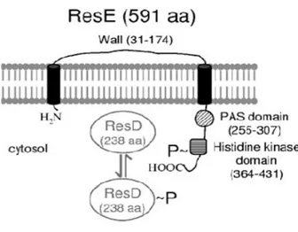
many cases, signalling through a single two-component system results in a coordinated change in expression of multiple genes whose products play a role in adaptation to a particular environment. Several study focused on the importance of two-component signal transduction systems in controlling both metabolism and virulence factors in B. cereus (Duport et al. 2006). One of the major controlling factors of gene expression in B. subtilis during fermentative (Nakano et al 1997, Cruz Ramos et al 2000) microaerobic and aerobic growth (Hartig et al 2004) is ResDE two-component system; moreover it regulates virulence in Staphylococcus aureus under low-oxygen conditions (Yarwood et al 2001). Homologs of the B. subtilis ResDE was found in B. cereus. It was demonstrated that ResDE, play an important role in the regulation of enterotoxin expression in B.
cereus. This two-component regulatory system consists of a histidine sensor kinase
(ResE), bound to the cell membrane, and a cytoplasmic response regulator (ResD), (Fig.2). Signals related to oxygen limitation are perceived by ResE that undergoes autophosphorylation at a conserved histidine residue. ResD phosphorylation level is determined by the balance between both activities of ResE as phosphate donor for ResD, and phosphatase of phosphorylated ResD. In B. cereus resE mutant strain, abolition of enterotoxin production was observed in all the conditions examined (Duport et al 2006).
Fig. 8 Gene organization of the B. cereus chromosome region containing resDE. (Duport et al 2006.)
Subsequent studies described another redox regulator that may act in synergy with ResDE to control the expression of fermentation and enterotoxin genes, demonstrating that,
Chapter 1
although important, ResDE is not essential for both fermentative metabolism and enterotoxin expression. This transcriptional regulator, known as CRP-Fnr (fumarate and nitrate reduction regulator), is member of the cyclic AMP receptor protein, and play an important role in modulating the expression of many metabolic genes in several facultative or strictly anaerobic bacteria (Korner et al 2003). Their functions also include the control of virulence factors (Baltes et al 2005, Bartolini et al 2006, Schmiel et al 2000). Furthermore, the one-component CRP-Fnr regulators are known to act coordinately with two-component regulators homologous to ResDE in response to two environmental signals: oxygen availability and the presence of alternative electron acceptors. CRP-Fnr family proteins, are characterized by a nucleotide-binding domain that extends from the N terminus over 170 residues to a C-terminally located helix-turn-helix structural motif. A short C-terminal sequence with four cysteine residues follow this DNA-binding domain. Three Cys residues from this C terminus together with one Cys residue from the central part of the protein bind a [4Fe-4S]2+ center that serves as a redox sensor (Reents et al 2006). Transcription of hbl and nhe was dramatically (90%) down-regulated after CRP-Fnr mutation experiment in B. cereus strains (Duport et al. 2007). The production of major virulence factors hemolysin BL (Hbl) and nonhemolytic enterotoxin (Nhe) in the food-borne pathogen B. cereus, seems to be regulated through complex mechanisms. A recent study led by Esbelin and colleagues (2009) clarified some aspects of the B. cereus virulence regulation, suggesting a strict interaction between the three system previously described. The response regulator ResD was shown to interact directly with promoter regions of the enterotoxin regulator genes resDE, fnr and plcR and the enterotoxin structural genes nhe and hbl, but with different affinities. Moreover, phosphorylation state of ResD results in a different target expression pattern. This finding led to the conclusion that enterotoxin expression and fermentative metabolism may be controlled coordinately at the transcription level. It was also clearly defined the role of ResDE two component system, as a sentinel capable of sensing redox changes, and coordinating responses that modulates B. cereus virulence.
3.3 Presence of toxin in other Bacillus spp.
In some instances, enterotoxin production from non-B. cereus species has been reported. Isolates of B. circulans, B. lentus, B. licheniformis, B. mycoides, B. subtilis, and B.
Chapter 1
thuringiensis demonstrated positive results using a commercial RPLA assay (Bacillus
cereus enterotoxin reverse passive latex agglutination) to detects the L2 component of the HBL-complex (Beattie & Williams 1999). Toxin production by two environmental strains of B. pumilus) were reported by Hoult & Tuxford (1991).
Toxins production by other Bacillus spp. has largely been limited to that of B.
thuringiensis, a member of the B. cereus group.
The species B. thuringiensis, B. anthracis, B. cereus, differ in 16S rRNA sequence by only nine nucleotides, leading many to the conclusion that these could be considered a single species (Ash et al. 1991). Phenotypic differences within this group are very few, but the pathogenicity patterns differ significantly. B. thuringiensis, is characterized by the presence of large crystalline endotoxin molecules which form during sporulation. The toxin is insect-specific, and several classes of these toxin molecule, that target particular order of insects, were isolated (Schnepf et al. 1998). However conjugative transfers of many plasmids among Bacillus cereus and B. thuringiensis are demonstrated (Yuan et al. 2007, Van der Auwera et al. 2007). B. cereus and B. thuringiensis are not able to be differentiated strictly on the basis of biochemical characteristics (Carlson et al. 1994, Damgaard et al. 1996, Yamada et al. 1999). Standards method for detection of B. cereus, have been failed to distinguish these two organisms. Damgaard et al. (1996) isolated several enterotoxin-producing strains of B. thuringiensis from pasta, bread, and milk. Perani et al. (1998) found that 29% of B. thuringiensis strains isolated from the environment produced B. cereus-like enterotoxins. From the reports mentioned above emerged that an exhaustive investigation into the ubiquity of enterotoxin genes in various
Bacillus spp. has not been done. Little attention has been paid, either in model systems or
in food environment, to asses the conditions that could support toxin expression in non-B.
cereus isolates.
4. Bacillus thuringiensis 4.1 General characteristics
Bacillus thuringiensis, like the food-borne and opportunistic pathogen Bacillus cereus,
belong to the Bacillus cereus sensu latu family. In 1901, a Japanese biologist, Ishiwata Shigetane, discovered a previously not described bacterium as the causative agent of a disease in silkworms. B. thuringiensis was originally considered a risk for silkworm rearing but it has become the heart of microbial insect control. It can form a parasporal
Chapter 1
crystal during the stationary phase of its growth cycle and was initially characterized as an insect pathogen. In 1956, T. Angus demonstrated that the insecticidal activity was attributed largely or completely (depending on the insect) to crystalline protein inclusions formed in the course of sporulation. This observation led to the development of bioinsecticides based on B. thuringiensis to control certain insect species, especially among the orders Lepidoptera, Diptera, and Coleoptera. The earliest commercial production began in France in 1938, under the name Sporeine. In 1982, Gonzalez et al. revealed that the genes coding for crystal proteins were harboured on transmissible plasmids. Schnepf and Whiteley (1981) first cloned and characterized the genes coding for crystal proteins (cry) from plasmid DNA of B. thuringiensis subsp. kurstaki HD-1, toxic to larvae of tobacco. This bacterium has quickly become of commercial interest as useful alternative or supplement to synthetic chemical pesticides in forestry and agriculture, and it is now the most widely used biologically produced pest control agent. In 1995, B. thuringiensis-based bioinsecticides represented about 2% of the total global insecticide market.
4.2 Ecology and serotyping
B thuringiensis seems to be indigenous to many environments (Chaufaux et al. 1997
Martin et al. 1989). Strains have been isolated worldwide from many habitats, including soil (Hastowo et al.1992, Martin et al. 1989), insects (Carozzi et al 1991), stored-product dust (Burges et al. 1977, Meadows et al. 1992). Isolation typically involves heat treatment for spores selection. Studies on B. thuringiensis spores persistence in the laboratory, field or forest environment revealed that, although rapid declines in population and toxicity have been noted, B. thuringiensis spores can survive for many years after spray applications (Addison et al. 1993).
For the identification and classification ofB. thuringiensis strains H serotyping, based on
the immunological reaction to the bacterial flagellar antigen, flagellin, has been established as a typing method (de Barjac et al. 1962). Today, the widely diverse B.
thuringiensisstrains are classified into more than 69 different H serotypes (Lecadet et al. 1999) and 13 sub-antigenic groups, giving 82 serovars, have been defined as subspecies. Although serotyping is the most common classification method used throughout the world, it has limitations since only reflects one characteristic of the species and prove unreliable as a predictor of insecticidal activity. The production of the parasporal crystal,
Chapter 1
which defines the quality of B. thuringiensis, is rather too narrow a criterion for taxonomic classification (Lysenko et al. 1983). The frequently isolation from the same serotype strain, of several new strains, having innate cry genes that were not known previously, also demonstrated that H-serotyping might not be enough to represent the molecular characteristics of B. thuringiensis species.
4.3 B. thuringiensis Cry proteins
Individual Cry toxin has a defined spectrum of insecticidal activity, usually restricted to a few species in one particular order of Lepidoptera (butterflies and moths), Diptera (flies and mosquitoes), Coleoptera (beetles and weevils) and nematodes.
Natural isolates of B. thuringiensis can produce several different crystal proteins; certain combinations of Cry proteins have been shown to exhibit synergistic effects. On the other hand different target specificity could be perhaps even undesirable, (Hofte et al. 1989, Lambert et al. 1992).
The toxins were originally classified into four classes according to their amino acid sequence homology and insecticidal specificities (Hofte et al. 1989). CryI toxins are toxic to lepidopterans; CryIIs are toxic to lepidopterans and dipterans; CryIIIs are toxic to coleopterans; CryIVs are toxic to dipterans. CryV and CryVI classes, were added for the toxins active against nematode (Feitelson et al. 1992). Each new protoxin discovered, acquires a name consisting of the mnemonic Cry and four hierarchical ranks (consisting of numbers) (e.g., Cry25Aa1), depending on its place in a phylogenetic tree (Crickmore et al. 1998).
The ongoing discovery of new B. thuringiensis toxin genes and rapid accumulation of information on their insecticidal activities has prompted the construction of a database on
“Bt toxin specificity”: "The Bacillus thuringiensis toxin specificity database"
http://www.glfc.cfs.nrcan.gc.ca/Bacillus. A 500 delta-endotoxin list with corresponding access name for NCBI database sequences, is available in this site; biological specificity is also a component of the orginal nomenclature.
4.4 Mode of action of B. thuringiensis Cry proteins
During the sporulation process, B. thuringiensis cells produces parasporal crystalline inclusions containing polypeptides (δ-endotoxins). These protoxins, plasmid encoded by
Chapter 1
insect species (Angsuthanasombat et al. 1993 ). Upon ingestion by the susceptible insect larvae, these inclusions are solubilised in the alkaline environment of the midgut and proteolytically digested to release the toxic fragments (Brown et al. 1990). During this proteolytic activation, δ-endotoxins undergo extensive proteolysis at both their C and N termini to produce a mature toxic moiety that has a molecular mass of approximately 60 kDa. A multistage process is generally accepted to describe the mode of action of Cry toxin. First, the activated toxins, then pass through the peritrophic matrix, binds to highly specific receptors located on the apical microvillus membrane of epithelial midgut cells (Bravo et al. 1992, Hofmannet al. 1988). After toxin binding to the receptor, a change in the toxin’s conformation, allow toxin insertion into the membrane. Perhaps, following an oligomerization, the toxin oligomer induces the formation of a lytic pore in the midgut epithelial membrane that that leads to osmotic cell lysis , cessation of feeding, and death of the larva (Sacchi et al. 1986, Lorence et al. 1995). Receptor binding is a key factor in specificity of activated Cry toxins The activated toxin readily binds to specific receptors on the apical brush border of the midgut microvilli. Two different insect proteins have been identified as receptors for Cry toxins: a 120-kDa aminopeptidase N (APN), of also called Cry1Ac toxin-binding protein, and the 210-kDa cadherin-like glycoprotein, called Cry1Ab toxin-binding protein, each purified from brush border vesicles of susceptible. Insect glycolipids were additionally suggested as a receptor in nematodes (Griffitts et al. 2005). Recent data suggest that toxicity is correlates with irreversible binding that could reflect a tighter interaction of the toxin with the receptor or might be related to insertion of the toxin into the membrane (Liang et al. 1995).
Chapter 1
Fig.9 Proposed mode of action for cry toxin
4.5 Transcriptional mechanisms of cry gene
The expression of cry genes is considered to be largely sporulation dependent. The development of sporulation, is controlled at the transcriptional level by the successive activation of σ-factors, which bind the core RNA polymerase to allow the transcription of sporulation-specific promoters. σA are the primary sigma factor of vegetative cells; five factors called σH, σF, σE, σG, and σK, appear in that order in a temporally regulated fashion during development of B. thuringiensis cell cycle. Several cry gene promoters have been identified, and their sequences have been previously determined (Yoshisue et al. 1993, Dervyn et al. 1995, Brizzard et al. 1991, Brown et al. 1993). Consensus sequences recognized by B thuringiensis RNA polymerase containing σE or σK, were found after alignment of promoter regions of these genes (Agaisse et al. 1995, Baum et al. 1995). The results are that is likely to be σE or σK -dependent. Low-level of cry genes transcripts has been also detected during the transition phase of B. thuringiensis biological cycle, lasting until the onset of sporulation (Poncet et al. 1997, Yoshisue et al. 1995). It is thought that this expression may be due to the σH RNA polymerase, and it is suggested that Spo0A represses this weak transition phase expression, when the cells enter the sporulation phase (Poncet et al. 1997). One case of cry gene expressed during vegetative growth was described (Malvar et al. 1994, Sekar et al. 1988). The cry3Aa gene expression, isolated from the coleopteran-active B. thuringiensis var. tenebrionis seems to
Chapter 1
be activated by a non-sporulation-dependent mechanism. The cry3Aa gene promoter, resembles promoters recognized by the primary sigma factor of vegetative cells, σA.
4.6 Development of B. thuringiensis biopesticides
First Insecticidal B. thuringiensis products were commercialized in France in the late 1930s (Lambert et al. 1992). By 1995, the U.S. Environmental Protection Agency (EPA), registered 182 Bt-based products, but in 1999 constituted less than two percent of the total sales of all insecticides (Carpenter et al. 2001, EPA et al 2001). As insect pests have become resistant to chemical insecticides. The use of Bt has strongly increased. As reported by Beegle and Yamamoto (Beegle et al. 1992), the early Bt formulations presented several problems. Standardization was based on spore count rather than potency, the products often contained subsp. thuringiensis of low potency.
After serotyping of Kurstaki HD-1 by Barjac and Lemille, this B. thuringiensis supsp. became the basis for products competitive with chemical insecticides for performance and cost. For many years, all of the B. thuringiensis companies produced only subsp. kurstaki. However, other varieties, such as the Coleoptera-active Bt subsp. Tenebrionis (Krieg et al. 1983) and the Diptera-active subsp. israelensis (Goldberg et al. 1977), have come to be used worldwide for the control of larvae of pest. Today B. thuringiensis subsp.
israelensis applications comprise up to 50% of all insecticide applications. The relevant
works of screening and isolation of new B. thuringiensis strains performed during years, finally resulted in the production of insect specific commercial products. Some of the most frequently used are listed in table 2.
Chapter 1
BIBLIOGRAPHY
Addison, J. A. 1993. Persistence and non-target effects of Bacillus thuringiensis in soil: a review. Can. J. For. Res. 23:2329–2342.
Agaisse, H., and D. Lereclus. 1995. How does Bacillus thuringiensis produce so much insecticidal crystal protein? J. Bacteriol. 177:6027–6032.
Agata, N., Mori, M., Ohta, M., Suwan, S., Ohtani, I. and Isobe, M. (1994) A novel dodecadepsipeptide, cereulide, isolated from Bacillus cereus causes vacuole formation in HEp-2 cells. FEMS Microbiology Letters 121, 31-34.
Agata, N., Ohta, M., Mori, M. and Isobe, M. (1995b) A novel dodecadepsipeptide, cereulide, is an emetic toxin of Bacillus cereus. FEMS Microbiology Letters 129, 17-20.
Angsuthanasombat, C., N. Crickmore, and D. J. Ellar. 1993. Effects on toxicity of eliminating a cleavage site in a predicted interhelical loop in Bacillus thuringiensis CryIVB delta endotoxin. FEMS Microbiol. Lett. 111: 255–261.
Angus, T. A. 1956. Association of toxicity with proteincrystalline inclusions of Bacillus sotto Ishiwata. Can. J. Microbiol. 2: 122-131
Ash C, Farrow JAE, Dorsch M, Stackebrandt E & Collins MD (1991) Comparative analysis of
Bacillus anthracis, Bacillus cereus, and related species on the basis of reverse
transcriptase sequencing of 16S rRNA. Intl. J. Syst. Bacteriol 41: 343–346
Ash, C., and M. D. Collins. 1992. Comparative analysis of 23S ribosomal RNA gene sequences of
Bacillus anthracis and emetic Bacillus cereus determined by PCR-direct sequencing.
FEMS Microbiol. Lett. 73:75–80.
Assia Zigha,1 Eric Rosenfeld, Philippe Schmitt, and Catherine Duport1 The Redox Regulator Fnr Is Required for Fermentative Growth and Enterotoxin Synthesis in Bacillus cereus F4430/73_ JOURNAL OF BACTERIOLOGY, Apr. 2007, p. 2813–2824
Atrih, A. and Foster, S.J., 1999. The role of peptidoglycan structure and structural dynamics during endospore dormancy and germination. Antonie Van Leeuwenhoek Int J Gen Mol Microbiol 75, 299–307.
Baltes, N., M. NDiaye, I. D. Jacobsen, A. Maas, F. F. Buettner, and G. F. Gerlach. 2005. Deletion of the anaerobic regulator HlyX causes reduced colonization and persistence of
Actinobacillus pleuropneumoniae in the porcine respiratory tract. Infect. Immun.
73:4614–4619.
Barak, I., and A. J. Wilkinson. 2005. Where asymmetry in gene expression originates. Mol Microbiol 57:611-20.
Barlass, P.J., Houston, C.W., Clements, M.O. and Moir, A. (2002) Germination of Bacillus cereus spores in response to L-alanine and to inosine: the roles of gerL and gerQ operons. Microbiol 148, 2089–2095.
Bartolini, E., E. Frigimelica, S. Giovinazzi, G. Galli, Y. Shaik, C. Genco, J. A. Welsch, D. M. Granoff, G. Grandi, and R. Grifantini. 2006. Role of FNR and FNR-regulated, sugar fermentation genes in Neisseria meningitidis infection. Mol. Microbiol. 60:963–972.