UNIVERSITA’ DEGLI STUDI DI VERONA
DEPARTMENT OF Medicine
GRADUATE SCHOOL OF Life and Health Sciences DOCTORAL PROGRAM IN Clinical and Experimental Biomedical Sciences WITH THE FINANCIAL CONTRIBUTION OF Banca Popolare di Verona – Società Italiana di Diabetologia (SID)
XXX Cycle /2014
TITLE OF THE DOCTORAL THESIS
Exercise, inflammation and vascular function in aging and obesity.
S.S.D. M-EDF/02
Coordinator: Prof. Paolo Moghetti
Signature __________________________
Tutors: Prof. Paolo Moghetti
Signature__________________________ Prof. Massimo Lanza
Signature __________________________
Doctoral Student: Dott.ssa Anna Pedrinolla Signature _____________________
Quest’opera è stata rilasciata con licenza Creative Commons Attribuzione – non commerciale Non opere derivate 3.0 Italia. Per leggere una copia della licenza visita il sito web:
http://creativecommons.org/licenses/by-nc-nd/3.0/it/
Attribuzione. Devi riconoscere una menzione di paternità adeguata, fornire un link alla licenza e indicare se sono state effettuate delle modifiche. Puoi fare ciò in qualsiasi maniera ragionevole possibile, ma non con modalità tali da suggerire che il licenziante avalli te o il tuo utilizzo del materiale.
Non Commerciale. Non puoi usare il materiale per scopi commerciali.
Non opere derivate —Se remixi, trasformi il materiale o ti basi su di esso, non puoi distribuire il materiale così modificato.
Exercise, inflammation and vascular function in aging and obesity – Anna Pedrinolla Doctoral Thesis
Verona, XX.XX.XX ISBN XXXX-XXXX-X
3 Abstract
Background. While aging is a non-modifiable process, obesity is a reversible condition. However, both are characterized by a low-grade inflammatory profile and vascular dysfunction. Exercise is a non-pharmacological strategy able to counteract the negative effect of aging as well as of obesity. Nonetheless, its short-term and long-term effect on inflammation and vascular function in obese and non-obese elderly individuals are still matter of debate. Still not clear are the differences in the acute inflammatory and vascular response to different types and intensities of exercise in sedentary subjects.
Aims. The primary aim was to examine how the inflammatory profile and vascular function in non-obese and obese elderly individuals is affected. A secondary aim was to understand the acute inflammatory and vascular response to different exercise types (i.e. aerobic, A; resistance, R) and intensity (i.e. high, H; low, L) in sedentary obese individuals (OB) compared with normal weight (NW) subjects.
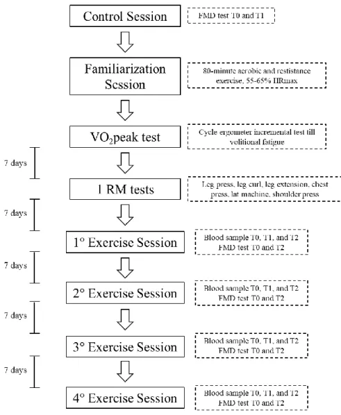
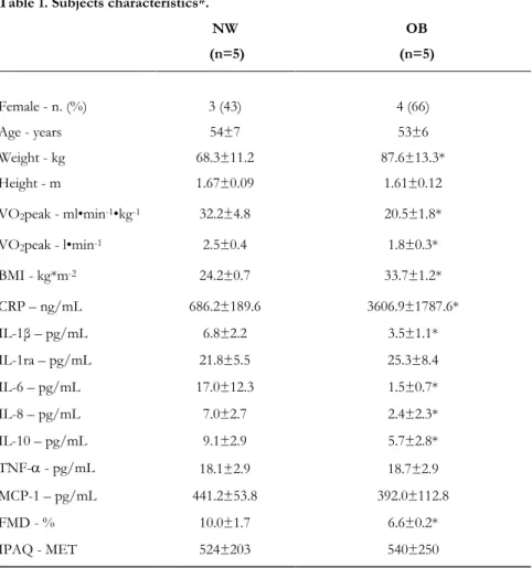
Methods. Seventy individuals who attended a structured exercise program (30/40-f/m; 75±5 years; 5±2 years of regular training) were enrolled in study 1 and tested for vascular function (flow-mediated dilation; FMD) and inflammatory profile (plasma CRP, IL-1β, IL-1ra, IL-6, IL-8, IL-10, TNF-, MCP-1). Subjects were stratified for age and BMI. Correlations between age, BMI and the measured variables were investigated. In study 2, still ongoing, 5 NW (54±7 years; 24.2±0.7 BMI) and 5 OB (53±6years; 33.7±1.2 BMI) subjects were included and tested for FMD and inflammatory profile before and after 4 different exercise sessions.
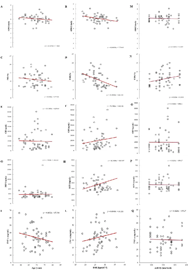
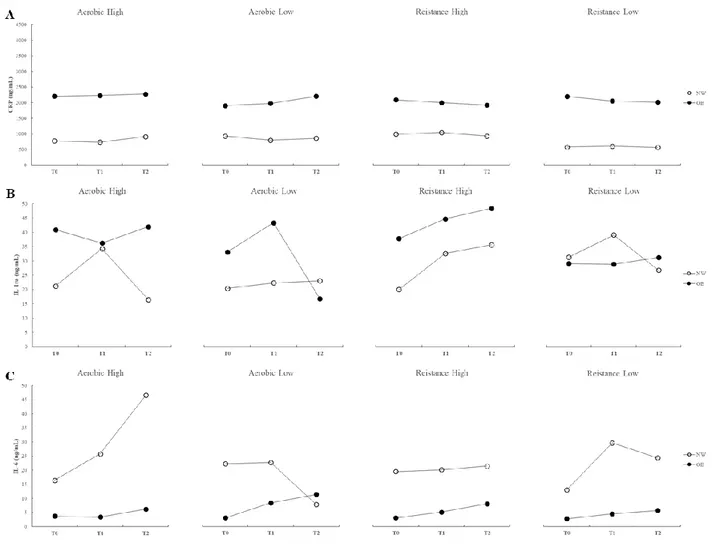
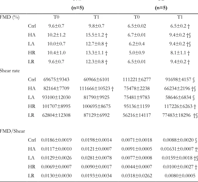
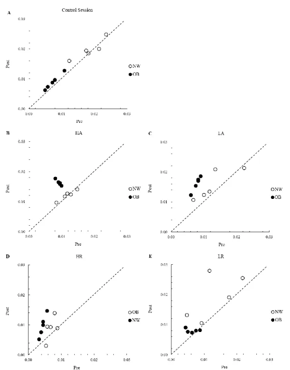
Results. In study 1 inverse correlations were found between age and IL- β (r -0.232; p<.05); IL-1ra (r -0.181; p<.05); IL-6 (r -0.255; p<.05); and IL-8 (r -0.248; p<.05). Direct correlations were found between BMI and CRP (r 0.155; p<.05), MCP-1 (r 0.217; p<.05); and TNF- (r 0.184; p<.05). An inverse correlation was also found between BMI and FMD (r -0.433; p<.01). In a preliminary analysis of data of study 2, different types and intensities of exercise seem to elicite different acute inflammatory responses in NW and OB. However, most differences did not reach statistical significance. FMD showed a significant increase in the post exercise period for all the
4
exercise sessions in both groups (p<.05) with smaller increases in OB as compared with NW. No significant differences between exercise sessions were found.
Conclusion. Sustained, regular exercise can counteract the deleterious effects of aging but not of obesity on vascular function and inflammatory profile. Preliminary results of study 2 lead us to speculate that exercise, both aerobic and resistance exercise as well as both high and low intensity, do not seem to affect adversely the inflammatory profile and the acute vascular response in either obese or non-obese individuals. However, further research is needed to confirm these findings.
5 Index Aging, Obesity and Physical Activity 1. Aging 1.1 Definition 1.2 Epidemiology 1.3 Physiological aspects 2. Obesity 2.1 Definition 2.2 Epidemiology 2.3 Pathophysiological aspects 3. Physical Activity 3.1 Definition
3.2 Epidemiology of physical (in)activity 3.4 Benefits of physical activity
3.4 General Recommendations
Inflammatory response 1. Definition
2. Physiology of inflammation 3. Causes/Inducers of inflammation
4. Most important mediators of inflammation 5. Inflammation and aging: “inflamm-aging” 6. Inflammation and obesity
6 7.1 Exercise-induced chronic adaptations
• General considerations • In Aging
• In Obesity 7.2 Exercise-induced acute response
• General considerations • In Aging • In Obesity Endothelial Function 1. Endothelium 2. Endothelial function 3. Endothelial dysfunction
4. Assessment of endothelial function 5. Endothelial function in Aging 6. Endothelial function in Obesity 7. Endothelial function and Exercise
7.1 Exercise-induced chronic adaptations • General considerations • In Aging
• In Obesity 7.2 Exercise-induced acute response
• General considerations • In Aging
7
Experimental study - Inflammation and vascular function in aging and obesity: chronic and acute exercise-induced adaptations.
Exercise-induced chronic adaptations of inflammatory profile and vascular function during aging and in obesity.
1. Hypothesis 2. Methods
• Subjects
• Blood samples and analysis • Flow-mediated dilation (FMD)
• International Physical Activity Questionnaire (IPAQ) • Supervised exercise training
• Statistical analysis 3. Results
• Characteristics of the participants • Subgroups analysis
• Correlations 4. Discussion
5. Conclusion
Acute inflammatory and vascular response to different type and intensity of exercise in obese subjects.
1. Hypothesis 2. Methods
• Subjects
• Experimental design • Control session
8 • Familiarization session • Maximal exercise testing • Blood samples analysis
• Flow-mediated dilation (FMD) • Exercise sessions • Statistical analysis 3. Results • Inflammatory response • Vascular response 4. Discussion 5. Conclusion References
9
Aging, Obesity, and Physical Activity
1. Aging
1.1 Definition
Aging is inevitable (1). By definition, aging includes a progressive physiological degeneration resulting in decline in function of organ systems and reduction of physiological reserve. It can be thought of as “primary aging” or “secondary aging”. The former refers to the inevitable deterioration of cellular structure and function, independent of the disease and environment. The latter refers to aging caused by disease and environmental factors (2).
However, aging affects all physiological processes by means of progressive deterioration involving delicate and irreversible changes in the function of most organs (3). The rapidity of the decline in function varies with the organ system under consideration but is relatively constant within a given system. Therefore, the rate of aging is the same for a 45-year-old man as it is for an 85-year-old man. The difference is that by 85 years of age more age-related changes have accumulated (3).
1.2 Epidemiology
Demographics. Over the past century, remarkable changes have been observed in the health of older persons throughout the World, and these changes have strongly affected society. The growth of the older population has resulted mostly from a general increase in overall population size but is also strongly influenced by major declines in leading causes of mortality (4). This demographic transformation resounds in society, increasing medical care and social needs, which are expected to increase sharply in the next years (5). In 1900 only 4.1% of the 76 million persons in the United States were aged 65 years or older, and among those in this age group only 3.2% were aged 85 years and older. By 1950 more than 8% of the population was aged 65 years and older, and by 2000 this percentage had increased to 12.6% (4). Change in the proportion of
10
a population that is elderly depends on changes in the survival of older persons and in the birthrate. Improved survival at older ages and a low birthrate have resulted in European countries, such as Germany and Italy, having oldest populations in the World, and almost one of the four Europeans projected to be aged 65 years or older by 2030 (6).
Mortality. Life expectancy at birth in Europe was only 47.3 years in 1900 and rose to 68.2 years by 1950, affected largely by improvements in infant and child mortality. Life expectancy continued to rise through the second half of the twentieth century, driven mainly by increases in survival in middle and old age (4). The increasingly greater life expectancy of the population has been driven in part by reduced mortality at older ages. The five leading causes of death (heart disease, cancer, stroke, chronic lower respiratory tract disease, and Alzheimer’s disease) account for 69.5% of all death (7). However, an exponential increase is present for all causes of death, and parallel increases are seen for heart disease, cerebrovascular disease, pneumonia, and influenza. Diabetes mortality rates also do not show an exponential increase with advancing age (4).
Disease status. Among people aged 65 years and older, the most common reported condition is hypertension, followed by coronary heart disease and stroke. Arthritis and chronic joint symptoms are reported by a large proportion of older persons, and these conditions have a large impact on overall health and quality of life but do not appear on the list of the most common conditions causing death (8). An important consideration of disease status that distinguishes the older population from the younger population is the high rate of co-occurrence of multiple chronic conditions, comorbidities. The concept of comorbidity is useful in considering the burden of disease in older people. However, the standardization of a definition for comorbidity depends on the number of conditions being ascertained and the intensity of the diagnostic effort to identify prevalent disease. The longer the list of conditions of prevalent diseases, the greater the prevalence of comorbidity (4).
Disability. The National Long-Term Care Survey performed assessments disability from 1982 to 2005. The findings show that the decline in disability observed for the first 12 years of the study continued and actually accelerated from 1994 to 2005 (9). Physical limitations include basic tasks such as standing, reaching, and grasping. These
11
tasks represent the building blocks of functional but are not specific measures of disability. Activities of daily living (ADLs) are basic self-care tasks. Instrumental ADL (IADLs) are tasks that are physically and cognitively somewhat more complicated and difficult than self-care tasks and are necessary for independent living in the community. ADLs and IADLs are measures of disability and reflect how an individual’s limitations interacts with the demands of the environment (10). The prevalence of ADLs disability, defining as needing help with three or more ADLs, increases with increasing age. Because disability status is a good way of representing overall health status in older persons with complex patterns of disease, and because disability also has direct implications for the long-term care needs of an older person, there has been much interest in evaluating disability trends over the time.
1.3 Physiological aspects.
In the last decades, the study of aging has expanded rapidly both in depth and width. Biological, epidemiological, and demographic data have generated a number of theories that attempt to identify a cause or process to explain aging. In recent years, the search for a single cause of aging, such as a single gene or the decline of a key body system, has been replaced by the view of aging as an extremely complex, multifactorial process (11). However, since aging is characterized by the declining ability to response to stress and by increasing homeostatic imbalance and incidence of pathology, death remains the ultimate consequence of aging (11). The biological basis of aging is incompletely understood with many theories being divided into two main categories: “programmed” theories and “damage” theories (2). According to the “programmed” theories, aging depends on biological clocks regulating the timetable of the life span through the stages of growth, development, maturity, and old age. This regulation would depend on genes sequentially switching on and off signals to the nervous, endocrine, and immune systems responsible for maintenance of homeostasis and for activation of defense responses (2). The “damage” theories identifies environmental insults to living organisms that induce progressive damage at various levels, such as mitochondrial DNA damage, oxygen radicals accumulations, cross-linking [2,11]. However, during aging several processes may interact simultaneously and may operate at many levels of functional organization. Consequently, different theories are not
12
mutually exclusive and may adequately describe some or all features of the aging process alone or in combination with other theories (11).
Below some of the physiological changes associated with the aging process on specific organ systems will be outlined
Respiratory system. The structure of the upper airway changes little with aging, however there are age-related structural lung changes including decreased elastic rebound of the lung, increased chest wall rigidity and decreases force-generation capacity of the respiratory muscles. These changes lead to a reduction in forced vital capacity, forced expiratory volume in 1 second (FEV1) and vital capacity, and an increase in functional residual capacity (FRC) [2,3]. Furthermore, arterial oxygen pressure shows a progressive decrease with aging, thus increasing the alveolar-arterial oxygen difference
(A-a)O2. Most of this decrease in arterial oxygen pressure results from a mismatch of
ventilation and perfusion that together with diminished elastic recoil of the lungs leads to a greater tendency for airways to collapse (3).
Cardiovascular system. Cardiac output decreases linearly with the aging process, about 1% per year in normal subjects or subjects free of cardiac disease. Consequently, the cardiac output of an 80-year-old subject is approximately half that a 20-year-old. This may be due to one of several factors. First, senescent cardiac muscle has decreased inotropic response to catecholamines, both endogenous and exogenous. Second, with aging there is an associated increase in diastolic and systolic myocardial stiffness, which might be due to increased interstitial fibrosis in the myocardium. Third, there is a progressive stiffening of the arteries with age leading to an increased afterload of the heart (Table 1) (3).
Another aspect to consider in the aging process is hypertension. A progressive increase in blood pressure after the first decade of life has long been regarded as normal consequences of aging and was the basis for ignoring the presence of hypertension in the elderly. The elevation with age is more pronounced for systolic than diastolic pressure (3). Aging is associated with stiffening of the large elastic arteries (i.e. aorta and carotid arteries). Enhanced pulse wave velocity, about 40-50%, and prolonged ejection augment ante grade and retrograde arterial waves, thus elevating systolic and
13
pulse pressure, cardiac work and oxygen demand (2). Left ventricular hypertension ensues as does tissue damage as a result of the increase in pulsatile flow, especially in high-flow organs, resulting in cerebrovascular events and renal impairment (2). Delayed or impaired baroreceptor response causes blood pressure lability, postural and postprandial hypotension and loss of sinus arrhythmia. In addition, with aging there is a reduction in adrenoceptor sensitivity with a reduced response to exogenous β-agonists. This baroreflex deterioration is multifactorial: reduced arteriole compliance, blunted transduction of stretch signals, altered central neural processing, altered baseline efferent autonomic outflows, and dampened end-organ responsiveness (Table 1) (2).
Furthermore, as consequences of all this processes arteriosclerosis and coronary artery disease often develop during the aging process. Indeed, thickening of the arteries walls with hyperplasia of the intima, collagenization of the media and accumulation of calcium and phosphate in elastic fibers progressively occurs with aging. Additionally, the lipid content of nonatherosclerotic portion of vessels increases, particularly of cholesterol (2).
Aging effects on major structural and functions characteristics of the cardiovascular system.
Cardiac changes Vascular changes
Heart weight ↑ Arterial wall thickness (intima-media) ↑ Cardiomyocyte dimensions ↑ Sub-endothelial collagen ↑ Cardiomyocytes number ↓ Arterial distensibility ↓ Ejection fraction = Pulse wave velocity ↑ Stroke volume = Total peripheral resistance ↑ Cardiac output = Endothelial permeability ↑ Early diastolic filling ↓ Endothelial nitric oxide release ↓ End-diastolic filling ↑ Inflammatory markers/mediators ↑
Table 1. ↓, diminished; ↑, augmented; =, unchanged (2).
Endocrine system. Increasing age results in a progressive deterioration in the number and the function of insulin-producing beta cells. As a consequence, the capacity of these cells to recognize and respond to changes in glucose concentration is impaired (3). In the elderly subjects, a greater proportion of the insulin released into the circulation in
14
response to a glucose challenge is in the form of the inactive precursor proinsulin than in younger individuals. In addition, with age there is a progressive peripheral insulin resistance. Compared with younger persons the elderly have a relative decrease in lean body mass with a relative increase in adiposity. Since little change in the total number of fat cells occurs with age, the increased adiposity appears due to an increase in fat cell size. In general, as adipocytes enlarge they turn off some insulin receptors. Consequently, even in non-obese elderly persons there is peripheral insulin resistance due to increased size of adipocytes with a relative decrease in insulin receptors. The combination of abnormal beta cell function with peripheral insulin resistance leads to increased glucose intolerance in normal aged persons (3).
Musculoskeletal system. The age-related decline in lean body mass is well known and is primarily due to loss and atrophy of muscle cells. Age-dependent changes also occurs in the innervation of muscles but the exact pathologic process is not well understood. Furthermore, degenerative joint disease occurs in 85% of persons older than 70 years of age and is a major cause of disability. It affects both, peripheral and axial skeleton and is characterized by degeneration of cartilage, subchondral bone thickening and remodeling of bone with formation of marginal spurs and subarticular bone cysts (3). Summary. Age-dependent changes occurs at structural, functional and molecular level throughout the body systems declining linearly with the aging process. These changes result in reduced physiological functional capacity, diminished cardiovascular responsiveness and decreased autonomic homeostasis.
15 2. Obesity
2.1 Definition
The word obesity (from the Latin ob-esum, meaning on account of having eaten) is a lay term which mean the same as fatness but with moderately abusive overtones. Obesity is a condition with International Classification of Disease code E66 (WHO) (12). Obesity generally is defined as excess body fat (13). However, the definition of excess is not clear-cut. Adiposity is a continues trend not marked by a clear division into normal and abnormal. Moreover, it is difficult to measure body fat directly. Consequently, obesity often is defined as excess body weight rather than as excess of fat (13). Conventionally, obesity is measured and classified by means of the body weight index (BMI) calculated as weight in kilograms divided by eight in meters squared. In adults, the BMI of 25 was approximately equivalent to the upper end of the wright range for large frame size in the 1959 Metropolitan Life tables, defined obesity as a BMI of 30 or more for men and of 28.6 or more for women (Table 2)(13).
BMI Classification <18.5 Underweight 18.5-24.9 Normal weight 25-29.9 Overweight 30-34.9 Obesity grade I 35-39.9 Obesity grade II
>40 Obesity grade III
Table 2. Classification of obesity based on the BMI
2.2 Epidemiology
In United States in 2003-2004, 32.9% of adults 20-74 years old were obese. In the early 1960s, the prevalence of obesity was 11& among men and 16% among women. The prevalence changes relatively little over the period from 1960 to 1980. However, the prevalence of obesity between 1980 and 1994 increased considerably, to about 21% in
16
men and to about 26% in women. By 2003-2004 the prevalence had increased to almost 32% in man and 34% in women (13). The prevalence of overweight and obese appears to increase with age. In 1999-2004, older adults were more likely to be obese than their younger counterparts. Among adults 20-39 years of age, 26.8% were obese. Among 40-to 59 year-old-adults 34.8% were obese, and among 60-70 year-old adults 35.2% were obese (13).
At global level, United States is not the only country experiencing increases in the prevalence of obesity. The current epidemic of obesity has been reported in several but not all regions globally. The highest rate of obesity has been reported in the Pacific Islands and the lowest rates have been reported in Asia (14). In England, the prevalence of obesity among women 25-34 years of age increased from 12% to 24% in only 9 years between 1993 and 2002. In Portugal, increases in overweight among school-age children also have been reported and less-developed countries have seen increases in obesity as well [13,14]. Differences in the prevalence of obesity between countries in Europe or between race-ethnic groups in the United States tend to be more pronounced for women than for men. However, the WHO estimates that in 2005 approximately 1.6 billion people worldwide were overweight and that at least 400 million adults were obese (14).
Health implications. Obesity is associated with increased risk of death. Studies estimated the risk of death in a prospective cohort of more than 500.00 U.S. men and women after 10 years of follow-up, and reported that, among patients who had never smoked, the risk of death is increased by 20% to 40% in overweight patients and by 2- to 3-fold in obese compared with normal- weight patients (14). Obesity is also associated with increased risk for numerous chronic diseases, including diabetes, hypertension, heart disease, and stroke and is further linked to several digestive disease, including gastroesophageal reflux disease and its complications, colorectal polyps and cancer, and liver disease [13-15].
2.3 Pathophysiological aspects
The development of obesity occurs when the caloric intake is disproportionate to the energy expended. Three metabolic factors have been reported to be predictive of
17
weight gain: a low adjusted sedentary energy expenditure, a high respiratory quotient (carbohydrate-to-fat oxidation ratio), and a low level of spontaneous physical activity (15). Several other factors are associated with being overweight, but it is not clear why or how they have an impact. Sex, age, race, and socioeconomic status have an impact on weight gain, with overweight and obesity being more likely among women, older individuals, ethnicity and those of low socioeconomic status (15).
Obesity appears to have a pivotal role in the dysregulation of cellular metabolism increasing the risk of many disorders that are associated with high mortality and morbidity including diabetes mellitus type 2, hypertension, coronary heart disease, dyslipidemia, gallbladder disease, and certain malignancies [16,17]. Moreover, excess adipocytes secrete numerous cytokines that contribute to vascular dysfunction in hypertension and dyslipidemia, as manifested by hypercholesterolemia and triglyceridemia. These conditions may contribute to significant atherosclerosis, and when associated with obesity and/or diabetes and insulin resistance, they contribute to metabolic syndrome (16).
Below some of the pathophysiological aspects associated with the obesity will be outlined.
Lipotoxicity and Insulin resistance. Excessive fat storage that creates obesity eventually leads to the release of excessive fatty acids from enhanced lipolysis, which is stimulated by the enhanced sympathetic state existing in obesity. The release of these excessive free fatty acids then incites Lipotoxicity, as lipids and their metabolites create oxidant stress to the endoplasmic reticulum and mitochondria. This affects adipose as well as nonadipose tissue, accounting for its pathophysiology in many organs, such as liver and pancreas, and in the metabolic syndrome. The free fatty acids released from excessively stored triacylglycerol deposits also inhibit lipogenesis, preventing adequate clearance of serum triacylglycerol levels that contributes to hypertriglyceridemia. Release of free fatty acids by endothelial lipoprotein lipase from increased serum triglycerides within elevated β lipoproteins causes Lipotoxicity that results in insulin-receptor dysfunction. The consequent insulin-resistant state creates hyperglycemia with compensated hepatic gluconeogenesis. The latter increases hepatic glucose
18
production, further accentuating the hyperglycemia caused by insulin resistance. Free fatty acids also decrease utilization of insulin-stimulated muscle glucose, contributing further to hyperglycemia. Lipotoxicity from excessive free fatty acids also decreases secretion of pancreas β-cell insulin, which results in β-cell exhaustion (16).
Inflammation and immune dysfunction. Adipocytes not only store triacylglycerol in fats depots in various body sites to provide energy reserve, but in aggregate constitute the largest endocrine tissue that constantly communicates with other tissues by adipocytes-released secretagogues, such as the proteohormones lectin, adiponectin, and visfatin. Visceral fat depots release inflammatory adipokines that along with free fatty acids provide the pathophysiologic basis for comorbid conditions associated with obesity. Visceral adipokines are transported by the portal vascular system to the liver, enhancing nonalcoholic steatohepatitis (NASH), and also by systemic circulation to another diverse site. Along with fatty acid Lipotoxicity, visceral adipokines also contribute to the adipokines inflammatory injury that leads to pancreatic β-cell dysfunction [17,18].
Dyslipidemia, hypertension, and atherogenesis are comorbid conditions, in addition to insulin resistance, that are associated with obesity and adversely influenced by the secretion of diverse inflammatory adipokines, particularly from the white adipose tissue in visceral fat depots. Specific adipokines enhance endothelial vasomotor tone by secreting renin, angiotensinogen, and angiotensin II, but when secreted from adipocytes, enhance hypertension in obese subjects. Tumor necrosis factor (TNF)- secretion increases in proportion to increased total body-fat mass and enhances inflammation in fatty livers and fat depots elsewhere, particularly pancreas, mesentery, and gut visceral fat. Furthermore, inflammatory markers that are increased in obesity commonly contribute to inflammatory conditions such as NASH and in the bronchial tree of patients with obstructive sleep apnea [16-18].
The progressive inflammatory state resulting from increased obesity that promotes insulin resistance also perpetuates atherogenesis throughout its development, from endothelial fatty streaks to late-plaque formation, rupture and thrombosis. Moreover, endothelial and adipose cell lipoprotein lipase activity are also decreased by inflammatory cytokines such as interleukin-6 (IL-6), so that by inhibiting lipolysis they increase serum triacylglycerol levels accentuating hyper-triglyceridemia. Furthermore,
19
as atherosclerosis progresses with macrophage and smooth-muscle cell infiltration, there is additional secretion of other cytokines, such as monocytes chemoattractant protein 1 (MCP-1), macrophage migration inhibiting factor (MMIF), and endothelin-1, that enhance the evolving inflammatory lesions of atherosclerotic plaques within the vascular wall. Progression of atherosclerotic plaque formation and remodeling of collagen results from the action of matrix metalloproteinases also secreted by adipocytes. This activity causes atheroma cap thinning and plaque rupture that precipitates release of the tissue factor, also promoting intravascular thrombosis (16). Clinical manifestation. Comorbidities result from the burden of weight and space-occupying effects of obesity. This include: diabetes mellitus type 2, endothelial dysfunction and hypertension, dyslipidemia. As explained before, these comorbidities and the effects of fatty acid Lipotoxicity culminate to promote atherogenesis, including coronary artery disease. All these disorders are adversely affected by enhanced upregulation of NF-kB from visceral white adipose tissue inflammatory adipokines. Other conditions that are linked to obesity include chronic renal disease, obstructive sleep apnea, and non-alcoholic fatty-liver disease [12,16,17].
Furthermore, obesity is a major risk factor for many forms of cancer, including breast, colon, endometrial, esophageal, hepatocellular, renal, and prostate cancer. Mechanisms of carcinogenesis or tumor growth include perturbed cellular proliferation, dedifferentiation and/or apoptosis, angiogenesis, and chronic adipokines-associated inflammation, along with effects of cancer genes and/or environmental toxins that enhance inflammation [12,17]. Other comorbidities related to obesity include joint disease, obstructive sleep apnea, asthma, pulmonary embolism, cholesterol gallstone disease, polycystic ovarian syndrome. Furthermore, obesity is a risk factor for preeclampsia and eclampsia of pregnancy. However, many of these disorders improve or even disappear with the elimination of obesity (16).
20 3. Physical Activity
3.1 Definition
Physical activity, exercise, and physical fitness are terms that describe different concepts. However, they are often confused with one another, and the terms are sometimes used interchangeably. Physical activity is defined as any bodily movement produced by skeletal muscles that results in energy expenditure. Physical activity in daily life, can be categorized into occupational, sports, conditioning, household, or other activities. Exercise, is a subset of physical activity that is planned, structured, and repetitive and has as a final or an intermediate objective the improvement or maintenance of physical fitness. Physical fitness is a set of attributes that are either health- or skill-related (17).
Physical activity is complex behavior, and may be meaningfully partitioned into other categories mutually exclusive of each other. For examples one might divide them according to intensity: light, moderate, or heavy intensity; those that are intentional or compulsory; or regularly such as: daily or weekend activities [19,20].
3.2 Epidemiology of Physical (in)Activity
Since the industrial revolution, the development of new technologies has enabled people to reduce the amount of physical labor needed to accomplish many tasks in their daily lives. As the availability of new devices has continued to increase, the effects of physical labor and human energy expenditure have grown to include many aspects of the lives of more and more people. The use of many of these technologies has been driven by the goal of increased individual worker productivity and reduces physical hardships and disabilities caused by jobs entailing continuous heavy labor. However, the human body has evolved in such a way that most of its systems (skeletal, muscle, metabolic, and cardiovascular) do not develop and function in an optimum way unless stimulated by frequent physical activity (18). Although the technological revolution has been of great benefit to many populations throughout the World, it has come at a major cost in terms of the contribution of physical inactivity to the worldwide epidemic of non-communicable diseases. In 2009, physical inactivity was identified as the fourth
21
leading risk factor for non-communicable diseases and accounted for more than 3 million preventable deaths (18).
From the WHO global health observatory repository. In 2012, the WHO obtained comparable estimates for physical inactivity in adults from 122 countries worldwide. The combined population of these 122 countries represents 88.9% of the World’s population. Physical inactivity was defined as not meeting any of three criteria: 30 min of moderate-intensity physical activity on at least 5 days week, 20 min of vigorous-moderate-intensity physical activity on at least 3 days week, or an equivalent combination achieving 600 metabolic equivalent (MET)-min per week. Results shown that worldwide, 31.1% of adults are physically inactive. The frequency of inactivity varied greatly between WHO regions: 27.5% of people are inactive in Africa, 43.4% in the Americas, 43.3% in the eastern Mediterranean, 34.8% in Europe, 17.0% in Southeast Asia, and 33.7% in the western Pacific. Women are generally more inactive than are men, 33.9% versus 27.9%. Inactivity increases with age in all WHO regions, which is a pattern known to have a strong biological basis. Moreover, physical inactivity is more common in countries of high income than in those of low income (18).
Walking is a common, accessible, inexpensive form of physical activity and is an important component of total physical activity in adult populations. It is aerobic and necessitates use of large skeletal muscles, and confers the multifarious health benefits of physical activity with few adverse effects. Interventions have been implemented to increase population levels of walking and have proven this activity’s effectiveness. Indeed, 64.1% of adults report walking for at least 10 min consecutively on 5 or more days per week. Variation between WHO regions is modest: 57.0% report such walking in Africa, 65.6% in the Americas, 66.9% in the eastern Mediterranean, 66.9% in Europe, 67.2% in southeast Asia, and 65.0% in the western Pacific. Additionally, patterns of walking hardly differ between men and women and between age groups. Another aspect of the human movement range that has received attention is sedentary behavior, which is usually defined as the time spent sitting. Overall, the proportion of adults spending 4 or more hours per day sitting is 41.5%. The value varied greatly in WHO regions: 37.8% of individuals sit for 4 or more hours per day in Africa, 5.2% in the Americas, 41.4% in the eastern Mediterranean, 64.1% in Europe, 23.8% in southeast Asia, and 39.8% in the western Pacific. For adults aged 15-59 years, the
22
proportions spending 4 hours of more sitting does not vary substantially, and both sexes are similar; for individuals aged 60 years or older, the frequency increased (18). Direct cost of inactivity to a health plan. In 2004, Garret and colleagues published an interesting study aimed to estimate the total medical expenditures attributable to physical inactivity patterns among members of a large health plan, Blue Cross Shield of Minnesota (19). This study used a cost-of-illness approach to attribute medical and pharmacy costs for specific diseases to physical inactivity in 2000. Relative risk calculated from patient data confirmed that heart disease, stroke, hypertension, type 2 diabetes, colon cancer, breast cancer, osteoporosis, depression, and anxiety are directly related to individual physical activity patterns in adults. Results of the study shown that nearly 12% of depression and anxiety and 31% of colon cancer, heart disease, osteoporosis, and stroke cases were attributable to physical inactivity. Heart disease was the most expensive outcome of physical inactivity within the health plan population, costing 35.3$ million in 2000. Total health plan expenditure attributable to physical inactivity were 83.6$ million, or 56$ per member. This study confirmed the growing body of evidence quantifying physical inactivity as a serious and expensive public health problem (19).
3.3 Benefits of Physical Activity
The benefits of regular physical activity are extensive. Indeed, physical activity reduces risk of cardiovascular disease, thromboembolic stroke, hypertension, type 2 diabetes mellitus, osteoporosis, obesity, colon cancer, breast cancer, anxiety, and depression (20). Moreover, of particular interest to older adults, there is substantial evidence that physical activity reduces risk of falls and injuries from falls, prevents of mitigates functional limitations, and is effective therapy for many chronic diseases. Additionally, clinical practice guidelines identify a significant therapeutic role of physical activity in coronary artery disease, hypertension, peripheral vascular disease, type 2 diabetes, obesity, elevated cholesterol, osteoporosis, osteoarthritis, claudication, and chronic obstructive pulmonary disease. Also, physical activity has a role in the management of depression and anxiety disorders, dementia, pain, congestive heart failure, syncope, stroke, prophylaxis of venous thromboembolism, back pain, and constipation. Other
23
evidence shown that physical activity may help in prevent or delaying cognitive impairment and disability, and improves sleep [20,24].
3.4 Recommendations
Physical activity for healthy adults and older adults. Several studies have supported a dose-response relationship between chronic physical activity levels and health outcomes; such greater benefit is associated with higher amounts of physical activity. Epidemiologic studies have estimated the volume of physical activity needed to achieve specific health benefits typically expresses as kilocalories per week, Metabolic Equivalent for Task (MET)-minute per week, or MET-hour per week. Studies of diverse populations clearly show that an energy expenditure of approximately 1000 kcal•week-1 of moderate-intensity physical activity is associated with lower rates of cardiovascular disease and premature mortality [20,25]. In the general population, this
1000 kcal•week-1 volume of physical activity is accumulated through a combination of
physical activities and exercise of varying intensities.
However, to promote and maintain health, all healthy adults aged 18-65 years need moderate-intensity aerobic physical activity for a minimum of 30 min on five days a week or vigorous-intensity aerobic activity for a minimum of 20 min on three days a week. Also, a combination of moderate-and vigorous-intensity activity can be performed to meet this recommendation. Furthermore, to promote and maintain good health and physical independence, adults will benefit from performing activities that maintain or increase muscular strength and endurance for a minimum of two days a week. It is recommended that 8-10 exercises be performed on two or more nonconsecutive days each week using the major muscle groups. To maximize strength development, a weight should be used that allows 8-12 repetitions of each exercise resulting in volitional fatigue [20,26].
Physical activity for weight loss and preventing weight regain in overweight and obese people. The clinical significance of weight maintenance and weight loss is often questioned in studies that provide marginal results. To provide context to a discussion of physical activity for weight maintenance, weight loss, or prevention or weight regain after weight loss weight maintenance was defined as a change of <2.3kg, or <3% change in
24
body weight (22). However, a primary prevention of obesity starts with maintenance of current weight, not weight reduction. The risk for weight gain may vary across time, and the need for physical activity to prevent weight gain may also vary. In general,
guidelines suggest that moderately vigorous physical activity of 150 to 250 min•week-1
with an energy equivalent of ~1200 to 2000 kcal•week-1 is sufficient to prevent a weight gain greater than 3% in most adults (22).
In order to promote weight loss, a negative energy balance generated by physical activity has to be applied, and the larger the negative energy balance, the greater the weight loss. Any increase in physical activity has the potential for weight loss, however, it seems that physical activity <150 min•week-1 results in minimal weight loss compared with greater amount. Physical activity > 150 min•week-1 results in modest weight loss of ~2-3kg, while physical activity between 225 and 420 min•week-1 results in 5-to 7.5kg weight loss (22). For weight maintenance after weight loss, some studies support the value of ~200 to 300 min•week-1 of physical activity in order to reduce weight regain. However, there are no correctly designed, adequately powered, energy balanced studies to provide evidence for the amount of physical activity to prevent weight regain (22). The American College of Sport and Medicine (ACSM) Position emphasized diet restriction and endurance exercise. Resistance training was not assigned a major role by the authors because it was believed that evidence was insufficient. Although the energy expenditure associated with resistance training is not large, resistance training may increase muscle mass which may in turn increase 24-hour energy expenditure. However, resistance training may increase loss of fat mass when combined with aerobic exercise compared to resistance training alone. Unfortunately, no evidence currently exists for prevention of weight regain after weight loss of for a dose effect for resistance training and weight loss (22).
25
Inflammatory response
1. Definition
The word inflammation come from the Latin inflammare (to set on fire) and reflects a physiological protective response which is generally tightly controlled by the body at the site of injury [1,2]. Most pathologists would agree that inflammation “represents a response of living tissue to local injury”; or “ is a basic way in which the body reacts to infection, irritation or other injury, the key feature being redness, warmth, swelling and pain”; or “the basic process whereby tissues of the body respond to injury” (24) Indeed, at present inflammation is defined by the presence of five macroscopic pathological phenomena, four of them proposed by Celsus as early as 2000 years ago. There are: swelling of the tissue, elevated tissue temperature, blood-color like redness of vascularized tissue at the inflammation site, intensive sensation of a noxious stimulus, and impaired function of the organ affected (24). All signs have been regarded as secondary to one primary pathophysiological event: enhancement of vasculature permeability as a direct consequence of tissue injury (24). This definition of inflammation recognizes what we would today known as “classical” acute inflammatory response, defining inflammation according to clinical signs and symptoms. Yet, in most cases the cellular processes and signals that underlie the cardinal signals occur at a subclinical level and not give rise to any heat, redness, swelling or pain (25).
Two centuries after Celsus, Galen was important in promoting the humoral view of inflammation. In this model, inflammation was part of the beneficial response to injury, rather than a superimposed pathology (25). Later, in 1871with the advances in microscopy and cell biology, Virchow viewed inflammation as inherently pathological giving rise to a cell based definitions of inflammation, and by the end of the 19th century it was acknowledges that changing cell populations arising from both the blood and local proliferation were a key feature of many model of inflammation (25). Finally, a prominent German biologist, Neumann, defined inflammation more loosely as a “series of local phenomena developing as the result of primary lesions to the tissues and that tend to restore their health”, defining inflammation as a necessary phase in
26
the repair response after injury (25). However, the significance of this discovery has been largely neglected, leaving considerable confusion the cause-effect relationship in pathogenesis and in the approaches to treatment of inflammation disease. In particular, it is very often noted that, although inflammation is a defensive, therefore useful process, its exaggeration or prolonged action may harm the body (24).
2. Physiology of inflammation
The innate ability of the body to defend itself is based on three elements: external barriers against invasion and tissue injuries, non-specific systems against foreign pathogens and debris, and antigen-specific responses to foreign pathogens (23). Inflammation is the body’s initial non-specific response to tissues injury produced by mechanical, chemical or microbial stimuli and it is a highly amplified controlled humoral and cellular response: the complement, kinin, coagulation and fibrinolytic cascades are triggered in tandem with activation of phagocytes and endothelial cells. There are four major events in the inflammatory process: a) vasodilation, b) increased microvascular permeability, c) cellular activation/adhesion and e) coagulation. Vasodilation and increased microvascular permeability at the site of injury increase locally available oxygen and nutrients, and produce heat, swelling and tissue edema, giving the rise to the five classical symptoms previously mentioned (23). Furthermore, the normal physiological response to stress and injury results in a series of cardiovascular changes and neuroendocrine changes, such as increases in heart rate, contractile and cardiac output, increased release of catecholamines, cortisol, antidiuretic hormone, growth hormone, glucagon, and insulin. The major metabolic change that occurs in response to inflammation is an initial increase in oxygen consumption [1,4].
3. Causes/inducers of inflammation
Inducers of inflammation can be exogenous or endogenous (26). Exogenous inducers can be classified into two groups: microbial and non-microbial. However, we will focus on the endogenous inducers which are signals produced by stressed, damaged or otherwise malfunctioning tissues. The identity and characteristics of these signals are
27
poorly defined, but they probably belong to various functional classes according to the nature and the degree of tissue anomalies in which they report. One common theme detecting acute tissue injury is the sensing of the desequestration of cells of molecules that are normally kept separate in intact cells and tissues. The desequestration of these components is afforded by the various types of compartmentalization that occur in normal tissue.
Cell-derived inducers. During necrotic cell death, for example, the integrity of the plasma membrane is disrupted, resulting in the release of certain cellular constituents such as ATP, K+ ions, uric acid, and several others. ATP binds to purinoceptors at the surface of macrophages, resulting in K+ efflux, and can cooperate with other signals to activate other inflammasomes. Also, ATP activates nociceptors reporting tissue injury to the nervous system (26).
Tissue-derived inducers. In intact tissues, epithelial cells and mesenchymal cells are normally separated from each other by the basement membrane, and the disruption of this barrier results in “unscheduled” epithelial-mesenchymal interactions. These interactions indicate the presence of tissue damage and consequently initiate tissue-repair responses (26).
Plasma-derived inducers. Damage to the vascular endothelium allows plasma proteins and platelets to gain access to extravascular spaces. A key plasma-derived regulator of inflammation, the Hageman factor, becomes activated by contact with collagen and other components of the extracellular matrix acting like a sensor of vascular damage and initiates the four proteolytic cascades that generate inflammatory mediators: the kallikrein-kinin cascade, the coagulation cascade, the fibrinolytic cascade and the complement cascade. Platelets are also activated by contact with collagen and produce various inflammatory mediators, including thromboxane and serotonin (26).
Reactive oxygen species (ROS). ROS produced by phagocytes in tissue injury also have a role inflammatory process (26). When tissues are injured by ischemia or anoxia, their ability to control the metabolism of oxygen is compromised and the species that are generated activate a superoxide-dependent chemoattractant process. This leads to an influx of leukocytes which generates still more ROS. Yet, ROS can initiate and amplify
28
the inflammatory process by upregulation of several proinflammatory cytokines (IL-2, IL-6 and TNF-) and adhesion molecules (23).
4. Most important mediators of inflammation
Cytokines are the physiological mediators of the inflammatory response. Cytokines are the physiological messenger of the inflammatory response and the principal molecules involved are tumor necrosis factor (TNF)-, interleukins, interferons and colony stimulating factors (CSFs), while monocytes/macrophages and endothelial cells are the cellular effectors of the inflammatory response (23). Leukocyte activation leads to increased leucocytes aggregation and tissues infiltration within the microcirculation where these leukocytes undergo respiratory burst, with an increase in their oxygen consumption and production of cytokines and other inflammatory mediators. Endothelial cells, exposed to this milieu of humoral and leucocytes-derived factors also become activated, and commence the expression of several adhesion molecules and receptors in their surface along with the synthesis and secretion of additional cytokines and secondary inflammatory mediators, including prostaglandins, leukotrienes, thromboxane, platelet activating factor, oxygen free radicals, nitric oxide and proteases. The presence of activated endothelial cells and the enhanced cytokines milieu results in activation of the coagulation cascades which leads to local thrombosis minimizing blood loss and the walling off of injured tissues, attempting physiologically to isolate the inflamed areas[1,4].
Of the multitude of mediators operating in the inflammatory response there are some that appears to be more influential than others, as follows:
TNF-. Several cells produce TNF-. Its expression is tightly controlled both at transcriptional and translational levels. Specific receptors for this cytokine are found on a wide variety of cells and a maximal biological response is elicited by occupancy of as few as 5% of these receptors. The systemic and tissue-specific cellular mechanisms of TNF- are dependent on its direct effect
as well as the release of other soluble mediators from host cells. TNF- elicits the release of neutrophil margination by inducing expression of adhesion molecules,
29
promoting their trans-endothelial passage and activation. It promotes differentiation of monocytes and macrophages, and induces the activation of macrophages. It stimulates the synthesis of acute-phase proteins and activates the common pathway of the coagulation and complement systems. TNF- produces a dose-dependent increase in endothelial procoagulant activity and may inhibit thrombomodulin expression at the endothelial cell surface. It induces IL-1 release from endothelial cells and macrophages, while IL-1 subsequently stimulates the biosynthesis of other cytokines (23).
IL-1. This citokine appears to be released either in parallel or in response to TNF-. IL-1 consists of two different molecules, IL-1 and IL-1β which are structurally related polypeptides. Most IL-1 remains in the cytosol in a precursor form or is associated with the cell membrane in a biologically active form. The presence of a cell-associated form of IL-1 can explain the capability of activated macrophages to induce natural killer cell cytoxicity, T cell proliferation and other functions by cellular contact in the absence of any releasable IL-1. It is a strong inducer of granulocytes, macrophages, and hepatic acute-phase protein synthesis. Excessive IL-1 release produces excessive margination of activated neutrophils into the vascular wall, stimulates endothelial cell procoagulant activity and increases leucocytes binding (23).
IL-6. IL-6 is a family of at least six differentially modified phosphoglycoproteins that are released rapidly within an hour in response to injury. IL-6 interacts synergistically with IL-1to affect thymocyte proliferation. The temporal relationship of IL-6 appearance within the cytokine cascade suggests a strong relationship to antecedent TNF- or IL-1 activity stimulation. Transcription and production are enhanced in response to TNF- and IL-1. When TNF- or IL-1 activity is attenuated, the subsequent IL-6 response is decreased (23).
IL-4 and IL-8. IL-4 synergistically increases TNF- or IL-1-induced antigen expression in endothelial cells, but inhibits the increased expression of adhesion molecules by TNF-, IL-1, or IFN-γ. IL-4 enhances lymphocytes adhesion to the endothelial cells and regulates growth and differentiation of T cells. Furthermore, it induces antigen expression on macrophages and suppress IL-8 expression from stimulated monocytes but not from stimulated fibroblasts or endothelial cells. IL-8 is produced by endothelial cells and is chemotactic for both neutrophils and lymphocytes (23).
30
IFN-γ. It promotes the release of TNF-, IL-1 and IL-6 by augmenting the effects of endotoxin on macrophages, thereby increasing the expression of adhesion molecules and cellular receptors for TNF-. It may act synergistically with TNF- to produce cytotoxic and cytostatic activity, and promotes B cell activation to increase antibody production. Also, IFN-γ enhances adhesion of lymphocytes to endothelial cells, and promotes maturation of macrophages and enhances their activity (23).
5. Inflammation and Aging: “inflamm-aging”
The term “Inflamm-aging” was invented by Franceschi and colleagues to describe the up-regulation of the inflammatory response at older ages resulting in the low-grade chronic systematic pro-inflammatory state that underline the most age-associated diseases (27). This process seems to be mediated by increased circulating levels of pro-inflammatory cytokines (primarily IL-1, IL-6, TNF-, and IL-1) and it results from the counterbalance between pro- and anti-inflammatory cytokines (Il-4, IL-6, IL-13, and IL-10) that ultimately sees an upregulation of the pro-inflammatory response [6-8]. The concept of “Inflamm-aging” determines that aging, either physiologically or pathologically, can be drive by the pro-inflammatory cytokines and other inflammatory mediators and this potentially harmful pro-inflammatory signals at a later stage of life may act antagonistically to the beneficial role they had in earlier stage of life (28). Moreover, this concept is based on an antagonistic pleiotropy theory programmed during evolution. In fact, aging is accompanied by chronic low-grade inflammation state, showed by a 2 to 4-fold increase in serum levels of inflammatory mediators which act as predictors of mortality independent of pre-existing morbidity. Low-grade inflammation has emerged as critical in the pathogenesis of several age-related chronic disease as Alzheimer’s disease, cardiovascular disease, type 2 diabetes, sarcopenia, frailty and functional disability. These diseases appear to be correlated, leading to the concept of age-related disease, which may be linked by the process of inflammation (28).
Chronic low-grade inflammation may have a rapid or slow onset but it is characterized primarily by its persistence and lack of clear resolution, occurring when the tissue are unable to overcome the effects of the harmful agent. During these chronic
31
inflammation events, immune responses, tissue injury and healing proceed simultaneously. The inflammatory side effects accumulate slowly and can lead to severe tissue deterioration without any relevant symptoms for years (29). Interestingly, the chronic inflammation state is characterized by the infiltration of various migratory inflammatory cells of macrophage, lymphocytes, and plasma cells due to sustained ROS production, implicating a major role of ROS in the inflammatory reactions (30). ROS causes both oxidative damage and elicit the release of additional inflammatory cytokines, perpetuating a vicious cycle resulting in a chronic pro-inflammatory state where pathophysiological changes, tissue injury and healing mechanisms proceed simultaneously and damage slowly accumulates asymptomatically over decades (27).
6. Inflammation and Obesity
Over the past decades, the search for a potential unifying mechanism behind the pathogenesis of obesity-related diseases has revealed a close relationship between nutrient excess and derangements in the cellular and molecular mediators of immunity and inflammation. This has given birth to the concept of “metainflammation” to describe the chronic low-grade inflammatory state of obesity [12,13]. The inflammatory response triggered by obesity involves many components of the classical inflammatory response to pathogens and include systemic increases in circulating inflammatory cytokines and acute phase proteins (i.e., C-reactive protein), recruitment of leukocytes to inflamed tissues, activation of tissues leukocytes, and generation of reparative tissue response (31).
In the recent years, it has become clear that obesity is a chronic and mild systemic inflammatory condition, and there is much evidence that chronic inflammation of White Adipose Tissue (WAT) contributes to the development of insulin resistance, as well as is at the molecular basis of diabetes (32). In the past, white adipose tissue (WAT), was considered to be simply a site for energy storage, however in the recent years it has become better understood at the molecular level (32). Indeed, WAT secretes physiologically active substances, collectively known as adipokines. Thus, WAT is now considered to be one of the tissue that play a critical role in the onset of life-style related disease (32). When adipocytes hypertrophy occurs due to excessive
32
energy intake or lack of exercise, infiltration of macrophages is observed in WAT increasing the production of pro-inflammatory adipokines, such as TNF- and monocyte chemoattractant protein-1 (MCP-1), IL-6, and leptina, and decreasing the production of anti-inflammatory adiponectin, causing WAT’ chronic inflammation (32). Specifically, changes in adipocyte and fat pad size lead to physical changes in the surrounding area and modifications of the paracrine function of the adipocyte. Therefore, adipocytes begin to secrete low levels of TNF-, which can stimulate preadipocytes to produce MCP-1 (33). At the same time, endothelial cells also secrete MCP-1 in response to cytokines and the increased secretion of leptin by adipocytes contribute to macrophage accumulation by stimulating macrophages to adipose tissue and promoting adhesion of macrophages to endothelial cells. It is conceivable also that physical damage to endothelium, could also play a role in macrophage recruitment, similar to that seen in atherosclerosis. Whatever the stimulus to recruit macrophages into adipose tissue is, once these cells are present and active, along with adipocytes ad other cell types, could perpetuate a vicious cycle of macrophage recruitment, production of inflammatory cytokines, and impairment of adipocytes function [15,16]. Furthermore, increasing adiposity activates both c-Jun N-terminal Kinase (JNK) and IKappaB kinase (IKKβ) as well as many of the typical inflammatory stimuli simultaneously activate JNK and IKKβ pathway that in addition to pro-inflammatory cytokines and ROS production play a pivotal role in the development of obesity-induced insulin resistance (34). One potential mechanisms in though the activation of NADPH oxidase by lipid accumulation in the adipocytes, which increase ROS production. This mechanism was shown to increase the production of TNF-, IL-6, and MCP-1 and decrease the production of adiponectin. Lipid accumulation also activates the unfolded protein response to increase endoplasmic reticulum stress which was shown to activate JNK to lead to serine phosphorylation of insulin receptor substrate-1 (34).
7. Inflammation and Exercise
In the past 20 years, research has demonstrated that exercise induces considerable changes in the immune system. The interaction between exercise and the immune
33
system provided a unique opportunity to evaluate the role of underlying endocrine and cytokine mechanisms. Recent studies showed that exercise provokes an increase in certain cytokines and it was demonstrated that active, but not resting, leg of human released significant amounts of IL-6 into the circulation during prolonged exercise (35). In light of that, it was theorized that IL-6 response may acts as an indicator that muscle glycogen stores are reaching critically low levels and that active muscles reliance on blood glucose as a source of energy is on the increase (36). Furthermore, recent research demonstrated that skeletal muscles might produce and express cytokines belonging to distinctly different families. Since skeletal muscle has the capacity to express cytokines, and muscle contractions play a regulatory role in the muscular expression of these cytokines, the term “myokines” was suggested (35). A bout of exercise provokes the appearance of several cytokines, including IL-6, IL-1ra, IL-8, I-10, whereas TNF- is only stimulated by very intense exercise (35).
Myokines. All cytokines and other peptides expressed, produced, and released by muscle fibers and which exert either paracrine or endocrine effects should be classified as myokines (35).
Increased circulating levels of IL-6 have been seen after prolonged exercise that seem to be independent on concomitant muscle damage. The level of IL-6 increases in an exponential fashion in response to exercise, and it declines in the post exercise period. The magnitude by which plasma IL-6 increases, is related to exercise duration, intensity, the muscle mass involved in the mechanical work, and the endurance capacity. IL-6 is most often classified as a proinflammatory cytokine, although data also suggest that IL-6 regulates acute-phase proteins are anti-inflammatory and immunosuppressive and that they may negatively regulate the acute phase response [20,21]. In response to exercise, IL-6 may be released in significant amounts from the working muscles in the circulation, where it can exert its effect in other organs in a hormone-like fashion. The exercise-induced increase in plasma IL-6 is followed by increasing levels of anti-inflammatory cytokines such as IL-1ra and IL-10 meanwhile TNF- appears to be suppressed [18,21]. Also, IL-6 gene is silent in resting muscles, but it is rapidly activated by contractions. The transcription rate is faster than reported for any other gene in muscles, and the fold increase of the transcript is massive. IL-6 production is modulated by the carbohydrate availability in skeletal muscles, suggesting
34
that IL-6 acts as an “energy sensor”. IL-6 released from contracting muscles into the circulation may enhance lipolysis and gene transcription in abdominal subcutaneous fat via its effect on adipose tissue. Lastly, muscle-derived IL6 is likely to inhibit low-grade TNF- production and thereby TNF--induced insulin resistance and it may be a player in mediating the beneficial health effects of exercise [18,21].
IL-8 is a known chemokine that attracts primarily neutrophils as well as acts as an angiogenic factor. IL-8, like IL-6, responds to exercise and its plasma concentration increases in response to exhaustive exercise such as running, which involved eccentric muscle contractions. The physiological function of IL-8 within the muscle is still unknown. The main part of the systemic increase in IL-8 as seen during exercise with eccentric component is most likely due to an inflammatory response. However, just a small and transient release of IL-8 was noted after a concentrically exercise limb, which did not result in an increase in the systemic IL-8 plasma concentration. A high local IL-8 expression take place in working muscle with only a small transient release could indicate that muscle-derived IL-8 acts locally and exerts its effects in an endocrine or paracrine fashion. A plausible function of IL-8 would be chemoattraction of neutrophils and macrophages when in concentric exercise is little or no accumulation of neutrophils or macrophages in skeletal muscle [18,22]. A more likely function of muscle-derived IL-8 is to stimulate angiogenesis. Indeed, IL-8 associated with the CXC receptor 1 and 2 induces its chemotactic effects via CXCR1, whereas CXCR2 is the receptor responsible for IL-8-induced angiogenesis (35).
IL-15 has been identified as an anabolic factor, which is highly expressed in skeletal muscle. Moreover, IL-15 seems to play a role in muscle-adipose tissue interaction. In human skeletal muscle myogenic culture, IL-15 indicates an increase in accumulation of the protein myosin heavy chain in differentiated muscle cells, suggesting that IL-15 is an anabolic factor in muscle growth and it stimulates myogenic differentiation independently of insulin-like growth factors (IGFs). Furthermore, in opposition to IGF-1, IL-15 has effects on fully differentiated myoblast (35).
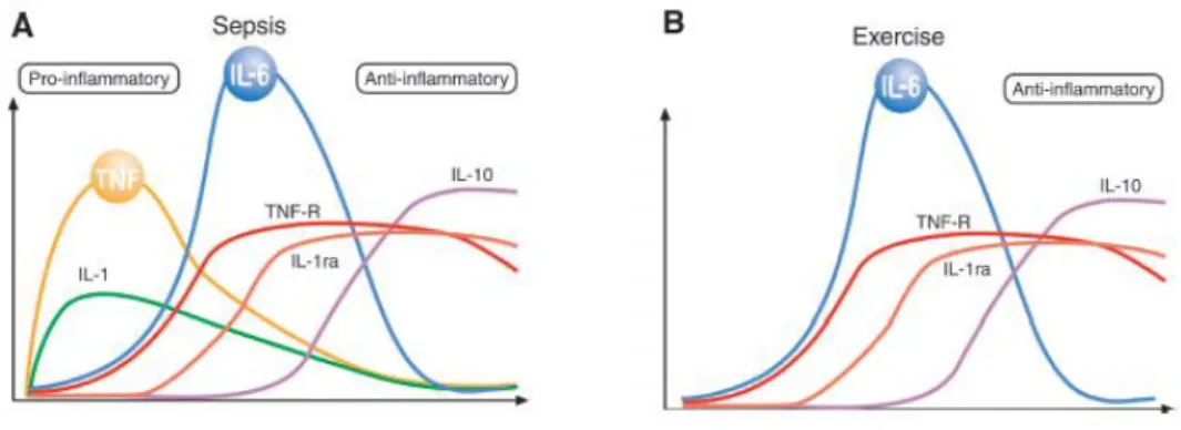
Cytokines responses to exercise. In severe infections, the cytokine cascade consist of: TNF-, IL-1β, IL-6, IL-1ra, sTNF-R, and IL-10 (Figure 1). The first two cytokines in the
35
cytokines cascade are TNF- and IL-1β, which are produced locally. These cytokines are usually referred to as proinflammatory cytokines. TNF- and IL-1 stimulate the production of IL-6. The cytokine response to exercise vary from that elicited by severe infections. Indeed, the classic pro-inflammatory cytokines, TNF- and IL-1β in general do not increase with exercise (Figure 1) (37). Typically, IL-6 is the first cytokine present in the circulation during exercise and its level increases in an exponential fashion. Furthermore, in response to exercise circulating levels of anti-inflammatory cytokines and cytokines inhibitors such as IL-1ra and TNF-R are increased. Taken together, exercise provokes an increase primarily in IL-6, followed by an increase in IL-1ra and IL-10 (37).
Figure 1. Different pattern of inflammation cascade during severe infection (A) (i.e. sepsis) and exercise (B) (37).
Anti-inflammatory effects of myokines. Data suggest that IL-6 exerts inhibitory effects on TNF- and IL-1 production. IL-6 inhibits lipopolysaccharide induced TNF- production both in cultured human monocytes and in the human monocytic line, indicating that circulating IL-6 is involved in the regulation of TNF- levels. The anti-inflammatory effects of IL-6 are also demonstrated by the fact that IL-6 stimulates the production of IL-1ra and IL-10. As well as it stimulates the release of TNF- receptors, but not IL-1β or TNF-, and appears to be primary inducer of the hepatocyte-derived acute phase proteins, many of which have anti-inflammatory properties [20,23].
IL-10 and IL-1ra in response to exercise also contribute to mediate the anti-inflammatory effects of exercise. IL-10 inhibits the production of IL-1, IL-1β, and TNF- as well as the production of chemokines, including IL-8 and macrophage