A
A
l
l
m
m
a
a
M
M
a
a
t
t
e
e
r
r
S
S
t
t
u
u
d
d
i
i
o
o
r
r
u
u
m
m
–
–
U
U
n
n
i
i
v
v
e
e
r
r
s
s
i
i
t
t
à
à
d
d
i
i
B
B
o
o
l
l
o
o
g
g
n
n
a
a
DOTTORATO DI RICERCA IN
BIOINGEGNERIA
Ciclo XXIII
Settore scientifico-disciplinare di afferenza:
ING-INF/06 BIOINGEGNERIA ELETTRONICA E INFORMATICA
TITOLO TESI
Foetal heart rate recording: analysis and
comparison of different methodologies
Presentata da: Ing. Mariano Ruffo
Coordinatore Dottorato
Relatori
Prof. Angelo Cappello
Prof. Mario Cesarelli
Prof. Paolo Bifulco
Ing. Maria Romano
INDEX
Abstract ... 6
1. INTRODUZIONE ... 9
1.1 Historical Highlights ... 9
1.2 Work task... 11
2. FOETAL AND UTERINE PHYSIOLOGY ... 17
2.1 Embryo development overview ... 17
2.1.1 Foetal heart development... 17
2.1.2 Foetal blood circulation... 20
2.1.3 Heart electrical activity... 22
2.1.4 Heart beat regulation ... 25
2.1.5 Uterus anatomy... 28
2.1.6 Uterine contractions ... 29
3. FOETAL SURVEILLANCE ... 33
3.1 Cardiotocography Measurement ... 33
3.2 Clinical relevance of cardiotocography ... 34
3.3 FHR signal... 37
3.3.1 FHR patterns... 43
3.3.2 Foetal behavioural states ... 44
3.3.3 Interpretation of EFM... 47
3.4 UC signal ... 49
3.4.1 Physiological conditions ... 49
3.4.2 Pathological conditions ... 50
3.5.1 Ultrasonographic Doppler cardiotocography... 53
3.5.2 Foetal magnetocardiography ... 55
3.5.3 Foetal phonocardiography ... 56
3.5.4 Foetal electrocardiography ... 57
4. ULTRASONOGRAPHIC DOPPLER CARDIOTOCOGRAPHY ... 59
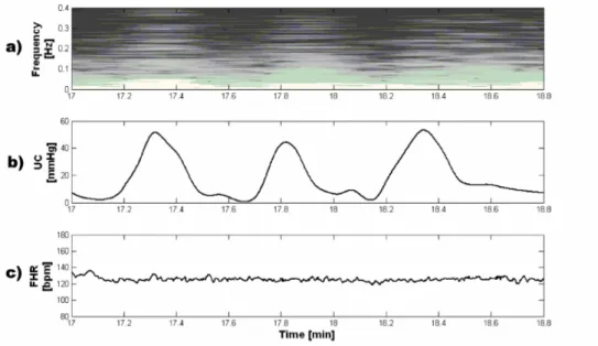
4.1 Time-frequency analysis of US-CTG signals ... 61
4.1.1 Processing (UCII)... 63
4.1.2 Time-frequency analysis results (UCII) ... 68
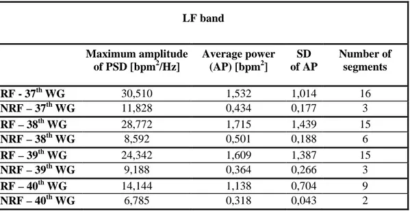
4.1.3 Foetal reactivity (CTG –TF) ... 71
4.2 PSD modifications of FHRV due to interpolation and US-CTG storage rate... 76
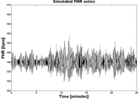
4.2.1 Simulation of FHR signals ... 78
4.2.2 Signal organization and processing ... 80
4.2.3 The Lomb Method ... 81
4.2.4 Results ... 82
5. Foetal phonocardiography... 88
5.1 FPCG signal ... 88
5.2 Time-frequency analysis of FPCG signals... 93
5.2.1 Data collection... 94
5.2.2 Pilot study... 95
5.2.3 Analysis results ... 96
5.3 Algorithm for FHR estimation... 99
5.3.1 Patients’ selection and equipment ... 100
5.3.2 Algorithm description... 100
5.4 Simulation of foetal phonocardiographic recordings... 129 5.4.1 Signal simulation ... 130 5.4.2 FHR signal simulation ... 130 5.4.3 FHS signal simulation... 132 5.4.4 Noise simulation... 134 5.4.5 Simulation results... 140
5.5 Assessment of FHR extraction algorithm ... 142
5.5.1 Statistical analysis... 144
5.5.2 Assessment results ... 145
5.6 Home care phonocardiography ... 146
6. Foetal Electrocardiography and combined FECG-FPCG monitoring ... 153
6.1 FECG signal... 154
6.2 FHR extraction from FECG ... 158
6.3 Combined FECG-FPCG monitoring... 162
7. Discussion and conclusion... 166
7.1 Analysis of US-CTG signals ... 166
7.2 PSD modifications of FHRV due to interpolation and US-CTG storage rate... 171
7.3 The algorithm for FHR estimation ... 173
7.4 FPCG simulation and assessment of the FHR extraction algorithm ... 176
7.5 Foetal home monitoring experience ... 177
7.6 Combined FECG-FPCG monitoring... 178
7.7 Further considerations and future developments ... 180
Abstract
Monitoring foetal health is a very important task in clinical practice to appropriately plan pregnancy management and delivery. In the third trimester of pregnancy, ultrasound cardiotocography is the most employed diagnostic technique: foetal heart rate and uterine contractions signals are simultaneously recorded and analysed in order to ascertain foetal health.
Because ultrasound cardiotocography interpretation still lacks of complete reliability, new parameters and methods of interpretation, or alternative methodologies, are necessary to further support physicians’ decisions.
To this aim, in this thesis, foetal phonocardiography and electrocardiography are considered as different techniques.
Further, variability of foetal heart rate is thoroughly studied. Frequency components and their modifications can be analysed by applying a time-frequency approach, for a distinct understanding of the spectral components and their change over time related to foetal reactions to internal and external stimuli (such as uterine contractions). Such modifications of the power spectrum can be a sign of autonomic nervous system reactions and therefore
However, some limits of ultrasonic cardiotocography still remain, such as in long-term foetal surveillance, which is often recommendable mainly in risky pregnancies. In these cases, the fully non-invasive acoustic recording, foetal phonocardiography, through maternal abdomen, represents a valuable alternative to the ultrasonic cardiotocography. Unfortunately, the so recorded foetal heart sound signal is heavily loaded by noise, thus the determination of the foetal heart rate raises serious signal processing issues. A new algorithm for foetal heart rate estimation from foetal phonocardiographic recordings is presented in this thesis.
Different filtering and enhancement techniques, to enhance the first foetal heart sounds, were applied, so that different signal processing techniques were implemented, evaluated and compared, by identifying the strategy characterized on average by the best results. In particular, phonocardiographic signals were recorded simultaneously to ultrasonic cardiotocographic signals in order to compare the two foetal heart rate series (the one estimated by the developed algorithm and the other provided by cardiotocographic device). The algorithm performances were tested on phonocardiographic signals recorded on pregnant women, showing reliable foetal heart rate signals, very close to the ultrasound cardiotocographic recordings, considered as reference.
The algorithm was also tested by using a foetal phonocardiographic recording simulator developed and presented in this research thesis. The target was to provide a software for simulating recordings relative to different foetal conditions and recordings situations and to use it as a test tool for comparing and assessing different foetal heart rate extraction algorithms.
Since there are few studies about foetal heart sounds time characteristics and frequency content and the available literature is poor and not rigorous in this area, a data collection pilot study was also conducted with the purpose of specifically characterising both foetal and maternal heart sounds.
Finally, in this thesis, the use of foetal phonocardiographic and electrocardiographic methodology and their combination, are presented in order to detect foetal heart rate and other functioning anomalies. The developed methodologies, suitable for longer-term assessment, were able to detect heart beat events correctly, such as first and second heart sounds and QRS waves. The detection of such events provides reliable measures of foetal heart rate, potentially information about measurement of the systolic time intervals and foetus circulatory impedance.
1.
INTRODUZIONE
1.1
Historical Highlights
It is of great interest to obtain clinical information about foetal health during pregnancy and labour. Auscultation of the foetal heart by applying the ear to the pregnant woman's abdomen it is an old practice that was reinforced after the invention of the stethoscope in the early 19th century, increasing its diagnostic capability. Stethoscopic auscultation of the foetal heart developed throughout the century, as its potential to recognise foetal well-being was realised. Interest grew in how to recognise changes in Foetal Heart Rate (FHR) that might predict and prevent foetal distress and/or intrapartum foetal death through obstetric intervention. Pinard's version of the foetal stethoscope appeared in 1876. Criteria for the normal FHR set in the latter part of the 19th century remained virtually unchanged until the 1950's. During the same period, interest and research increased into the significance of meconium staining of the amniotic fluid as a means of predicting foetal well-being. By the beginning of the 20th century, auscultation of the foetal heart was an established practice.
Advances in the techniques of auscultation were limited, mainly by inability to detect subtle changes or provide continuous surveillance, until the advent of audiovisual technologies in the early 20th century. These promised the possibility of a continuous form
of monitoring. Early electrocardiographic techniques were limited by their inability to sufficiently eliminate maternal ECG signal. This problem was addressed by the use of the foetal scalp electrode in 1960.
A considerable advance in technology with which to detect the foetal heartbeat came in 1964 when the Doppler principle was applied. In 1968, the first commercially available Electronic Foetal Monitor (EFM) applied Doppler's principle of a distinct change in frequency when a Ultra Sonique (US) waveform is reflected from a moving surface. Subsequently, the use of EFM increased rapidly.
The monitoring of foetal scalp blood acid-base was developed in Germany in the 1960s and was introduced clinically as an adjunct to continuous electronic foetal heart-rate monitoring to increase its specificity.
Medical and socio-economic advances transformed maternal birth outcomes in the 19th and 20th centuries. While the original aim of intrapartum EFM was to prevent harm, it was introduced on to the labour wards in the 1950s with the emphasis on improving foetal birth outcomes by detecting foetal hypoxia, before it led to death or disability. Like intermittent auscultation in the 19th century, continuous EFM was introduced clinically before its effectiveness had been fully evaluated scientifically.
A number of retrospective observational studies published in 1972-76 reported a decrease in perinatal mortality in those women who had continuous EFM as opposed to those who had selective EFM or no EFM at all [1]. While these studies were encouraging, the methodological biases of observational studies (they may overestimate the true effects of a
1.2
Work task
Foetal diagnostics is very important in clinical practice to appropriately plan pregnancy management and delivery. Unlikely, non-invasive monitoring of the foetus well-being is not an easy task to achieve and some limits in the most diffused pre-natal diagnostic techniques, still remain.
In this thesis, new parameters or methods of interpretation and alternative methodologies for non-invasive foetal monitoring are presented in order to further support physicians’ decisions. In particular, different methodologies of foetal heart rate monitoring were analysed and compared: ultrasonographic Doppler cardiotocography (US-CTG), phonocardiography (PCG) and electrocardiography (ECG).
Concerning Doppler cardiotocography, US-CTG data provide physicians relevant information about foetal development and permit to assess conditions such as foetal distress. Hence, an incorrect evaluation of the foetal status can be of course very dangerous. However, although US-CTG is the most spread diagnostic technique in clinical environments, mainly in the third trimester of pregnancy, its interpretation still lacks of complete reliability so that new methods for a complete and certain assessment of foetal well-being could be very helpful.
To this aim, indexes related to variability of foetal heart rate (FHRV) are particularly advocated in bibliography as useful, since, already in adult subjects, FHRV is acknowledged as an important parameter for the evaluation of healthy conditions. Thus, time dependent spectrum analysis, from US-CTG recordings, of the FHRV frequency components and their modifications were carried on by applying a time-frequency
approach, in order to better identify the spectral components related to foetal reactions to internal and external stimuli and their change over time. Being UC strong stimuli for the foetus and his autonomic nervous system (ANS), the FHRV response to UC was analyzed and characterized. Such modifications of the FHRV power spectrum can be a sign of ANS reaction and therefore represent additional, objective information about foetal reactivity and health during labour.
However, it is important to underline that since foetal heart rate is intrinsically an uneven series, in order to produce an evenly sampled series a zero-order, linear or cubic spline interpolation are usually employed in some cardiotocographic devices on the market. This is not suitable for frequency analyses because interpolation process can produce alterations of the power spectral density that, for example, affects the estimation of the sympatho-vagal balance (computed as low-frequency/high-frequency ratio), which represents an important clinical parameter. In order to estimate the frequency spectrum alterations of the foetal heart rate variability signal due to interpolation and cardiotocographic storage rates, the Lomb method, as suggested by other authors to study the uneven heart rate series in adults, was used to prove an overestimation due to the interpolation process and to the storage rate.
This thesis aim was also to analyse a valuable alternative to traditional US-CTG: the phonocardiography (FPCG), a passive and low cost recording of foetal heart sounds. Firstly an analysis of FPCG mean characteristics in the time and frequency domains is presented. In particular, a data collection pilot study was conducted with the purpose of
available literature is not rigorous in this area. Those data were useful to understand better FPCG signals and to obtain precious information for software developing.
After, a simulating software of FPCG signals relative to different foetal states (physiological and pathological) and recording conditions (for example different kinds and levels of noise) and a new algorithm for FHR estimation from FPCG recordings are here presented. Both software were developed utilising information obtained by means of the data collection pilot study.
The described simulating software can be useful as a teaching tool for demonstration to medical students and others but also for testing and assessment of foetal heart rate extraction algorithms from foetal phonocardiographic recordings.
Concerning the developed algorithm for FHR estimation, owing to the extremely noisy nature of FPCG signals, different filtering, signal enhancement techniques and logic blocks for reliable detection of heartbeats were applied, so that different signal processing paths were implemented. The performances of the different paths were tested comparing the estimated FHR signals with those of simultaneously recorded US-CTG (currently considered reference technique), in order to identify the most reliable path. Beside, the FPCG simulator software was used to test the algorithm developed in order to estimate its performance in cases of different signal to noise recordings.
Inside the FPCG monitoring described in this thesis, an Italian experience of foetal home monitoring is presented to show the advantages of this methodology (such as the reduction of the need of travel for patients and consequently of their stress). It is based on a telemedicine system (consisting of phonocardiograph home monitors able totransfer data by GPRS to a remote server where gynaecologists can consult patient recordings) that
increases the possibility of foetal long-term home surveillance which in turn could raise the efficiency of the service offered to pregnant women.
Finally, the use of foetal phonocardiographic and electrocardiographic methodologies and their combination was described in this thesis, in order to detect the FHR and other functioning anomalies. Software processing methodologies, suitable for longer-term assessment, were presented to detect heart beat events, such as first and second heart sounds and QRS waves, which provide reliable measures of heart rate, potentially information about measurement of the systolic time intervals and foetus circulatory impedance.
The research activities, described in this thesis, have produced several scientific results published on scientific journal and book chapters or presented at national and international congresses. Below the list of all the publications is reported:
-Papers published on scientific journal:
• M. Ruffo, M. Cesarelli, M. Romano, P. Bifulco, A. Fratini. An algorithm for FHR estimation from foetal phonocardiographic signals. Biomedical Signal Processing and Control, Vol. 5, Issue 2, April 2010, pp. 131-141
• M. Romano, M. Cesarelli, P. Bifulco, M. Ruffo, A. Fratini, G. Pasquariello: Time-frequency analysis of CTG signals. Current Development in Theory and Application of Wavelets, Vol. 3, Issue 2, August 2009, pp. 169 - 192
Monitoring of Athletes. Medical Devices: Evidence and Research, Vol. 2010:3, July 2010, pp. 1 – 9
• M. Cesarelli, M. Romano, M. Ruffo, P. Bifulco, G. Pasquariello; Foetal heart rate variability frequency characteristics with respect to uterine contractions. Journal of Biomedical Science and Engineering, Vol. 3, Issue 10, Oct. 2010, pp. 1014 – 1021
• M. Cesarelli, M. Romano, M. Ruffo, P. Bifulco, G. Pasquariello, A. Fratini; PSD modifications of FHRV due to interpolation and CTG storage rate. In press by Biomedical Signal Processing and Control (Available online 18 November 2010) - Abstract of papers presented at National and International congresses:
• M. Ruffo, M. Romano, M. Cesarelli, P. Bifulco: Extraction of heart rate from fetal phonocardiographic signals. Primo Congresso Nazionale di Bioingegneria – GNB-2008
• M. Cesarelli, M. Ruffo, M. Romano, P. Bifulco, F. Kovacs, S. Iaccarino: An Algorithm for fetal heart rate extraction from maternal abdomen sounds. 4th European Congress For Medical and Biomedical Engineering 2008, November 23-27, Antwerp – MBEC 2008
• M. Ruffo, M. Cesarelli, M. Romano, P. Bifulco, A. Fratini, G. Pasquariello, S. Iaccarino: Comparison of software developed for FHR extraction from PCG signals. Medical Physics and Biomedical Engineering World Congress 2009, September 7-12, Munich
• M. Cesarelli, M. Romano, M. Ruffo, P. Bifulco, G. Pasquariello, A. Fratini: PSD modifications of FHRV due to CTG storage rate. 9th International Conference on
Information Technology and Applications in Biomedicine- ITAB 2009, November 5-7, Larnaca, Cyprus
• M. Cesarelli, M. Romano, M. Ruffo, P. Bifulco, M. Iaccarino, S. Iaccarino: Home care phonocardiography: an Italian experience. 9th International Conference on Information Technology and Applications in Biomedicine- ITAB 2009, November 5-7, Larnaca, Cyprus
• M. Ruffo, M. Cesarelli, M. Romano, P. Bifulco: Testing a FHR extraction algorithm by means of a simulating software of foetal phonocardiographic recordings. Secondo Congresso Nazionale di Bioingegneria – GNB-2010
• G. Donadono , M. Ruffo, M. Romano, P. Bifulco, M. Cesarelli, D. Gargiulo, A. Fratini: Recording of fetal heart sound using a PVDF piezoelectric film sensor. Secondo Congresso Nazionale di Bioingegneria – GNB-2010
• M. Ruffo, M. Cesarelli, M. Romano, P. Bifulco, A. Fratini; A simulating software of fetal phonocardiographic signals. The 10th IEEE International Conference on Information Technology and Applications in Biomedicine, ITAB 2010, Corfu, Greece, November 2-5, 2010
- Book chapters
• M. Ruffo, M. Cesarelli, C. Jin, G. Gargiulo, A. McEwan, C. Sullivan, P. Bifulco, M. Romano, R. W. Shephard, A. van Schaik: Non invasive foetal monitoring with a combined ECG - PCG system. “Biomedical Engineering, Trends, Researches and
2.
FOETAL AND UTERINE PHYSIOLOGY
2.1
Embryo development overview
2.1.1 Foetal heart development
Cardiovascular system very early reaches an adequate functional state in order to support a complex organism like embryo. The primordium of the heart forms in the cardiogenic plate located at the cranial end of the embryo. The most critical period of foetal heart development is between three and seven weeks after fertilization, when a simple heart tube assumes the shape of a four-chambered heart. In fact, the heart actually begins beating by the 22nd day of life (or the fifth week of gestation). In more detail, in the human embryo, the mesodermal germ layer gives rise to the entire cardiovascular system (heart, blood vessels and blood cells). The heart develops from two simple epithelial tubes which fuse to form a single chambered heart that is efficiently pumping blood by the fourth week of embryonic development.
Twenty-three days following conception, the single, simple epithelial heart tube lies within the embryo's pericardial cavity. At this time there are three cell layers present within the
heart tube. The inner layer, known as cardiac jelly, is a structureless mass of cells which contain very few nuclei. The second and third layers are known as the cardiac mantle and will eventually give rise to the epicardium and myocardium.
The heart tube contains three specific areas: the cranial portion, the caudal portion and the bulbus cordis. As development progresses, the cranial dilates to form the aortic sac which will give rise to the aortic arches. The caudal also dilates to form the early embryonic ventricle. The remaining mid-portion forms the bulbus cordis which has three distinct areas of development. Two of these areas give rise to the body of the right ventricle, the aortic root and the parts of the ascending aorta. The remaining portion connects the primitive right ventricle to the truncus arteriosus. In this phase, the two atria are partly separate but there is just one big ventricle. As the heart tube grows and becomes longer it usually bends to the right, because of its need space. Rightward bending is responsible for the initial positioning of the primitive ventricle.
The cardiac jelly now acts as a valve for movement of blood from the atrial end of the heart tube to the distal end. By the 24th day of gestation, the primitive ventricles have expanded and the cardiac jelly contains trabecula or supporting structures. As growth quickly continues, the conotruncal region (area distal to the primitive right ventricle) moves centrally with torsion and twisting, giving rise to the anatomical curve of the aorta and the pulmonary artery.
Between 4th and 5th week, external form of the heart is established and interatrial and interventricular septi form. By the end of the 8th week partitioning is completed and the
It is nice to observe that during the foetal heart's developmental stages, the heart actually takes on several distinct appearances. These heart structures resemble other animal hearts. During the first phase, the tube-like heart is much like a fish heart. The second phase, with two chambers, resembles a frog heart. The three-chambered phase is similar to a snake or turtle heart. The final four-chambered heart structure distinguishes the human heart.
Epithelial tubes Specific areas of the heart tube
Atria are separate but there is one ventricle
All parts of the heart are recognisable Aorta has its anatomical curve
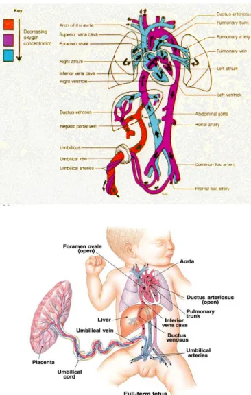
2.1.2 Foetal blood circulation
The foetal circulation is one of the first organ systems to need to be able to function properly in order to sustain the foetus. Before a circulatory system has developed, nutrients and oxygen diffuse through the extraembryonic coelem and yolk sac from the placenta. As the embryo increases in size, its nutrient needs increase and the amount of tissue easily reached by diffusion decreases. Hence the circulation must develop quickly and accurately. However, throughout the foetal stage of development, the maternal blood supplies the foetus with O2 and nutrients and carries away its wastes. These substances diffuse between the maternal and foetal blood through the placental membrane.
In the foetal circulatory system, the umbilical vein transports blood rich in O2 and nutrients from the placenta to the foetal body. The umbilical vein enters the body through the umbilical ring and travels along the anterior abdominal wall to the liver. About 1/2 the blood it carries passes into the liver. The other 1/2 of the blood enters a vessel called the ductus venosus of Aranzio which bypasses the liver. The ductus venosus travels a short distance and joins the inferior vena cava. There, the oxygenated blood from the placenta is mixed with the deoxygenated blood from the lower parts of the body. This mixture continues through the vena cava to the right atrium. In the adult heart, blood flows from the right atrium to the right ventricle then through the pulmonary arteries to the lungs. In the fetus the lungs are non-functional and the blood largely bypasses them. As the blood from the inferior vena cava enters the right atrium, a large proportion of it is shunted directly
the right atrium, including a large proportion of the deoxygenated blood entering from the superior vena cava, passes into the right ventricle and out through the pulmonary trunk. Only a small volume of blood enters the pulmonary circuit, because the lungs are collapsed, and their blood vessels have a high resistance to flow. Enough blood reaches the lung tissue to sustain them. Most of the blood in the pulmonary trunk bypasses the lungs by entering a foetal vessel called the ductus arteriosus of Botallo which connects the pulmonary trunk to the descending portion of the aortic arch. The more highly oxygenated blood that enters the left atrium through the foramen ovale is mixed with a small amount of deoxygenated blood returning from the pulmonary veins. This mixture moves into the left ventricle and is pumped into the aorta. Some of it reaches the myocardium through the coronary arteries and some reaches the brain through the carotid arteries. The blood carried by the descending aorta is partially oxygenated and partially deoxygenated. Some of it is carries into the branches of the aorta that lead to various parts of the lower regions of the body. The rest passes into the umbilical arteries, which branch from the internal iliac arteries and lead to the placenta. There the blood is reoxygenated.
Both ductus venosus of Aranzio and ductus arteriosus of Botallo are completely closed after birth.
It is worth mentioning that the concentration of haemoglobin in foetal blood is about 50 % greater than in maternal blood. Foetal haemoglobin is slightly different chemically and has a greater affinity for O2 than maternal haemoglobin. This is a sort of safety mechanism; in fact because of this characteristic, foetus can overcome relatively short lacks of oxygen.
Figure 2: on the top and on the bottom, pictures of foetal blood circulation
2.1.3 Heart electrical activity
elongated mass of specialized muscle tissue just beneath the epicardium. Fibres are continuous with those of the atrial muscle fibres. Membranes of the nodal cells are in contact with each other and have the ability to excite themselves. Without being stimulated by nerve fibres or any other outside agents, the nodal cells initiate impulses that spread into the surrounding myocardium and stimulate the cardiac muscle fibres to contract; this activity is rhythmic. As a cardiac impulse travels from the S-A node into the atrial myocardium, the right and left atria contract almost simultaneously. Cardiac impulses pass along fibres to the atrioventricular node (A-V node), which is located in the floor of the right atrium just beneath the endocardium. Fibres that conduct the cardiac impulse into the A-V node have very small diameters and conduct impulses slowly and cause the impulse to be delayed. Impulse is delayed still more as it travels through the A-V node. This delay allows time for the atria to empty and the ventricles to fill with blood. Impulse now passes into a group of large fibres that make up the A-V bundle (Bundle of His) and the impulse moves rapidly through them. A-V bundle divides into the right and left bundle branches. About 1/2 way down, the branches give rise to enlarged Purkinje fibres. Purkinje fibres spread from the interventricular septum into the papillary muscles and then continue downward to the apex of the heart (see Figure 3: electrical conduction system of the heart). Stimulated by the impulses on the Purkinje fibres, the ventricular walls contract with a twisting motion which squeezes blood out of the ventricular chambers and forces it into arteries.
The heart can be viewed as two separate pumps represented by the right and left halves of the heart. Each pump consists of a primer pump (the atrium) and a power pump (the ventricle). Both atrial primer pumps complete the filling of the ventricles with blood and both ventricular power pumps produce the major force that causes blood to flow through
the pulmonary and systemic arteries. The cardiac cycle refers to the repetitive pumping process that begins with the onset of cardiac muscle contraction and ends with the beginning of the next contraction. The duration of the cardiac cycle varies among people and also varies during an individual's lifetime. In an adult subject, the normal cardiac cycle (0.7-0.8 sec.) depends on two factors: capability of cardiac muscle to contract and functional integrity of the conducting system. Abnormalities of cardiac muscle, the valves, or the conducting system of the heart may alter the cardiac cycle and compromise the pumping effectiveness of the heart.
The described impulse transmission through the conduction system generates electrical currents that can be detected on the body's surface. In a typical ECG record, three clearly recognisable waves accompany each cardiac cycle.
P-Wave (small upward wave) which indicates atrial depolarization, which is the spread of the impulse from the S-A node through the two atria. A fraction of a second after the P-wave begins, the atria contract.
QRS-Wave or Complex. It begins as a downward deflection, continues as a large, upright, triangular wave and ends as a downward wave at its base. Represents ventricular depolarization, the spread of the electrical impulse through the ventricles. Shortly after the QRS wave begins, the ventricles undergo contraction. T-Wave (dome-shaped upward wave) indicates ventricular repolarization.
Figure 3: electrical conduction system of the heart
Through labour and delivery, we can invasively record foetal heart electrical activity by means of direct scalp foetal ECG, attaching electrodes to the presenting part of the foetus after membrane rupture. Otherwise, after 16th week’s gestation, we can adopt the external abdominal ECG, putting electrodes on the maternal abdomen. With this technique, there is an overlap between foetal (typical amplitude ranges 100-600 µV [2] ) and maternal (whose amplitude is greater of 1 or 2 orders) ECG, so a subtraction algorithm is necessary.
2.1.4 Heart beat regulation
Venous return is the amount of blood that returns to the heart during each cardiac cycle. It determines an intrinsic regulation of the heart beat. An increase in venous return causes an increase in cardiac output and stretches the S-A node, so the heart rate increases. However, foetal heart rate variability is also intimately related to foetal central nervous system;
particularly, the most important mechanism immediately involved in producing heart rate variability is the autonomic innervations of the heart Errore. L'origine riferimento non è stata trovata.. The cardioregulatory centre in the medulla oblongata regulates the parasympathetic and sympathetic nervous control of the heart.
Parasympathetic stimulation is supplied by the cardiac branches of the vagus nerve. It is of primary importance in producing beat-to-beat variability Errore. L'origine riferimento non è stata trovata.. It decreases heart rate and can cause a small decrease in the force of contraction (stroke volume). This component of cardiac innervations is well suited to a role of fine tuning the heart rate on a beat-to-beat basis because of the very rapid decrease in heart rate which occurs whit vagal nerve stimulation, and the nearly equally rapid recovery after the end of a series of impulses. Moreover, postganglionic neurones secrete acetylcholine which increases membrane permeability to K+, producing hyperpolarization of the membrane.
Sympathetic stimulation is supplied by the cardiac nerves which are projections of the cervical sympathetic chain ganglia (spinal nerve). Sympathetic stimulation increases heart rate and force of contraction (stroke volume). Changes in heart rate with stimulation of cardiac sympathetic innervation are slower compared to stimulation of cardiac vagal innervations. Moreover, it dilates vessels in skeletal and cardiac muscle.
Other specific control mechanisms play an important role in heart beat regulation, such as the effect of blood pressure, pH, CO2, O2, extracellular ion concentration and body temperature.
indirectly blood pressure. In response to an increase in blood pressure, the baroreceptor reflexes decrease sympathetic stimulation and increase parasympathetic stimulation (see
Figure 4).
Figure 4: on the left cardiocirculatory regulation in case of pressure increase and on the right description of the feed-back related to blood pressure
Chemoreceptors in the brain, aortic arc and carotids monitor blood CO2, O2, and pH levels. In response to increased CO2, decreased pH, or decreased O2, autonomic nervous system reflexes increase sympathetic stimulation and decrease parasympathetic stimulation.
An increase or decrease in extracellular K+ decreases heart rate. Increased extracellular Ca2+ increases the force of contraction of the heart and decreases the heart rate. Decreased Ca2+ levels produce the opposite effect.
Finally, heart rate increases when body temperature increases and decreases when body temperature decreases.
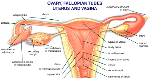
2.1.5 Uterus anatomy
The human uterus is a massive, hollow, pear-shaped organ with a thick wall, situated deeply in the pelvic cavity between bladder and rectum. It is composed of two distinct anatomic regions: the cervix and the corpus. The corpus is further divided into the lower uterine segment and the fundus. The cervix is a narrow cylindrical passage which connects at its lower end with the vagina. At its upper end, the cervix widens to form the lower uterine segment (isthmus); the lower uterine segment in turn widens into the uterine fundus. The corpus is the body of the uterus which changes in size and structure during pregnancy to accommodate itself to the needs of the growing embryo. Extending from the top of the uterus on either side are the fallopian tubes (oviducts); these tubes are continuous with the uterine cavity and allow the passage of an ova (egg) from the ovaries to the uterus where the egg may implant if fertilized.
Spatial organisation of the smooth muscle fibres in the uterine wall is complicated and still remains the matter of debate. The thick wall of the uterus is formed of three layers: endometrium, myometrium, and serosa or perimetrium. The endometrium (uterine mucosa) is the innermost layer that lines the cavity of the uterus. Throughout the menstrual cycle, the endometrium grows progressively thicker with a rich blood supply to prepare the uterus for potential implantation of an embryo. In the absence of implantation, a portion of this layer is shed during menstruation. The myometrium is the middle and thickest layer of the uterus and is composed of smooth (involuntary) muscle. The myometrium contracts during
contiguous with extrauterine connective tissue structures such as ligaments that give mechanical support to the uterus within the pelvic cavity.
Non-pregnant uterine size and position varies with age and number of pregnancies.
Figure 5: uterus or womb
2.1.6 Uterine contractions
Uterine wall structure is aimed to effective expulsion of foetus if pregnancy is about to terminate. Although biological mechanisms prevent massive contractions of uterus during pregnancy, the uterine wall never remains quiet. Every single muscle fibre possesses the possibility to change its membrane potential slowly, which results in depolarisation. Working potential it generates may be transmitted to other cells in a close neighbourhood,
but the area it can spread on strongly depends on local properties of signal propagation. In course of a physiological pregnancy the intercellular communication is poorly developed, which seems to be a mechanism of a foetus’ safety. This leads to the lack of coordination between muscle fibres which produces a kind of fibrillation of uterine wall with almost no significance rise of pressure inside its cavity. In a full term pregnancy, or in some pathological circumstances even sooner, uterine wall becomes well coordinated and uterine contractions frequent, intense, persistent and painful. Low resistance intercellular connections – gap junctions appear in a smooth muscle tissue enhancing trigger wave propagation. Even though there is no specialised trigger wave conducting system in uterus, gap junctions enable it to contract as a whole, presenting a specific pattern of contraction. The certain degree of synchronisation of smooth muscle cells amplifies uterine working potentials, since their appearance results from spatial and temporal summation of electrical activity of single fibres [4].
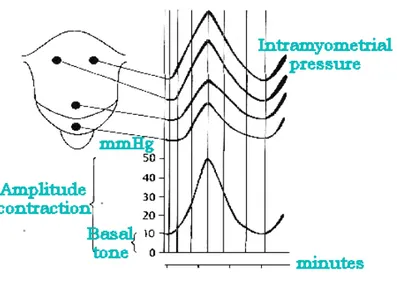
In spite of the fact that the uterine contractility is predominantly commanded by hormonal and biochemical factors (estrogens, oxytocin, prostaglandins), there are indications that the sympathetic and parasympathetic innervation of the uterus may also have a considerable influence upon it. Independently of the majority of the uterine contractions being endocrinally and biochemically triggered, the myometrial contractile activity exhibits a very peculiar characteristic that seems to demonstrate the existence of a precise nervous coordination: it is the "triple descending gradient." This gradient gives the uterine contractility its typical expulsive pattern.
on the left. This author describes the "triple descending gradient" with the following characteristics (seeFigure 1 figure 6):
1) the propagation of the contractile wave along the uterus has a descending direction; 2) the systolic phase of the contraction lasts more at the uterine fundus and less at the inferior parts of the organ;
3) the contractions are stronger in the upper parts of the uterus than in the lower ones. It seems somewhat difficult to explain this so precise and symmetric coordination of the myometrium contractile waves exclusively by means of the hormonal mechanisms that trigger them. Even the double spiral arrangement of most myometrial fibres throughout the uterus does not seem to be, only by itself, capable of entirely explaining the "triple descending gradient" - regardless of being an essential condition for its occurrence. Several facts suggest that the neurovegetative system may have some coordinating activity upon the uterine contractions.
Concerning their characteristics, uterine contractions become very rhythmic and regular in shape during labour, when the hypophysis releases a large dose of oxytocin. The contraction length ranges between 15 and 20 seconds at the begin of the labour and between 60 and 70 seconds at the end (expulsive period).
Approximately at 20th week of gestation irregular contractions with very small amplitude, called Alvarez’s waves, are present. They represent a located muscular contraction. In physiological conditions, their frequency decreases and their amplitude increases with gestation progress. In the second period of gestation, a large part of the uterus contracts itself giving rise to Braxton-Hicks’ contractions, also slang called “preparations contractions”. After 30th week, these contractions gradually become more frequent and strong [6].
3.
FOETAL SURVEILLANCE
3.1
Cardiotocography Measurement
The cardiotocography (CTG) techniques adopted to record the FHR and Uterine Contractions (UC) signals can be either direct (e.g. using the ECG signal recorded by means of foetal scalp electrodes and direct intrauterine pressure measurement by means of appropriate catheter or sensor insertion) or indirect (trough the maternal abdomen).
Direct measurements provide more accurate values: direct access to amniotic fluid permits to evaluate absolute intrauterine pressure; foetal scalp ECG availability allows a good evaluation of inter-heartbeats rate. However, direct methods can only be used during labour, after membranes rupture; moreover, this implies ethical problems and infection risk.
Therefore, the most employed methods in clinical practice are those indirect, such as US-Doppler cardiography, abdominal ECG, phonocardiography to measure the HRV and extra-abdominal pressure detection or abdominal EMG to detect changes relates to UC. Using the latter techniques, it is possible to record FHR and UC by means of probes (or electrodes) placed onto the maternal abdomen. For example, a US-Doppler probe can be utilised to record FHR signal by detecting heart motion and blood flow. While, another
probe, incorporating a pressure transducer, can record the increased pressure exerted by uterus on abdominal wall, which is an indirect effect of uterine contraction.
3.2
Clinical relevance of cardiotocography
Among many techniques to provide information about foetal health, external Cardiotocography (CTG) is the most diffused indirect, diagnostic method in clinical practice, during last pregnancy stage and labour [2].Often CTG or EFM terms are used as synonymous. CTG is based on the simultaneous recording of FHR and UC (also referred as toco signal). Cardiotocographic recordings (CTGs) provide physicians some information about foetal development and well-being; particularly, FHR signal permits also to assess maturation of Autonomous Nervous System (ANS) of the foetus; in fact, FHR modifications mean ANS reactions to stimuli [7]. Toco signal evaluation allows mainly to check labour progress and to avoid, for example, premature births, although it is also an additional source of information for estimating foetal well-being.
CTG usefulness is widely demonstrated in literature [2], especially in antepartum period; since its introduction in the ‘60s, EFM led to a considerable reduction of perinatal morbidity and mortality. Moreover, cardiotocography recording is the only medical report having legal value in Italy and in some other countries. Nevertheless, physicians generally
Much important factors are, for example, physiological mechanisms, like thermoregulatory oscillations; maturational changes with advancing gestational age; maternal medication; foetal behavioural states (foetus alternates sleep and awake states and, in each state, periods of more or less activity, giving rise to four different behavioural states) [8].
Still nowadays, there is a very high intra- and interobserver variation in the assessment of FHR patterns, which could lead to an incorrect evaluation of foetal status [8][9][10] and a lack of standardised definitions [7].
The concept of routine FHR monitoring has been subject to much criticism and debate. Nevertheless, there is general agreement concerning the urgency for the detection of foetal distress. Electronic monitoring of the FHR is the most commonly employed method to detect foetal distress and continuous EFM should be recommended particularly for high-risk pregnancy. CTG can be utilised from the 24th week of gestation to delivery; however, in clinical routine, it is generally employed from the 35th week’s gestation, once or twice in a week. Cardiotocography recording can be simply carried out also by midwives, it supplies data in real-time and allows also off-line revisions [2].
To assess foetal health and reactivity, gynaecologists and obstetrics evaluate specific clinical signs, generally by an eye inspection of recorded signals. Parameters of great interest are: FHR mean value (related to week’s gestation), FHR variability, FHR accelerations, FHR decelerations, and foetal movements. During labour, physicians pay also attention to shape, intensity and frequency of UCs, correlating them to changes induced in the FHR. It is worth reminding that if a distress situation is falsely diagnosed for a foetus, an unnecessary intervention may occur; on the other side, if the foetus is erroneously diagnosed to be in a healthy situation, the care that it needs may be denied
[10]. Thus, a reliable, objective and reproducible method for CTGs interpretation is of crucial importance [10].
Numerical analysis of CTGs was found essential as a more accurate tool in the assessment of foetal conditions, consequently, some authors developed computerized systems to analyse CTGs [11][12][13][14], in order to provide more objective means of CTGs reporting and quantitative evaluation of specific parameters [15]. Generally, automatic classifiers presented in literature base their analysis on the same parameters adopted by clinicians (accelerations, decelerations, contractions detection, etc.). This kind of approach, in the time domain, led to a partial reduction of intra- and interobserver variation but did not show significant clinical improvements with respect to the traditional analysis achieved by visual inspection. Indeed, the selected set of parameters is not always able to highlight important risky conditions as foetal hypoxia or academia [16][17].
It is well known, from literature, that, in adults, the HR Variability (HRV) frequency analysis is a useful, non-invasive and powerful means to investigate ANS activity [18]. The powers of the different components characterizing HRV spectrum seem reflect, in their reciprocal relationship, changes in the sympathovagal balance both in physiological and pathological conditions [19]. Perinatal foetal monitoring was the first area of clinical medicine in which heart rate variability is used as an important clinical variable. Moreover, it has been demonstrated that also for the foetus, the Variability (often referred as fluctuations), of the HR around its baseline (FHRV) could be a valid support for a more objective analysis and for a better knowledge of ANS reactions and its functional state
be sometimes confused with pathological conditions); to study and understand in depth physiological mechanisms underlying some FHR modifications.
3.3
FHR signal
Some of the FHR signal features and their variation are very important in FHR analysis and recording, in order to monitor the foetal well-being.
According to the FIGO (International Federation of Gynaecology and Obstetrics) guidelines, the baseline of a FHR recording is defined as the mean level of the FHR, when this is stable, accelerations and decelerations being absent, determined over a period of 5 to 10 min [22] [13]. The FHR is under constant variation from the baseline. This variability reflects a healthy nervous system, chemoreceptors, baroreceptors and cardiac responsiveness.
Foetal hypoxia, congenital heart anomalies and foetal tachycardia cause decreased variability. However, reduced baseline variability is common also during foetal sleep cycles.
The minor fluctuations in baseline FHR occurring at 3 to 5 cycles per minute. It is measured by estimating the difference in beats per minute between the highest peak and lowest trough of fluctuation in a one-minute segment of the trace [13].
Beat-to-beat or short term variability is the oscillation of the FHR around the baseline in amplitude of 5 to 10 bpm [23][23].
Long term variability is a somewhat slower oscillation in heart rate and has a frequency of 3 to 10 cycles per minute and amplitude of 10 to 25 bpm [23][23]. Clinically, loss of beat-to-beat variability is more significant than loss of long-term variability [23]. Statistically, variability is commonly expressed by the width of the distribution of either RR intervals or heart rates [8].
Other authors proposed a modification to FIGO’s ambiguous interdependence of definitions: baseline should be defined as the line that corresponds to the mean FHR level in the absence of foetal movements and uterine contractions rather than in the absence of accelerations and decelerations, as it is necessary to define a baseline rate before identification of an acceleration or deceleration is possible [22]. Also this definition is ambiguous and difficult to apply, mainly in automated analysis software. Some authors adopted another definition and consider the baseline as the running average of HR in the absence of accelerations and decelerations [11][13], without specifying a time interval (for the average).
However, regardless of the way of calculating it, normal range of baseline is 120-160 bpm [23][23]. Prematurity, maternal anxiety and maternal fever may increase the baseline rate, while foetal maturity decreases the baseline rate, since progressive vagal dominance occurs as the foetus approaches term [23] (see Figure 7). A baseline of between 110 and 100 is considered to be suspicious and one below 100 as pathological [8].
Figure 7: mean FHR versus week of gestation
Foetal tachycardia is defined as a baseline HR greater than 160 bpm [23][23] for more than 10 min [6]. Tachycardia is considered mild when the HR is 160 to 180 bpm and severe when greater than 180 bpm [6] [23]. Some of the possible causes of foetal tachycardia are foetal hypoxia, maternal fever, parasympatholytic drugs, sympathomimetic drugs and prematurity [23]. On the other hand, foetal bradycardia is defined as a baseline HR less than 120 bpm [23][23] for more than 3 min [6]. Bradycardia is severe if FHR is less than 100 bpm [6]. Some of the possible causes of foetal severe bradycardia are prolonged cord compression, cord prolapse, tetanic uterine contractions, epidural and spinal anaesthesia, maternal hypotension and post-maturity [8] [23]. However, it is possible to say that almost any stressful situation in the foetus evokes the baroreceptor reflex, which elicits selective peripheral vasoconstriction and hypertension with a resultant bradycardia [23]. Both these patterns (tachycardia and bradycardia) often are not associated with severe foetal distress unless decreased variability or another abnormality is present [23][23].
FHR patterns present also periodic changes as accelerations and decelerations. Both are defined as deviations from baseline with a certain amplitude and duration and can be present also in conditions of tachycardia or bradycardia.
Accelerations are transient increases of the FHR from the baseline of at least 10 bpm for at least 15 bpm [6]. They are usually associated with foetal movements, vaginal examinations, uterine contractions, umbilical vein compression, foetal scalp stimulation, external acoustic stimulation or transient hypoxia, which actives sympathetic system by means of chemoreceptors. The presence of accelerations is considered a reassuring sign of foetal well-being [23] and a good indicator of good perinatal outcome [13]. Vice versa, the significance of no accelerations on an otherwise normal CTG is unclear [13]. However, a series of accelerations may create confusion. If one acceleration immediately follows another during a series of gross body movements, there is insufficient time for the FHR to return to the baseline level and the accelerations may fuse into tachycardia, as can regularly be observed during the 4F state (see following paragraphs). The number of accelerations in associations with foetal movements increases with advancing gestational age and has been related to advancing maturity of the foetal nervous system [8].
Recapitulating, it is possible to say that some studies evaluated changes in FHR pattern with advancing gestation and found a gradual fall in baseline with advancing gestational age up to 30 weeks corresponding to the progressive vagal dominance [23]. Similarly, an increase in variability was seen and an increase in the number of accelerations [13], which become larger in amplitude and duration [8]. Transient decrease of the FHR below the baseline level of at least 10 bpm for at least 15 bpm [6].
Early decelerations: they are caused by foetal head compression during uterine contractions, resulting in vagal stimulation and slowing of the HR. They represent uniform, repetitive, periodic slowing of FHR corresponding to the contractions. This type of deceleration has a uniform shape, with a slow onset that coincides with the start of the contraction and a slow return to the baseline that coincides with the end of the contraction. Thus, it has a characteristic mirror image of the contraction. Although these decelerations are not associated with foetal distress and thus are reassuring, especially during the second stage of labour, they must be carefully differentiated from the other, non-reassuring decelerations [23].
Late decelerations: they are associated with uteroplacental insufficiency and are provoked by uterine contractions. Any decrease in uterine blood flow or placental dysfunction can cause late decelerations. A late deceleration is a symmetric fall in the foetal heart rate, beginning at or after the peak of the uterine contraction and returning to baseline only after the contraction has ended. The descent and return are gradual and smooth. Regardless of the depth of the deceleration, all late decelerations are considered potentially ominous [23]. They are particularly found in association with severe intrauterine growth retardation, a reduction in the amount of amniotic fluid and abnormal flow-velocity waveforms in foetal or umbilical vessels [8]. Moreover, in some studies, a marked increase in the number of cerebral palsy was found in association with multiple late decelerations. This risk was further increased if both late decelerations and reduced baseline variability were present [13].
Figure 8: on the top example of late deceleration, on the bottom relative UC
Variable decelerations: they are shown by an acute fall in the FHR with a rapid downslope and a variable recovery phase. They are characteristically variable in duration and intensity. Time relationships with contraction cycle are variable and may occur in isolation. Variable decelerations are baroreceptor mediated and reflect changes in the blood pressure of the foetus due to compression of the umbilical cord [8]. Pressure on the cord initially occludes the umbilical vein, which results in an acceleration and indicates a healthy response. This is followed by occlusion of the umbilical artery, which results in the sharp downslope. Finally, the recovery phase is due to the relief of the compression and the sharp return to the baseline, which may be followed by another healthy brief acceleration or shoulder. Variable decelerations may be classified according to their depth and duration as mild, moderate and severe (depth below 70 bpm and duration longer than 60 s) [23].
poor neonatal outcome (reduced five-minute Apgar scores – see chap. 6- or metabolic acidosis) [13].
Prolonged decelerations: they are abrupt decreases in FHR values to levels below the baseline that lasts at least 60-90 seconds [13]. These decelerations become pathological if they cross two contractions.
3.3.1 FHR patterns
Silent trace: it corresponds to an oscillation amplitude of 5 bpm or less; sometimes, oscillations in the range of 3 to 5 bpm with normal oscillation frequency are observed during rest periods in normal foetuses [6][3].
Reduced undulating trace: it has an oscillation amplitude of 5-10 bpm [6].
Undulating trace: it presents an oscillation amplitude in the range 11-25 bpm. This kind of trace represents the physiological reactions to internal and/or external stimuli of a healthy foetus [6].
Saltatory trace: it corresponds to oscillations greater than 25 bpm [3]. It is common in presence of frequent and large foetal movements.
Figure 9: example of saltatury FHR trace (on the top) andUC signal (on the bottom)
Sinusoidal trace: the definition of sinusoidal FHR patterns varies in the literature. However, it is generally considered as a regular, smooth, oscillation of the baseline long-term variability resembling a sine wave [13]. It occurs with a frequency of 2 to 5 cycles per minute and an amplitude range of 5 to 15 bpm. It is also characterized by a stable baseline FHR of 120 to 160 bpm and absent beat-to-beat variability [23]. It is rare but ominous and associated with high rates of foetal morbidity and mortality.
metabolism and response to stimulation [25]. Characteristic behavioural states do exist for the human foetus. These states have been called 1F to 4F and resemble states in the neonate. States 1F and 2F are similar to non-REM sleep or quiet sleep and REM sleep or active sleep respectively. The foetus spends most of the time in these two states.
Behavioural states are defined as combinations of physiological and behavioural variables, repeatedly recurring, not only in the same subject [25]. In particular, each state can be characterized by a particular combination of 3 variables: presence or absence of foetal eye movements and body movements, and FHR patterns. From about 36 weeks these combinations can be recognized during longer periods without interruptions, and with clear state transitions [26].
The four foetal behavioural states were defined as follows [26]:
State 1F: quiescence, which may be regularly interrupted by brief gross body movements. Eye movements absent. Stable FHR pattern, with a narrow oscillation bandwidth. Isolated accelerations occur, strictly related to movements.
State 2F: frequent and periodic gross body movements, mostly stretches and retroflexions, and movements of the extremities. Eye movements continually present. The FHR shows a wider oscillation bandwidth with frequent accelerations in association with movements.
State 3F: gross body movements absent, and eye movements continually present. The FHR is stable, but has a wider oscillation bandwidth than in state 1F and a more regular oscillation frequency than in state 2F. No accelerations.
State 4F: vigorous, continual activity with many trunk rotations. Eye movements present. The FHR pattern is unstable, showing large and long-lasting accelerations, often fused into sustained tachycardia.
It is important to emphasize that these states are clearly established only near term, by about 36 week of gestation [3] [25].
3.3.3 Interpretation of EFM
First of all, interpretation of EFM requires a definition of what is normal. The definition of normal should be derived by the identification of cases where values outside a given normal range increase the likelihood of the adverse outcomes. Clearly, the impact of individual FHR features on perinatal outcome is varied. In clinical practise, CTGs are not analysed on individual features. Instead, an overall assessment of a number of features is made and these are used to make clinical decisions in the light of clinical factors and the stage of labour [13].
It is possible to refer to the following categorisation of FHR features.
Feature Baseline (bpm) Variability (bpm) Decelerations (dec) Accelerations
Reassuring 110-160 ≥5 None Present
Non-reassuring
100-109 161-180
<5
for ≥40 but less than 90 min.
Early dec. Variable dec.
Single prolonged dec up to 3 minutes. Abnormal <100 >180 Sinusoidal pattern ≥ 10 min. <5 for ≥90 min.
Atypical variable dec. Late dec.
Single prolonged dec >3 minutes. The absence of accelerations with an otherwise normal CTG is of uncertain significance
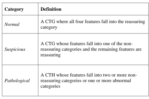
Based on this categorisation of FHR features, it is possible to make the following categorisation of FHR traces
Category Definition
Normal A CTG where all four features fall into the reassuring category
Suspicious
A CTG whose features fall into one of the non-reassuring categories and the remaining features are reassuring
Pathological
A CTH whose features fall into two or more non-reassuring categories or one or more abnormal categories
Table 2: categorisation of Foetal Heart Rate traces
However, the CTG trace should be interpreted only in the context of the clinical scenario, and any therapeutic intervention should consider the maternal condition as well as that of the foetus [23]. Generally, the agreement between experts on normal FHR traces is significantly better than that seen with suspicious or pathological traces.
3.4
UC signal
About uterine contractions signal, the main features are: frequency, intensity and resting tone [6]: the frequency is the number of uterine contractions in time unit or number of contractions in 10 min; the resting tone or Basal tone is the lowest pressure recorded in absence of uterine contractions, its value is about 10 mmHg (this definition and value refer to direct measurements of intrauterine pressure); the intensity is the difference between recorded maximum pressure and resting tone.
3.4.1 Physiological conditions
In physiological conditions, the uterine activity is characterised by a resting tone <= 15 mmHg and UC with intensity of 30-50 mmHg and frequency of 2-5 contractions in 10 min. [6]. During delivery, three kinds of contractions can be observed.
Kind I, 80% of contractions at the labour onset, is characterized by a slow upslope and a fast downslope after contraction’s apex.
Kind II, less than 30%, is bell shaped, symmetrically respect to the contraction’s apex. Kind III, from 20% at the onset of labour to 90% or more in expulsive period, is symmetric respect to kind I, fast upslope and slow downslope.
Figure 11: different kinds of uterine contractions
3.4.2 Pathological conditions
Only some of the most common kinds of pathologies will be reported in this study. Some of them are uterine hypokinesia or hyperkinesia. In the first case the contractions have low intensity and frequency, for example due to sedatives. In the second case, the contractions have high intensity and frequency; in this situation, for example due to excessive dosage of oxytocin, between two subsequent contractions there is not sufficient time for an adequate recovery of oxygen, so a respiratory acidosis can rise.
3.5
FHR recording
The most important aim of foetal surveillance is to avoid intrauterine death or permanent damages to the foetus. So, in industrialised countries, all pregnant women periodically take pregnancy and foetal well-being controls. It is very important to collect right information about foetus’ health also in order to correctly plan successive diagnostic tests. The widespread diagnostic tool, ultrasonographic Doppler cardiotocography (US-CTG), has some limitations: some pathologies and some anomalies of cardiac functioning are not detectable. Moreover, although frequent and/or long-term FHR monitoring is recommendable, mainly in risky pregnancies, there is no strong evidence how long application of ultrasound irradiation can be taken as absolutely harmless for the foetus. Finally, the high quality ultrasound devices are so expensive that they are not available for home care use. Therefore, in the last years, many efforts are been paid by the scientific community to find a suitable alternative.
The development of new electronic systems and sensors now offer the potential of effective monitoring of foetal phonocardiography (FPCG) and foetal electrocardiography (FECG) with passive, fully non-invasive low cost digital recording systems that could be suitable also for home monitoring. These advances provide the opportunity of extending the recordings of the current commonly used US-CTG from relative short to long term, and provide new previously unavailable measures of cardiac function.
The objective of foetal monitoring is to assess foetal well-being and status including the pattern of foetal growth and maturation, oxygen availability, neurogical function and
cardiac function. There are two situations for which FHR provides important information about the condition of the foetus: a normal reactive foetal monitor tracing identifies a foetus that has no trouble with the events of labour and a non-reactive, non-reassuring tracing with complete loss of reactivity and variability identifies a foetus that is unable to respond [27]. It is expected that as the foetus moves the heart rate will temporarily accelerate in response to stimulation by the nervous system. If after foetal movement the physician observes two to three accelerations of greater than 15 beats per minute for 15 seconds the result of the non stress test is indicated as Reactive. Alternatively the test is deemed to be Non-reactive if no accelerations were observed or the observed accelerations did not meet the criteria for a reactive test. It is interesting to compare the clinical significance of both the reactive and non-reactive results; the reactive result is considered a reasonably reliable indicator of reassuring foetal development and well-being whereas the non-reactive result can have more than one explanation and consequently isn’t as useful. If the non stress test was deemed to be non-reactive it could be concluded that: the foetus is compromised, the patient is incorrectly positioned, the test is being effected by drugs in the maternal system or the result is false [28].
FHR can be monitored by means of different techniques: CTG, magnetocardiography, electrocardiography (ECG), phonocardiography (PCG), described in the following paragraphs.