0
Supervisor: Candidate:
Prof.ssa Isabella Screpanti Álvaro Carmona Pestaña
Co-supervisor: Matricola:
Dr. Rocco Palermo 1742190
Histone modifiers driving Notch
context-dependent activity in cancer.
PhD in Molecular Medicine
XXXII Cycle
1 Index
Abstract ... 1
1. Introduction ... 3
1.1 Insights of the molecular signaling of Notch ... 3
1.1.1 General aspects of Notch signaling ... 3
1.1.2 Ligands and receptors from the Notch family ... 6
1.1.3 Notch receptor structure ... 6
1.1.4 Molecular mechanisms of the Notch-signaling pathway ... 8
1.1.5 Notch mutations ... 11
1.2 Notch signaling in development and tissue homeostasis. ... 13
1.2.1 Pleiotropic roles of Notch ... 13
1.2.2 Notch and Hematological Stem Cells (HSCs)... 16
1.2.3 Notch in T-cell development ... 17
1.2.4 Notch in epithelial tissue homeostasis ... 21
1.3 Notch pathway dualism in tumor fate... 27
1.3.1 Notch and T cell acute lymphoblastic leukemia (T-ALL) ... 27
1.3.2 Notch signaling in cervical cancer ... 29
1.3.3 Notch and Epithelial to Mesenchymal Transition (EMT) ... 32
1.4 Epigenetics ... 34
1.4.1 Eukaryotic organization of the chromatin ... 34
1.4.2 Post-traductional modifications of the histones... 37
1.4.3 The Polycomb repressor Complex (PRC) ... 41
1.4.4 Demethylase Jumonji domain-containing 3 (JMJD3) ... 45
1.4.5 Epigenetic regulation of Notch signaling ... 46
1.4.6 Epigenetic control of Notch ... 51
1.3.7 Misregulation of EZH2 in cancer ... 53
2. Materials and Methods ... 58
3. Results ... 62
3.1 JMJD3 down-regulation abrogates Notch signaling in T-ALL... 62
3.2 Pharmacological inhibition of JMJD3 decreases viability of T-ALL cell lines ... 64
3.3 Enforced Notch-cMyc axis partially rescues T-ALL cells from anti-growth effects of JMJD3 inhibition ... 66
2
3.4 EZH2 blockade stimulates Notch pathway activation in cervical cancer cells ... 68
3.5 Notch signaling blockade partially abrogates anti-viability effects of EZH2 inhibition in cervical cancer cell lines... 70
3.6 EZH2 sustains EMT program in cervical cancer cells through Notch signaling repression ... 72
4. DISCUSSION ... 75
5. CONCLUSIONS ... 79
1
Abstract
Deregulated Notch signaling is linked to onset and progression in various cancers. Epigenetic machinery writing or erasing chromatin status of NOTCH genes has been vigorously studied but not fully described yet. In particular, methylation status of H3K27 and availability of corresponding DNA loci for transcription is based on balanced work of antagonizing chromatin modifiers including the EZH2 methyltransferase component of Polycomb-Repressive Complex 2 (PCR2) and demethylases like JMJD3 or UTX. In the current work, we aimed to contribute to the discovery of additional mechanisms regulating transcription of NOTCH1 and NOTCH3 genes in contrasting cancer contexts: T-cell acute lymphoblastic leukemia (T-ALL) where Notch is a well-known oncogene and cervical cancer where Notch proteins are supposed to act as oncosuppressors, concentrating on roles of demethylase JMJD3 and methyltransferase EZH2 favoring chromatin opening and condensation, respectively.
In case of T-ALL where low methylation status of H3K27 at NOTCH-gene corresponding loci favors transcription of NOTCH genes and their targets, we found that pharmacological inhibition of JMJD3 by GSKJ4 decreased the expression levels of NOTCH3, NOTCH1, the target gene DELTEX1, and c-Myc, and abrogated cell viability in both Notch1- and Notch3-dependent T-cell contexts, as confirmed with the accumulation of anti-proliferative factor p27 and apoptosis-associated cleaved form of PARP. Anti-growth effects of GSKJ4 were partially rescued by exogenous Notch1, Notch3, and c-Myc expression indicating a possible involvement of Notch/c-Myc regulatory axis.
Specularly, in cervical cancer, EZH2 is responsible for the epigenetic silencing of tumor-suppressor genes. Supporting the anti-cancerous role of Notch1 protein in this context, we found that EZH2 inhibition upregulated the levels of the Notch receptors, ligands, and target genes, potently suppressed the growth of cervical cancer cells in vitro, and was associated with upregulation of cell cycle blockers p21cip, p27kip, and p53 and reduced expression of c-Myc. Similar effects on cell viability and expression of cell cycle regulators were achieved through transient ectopic introduction of Notch1 in model cell lines. Confirming the partial dependence of observed anti-growth effects of EZH2 inhibition on Notch activation, combined treatment with GSK126 and γ-secretase
2 inhibitor (GSI) abrogated GSK126 effects on cell viability by restoring the number of viable cells and p21cip, p27kip, and p53 levels. Furthermore, EZH2 inhibition suppressed the motility of human cervical cancer cells and upregulated epithelial phenotype marker E-cadherin (E-Cadh) and/or reduced the expression of promesenchymal vimentin in cell-line dependent fashion. Proving the relevance of Notch signaling in the epithelial phenotype maintenance, the combination of GSK126 with GSI restored E-Cadh and/or vimentin levels and partially rescued cell migratory capacity.
The results of our work contribute to the increasing amount of evidence that pharmacological inhibition of histone methyltransferases and demethylases might be a promising strategy for controlling oncogene activation, malignant cell growth and metastatic capacity thus opening new roads for creation of novel targeted cancer therapies.
3
1. Introduction
1.1 Insights of the molecular signaling of Notch 1.1.1 General aspects of Notch signaling
Notch signaling is an evolutionarily conserved cell communication pathway regulating many biological processes in different tissues during embryonic and adult life. Several studies carried out in Drosophila Melanogaster, Caenorhabditis Elegans and in vertebrates have characterized the molecular functions of the main components and secondary factors of the Notch signaling pathway and it has been reported that its activation has varied outcomes depending on the tissue context as well as on the differentiative state of the cells. Proper functioning of Notch signaling determines cell fate in a tissue-dependent fashion by regulating cell proliferation apoptosis, stem cell self-renewal and differentiation. Notch signaling is a short-range intercellular communication system, wherein a Notch ligand expressed on the signal-sending cell interacts with a membrane-tethered Notch receptor on the juxtaposed signal-receiving cell. Notch receptors are highly conserved type I transmembrane multidomain proteins that are synthesized as single precursors and which function, besides the ligand interaction, depends on the activity of some central components of the pathway which drives their proper maturation and activation. The main components of the Notch pathway include:
- Furin convertase, that is involved in the first proteolytic cleavage (S1) of the immature Notch receptor which takes place at the endoplasmic reticulum (Logeat F, et al. 1998; Gordon WR, et al. 2009);
- Fringe Glycosyl-transferases (Lunatic, manic, radical) that catalyze the Notch receptor glycosylation which occurs at Golgi level (Moloney DJ, et al. 2000); - ADAM 10 and 17 Metalloproteases which are implicated in the second cleavage
(S2) of the Notch receptor, which follows the ligand-receptor contact (Brou C, et al. 2000; Mumm JS, et al. 2000; Lieber T, et al. 2002);
- γ-Secretase Complex (Presenilin-Nicastrin-APH1-PEN2) that performs the final proteolytic cleavage (S3), which releases the Notch-activated intracellular domain that translocates into the nucleus in order to induce the transcription of its target genes (De Strooper et al. 1999; Fortini ME, 2002; Tagami S, et al. 2008).
4 Apart from the above mentioned central components of the pathway, other factors, are involved in the Notch signaling regulation by acting at several levels of the pathway activation, starting from the ligand interaction, to the subsequent receptor cleavages and Notch nuclear translocation and transcriptional activity as well as to its protein stability. Some of these proteins are E3 ubiquitin ligases that regulate the amount of the receptor available on the cell surface by controlling its trafficking towards degradation or recycling (Bray SJ. 2006). The E3-Ubiquitin Ligase NEDD4/Su(dx), Numb and Notchless are negative regulators of Notch, whereas Deltex, which is a direct target gene of Notch, is considered a Notch activator as antagonize NEDD4/Su(dx) activity (Xu T, et al. 1990). On the other hand, Strawberry Notch (Sbno in vertebrates, Sno in Drosophila) is a helicase-related nuclear factor that belongs to the SF2 superfamily. It is highly conserved among species and it is involved in the transcriptional control of Notch gene expression by acting as a chromatin remodeler. Although its role is still unclear studies suggest that it functions downstream of Notch
acting through the Epstein-Barr virus latency C promoter binding factor 1, Suppressor of Hairless, and Lag1 (CSL) complex inhibition removal (Watanabe Y, et al. 2017). Notch signaling regulation may occur also by controlling ligands activity. Instead, Neutralized and Mindbomb are proteins containing multiple ankyrin repeats and RING finger domains that functions as an E3 ubiquitin ligase which interacts with the intracellular domain of the Notch ligand Delta to promote its ubiquitination and internalization, therefore their function in the signaling cell is essential for efficient activation of Notch in neighboring cells (Lai EC, et al. 2001). Finally, Notch signaling regulation occurs also subsequently to the ligand-binding activation step Indeed, the E3-ligase FBXW7/hCdc4 (FBXW7) negatively regulates Notch signaling by targeting the intracellular domain of Notch to the proteasomal degradation in the cytoplasm and therefore regulating the potency and the intensity of the signaling (Matsumoto A, et al. 2011). The following figure 1.1 shows a complete protein map that resumes the Notch signaling pathway since its maturation to the proper degradation after completing its function.
5 Figure 1.1: Complete cycle of the Notch signaling pathway. (A) The Notch receptor becomes mature
after its processing by Furin and Fringe (S1 cleavage). (B) Metalloproteases ADAM 10 and 17 perform the second cleavage (S2) of the Notch receptor, which follows the ligand-receptor contact and (C) the extracellular domain of Notch, which remains attached to the ligand, become endocytosed by the ligand presenting cell. (D)After the γ-Secretase Complex S3 cleavage, the intracellular domain of Notch (NICD) becomes translocated into the nucleus (F) where it develops the induction of the target genes transcription. (E) In the absence of activated NICD, the target region of the chromatin is silenced by several repressing factors. Adapted from: Kopan R, 2012.
6 1.1.2 Ligands and receptors from the Notch family
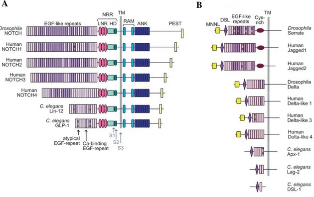
There is just one characterized Notch receptor in Drosophila (dNotch), while four different Notch receptors (Notch1-4) in vertebrates. As it is faithfully described, mammalian Notch1 and Notch2 are pretty much similar between them under a structural point of view, while Notch3 and Notch4 differ from these two on both extracellular and intracellular domains. Serrate and Delta are the only Notch ligands that exist in Drosophila, while in vertebrates, have been described 5 different ligands: Jagged 1-2 (Jag1-2) and Delta-like 1/3-4 (Dll1/3-4). The output signal of the many different ligand-receptor interactions is context-dependent, further supporting the high functional complexity of the Notch-signaling pathway (Shimizu K, et al. 2002).
1.1.3 Notch receptor structure
The Notch gene codifies a 300KDa protein that undergoes post-translational modifications during its transport to the cell surface. In the trans-Golgi apparatus Notch precursor is cleaved by a furin-like protease (S1 cleavage) in an unstructured region of the hetero-dimerization domain generating a heterodimeric receptor composed of an extracellular domain (NECD) non-covalently linked to a transmembrane fragment (NTM) that is followed by an intracellular cytoplasmic domain (NICD). The heterodimeric mature receptor is then transported into the membrane through a Golgi vesicle system in which it is glycosylated by Fringe glycosyl-transferase.
Notch modular structure is highly conserved in all four Notch paralogues which have been identified in mammals; (Kopan R, et al. 2009; Aster JC, et al. 2017). The NECD comprises a series of epidermal growth factor-like (EGF) repeats (36 for Notch-1 and Notch-2, 34 for Notch-3, and 29 for Notch-4) ,which mediate the ligand-binding of the receptor (Gordon WR. et al. 2008) and the negative regulation region (NRR) consisting of three cysteine-enriched repeats (Notch/LIN12) and a heterodimerisation (HD) region which prevent the ligand-independent activation of the receptor. Indeed, mutations, occurring at the NRR module lead to the receptor destabilization and to the constitutive activation of the signaling (Sanchez-Irizarry C, et al. 2004). The NICD contains an RBP-jk- associated molecule domain (RAM) involved in the NICD/CLS complex formation (Tamura K, et al. 1995), and seven highly-conserved ankyrin/cdc10 repeats (ANK) that are required for protein/protein interactions. Flanking the ANK domain,
7 there are two nuclear localization signaling motifs (NLS) necessary for NICD nuclear translocation (Blank V, et al. 1992). In addition, Notch receptors 1 and 2 contain a trans-activation domain (TAD) that is required to properly assemble the Notch/Rbpj/Maml transcriptional complex. At the carboxyterminal region of Notch is located a glutamine-enriched (OPA) and a proline (P), glutamic acid (E), serine (S), threonine (T) enriched region. This aminoacid combination defines the NICD ubiquitination and degradation domain (PEST) (Rechsteiner M, 1988).
Figure 1.2: Characterized Notch receptors and ligands of Drosophila and mammals. (A) The domain
organization of Notch receptors including highly conserved EGF-like repeats, NRR, RAM, ANK and PEST domains. (B) The DSL-family ligands from fly, human, and worm. Adapted from: Gordon WR 2008.
8 1.1.4 Molecular mechanisms of the Notch-signaling pathway
In the absence of ligand binding, the conformational integrity of the NRR holds the basal activation of Notch receptors. The Notch-signaling pathway is triggered by the binding of NECD expressed on the membrane of a signaling receiving cells and a ligand located on the surface of neighboring cell sending signal The ligand-interaction leads to NRR unfolding that allows two sequential proteolytic cleavages of the receptor (S2 and S3) (Bray SJ, et al. 2006).:
1- The ADAM metalloproteases mediate the S2 Cleavage of 12/13 aminoacids at the early transmembrane domain, allowing the generation of a membrane-anchored intermediate fragment called Notch Extracellular Truncated domain (NEXT). 2- The γ-Secretase Complex (Presenilin-Nicastrin-PEN2-APH1) catalyzes the S3
cleavage, releasing the activated intracellular domain of the receptor.
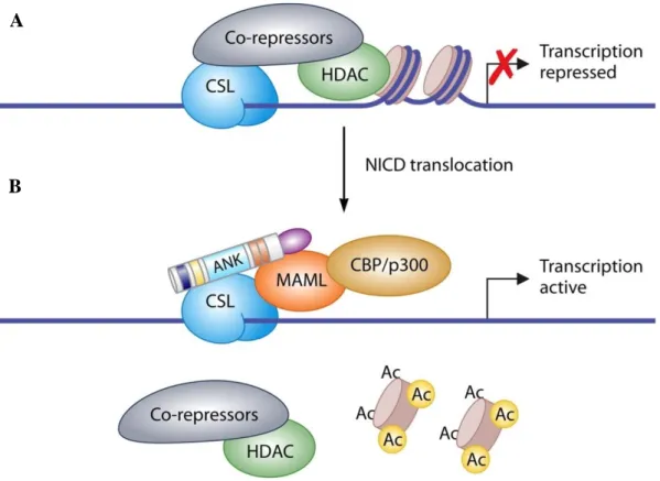
The NEC, released by the S2 cleavage, is endocytosed by the ligand-presenting cell. This process depends on the mono-ubiquitination of the cytoplasmic tail of the ligand. S3 cleavage allows the release of the NICD that it is driven to the intranuclear compartment thanks to its NLS motifs. In the nucleus, NICD promotes the transcriptional activation of its target genes (Bray SJ, et al. 2006). However, NICD does not bind directly the DNA, but, to drive target gene expression, it heterodimerizes with the Recombinant Signal Sequence-Binding protein jK (CSL, also known as CBF1, Su(H) or LAG-1 (Dou S, et al. 1994; Barrick D, et al. 2006). In the absence of nuclear NICD, CSL is bound to the regulatory regions of Notch target genes in association with a co-repressor complex, including the Histone Deacetylases 1 and 2 (HDAC1, HDAC2) (Morel V, et al. 2001), and several other co-repressors as SMRT/Ncor and SHARP/MINT/SPEN to keep Notch target genes expression switched off (Oswald F, et al. 2005). NICD interaction with CSL is crucial for the switch from repressed to the activated transcriptional state of Notch target genes. Although NICD carries a transcription activation domain (TAD), the incorporation of Mastermind Like (MAML) protein into the NICD/CSL complex is essential for the transcription initiation. Indeed, NICD/CSL complex formation recruits the co-activator (MAML), that promotes the displacement of the co-repressors and allows the engagement of additive co-activators (Kao HY, et al. 1998), including the SKI-interacting protein (SKIP) (Zhou S, et al. 2000), the acetyl-transferases p300 and PCAF (Wallberg AE, et al. 2002; Bray SJ. 2006), and the elongation factor CyclinT1:CDK9 (Chopra VS, et al. 2009). In the early
9 stage of the transcription activation, there is the formation of a multimeric complex; SKIP associates with the ANK domain of NICD and MAML interact with SKIP, forming the definitive transcription cluster which associates with the DNA-anchored CSL through the N-terminal domain of MAML. At this step, MAML protein is required for the recruitment of the epigenetic modulators CBP/p300 to the regulatory regions (Bray SJ. 2006). Complete complex promotes the transcription of Notch downstream genes, including the Enhancer of Split (ESpl) complex components in Drosophila, or the Hairy enhancer-of-split (HES) and Hairy/enhancer-of-split related with YRPW motif (HEY) in mammals (Davis RL, et al. 2001; Iso T, et al. 2003). The proper timing of Notch signaling is determined by mechanisms that control nuclear NICD levels by regulating its turn over. Indeed, MAML binds to CyclinC:CDK8, thus allowing NICD phosphorylation within the TAD and PEST domains and therefore promoting the NICD PEST-dependent degradation by the Fbw7/Sel10 ubiquitin ligase (Fryer CJ, et al. 2004). In addition, to PEST phosphorylation, NICD function is also regulated by other signaling pathways and several post-translational modifications, including arginine methylation and lysine acetylation (Palermo R, et al. 2014).
10 Figure 1.3. Overview of Notch signaling. Notch signaling is activated by the interaction of a transmembrane ligand of the Delta–Serrate Lag (DSL) family (1) to the EGF-like repeats of the NECD
(2) on a neighboring cell. Ligand/receptor interaction (3) induces a structural change in the receptor, exposing target sites for ADAM metalloproteases (S2) and γ-secretase (S3) cleavage of Notch (4). Cleavage of Notch outcomes in the release of the intracellular domain (NICD) from the membrane and its translocation to the nucleus (6) while the NECD is endocytosed by the ligand-bearing cell (5). In the absence of Notch signaling, CBF1 is associated with the regulatory regions of Notch target genes with the transcriptional co-repressors, such as SMRT and HDAC1, which actively keep gene expression switched off). Conversely, NICD binds CBF1, displaces the co-repressors and assembles an active transcriptional complex including the co-activators MAML, p300, and PCAF to promote the transcription of its responsive genes (7). Finally, Notch signaling is turned off by the recruitment of CDK8 by MAML to the active complex that phosphorylates NICD at the PEST domain. Phosphorylated NICD is then ubiquitinated by theE3 ubiquitin ligase Fbw7 (8) and subsequently degradated by the proteasome (9). As a consequence of the activator complex destabilization, CBF1 re-associates with the co-repressors. Adapted from: Boucher J, et al. 2012.
11 1.1.5 Notch mutations
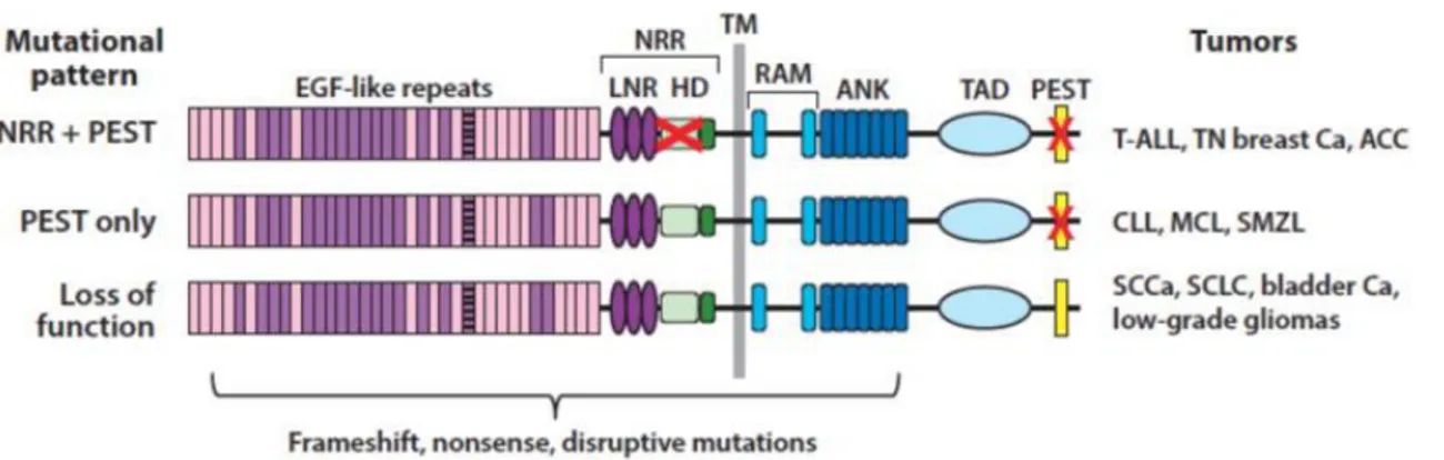
Genomic sequencing of different human tumors has revealed three different patterns of Notch gene mutations altering the signaling activity. The first discovered type of mutation occurring on Notch genes was identified in T-ALL patients and it is a rare chromosomal translocation (7;9)(q34;q34.3) that generates a chimeric gene consisting of the 3 'end of Notch1 bound to the TCRβ gene enhancers (Ellisen LW, et al. 1991). This type of chromosomal rearrangement results in the deletion of the coding sequence of the Notch1 NRR that leads to the expression of a constitutively and ligand-independent active Notch1 ICD (N1ICD). Similar to the above-described translocation, point substitutions and in-frame insertion/deletion mutations that disrupt the structure of the Notch1 NRR result in the constitutive ligand-independent activation of the receptor in T-ALL and in triple-negative breast cancer (Weng AP, et al. 2004; Robinson DR, et al. 2011). Moreover, in these type of cancers have been described also nonsense and frameshift mutations occurring at the C-terminal sequence of Notch1, that, by deleting the C-terminal PEST degron domain, result in increased stability of the N1ICD (Weng AP, et al.2004; Wang K, et al. 2015). Rarely, NRR and PEST mutations occur in cis in the same allele leading to very high levels of constitutive Notch activation (Weng AP, et al. 2004).
The second pattern of mutations is defined by tumors in which changes in the reading pattern, meaningless, or alternative splicing disrupt the PEST domain. These mutations are found in the absence of mutations affecting the NRR domain. PEST mutation only is mainly associated with B-cell tumors including chronic lymphocytic leukemia, marginal zone lymphoma of the spleen and mantle cell lymphoma (Fabbri G, et al. 2011; Trøen G, et al. 2008; Kiel MJ, et al. 2012; Kridel R, et al. 2012). Based on the logic of Notch receptor activation, it has been proposed that Notch activation in tumor cells bearing only PEST deletions could depend on ligands expressed in the tumor cells instead of the engagement with ligand-expressed by the microenvironment (Rosati E, et al. 2009). Although such interpretation is in disappoint with experimental observations indicating that coexpression of ligand and receptor in the same cell results to Notch inhibition rather than Notch activation, a phenomenon referred to as cis inhibition (del Alamo D, et al. 2011).
The third mutational pattern is characterized by nonsense or point substitutions mutations occurring at the N-terminal region of the Notch receptor, all of which are predicted to result in loss of function of Notch. Some of these mutations simply lead to
12 a failed production of the protein, while occasionally result in the expression of a negative dominant receptor with a deleted or useless intracellular domain. Mutations of loss of function in Notch receptors are particularly prevalent in squamous cell carcinomas of the skin (Wang NJ, et al., 2011).
Figure 1.4: Patterns of Notch mutations in various cancers. The red X in the negative regulatory region
(NRR) corresponds to point substitutions, in-frame indels and rare deletions that remove the 5′ coding exons of Notch receptors. The red X in the PEST domain corresponds to nonsense or frameshift mutations that lead to loss of the PEST domain. Adapted from: Aster JC, et al. 2017.
13 1.2 Notch signaling in development and tissue homeostasis.
1.2.1 Pleiotropic roles of Notch
The Notch signaling pathway is a cellular mechanism highly conserved throughout the animal kingdom that regulates proliferation, fate decisions and differentiation of cells during embryonic and postnatal development and it takes part also in other functional processes such as cell adhesion, epithelium-mesenchymal transition, and angiogenesis. Despite the relative simplicity of the core of this route, the final representation of Notch activation is different in varied tissues and during the stages of development and adulthood. The simplistic explanation for its pleiotropic function is that Notch is able to activate groups of target genes which vary depending on the epigenetic context, making the outcome of Notch signaling strictly tissue-dependent (Aster JC, et al. 2017). Therefore, Notch context-dependent transcriptional output is likely driven by the chromatin states dictated by upstream transcription factors capable to regulate the chromatin state or conversely by the ability of Notch to turn on some downstream factors, which then modify chromatin landscapes so as to enable Notch to drive different transcriptional outputs. In addition, Notch signaling frequently is integrated with the output from other pathways, such as Hegegogh, NFkB and Wnt and also this kind of interplay differs according to the context (Du Q, et al 2010). Indeed, although the comparison of genes that rapidly respond to Notch activation in T cell acute lymphoblastic leukemia (T-ALL), mantle cell lymphoma and triple-negative breast cancer identified about 100 genes that were stimulates by Notch only 5 of them were upregulated across all three cell types, and among them, only Nrarp, Hey1, and Notch3 overlapped with a set of direct Notch1 target genes in murine myoblasts. Similarly, to cancer contexts, lineage-dependent variation in downstream transcriptional responses to Notch activation was observed also in different Drosophila cell types. (Stoeck A, et al. 2014; Krejcí A, et al. 2009).
Currently, there is no strong evidence to suggest that Notch is a general factor of stemness. However, the evidence demonstrated that the pathway activation favors the maintenance or expansion of stem cells to the detriment of differentiation, an activity with a more than obvious potential relevance in cancer. Confirming these observations, studies in murine models have demonstrated that the activation of the Notch signal is involved in the maintenance of the populations of neuronal stem cells in the fetal brain
14 (Imayoshi I, et al. 2010). Since its discovery, the role of Notch in the differentiation, morphogenesis, and function in the Central Nervous System has become highly valued in developmental biology research. Through his work on Drosophila, Poulson and colleagues were the first to associate the lack of function of Notch to a lethal embryonic phenotype (Poulson, 1939), referring to them as “a kind of hopeless monster”. The main cause is the failure in the early neurogenic ectoderm when it comes to segregating epidermal cell lines from those destined to be neural. In vertebrates, Notch is necessary when the epidermal and neural lineages are segregated; its activation results in the "neurogenic phenotype", characterized by a premature differentiation of the neuronal progenitors, leading us to interpret the role of Notch in this context as an inhibitor of the differentiation that maintains the progenitor state of the cells (Poulson DF, 1968). The first gene of the Notch pathway to be disrupted by homologous recombination was Notch1; Mutant embryos died in the middle of gestation because of defective and deleterious somitogenesis and placentation (de la Pompa, JL, et al. 1997). Several conditional deletions of Notch1 support the idea that the activation of its signal inhibits neuronal differentiation and sustains the neural progenitor populations in the cerebellum and the telencephalon (Lütolf, et al. 2002; Yang X, et al. 2004; Yoon K, et al. 2004).
In contrast to its role in neuronal differentiation, Notch appears to have an instructive role in gliogenesis by directly promoting the differentiation of many glial subtypes. Activation of Notch signaling favors the generation of Müller glia cells at the expense of neurons, whereas reduced Notch signaling induces the production of ganglion cells, causing a reduction in the number of Müller glia (Furukawa T, et al. 2000; Del Debbio CB, et al. 2016). Notch signaling has been also linked to somitogenesis, as defects in somite morphology have been observed in mice with targeted mutations in the Notch1. These findings suggest that Notch is critical in the patterning process leading to somite boundary formation and the establishment of the Anterior-Posterior polarity of somites (Pourquié O, 2001).
Tissue interactions between the myocardium and endocardium in the atrioventricular canal and outflow tract regions lead to epithelial-mesenchyme transition (EMT) of endocardial cells, to participate in cardiac valves and membranous septa formation. Different Notch ligands and receptors, as well as their downstream effectors and target genes, are expressed in the vascular system. Evidence for a crucial function of Notch in
15 vascular development and homeostasis is the finding that the human disease CADASIL (cerebral autosomal dominant arteriopathy with subcortical infarction and leukoencephalopathy), which involves the NOTCH3 gene, causes stroke and vascular dementia (Joutel A, et al. 1996).
Specific Notch pathway elements and downstream effectors are expressed in the developing pancreas, suggesting a role for Notch in pancreatic development (Lammert E, et al. 2000). The physiological relevance of Notch in the exocrine pancreas has been shown in the mouse, where Notch is active in committed exocrine progenitor cells and whose ectopic activation in pancreatic bud explants represses acinar cell differentiation (Esni F, et al. 2004). The available data indicate that Notch regulates the progressive recruitment of endocrine and exocrine cell types from a common precursor pool in developing pancreas. The inhibitory effect of Notch signaling in exocrine differentiation has been well characterized in zebrafish, where endocrine and exocrine cells arise independently (Field HA, et al. 2003). Genetic data indicates that Notch-mediated Hes1 expression regulates a binary cell fate decision between adsorptive and secretory cell fates in gut development. However, Hes1-deficient mice do not show a change in the proliferative status of the intestinal precursor pool (Jensen J, et al. 2000), whereas Notch activation profoundly affects the proliferation potential of intestinal progenitors (Fre S, et al. 2005), suggesting that other Notch targets may be responsible for the increased in proliferation.
The potential of Notch pathway in osteoclastogenesis and osteoblastogenesis has been investigated in several in vitro experimentations. Evidence indicates that Notch down-regulates osteoclastogenesis activation in osteoclast precursor cells by reducing the surface expression of c-Fms, which is a receptor for macrophage colony-stimulating factor, and enhances the expression of osteoprotegerin (OPG) / osteoclastogenesis inhibitory factor in stromal cells (Yamada T, et al. 2003). Continuous NICD expression inhibited bone morphogenetic protein 2 and induced osteoblast differentiation in osteoblast precursor cells (Sciaudone M, et al. 2003). In contrast, transient expression of N1ICD in osteoblast precursor cells leads to an enhanced bone mineral deposition (Regan J, Long F. 2013). Notch1 is expressed in the mesenchymal condensation area and subsequently in the hypertrophic chondrocytes during chondrogenesis (Watanabe N, et al. 2003). Another study shows that Notch1, Delta1, and Jagged1 are expressed in
16 cultured osteoblast precursor cells as well as in differentiating osteoblasts during bone regeneration and that Notch1 is activated in these cells. These results suggest that Notch signaling plays an important role in the commitment of mesenchymal cells to the osteoblastic cell lineage (Nobta M, et al. 2005). Overall, this evidence suggests the therapeutic potential for Notch in bone regeneration as well as in osteoporosis. Moreover, it is clearly demonstrated that Notch signaling is crucial for generating the marginal zone B-cell population located within the spleen and there are convincing proofs that Notch signaling controls multiple stages of B-cell differentiation and that it takes part in the shaping of the antibody repertoire (Cruickshank MN, et al. 2010).
1.2.2 Notch and Hematological Stem Cells (HSCs)
The first definitive Haematological Stem Cells (HSCs) are defined as those capable of generating adult erythrocytes, myeloid and lymphoid cells. In mice, HSCs arise at around embryonic day 9.5 and express Notch1, Notch2, and Notch 4, but among them only Notch1 seems to be necessary for the final HSCs development, as.. Notch1 -/-models (Kumano K, et al. 2003) and CSL-/- (Robert-Moreno A, et al. 2005) exhibited low level of expression of the transcription factors Scl, Gata2 and Runx1 required for the hematopoietic differentiation. However, the role of Notch in the maintenance of the adult hematopoietic stem component is still unclear as .controversial conclusions have been found by in vitro and in vivo experimentations. The in vitro overexpression of Notch ligands and receptors (Varnum-Finney B, et al. 2000; Stier S, et al. 2002; Varnum-Finney B, et al. 2003) or the constitutive expression of their direct transcriptional targets like HES1 (Kunisato A, et al. 2003; Shojaei F, et al. 2005), inhibit the hematopoietic progenitors differentiation, thus suggesting that Notch pathway contributes to the stemness maintenance. Consistently, genetic modifications leading to an increased expression of the Notch ligand Jagged1 at the bone marrow level results in increased self-renewal of adult HSCs (Calvi LM, et al. 2003). However, in vivo loss-of-function experiments still does not fully support the above observations. Therefore, inducible Cre-loxP-mediated inactivation of the Jagged1 gene in bone marrow progenitors and stromal cells does not impair HSC self-renewal or differentiation in blood lineages and, similarly, the simultaneous inactivation of Notch1 and Jagged1 in mice does not affect their stemness maintenance or survival capability (Mancini SJ, et al. 2005). Moreover, blocking Notch by CSL silencing in HSCs, does not corrupt
17 Myeloid and B lineage differentiation in the bone marrow (Han H, et al. 2002) and Notch pathway inhibition based on the expression of a mutated MAML incapable to bind CSL, revealed that Notch signaling is not necessary for adult HSCs maintenance (Duncan AW, et al. 2005).
Although dispensable for HSCs maintenance, recent studies carried out in vitro and in vivo involved Notch in the early stages of myeloerithroid differentiation. Co-culture of HSCs and OP9 bone marrow stromal cells expressing Dll1 ligand undergoes megakaryocytic differentiation (Mercher T, et al. 2008) and Notch4, as well as many of Notch transcriptional targets, have been found expressed in ex vivo megacario-erythrocytic precursors However, in vivo studies have not yet identify the precise members of the Notch pathways involved in this process. Published evidence clearly linked the development of megakaryocytic leukemia to dysregulated Notch-CSL signaling activation (Mercher T, et al. 2008). In multipotent progenitors that have lost their erythro-megakaryocytic potential, the maintenance of Notch1 expression is part of a lymphoid specification program induced by specific transcription factors. The expression of Notch1 in lymphoid primed multipotent progenitors (LMPPs) appears to be specifically dependent on the transcription factors E2A (Dias S, et al. 2008; Ikawa T, et al. 2006) and Ikaros (Ng SY, et al. 2009).
1.2.3 Notch in T-cell development
As soon as the role of Notch in the development and differentiation of hematopoietic precursors remains doubtful, its implication during the intrathymic differentiation of T lymphocytes is clear (Pear WS, et al. 2003). T lymphocytes are a crucial cellular component of the adaptive immune system, actively engaged in the recognition and elimination of specific foreign antigens. T lymphocytes originate in the bone marrow but reach full specification (CD4+ or CD8+) in the thymus. Mature thymocytes express a unique receptor system capable of binding and recognizing the antigen, the T-cell receptor (TCR). This receptor contacts the major histocompatibility complex (MHC) expressed by a non-self antigen-presenting cell. During development, the reactivity of mature T lymphocytes to the subject's own antigens is examined. In case there is reactivity for self-molecules, the lymphocyte is pushed towards apoptotic programmed death according to a process known as negative selection. On the other hand, due to effective recognition of exogenous antigens, the activated lymphocyte proliferates, thus expanding the number of reactive lymphocytes; in this case, we speak of a positive
18
selection (Starr TK, et al. 2003). Thymocyte maturation consists of three fully described main phases:
1) Double negative state (DN): thymocytes in the early stages of differentiation do not have the classic markers CD4 and CD8 (CD4-CD8-). During the DN phase, the presence of other differentiation markers allows to distinguish 4 sub-phases (DN 1-4) depending on the expression of the adhesion molecule CD44 (CD44+), or of the α-chain of the interleukin receptor 2 (IL2- R) CD25 (CD25+
). The cell that reaches the thymus to begin its maturation (DN1) is, in fact, capable of giving life to any lymphocyte subtype and phenotypically stands out from the combination CD44hi/CD25-. When the DN1 cell enters the thymic environment and makes contact with it, it begins to actively proliferate and express the CD25, becoming CD44low/CD25+. With the beginning of the TCR gene rearrangement, they actually enter the DN2 sub-phase. Moving from DN2 to DN3, the cell becomes CD44- and stops proliferating. The rearrangement of the TCR β-chain begins, rapidly bringing the cell from DN3 to DN4 and completely shutting off the CD25.
2) Double positive state (DP): in this phase the thymocytes present contextual expression of the CD4 and CD8 markers (CD4+CD8+). The cells divide rapidly, contributing to the thymocyte cell diversity increase. While the ability of T cells to recognize antigen-MHC complex is vital for their ability to fight pathogens and other foreign cells, it is equally important that these T cells do not recognize and attack our own cells. This is where negative selection comes into play. After the proliferative phase, the TCR α-chain rearrangement begins; cells that manage to properly develop their own TCR, or are activated by antigens of the developing subject, are eliminated by apoptosis, while a moderate degree of self-antigen binding leads to survival. One of the most intriguing aspects of negative selection is that it occurs mainly in the thymus, which means that T cells depend exclusively on thymus cells to present self-peptides in MHC molecules. Due to this fact, a protein called autoimmune regulator or AIRE in the thymus induces the production of many different proteins that are not classically expressed in thymic cells, such as proteins characteristic of other tissues. Thanks to this, developing T cells are exposed to many peptide-MHC complexes, not only those typically present in thymic cells, which avoids autoimmunity once the T cells leave the thymus.
19 3) Single-positive state (SP CD4+ / SP CD8+): the cells mature from DP to SP extinguishing one of the two markers and maintaining the expression of the single CD4 or CD8 depending on the specification in T-Helper lymphocyte (CD4+), or T -cytotoxic (CD8+). Terminally differentiated lymphocytes migrate from the cortical thymus gland to the medulla, where they meet the blood system of the thymus and enter the bloodstream.
Notch signaling is implicated in the development of intrathymic T lymphocyte precursors, while it is dispensable in that of B cells and myeloid bone marrow. For this reason, the expression of Notch1 in pre-thymic progenitors such as LMPPs represents one of the earliest events in thymocyte specification. The high expression of Notch ligands in bone marrow (Yan XQ, et al. 2001), indicate that it might subsist a mechanism by which LMPPs-mediated Notch activation is inhibited, permitting the generation of T cells prior to thymic implantation. This mechanism involves a so-called lymphoma-associated repressor factor (LRF). It has been shown, that the retroviral expression of the Dll4 ligand in medullary cells could elude this repression, inducing the development of double-positive CD4 / CD8 (DP) T cells in bone marrow (Yan XQ, et al. 2001). It is important to underline that these studies, as well as others (Koch U, et al. 2001; Wilson A, et al. 2001), demonstrated that the bone marrow represents a suitable environment for the differentiation of T cells. Similarly, Notch/CSL signaling promotes the extrathymic development of DP cells in the spleen and lymph nodes of irradiated mice, but this process appears to be suppressed in cases of lymphopenia (Maillard I, et al. 2006). T-cell extrathymic development may have therapeutic relevance in bone marrow transplants. Further studies are needed to identify additional mechanisms that prevent the activation of the Notch pathway outside the thymus.
Notch1 receptor is present at diverse levels during the intrathymic differentiation stages and it is highly expressed in the early phases of DN, less so in DP cells, and then returns to be quite expressed in SP thymocytes (Izon DJ, et al. 2002). Upregulation of Notch1 is critical for early stages of T cell development, whereas downregulation of Notch1 past the β-selection checkpoint at the DN3 stage of T cell development may be important in avoiding sustained Notch signaling, which is strongly transforming in this lineage. The latter event may occur via signals transduced by the pre-TCR which upregulate Id3 and inhibits Tcf3 (also known as E2A). Tcf3 is proposed to be an important positive
20 regulator of Notch1 expression at the DN3 stage of thymocyte development (Yashiro-Ohtani Y. et al, 2009). Notch3 is more expressed than Notch1 in the phases of transition from DN to DP, after this transition, it is down-regulated or turned off in SP cells (Felli MP, et al. 1999; Bellavia D, et al. 2002). At the thymic level, Notch1 also has a role in the selection of the SP CD8+, favoring this T-cell specification at expense of the CD4+. In the same way, it would allow the rearrangement of the αβ-TCR with respect to the γδ type (Robey E, et al. 1996; Washburn T, et al. 1997; Fowlkes BJ, et al. 2002), besides being necessary in gene rearrangement VDJk (Wolfer A, et al. 2002). This is a mechanism of genetic recombination that occurs in vertebrates by which amino acids are selected and assembled randomly from genes that encode specific proteins with important roles in the immune system. This location-specific recombination generates a diverse repertoire of TCR and immunoglobulin (Ig) molecules that are necessary for the recognition of various bacterial, viral and parasite antigens, as well as dysfunctional cells, such as tumor cells (Wolfer A, et al. 2002). Notch3, induced to over-expression by the intrathymic stromal cells in the DN-SP transition, plays a decisive role in the pre-TCR control phase (Bellavia D, et al. 2003).
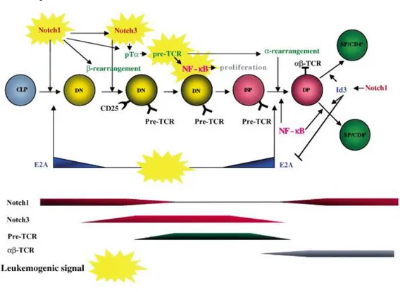
Figure 1.5: Schematic model of relationships between murine T cell development and leukemogenic events. The sequence of developmental stages and the main signaling pathways involved are shown.
Abbreviations: CLP, common lymphoid precursor; DN, CD4−CD8−double negative thymocytes; DP, CD4+CD8+double positive thymocytes; SP, CD4+and CD8+thymocytes. Adapted from: Campese AF, et al. 2003.
21 1.2.4 Notch in epithelial tissue homeostasis
The epidermis is a finely organized and continuously renewing squamous epithelium that consists of four functionally different layers: basal, spinous, granular and cornified (in ascending order from the basal membrane). The stratum corneum is the epithelial barrier with the environment that limits water loss and prevents the invasion of microorganisms. Epidermal homeostasis depends on the balance between proliferation and differentiation of keratinocytes (Fuchs E, 1990). Tissue renewal is sustained by a small population of stem cells located at the basal layer that undergoes proliferation into transiently amplifying cells with limited proliferative capacity.
In the early phase of the differentiation, the cells stop proliferating, detach from the basement membrane and migrate to the spinous and granular upper layers., Keratinocytes start to express a specific protein pattern including keratin 1, 10 and involucrin in the spinous layer, as well as filaggrin and loricrin in the granular layer. Finally, a series of biochemical and morphological changes take place that results in the conformation of the stratum corneum. This complex process during the epidermal differentiation requires the coordination among the molecular mechanisms that drive the generation of the stratified epithelium and in which Notch and its ligands play an important role (Okuyama R, et al. 2008).
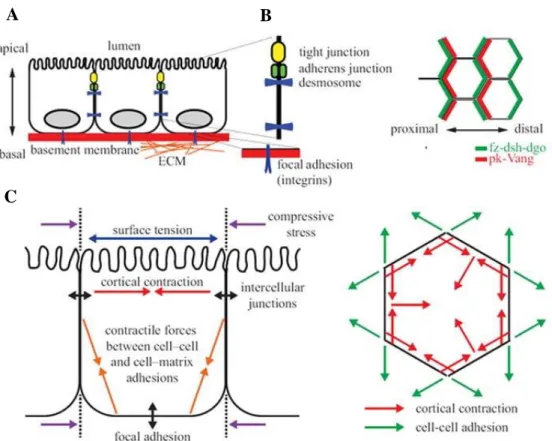
Epithelial cells make up the tissues through a paired structure with apicobasal polarity maintained stably thanks to extensive cell-cell contacts and adherence to the basement membrane (figure 1.6 A) (Cereijido M, et al. 2004; Shin K, et al. 2006). The cells of the epithelial tissues also have another type of axis, called planar cell polarity (PCP). This polarity presents an orthogonal structure between the apical and basal ends in the orientation of cell-cell and cell-extracellular matrix contact (figure 1.6 B, C) (Devenport D, et al. 2008). The signaling proteins of the PCP group include frizzled (fz), disheveled (dsh), Van Gogh (Vang), prickle (pk), diego (dg) and flamingo (fmi), which present different distribution to establish the proximal and distal sides of polarity (Wu J, et al. 2009). However, during the development stages, the epithelial cells show the ability to change to a mesenchymal phenotype in situations in which deep cellular readjustments are necessary. The mesenchymal cells are characterized by fusarium morphology due to the lack of cell-cell contacts; this confers the ability to migrate through the basement membrane. This process of phenotype change is called epithelial-mesenchymal
22 transition (EMT), and there is solid evidence that maintains its relevance in the progression of epithelial tumors since the transformed cells acquire the ability to escape from the extracellular matrix and invade the surrounding tissues (Moreno-Bueno G, et al. 2008; Onder TT, et al. 2008; Yang J, et al. 2008). The following figure shows the structure of an adherent tissue according to its vertical and horizontal polarity.
Figure 1.6: Detail of the morphology of a polarized epithelial monolayer. (A) Vertical epithelial
polarization towards the apical, basal and lateral domains of the plasma membrane. The apical surface presents specialized microvilli in the absorption and secretion. The basal surface adheres to the proteins of the extracellular matrix through integrins for the formation of focal adhesions. Lateral membranes contain complexes such as tight junctions, adherents, and desmosomes that maintain the cohesion of the tissue and favor diffusion barriers. (B) Horizontal polarization of the core planar cell polarity proteins. In the wings of Drosophila, the wing, fz, dsh, and dgo proteins are found in the distal chord domains, while pk and Vang are located in the proximal cortical domains. (C) The intracellular and intercellular tension maintains the shape of the cells. The constriction promotes forces of linear tension in the area of contact between cells and serves as an energy barrier for cell readjustments. Adapted from: Wang CC, et al 2012.
A B
23 The information is transmitted directly between adjacent cells through signaling receptors. The major homotypic receptor in epithelial cells is E-cadherin, a calcium-dependent receptor that is critical in cell-cell recognition and adhesion (Gumbiner BM, et al. 2005). The cytoplasmic domain of E-cadherin connects to the actin cytoskeleton via α and β-catenin to preserve cell shape and polarity. In Madin-Darby Canine Kidney (MDCK) cells, the cadherin-catenin complexes affect signaling predominantly through small GTPases. Rac-induced lamellipodia activity is repressed as E-cadherin amasses, and Rho-dependent actomyosin rises during the establishment of new contacts once the cytoskeletal networks are formed (Yamada S, et al. 2007) (figure 1.7 A). Computational models were used to investigate whether Delta-Notch negative feedback loops are sufficient to generate long-range signaling patterns during development. In some contexts, high expression of the Delta ligand in a cell inhibits the expression of the same ligand in the neighboring cell by a process called lateral inhibition (Kimble J, Simpson P. 1997). In other contexts, a lateral induction process may occur (de Celis JF, Bray S. 1997). Interestingly, Webb and Owen found evidence that both mechanisms were capable of generating long-range patterns for certain feedback forces. It is possible that these computational studies in the Delta-Notch signaling could be useful to identify disturbances in the signal or feedback that could distinguish whether the inhibition or lateral induction was the dominant non-cell autonomous mechanism (Webb SD, et al. 2004) (figure 1.7 B).
24 Figure 1.7: Detail of the intercellular communication between adjacent epithelial cells by cell-cell signaling receptors. (A) E-cadherin forms homophilic complexes between neighboring cells. Catenin
p120 binds to the juxtamembrane domain of E-cadherin and stabilizes it by preventing its clathrin-mediated endocytosis. B-catenin is bound to E-cadherin. The alpha-catenin acts in connection with the epithelial protein lost in neoplasm (EPLIN), finally connecting it with the F-actin. (B) Notch binds to its ligand (Delta or Jagged) by contact with a neighboring cell, releasing NICD by proteolysis that translocates to the nucleus and interacts with the RBPkJ DNA binding protein together with various coactivators for the regulation of the expression of Notch's target genes, such as Hes and lunatic fringe (Lfng), which could negatively regulate Notch. Adapted from: Wang CC, et al. 2012.
In normal skin, Notch receptors and ligands are expressed in keratinocytes within the epidermis as well as in adnexal structures. Notch1 is expressed in all epidermal layers, while Notch2 is expressed only in the basal layer (Rangarajan A, et al. 2001; Okuyama R, et al. 2004) and the Notch ligands Jagged and Delta-like family members are expressed in overlapping patterns with Notch in the epidermis. Former experimentation indicated that Notch signaling induces differentiation in human and murine keratinocytes (Rangarajan A, et al. 2001; Lowell S, et al. 2000; Nickoloff BJ, et al. 2002) (figure 1.8). Indeed, Notch1 signaling stimulates the expression of early differentiation markers such as keratin1 and involucrin in mouse keratinocytes. These results correlate with the epidermal phenotype observed in mice models of constitutive activation of Notch1 in the epidermis (Blanpain C. et al 2006). On the other hand, Notch1 deletion in mice results in decreased epithelial cell differentiation and increased proliferation of cell layers (Rangarajan A, et al. 2001).
In addition, it has been demonstrated that Notch1 signaling induces the expression of the cell cycle regulator p21Waf1 / Cip1 (Rangarajan A, et al. 2001) and promotes cell cycle arrest in proliferating keratinocytes thus allowing their terminal differentiation.
25 Conversely, it has been shown that Notch1 signaling activates caspase 3 in embryonic keratinocytes, thereby producing a decrease in proliferation and an increased differentiation by activating PKC-delta (Okuyama R, et al. 2004).
The different functions of Notch are complex and involve a cross-talk with other signaling molecules. Notch is linked to important factors controlling cell behavior such as p63 (also known as p51 or KET). The p63 gene was originally identified as a member of the p53 family (Osada M, et al. 1998; Yang A, et al. 1999) that codifies for a protein with high structural homology with p53. Furthermore, p63 binds to responsive elements of p53 and it acts as a positive regulator of p53 target genes, suggesting a functional analogy between these proteins. Mice lacking p63 show developmental deficiency, especially in the formation of the stratified epithelium (Mills AA, et al. 1999; Yang A, et al. 1999). Interestingly, the activation of Notch signaling suppresses the expression of p63, while, on the contrary, p63 inhibits Notch-induced p21 expression, cell cycle arrest, and epidermal differentiation (Nguyen BC, et al. 2006; Okuyama R, et al. 2007). Therefore, the proliferation or differentiation of keratinocytes is determined by a fine balance between the intracellular levels of Notch and p63. In addition to the p53 family members, it has widely demonstrated the existence of fine crosstalk between Notch signaling and the Wnt and Shh pathways. Both Wnt and Shh signaling pathways are known to be tumorigenesis regulators and abundant evidence indicates that are repressed by Notch1. Indeed, the deletion of Notch1 in skin of mice results in the constitutive activation of Wnt and Shh signaling and combined with the development of basal cell carcinoma and squamous cell carcinoma (Nicolas M, et al. 2003). The following figure 1.8 represents a diagram of the Notch interactions among the epidermal layers and its relevant crosstalk with other signaling pathways.
26 Figure 1.8: The Notch1signaling interplay within the epidermis. Each epidermal layer shows a different
state of differentiation and the expression of specific proteins. Notch1 enhances the expression of keratin1 and involucrin and prevents the induction of filaggrin and loricrin that are induced in later stages. Sonic hedgehog (Shh) and Wnt signaling are normally repressed by Notch1. In addition, p63 and Notch repress each other. Adapted from: Okuyama R, et al. 2008.
27 1.3 Notch pathway dualism in tumor fate
The biological function of the Notch signaling pathway is considered context-dependent as activated Notch exerts opposing effects like growth and differentiation in diverse cell types and in the same cell context in physiological versus pathological condition (Lefort K, et al. 2016; Ranganathan P, et al. 2011). Notch receptors can function as cell-autonomous oncoproteins, cell-cell-autonomous tumor suppressors, or microenvironment-dependent oncoproteins in different cellular contexts.
1.3.1 Notch and T cell acute lymphoblastic leukemia (T-ALL)
As mentioned in the previous section 1.2.3, the role of Notch in correct thymocyte development and differentiation has been partially clarified, making even more complex to describe the contribution of this signaling pathway in the pathogenesis of T-ALL. This is a hematopoietic malignancy characterized by the accumulation of T lymphoblasts in blood and primary lymphoid tissue that represents the 15% of pediatric acute leukemia and almost 25% in adult cases (Bongiovanni D, et al. 2017). This hematological cancer is characterized by a less favorable prognosis than B Cell Acute Lymphoblastic leukemia (B-ALL). The clinical evidence regards a high number of leucocytes in blood, adenopathy and marrow infiltration of immature lymphocytes, frequently followed by central nervous system colonization of these cells (Van Vlierberghe P, et al. 2012). The tumor etiopathogenesis is due to the malignant transformation of thymic lymphocitary precursors during their differentiation process. The crucial mechanisms causing this outcome have been demonstrated to be related to an expression and/or activation deregulation of molecular pathways involved in thymic cells differentiation channeling. The T-ALL is characterized by genetic modifications as chromosomic aberrations, altered gene expression profiles, mutations or immunoglobulin (Ig) genes rearrangement, and T cell receptors (TCR) (Kraszewska MD, et al. 2012). In fact, the first scientific evidence of the involvement of notch1 in leukemic pathology dates back to 1991, when chromosomal translocation with function gain t (7; 9) (q34; q34.3) was identified in some patients with T-ALL. This gene rearrangement involves the dosing of the N1ICD domain with elements of the promoter of the TCR gene, resulting in the expression of a constitutively active form of Notch1 (Ellisen LW, et al. 1991). Murine models investigations involving the overexpression of the intracellular domains of Notch1 and Notch3, placed them under the control of the specific promoter of the immature thymic stages Lck (Robey E, et al. 1996; Bellavia D,
28 et al. 2000). Furthermore, transgenic mice for incomplete TAD domain of N1ICD only occasionally present the disease, whereas those with a complete region develope T leukemia (Weng AP, et al. 2006). Bellavia and colleagues generated a transgenic mouse that overexpresses the intracellular domain of Notch3 (N3ICD), again located under the control of the Lck promoter. Contrary to what happens in the murine Lck-N1ICD models, the Lck-N3ICD transgenic mice develop a particularly early and aggressive form of T-cell leukemia, suggesting the oncogenic role of Notch3 which takes place in the thymus (Bellavia D, et al. 2000). Subsequent investigations have shown that, at a phenotypic level, leukemic cells present characteristics similar to those of immature thymocytes such as CD25+ and presenting the pTα chain. The Notch3-pTα axis would promote the constitutive activation of NF-kB, favoring the proliferation of these cells bypassing the apoptotic systems, and thus contributing to the neoplastic transformation of immature thymocytes (Bellavia D, et al. 2002). From this work, it has been shown how the activation of Notch3 and Notch1, together with NF-KB, is capable of inducing the transcription of several microRNAs, particularly of miR-223 (Kumar V, et al. 2014). This microRNA has been demonstrated to be an oncogene among whose targets it is the E3-ligase FBXW7, responsible for the ubiquitination and consequent degradation of the intracellular domain of Notch1 increasing its half-life and cytoplasmatic stability, together with its potential oncogenicity (Mansour MR, et al. 2013).
Recent experimental evidence stands out that these genetic aberrations join the effects of a deregulated expression of transcriptional oncogenic factors and their direct targets with a tumorigenic outcome, among which it has been described c-Myc and Cyclin-E (Szczepanski T, et al. 2011; Kumar V, et al. 2014). In murine models in which a stable viral insertion of a Notch1 allele has been performed, transcriptional induction of helix-loop-helix domain (bHLH) factors of the Hes family is observed as a result. Among these, Hes1 seems to be a potential inducer of leukemogenesis (Lee JS, et al. 1999). Similarly, the DELTEX oncogene is overexpressed in murine transgenic models for both Notch1 and Notch3, emphasizing the oncogenic role of this protein in T-ALL. The combined expression of Notch3, HES1, and DELTEX is characteristic of T-ALL since, interestingly, the isolated expression of these oncogenic mediators is not sufficient to induce the disease (Zúñiga-Pflücker JC, 2004), thus showing that Notch expression is an important requirement for the development of leukemia.
29 Another oncogenic mechanism dependent on Notch lies in the ability of the activated receptor to promote the G1 / S cell cycle progression by direct or indirect regulation of crucial proteins for this checkpoint, such as the CDK4 or CDK8 kinases or the cyclin-dependent kinase inhibitors p27KIP and p18INK4C (Dohda T, et al. 2007). All these deregulatory processes act in a synergistic manner with those Notch mutations mentioned in point 1.1.5 thus suggesting that an aberrant regulation that induces a constitutive and/or uncontrolled activation of the Notch pathway is decisive and necessary for the full development of the T-ALL.
1.3.2 Notch signaling in cervical cancer
Cervical cancer is one of the leading causes of cancer-related deaths among women in the developing world accounting for the fourth most common cause of cancer deaths in the world. However, in developed nations, cervical cancer deaths do not exceed 2% per year; this is due to the systematic use of the Pap test to detect cervical abnormalities and proceed to early treatment. Most cervical cancers (80 to 90 percent) are squamous cell cancers, while adenocarcinoma is the second most common type of cervical cancer, accounting for the remaining 10 to 20 percent of cases. Adenocarcinoma types develop from the glands that produce mucus in the endocervix. While less common than squamous cell carcinoma, the incidence of adenocarcinoma is on the rise, particularly in younger women. Solid evidence associates cervical cancer with the infection of squamous and columnar cells of the epithelium of the ectocervix and endocervix with high-risk human papillomavirus (HPVs) (Zur Hausen H, 2002). In the so-called transition zone located between squamous and columnar cells, there is a population of cells with differentiation capacity in one or another subtype (figure 1.9). The integration of the HPV genome, as well as the neoplastic transformation process, is believed to begin in this region (Reid R, 1983).
30 Figure 1.9: Cervical cancer initiates in the cervix, the narrow opening into the uterus from the vagina.
The region known as ectocervix is the portion of the uterus extending into the vagina. In a healthy state, it shows itself covered by the flat, thin squamous cells. The endocervix or cervical canal is built by columnar cells. The area between them is called the transformation zone (T-zone), considered most likely the location for abnormal or precancerous lesions. Adapted from: National Cervical Cancer Coalition (NCCC).
Decades of study have unveiled the molecular insights of the virus. It has been well described that the integration of the virus DNA results in the disruption of the viral open reading frame that encodes for the viral E2 inhibitory subunit. This erases the repression of the rest of the viral genome transcription which encodes for oncogenes E6 and E7 (Goodwin and DiMaio, 2000; Zur Hausen H, 2000; Parish JL, et al. 2006). The best-known function of these two oncogenes is to transcribe for the viral subunits E6 and E7 that interact and block host proteins p53 and Retinoblastoma (Rb), respectively. This causes deregulation of the control of the cell cycle and the response to DNA damage, which contributes to tumorigenesis. The different pleiotropic roles of E6 and E7 continue to be identified and documented extensively (Oh ST, et al. 2004; Yim EK, et al. 2005; Jones and Wells, 2006; Tomaić V, 2016). The detection of the intracellular form of Notch in certain types of cervical cancers aroused great interest since such forms of Notch are really difficult to detect in the context of neoplasms. There is currently a large and controversial bibliography around the role of Notch in the development of cervical cancer (figure 1.10). The first evidence of the relationship between Notch signaling and solid tumor development arose from the studies on the integration of the murine mammary tumor virus (MMTV) by Robbins and colleagues in
31 1992 (Robbins J, et al. 1992). Up to four genes were detected and analyzed in the context of the murine mammary tumor, of which two were members of the Wingless family. The other two genes characterized were members of the fibroblast growth factor gene family and the Notch family (Robbins J, et al. 1992).
Figure 1.10: The role of Notch signaling in the neoplasic transformation of the cervix epithelium associated with the authors who have collaborated to unveil it. The neoplasic transformation takes place
by the action of the human papillomavirus (HPV) oncoproteins that cooperate with Ras and Notch1. The moderate expression of Notch1 results in the positive regulation of c-Myc and the activation of PKB / Akt, causing neoplasia. On the other hand, it has been demonstrated that the high constitutive expression of Notch1 can induce apoptosis in keratinocytes transformed by HPV. Adapted from: Maliekal TT, et al. 2008.
32 Different to what observed in T-ALL in which activating mutations occur on Notch1 locus in about 50% of patients (Weng AP, et al. 2004), Notch1 mutations are very rare in human cervical cancers samples and Notch signaling activation is driven by a ligand-dependent mechanism sustained by elevated expression of its ligand Jagged1 and low levels of its negative regulator Manic Fringe (Veeraraghavalu K, et al. 2004). However, the role of Notch in cervical cancer is not clearly determined and several findings are controversial. It has been demonstrated that different levels of Notch activation are specific of the tumor phases and that Notch activation acts as oncogene by inducing cell growth, drug resistance and invasiveness through Epithelial-to-Mesenchymal transition (EMT) in some cervical cell lines (Yu H, et al. 2007; Wang L, et al. 2018). Conversely, it has been also shown that high levels of activated Notch can induce apoptosis and cell cycle arrest by stimulating the expression of markers such as p53 and p21 (Yousif NG, et al. 2015; Yun J, et al. 2015; Talora C, et al. 2005; Wang L, et al. 2007; Franko-Tobin LG, et al. 2012). For this reason, it is important to consider that a better and deeper understanding of the molecular mechanisms of the Notch pathway in cervical cancer could lead to the development of novel therapeutic strategies.
1.3.3 Notch and Epithelial to Mesenchymal Transition (EMT)
EMT is a highly conserved cellular process that permits polarized and fixed cells to lose their epithelial characteristics and gain motile attributes of mesenchymal cells. EMT occurs in a physiological way during embryonic development and wound healing, but it is also associated with cell migration and invasion and considered a critical step for cancer progression in solid tumors. This process alters the morphology of the healthy epithelium occupying the lumen and invading the basement membrane and neighboring vessels (figure 1.11) (Birchmeier C. et al, 1996; Gotzmann J. et al, 2004; Thiery JP. et al 2009). Not surprisingly, it has been widely described that the EMT program in epithelial cells is not only coordinated by multiple signaling pathways but also by epigenetic and post-translational modification (Serrano-Gómez, SJ. et al. 2016).
33 Figure 1.11: Invasion mediated by EMT and collective invasion in metastasis. Healthy epithelial cells
(orange) undergo a process of epithelial-mesenchymal transition (EMT) and give rise to the primary tumor (blue). Some primary tumors develop the ability to invade and migrate to nearby vessels as a multicellular strand (green). After filtering into circulating blood, these tumor cells can colonize other tissues where they undergo an inverse process of extravasation and settlement called mesenchymal-epithelial transition (MET). MAT: mesenchymal-ameboid transition. Adapted from Kawauchi T, 2012.
During the last decade, it has been reported increased expression of Notch signaling in certain pathological frames of cervical cancer cells and cervical carcinomas with high grade, lymph node involvement, and parametrial invasion, frequently accompanied by activation of the NF-κB pathway (Weijzen S, et al 2003; Ramdass B. et al 2007; Sun XM, et al. 2009). Song, et al. reported that the over-expression of Notch-1 stimulates NF-κB activity in CaSki metastatic cervical cancer cells by associating with the IKK signalosome through IKKα (Song LL, et al. 2008). Conversely, several researches demonstrated that Notch-1 activation has a dose-dependent effect and it leads to cell cycle arrest and growth repression in cervical cancer cells (Talora C, et al. 2002; Talora C, et al. 2005; Wang L, et al. 2007; Yao J, et al. 2007; Maliekal TT, et al. 2008). Overall these findings unveil the complexity of Notch's role in different contexts of human cervical cancers.