FACOLTA’ DI MEDICINA E CHIRURGIA
Dottorato di ricerca in tecnologie avanzate in Chirurgia
XXVII ciclo
Aneurysmal subarachnoid hemorrhage:
multivariate analyses for functional outcome and mortality
at a single institution from 2004 to 2015
Relatore Candidata
Prof. Vito D’Andrea Dott.ssa Biagia La Pira
Matricola 940130
A te papà, che sin da piccola mi hai spronata
alla lettura, alla cultura, alla curiosità, alla scienza.
INDEX
Abstract
1. Summary
2. Introduction
2.1 General features of aneurysmal subarachnoid hemorrhage (aSAH) 2.2 Clinical presentation
2.3 Diagnosis of aSAH 2.4 Management of aSAH
2.5 Complications of aSAH and treatment
3. Material and methods
3.1 Patient consent and study design 3.2 Study definitions
3.3 Collected variables 3.4 Statistical analyses
4. Results
4.1 Multivariate analysis for mortality
4.2 Multivariate analysis for functional outcome
5. Discussion
5.2 What we can improve in the management of aSAH 5.3 Study Limitations
6. Conclusion
References
Abstract
Objective: The aim of our study is to analyze which factors are mostly involved in mortality rates and functional outcome in patients who suffered of aneurysmal SubArachnoid Hemorrhage (aSAH) between January 1, 2005, and December 31, 2014 admitted and treated at Mayo Clinic, in Rochester.
Patients and methods: We conducted a multivariate analysis of consecutive and retrospectively collected patients suffering of aSAH in a study period from 2005 to 2014, taking into account specific factors as age, worse severity at presentation, ischemia, rebleeding and APACHE 3 (acute physiologic assessment and chronic health evaluation).
Results: A total of 438 patients with a diagnosis of aSAH within 48 hours from admission were retrospectively collected. Patients demographics, aneurysms features and treatment modalities were identified. Rebleeding, worse clinical at presentation, APACHE 3, ischemia (or symptomatic vasospasm), were analyzed in a multivariate analysis for the mortality rate, whereas age, rebleeding, worse clinical at presentation, APACHE 3, ischemia were considered in a multivariate analysis for the clinical outcome at last follow-up. After controlling for comorbidities, we observed that worse clinical severity and ischemia were the major factors influencing the mortality rate (OR 5.25, CI 2.54-11.13 and OR 5.16, CI 2.35-11.46 respectively). Similarly these same factors influenced the functional outcome (worse clinical severity: OR 4.43, CI 2.30-8.61 and rebleeding: OR 3.50, CI 1.66-7.40). Less contribution was given from ischemia/ vasospasm (OR 2.01, IC 1.08-4.06).
Conclusions: Management in patients with aSAH, and, as a consequence, mortality rates and functional outcome, have improved in our institution in the last decades. From our multivariate analyses we understood that rebleeding and worse clinical at presentation are the most important factors we can work on to improve good functional outcome and survival in our patients.
1. Summary
During the past decades, the management of aneurysmal subarachnoid hemorrhage (aSAH) has substantially changed. Progression in neurocritical care and the better understanding of all the pathophysiological mechanisms related to the SAH changed our knowledge and our behavior to effort this otherwise fatal event.1
Furthermore, while the development of coiling has provided a valid and safe alternative to clipping,2-3 the tendency to treat precociously a ruptured aneurysm had a positive effect on the incidence of rebleeding and, as a consequence, on mortality rate and neurological outcome.4
Besides these advances, literature data regarding the mortality rate and the overall functional outcome in based population-studies over time are scant.5-13 Some population studies described a decreasing-fatality rates, but no specific series on neurological outcome over a specific period range taking into account some important revolutionary facts are available. For this study population analyses, we consider patients admitted at our hospital from a 2005 to 2014, after the occurrence of the events that marked and changed substantially the management of aSAH: the increasing role of endovascular therapy for high-risk surgical patients in the mid 90’s and publication of the results of the ISAT study in 2002-2004.
Considering all the above mentioned factors, we conducted a retrospective review of consecutive patients with aSAH treated at Mayo Clinic, Rochester, Minnesota, between January 01, 2005 and December 31, 2014. The 438 patients identified were analyzed for demographics, aneurysms characteristics, treatment modalities. Our multivariate analyses stated that poor functional outcome at last follow-up and mortality rate were strongly influenced by worse clinical at presentation and rebleeding. Nevertheless, more detailed investigation is necessary to determine whether this or other factors may directly explain the trends of survival and functional recovery over time.
2. Introduction
2.1 General features of SAH
Subarachnoid hemorrhage (SAH) is defined as bleeding around the brain confined within the subarachnoid space between the arachnoid membrane and the pia mater. The etiology of SAH can be divided into two main categories: 1) spontaneous, and 2) traumatic. The term spontaneous is utilized to define SAH from causes other than traumatic SAH. The epicenter of the SAH can be at the cisterns of the base of the skull at or immediately adjacent to the Circle of Willis and from here diffuse to the convexity or can be limited to single of few sulci (sulcal SAH). Sulcal SAH, if spontaneous can be seen in CNS involvement due to vasculitis associated with systemic autoimmune diseases or as part of the spectrum of reversible vasoconstriction syndrome. Spontaneous SAH can be due : rupture of an intracranial aneurysm (75-89% of cases), cerebral arteriovenous malformations, dural and pail arteriovenous fistulas, dural venous sinus thrombosis, prentruncal/perimesencephalic non-aneurysmal SAH, cerebral artery dissection (for the internal carotid and the vertebral arteries), rupture of an infundibulum, pituitary apoplexy, coagulation disorders (bleeding dyscrasias and thrombocytopenia), central nervous system vasculitis, spinal arteriovenous malformations of the cervical or high thoracic region, rarely brain tumors, otherwise unknown/idiopathic. Unless otherwise specified, we will focus on aneurysmal SAH (aSAH) related to rupture of an intracranial aneurysm.
The incidence of aSAH varies in different geographic regions. It is higher in Japan and Finland while in the US its incidence is estimated to be between 6.9 to 9.4 per 100, 000. There are ethnic differences in the incidence of SAH in the US with higher rates observed among Hispanics and African Americans. Although the incidence of aSAH has been relatively constant over the years, there may be a trend toward a reduced incidence in the Western World in more recent years, probably related to reduced rates of smoking and better control of hypertension. The incidence of aSAH increases with increasing age and although more common in
women than men, gender distribution varies in different age groups. In the younger age groups (25-45 years), aSAH is more common in men while in the older age groups it is more prevalent among women.14
Established modifiable risk factors for aSAH include smoking and hypertension. The risk is higher for current rather former smokers. There is less strong evidence for a contributory role of other modifiable risks factors such as alcohol consumption, drugs (cocaine) and hormonal factors. There is interaction between current smoking and risk of aSHA between genders since current smoker women have higher risk than current smokers of male sex. The joint effect of hypertension and smoking is higher than the increased risk ascribed to each factor considered singly. This observation suggests an additive effect of smoking and hypertension on the risk of aSAH.15-16
Furthermore, posterior circulation aneurysms and giant size aneurysms are well known predisposing factors for their rupture.Because of aSAH sequele can lead to death and severe disability, with mortality rates reaching 45%, timely diagnosis and focused specialized management are of paramount importance to prevent rebleeding and the secondary effects of SAH, as vasospasm and cerebral ischemia.17
2.2 Clinical presentation
Severe headache of sudden onset (‘the worst headache of my life” ) is the clinical hallmark of SAH. The rapidity of onset, usually reaching a maximum within a few seconds rather than the severity itself, is the most important clinical criteria for a suspected diagnosis of aSAH.
Patients with the classic sudden thunderclap headache or “worst headache of their life” are considered to be suffering from spontaneous SAH until proven otherwise, despite SAH is responsible only for a small fraction of sudden headache at the emergency room (approximately 6%). 18
The differential diagnosis of a sudden-onset headache includes SAH, cerebral venous thrombosis, pituitary apoplexy, spontaneous intracranial hypotension, and hypertensive encephalopathy. Regardless the reasons that triggered the thunderclap headache , the sudden change in the pressure within the subarachnoid space causes two sequelae:
1. transmission of pressure to the rest of the intradural space, and to the dura, that has pain receptors
2. decrease of the cerebral perfusion pressure (leading to loss of consciousness, confusion, and/or delayed ischemic neurologic deficit).
The initial diagnostic workup includes a detailed history and physical exam. The history should focus on questions that relate to the patient’s characterization of the headache and associated symptoms.
Typically but not always, the headache lasts for prolonged period of times, usually days as opposed to more benign forms of thunderclap headache which may resolve within a few minutes. Additional symptoms include nausea, vomiting and loss of consciousness, photophobia, phonophobia, diplopia, focal neurological deficit, back pain, seizures, weakness, presence or absence of sentinel headaches, association with another activity (e.g., cocaina use and sexual activity). Associated signs like nuchal rigidity may take hours to develop. Loss of consciousness can be brief or prolonged in relation to the intensity of the hemorrhage. Loss of consciousness is a reliable differentiator between aneurysmal and non-aneurysmal SAH since patients with non-aneurysmal perimesencephalic SAH typically do not suffer loss of consciousness at presentation.
Seizures or better “seizures-like movements” are commonly observed at the onset of SAH and are related to the sudden increase in intracranial pressure which results in a transient arrest of flow (which is also one of the mechanisms by which the bleeding stops in survivors). In patients with severe SAH, loss of consciousness can be persistent. Gradual loss of consciousness after SAH onset is typical of hydrocephalus.
The diagnosis of aSAH can be difficult in patients with isolated headache and misdiagnosis of SAH in the acute phase is not uncommon. Patients with isolated headache can be misdiagnosed as having migraine
or tension headache. In the patients with history of benign headache and history of numerous ER visits it is to misdiagnose SAH although often the patient recognizes that this time the headache is different. In patients presenting with agitation or in a confusional state, the headache may not be noticeable and these patients can be misdiagnosed as having a hypertensive crisis because of the associated raise in systemic blood pressure seen in the acute phase of aSAH. Patients with acute and persistent loss of consciousness may also be misdiagnosed as having an acute coronary syndrome because of the associated ECG changes and arrhythmias.
Differentiation between spontaneous and traumatic SAH can be problematic. In a patient that may have fallen after the onset of SAH or suffered a trauma as a result of associated seizures or lost of consciousness, aSAH has to be suspected.
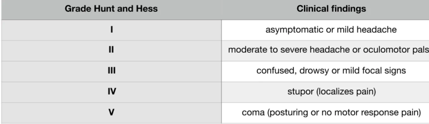
As we will describe in our analysis, the clinical condition at presentation is one of the most important predictor of final outcome. Numerous clinical scales have been proposed over the years to predict outcome. However, despite some more complex scales having better predictive value, only the Hunt Hess (HH) and the World Federation of Neurosurgical Societies (WFNS) scales have resisted the test of time (Table 1-2). Although not perfect, these scales are easy to remember and apply in clinical practice, have predictive value and good interobserver reliability. It is common to dichotomize either scale into good grades (Grades I through III) and poor grades (Grades IV and V) and this simple distinction is useful in practice.
Table 1: Hunt and hess grading system
Table 2: World Federation of Neurosurgical Societies (WFNS) scale
The symptomatology should lead to swift imaging (a computed tomography (CT) scan) that will serve as an initial diagnostic aid to determine the presence of subarachnoid blood. Timely exclusion of aneurysmal SAH is crucial to the patient’s subsequent management paradigm.
Grade Hunt and Hess Clinical findings
I asymptomatic or mild headache
II moderate to severe headache or oculomotor palsy
III confused, drowsy or mild focal signs
IV stupor (localizes pain)
V coma (posturing or no motor response pain)
WFNS scale Glasgow coma scale score Motor deficit
I 15 Absent
II 14-13 Absent
III 14-13 Present
IV 12-7 Present or absent
2.3 Diagnosis of aSAH
Non contrast head CT is the first line diagnostic study to confirm SAH. The sensitivity of non- contrast head CT is close to 100% in the first few hours after onset and progressively decreases over time (sensitivity of 98% when the scans are obtained within 1 to 5 days after bleed). 19 In the presence of a negative CT scan, because of the potential catastrophic consequences of misdiagnosing a ruptured aneurysm, lumbar puncture is indicated. In patients presenting days after onset, the lack of sulci or lack of visualization of the Sylvian fissure on head CT may be a useful indirect sign to suspect isodense subacute blood in the subarachnoid space.
Once the CT scan is positive for SAH, the patient should undergo further imaging to study the vasculature of the head and potentially the neck. The noninvasive vascular study of choice is a CT angiogram (CTA). The CTA can be performed efficiently with minimal risk to the patient (unless the patient has an iodine-related allergy or sensitivity). This modality is very sensitive for detecting the etiology of SAH. CTA has a specificity of 100% and a sensitivity of 96% to 99.7% for aneurysms that are 4 mm or larger. 20
The CTA allows effective assessment of the aneurysm’s morphology, nearby skull base anatomy, and intrasaccular thrombosis. However, CTA suffers from certain disadvantages, as it tends to underestimate the size of the aneurysm; reformatting of the images depends on the expertise of the available technician. The potential limitation of the CTA is to detect small aneurysms (< 4mm), especially within the crowded Sylvian cisterns. These small aneurysms require treatment among patients with a history of SAH from another aneurysm. A delayed detection on postoperative angiography can lead to un-necessary second surgery/ endovascular treatment. In specific cases of aneurysms, where there’s a bony artifact from the skull base and for their particular angioarchitecture, as for the posterior communicating artery aneurysm (PcomA) and anterior communicating artery aneurysm (AcomA) is justified the more frequent use of catheter angiography. Magnetic resonance imaging (MRI) is another alternative, but less efficient and cost effective, for initial
screening for detection of blood within the subarachnoid space. Fluid-attenuated inversion recovery (FLAIR) sequences have 100% sensitivity within the first 5 days after SAH, whereas T2-weighted gradient echo sequences have similar sensitivity within 6 to 30 days after the ictus. The sensitivity of magnetic resonance angiography (MRA) for detecting aneurysms >5 mm in size is 85% to 100%. 21
CTA is superior to MRA for detection and study of the aneurysm’s morphology and its intrasaccular contents. Patients with an iodine-based allergy or sensitivity may undergo MRI/MRA. Also, MRA is indicated for screening the patients who may suffer from the familiar forms of cerebral aneurysms.
If the CT scan is unremarkable for SAH or if the headaches occurred days before evaluation, but the patient’s symptoms are suspicious for occurrence of SAH, a lumbar puncture (LP) is indicated. The history and character of the headaches play an important role in the need to proceed with an LP whose purpose is to rule out the presence of xanthochromia.
Xanthochromia is found within hours (most likely after 12 hours) from the ictus and remains within the CSF for 3 to 4 weeks. If the CSF results are positive for xanthochromia, CTA is necessary, and if CTA is unremarkable, a catheter angiogram is indicated. When the CT and CTA results are unremarkable in a patient suspected of suffering from SAH, there is a 99% likelihood that SAH has been reliably ruled out. The 1% rate of misdiagnosis can be reduced based on effective history taking and performance of an LP. 22
Pitfalls leading to a misdiagnosis of aneurysmal SAH include failure to recognize the presenting symptoms and to understand the limitations of the CT scan following a sentinel hemorrhage, and failure to perform a lumbar puncture. Catheter angiography is the gold standard study for evaluation of aneurysms and occult arteriovenous malformations not detectable on other imaging modalities. This from of vascular imaging is unique in its therapeutic abilities via endovascular aneurysm embolization.
Catheter angiography is more sensitive to detect even smallest aneurysms and , in addiction, provides aneurysm characteristics, as aneurysm size, anatomic projection, neck-to-dome ratio, the robustness of the collateral circulation, and the anatomy of the neighboring vascular territories. Unlike CTA, it can also provide valuable information about flow dynamics within the corresponding vasculature.
The best way to perform an angiogram and to be considered negative has to respect these following three criteria:
1. Study of bilateral internal and external carotid arteries, bilateral vertebral arteries, including bilateral posterior inferior cerebellar arteries (this can be performed via a dominant vertebral artery).
2. Evaluation of the anterior communicating artery complex (it may be necessary to perform an internal carotid artery cross compression study)
3. Evaluate the presence of an atypical infundibulum near the focus of SAH. This minor vascular abnormality may occasionally require surgical exploration for its exclusion as the source of hemorrhage.
Repeat angiography is not necessary in patients with non-aneurysmal perimesencephalic SAH after a first negative catheter angiogram. While repeat delayed catheter angiography is indicated after 1-2 weeks if the initial angiogram is negative and there is a pattern of “diffuse” aneurysmal bleed on the initial CT. Overall, CTA and catheter angiograms play complementary rather than competitive roles in the management of complex intracranial aneurysms. CTA is invaluable for detection of calcium or thrombus within the aneurysm neck and dome, respectively. In addition, its analysis of the skull base provides indispensable information about the location of the relevant bony anatomy in relation to the components of the circle of Willis. For example, the anatomy of the anterior and posterior clinoid processes relative to the posterior
communicating artery and superior cerebellar/basilar bifurcation aneurysms is important for preoperative planning of the extent of bony removal and selection of the appropriate operative corridor, respectively.
2.4 Management of aSAH
Important considerations include the patient’s level of consciousness, time of onset of headache (and/or other symptoms) and focal neurologic deficits, as well as the presence of sentinel headaches, seizure, and use of anticoagulation/antiplatelet medications. In patients with an altered mental status, management of the airway, breathing, and circulation must be a priority. Patients who present with a Glasgow coma scale (GCS) score of 8 or worse should be considered for intubation and airway protection.
The patient is admitted to a neuro intensive care unit and appropriate studies are performed, including baseline electrocardiogram (to evaluate for arrhythmias), two- dimensional echocardiography to establish baseline heart function and screen for early cardiac dysfunction secondary to a catecholamine surge prompted by SAH, and relevant laboratory studies. Initial lab tests include a complete blood count and basic metabolic panel, as well as ionized calcium, magnesium, and phosphorous levels, and prothrombin time, partial thromboplastin time, blood type and screen, arterial blood gas, initial troponin level, and a urine drug screen.
An arterial and central venous catheter may be needed. The arterial line allows close monitoring of blood pressure and frequent blood draws, while the central line measures central venous pressure to assess volume status and determine the etiology of hyponatremia (which is commonly encountered in patients with SAH). Strict blood pressure control is necessary for patients presenting with hypertension and SAH who harbor an unsecured ruptured aneurysm. Typically, systolic blood pressure should be maintained below 140 mm Hg until the aneurysm is treated.
Blood pressure control can be effectively achieved using intravenous administration of a calcium channel blocker such as nicardipine or a beta-blocker such as labetalol. The presence of intraventricular blood and ventriculomegaly/hydrocephalus indicate the need for placement of an external ventricular drain (EVD). The laterality of the EVD should not interfere with the planned side of surgical approach. Liberal drainage of
CSF is not advised because this maneuver can cause abrupt changes in intracranial and transmural pressures, leading to aneurysmal rerupture. I usually leave the drain at 15 cm of water rather than the standard 10 cm. Statins are likely to decrease the risks of vasospasm, delayed neurologic deficits, and mortality; they are continued for 30 days. The absence of seizure activity limits the anticonvulsant medication dosing to 7 days. Calcium channel blockers (nimodipine) are administered for 21 days.
After the source of SAH has been established, further effort is focused on securing the ruptured aneurysm. The optimal treatment modality must be addressed on a case-by-case basis. However, there are four main considerations that influence treatment options (microsurgical versus endovascular):
1. Aneurysm morphology
2. Patient’s age, medical status, and presenting symptoms (e.g., HH grade)
3. The preferences of the patient and the family
4. The expertise of the treating surgeons/interventionalists
The long-term care of patients with microsurgically-treated aneurysms includes a CT or catheter angiogram at the 1- year postoperative visit. If a residual or recurrent aneurysm is not detected and the patient initially harbored only a single aneurysm, long-term surveillance imaging after clip ligation has a questionable utility because of the very low risk of recurrence. Patients who suffer from multiple aneurysms as patients with residual aneurysms should continue to undergo imaging every 5 years.
Furthermore, patients with an unruptured aneurysm who present with a sentinel headache (a severe nonthunderclap headache that is different from the patient’s usual headaches) should undergo urgent treatment of their aneurysm. These headaches indicate instability of the aneurysm and a pending risk of hemorrhage.
Screening of other family members should be considered if there are first-degree relatives with cerebral aneurysms. Diseases associated with aneurysms, such as coarctation of the aorta, polycystic kidney disease, fibromuscular dysplasia, and sickle cell disease, as well as cocaine use are other potential indications for prophylactic imaging.
2.5 Complications of aSAH and treatment
In patients with aSAH, the basic goal of treatment is focused on preventing secondary injuries which can occur as a result of neurologic (aneurysm rebleeding, increased intracranial pressure/hydrocephalus, and vasospasm) and systemic complications.
Rebleeding
Rebleeding is one of the most severe, potentially lethal and earliest complications of aSAH. The risk of rebleeding is maximal within the first 2-6 hours after onset. Once patients have been stabilized and/or transferred to a tertiary facility, the risk of rebleeding is approximately 4% within the first 24 hours and then 1.5%/day for the first 2 weeks. 41 Clinically, rebleeding is often associated (not preceded) by a sudden rise in systolic blood pressure, bradycardia, sudden loss of consciousness in an otherwise alert patients and tonic-clonic seizure. A tonico-tonic-clonic seizure in a patient with an unsecured ruptured aneurysm should be considered expression of rebleeding unless proven otherwise. The most effective measure to prevent rebleeding is securing the ruptured aneurysm. Antifibrinolytic drugs (i.e. epsilon-amino caproic and tranexamic acid) have been shown to reduce the incidence of rebleeding in the acute phase after aSAH.
Vasospasm
Vasospasm is a common complication of aSAH. Vasospasm usually starts between day 3 and 5, peaks between day 7 and 12 and resolves by day 14 to 21. A small subset of patients may suffer from ultra-early vasospasm (probably a “defensive” mechanism to limit the consequences of blood extravasation after the acute bleed). While angiographic vasospasm occurs in up to 70% of patients after aSAH, symptomatic
vasospasm occurs only in about 30%. 43 The neurological examination is by far the most important and reliable monitor for development of symptomatic vasospasm. Thus, after aneurysm treatment patients should be extubated as soon as safely possible and monitored by personnel trained in neurological examination. Transcranial Doppler is a non-invasive imaging tool which can be obtained at the bedside. Progressive elevated velocities should alert the clinician to be more vigilant in an individual patient.
During the period at risk for vasospasm patients are very sensitive to any kind of “secondary” insult which may precipitate cerebral ischemia. It is important to maintain an adequate volume status with the goal of euvolemia and avoid hypotension. The calcium antagonist nimodipine should be initiated as soon as the diagnosis of aSAH is confirmed. Induced hypertension is the first line of treatment in symptomatic patients along with maintenance of adequate volume status. In patients who fail to improve despite induced hypertension, imaging studies to confirm angiographic vasospasm such as CTA with or without perfusion or catheter angiography may be indicated. In refractory cases, pharmacological (through intra-arterial injection of vasodilatative agents) or mechanical (with endovascular balloons) angioplasty is helpful to counteract angiographic vasospasm. Prophylactic angioplasty is not indicated.
Hydrocephalus
Acute hydrocephalus and increased ICP are common in patients with aSAH. Although ventricle size is commonly used in clinical practice in patients with aSAH as a surrogate for hydrocephalus and potential need for CSF diversion, there is no association between ventricle size and the level of intracranial pressure. Similarly to patients with severe head injury, patients with high grade SAH (Grades IV and V) often have significant elevations of intracranial pressure without a correspondent increase in ventricle size. . 34
In patients with compromised level of consciousness or progressive worsening neurological condition because of acute hydrocephalus/increased ICP, CSF diversion, usually by placement of an external
ventricular drainage (EVD) is indicated. It is controversial whether placement of an EVD in a patient with an unsecured ruptured aneurysm increases the risk of rebleeding by decreasing the transmural pressure across the aneurysm wall. In patients without intraparenchymal hematomas and no evidence of tonsillar herniation on CT, a lumbar drain is a valid alternative to an EVD.
In addition to acute hydrocephalus, patients with aSAH are at risk for delayed communicating hydrocephalus probably related to a dysfunction of the mechanisms of CSF reabsorption. Risk factors for delayed hydrocephalus include increasing age, higher subarachnoid blood burden, and associated intraventricular hemorrhage. Delayed hydrocephalus is usually associated with decreased level of activity, sleepiness, gait ataxia, and general slowness in a patient who is otherwise recovering from aSAH. These patients benefit from permanent CSF diversion through placement of a ventriculo-peritoneal shunt. . 35
Systemic complications
Systemic complications are common after aSAH as arrhythmias and ECG changes. These are related to increased catecholamine release secondary to aSAH more than an underlying coronary pathology. Electrolyte imbalance and especially hyponatremia are also common. Deep venous thrombosis prophylaxis should be instituted in patients with impaired mobility once the aneurysm has been secured.4
Outcome and Follow-up
Case fatality rates after aSAH varies widely across different studies and have decreased over time. It is estimated that 35 to 55% of survivors of aSAH achieve a good functional outcome (modified Rankin 1-3) at follow-up. 6 Patients who survive aSAH without residual physical deficits often suffer from cognitive dysfunction and mood disorders which affect overall quality of life. Patients with aSAH are at risk for new
aSAH. Recurrent aSAH can be related to rupture of incompletely obliterated aneurysms, newly formed aneurysms, or regrowth of previously treated aneurysms. Younger age at the time of the first aSAH, current smoking and family history are associated with higher risk of recurrent aSAH. The interval and frequency of interval of follow-up in patients with history of aSAH remains a matter of controversy.
3. Material and methods
3.1 Patient consent and study design
The study was approved by the Mayo Clinic Institutional Review Board. We conducted a retrospective review of all the consecutive patients diagnosed with aneurysmal subarachnoid hemorrhage (aSAH) who were treated at Mayo Clinic (Rochester, MN) between January 2005 and December 2014. Cases with a final diagnosis of aSAH were identified using our electronic data search system and confirmed by manual review of the medical records. All included patients or their representative had signed a general informed consent form allowing their medical records to be used for research purpose.
3.2 Study definitions
We defined as suffering of aneurysmal SAH, patients with a confirmed aneurysmal bleeding pattern on CT scan or, in case of negative CT scan but with a strong history of SAH, patients with blood-positive lumbar puncture. Patients were included only if an aneurysm was documented by a vascular study, as CT-Angio, catheter angiography (the most used diagnostic procedure), AngioMRI (in rare cases, as any allergies to the iodate contrast). Furthermore, patients were included if they have been admitted to the hospital within 48 hours of symptoms onset. We excluded patients with angiographycally negative aSAH (as the perimesencephalic SAH) and those with SAH from other causes (as arteriovenous malformations). Patients under 18 age were also excluded.
Patients with a final diagnosis of aSAH were initially identified using electronic data research system, and the diagnosis was confirmed by manual review of the medical records.
3.3 Collected variables
Electronic health records of patients were abstracted for patient demographic characteristics, age, sex, worse severity at presentation, location of the aneurysm, type of aneurysm treatment, occurrence of rebleeding during the hospital staying, symptomatic vasospasm or delayed cerebral ischemia, hospital mortality, outcome at the discharge and at 3- to 6- months follow up. Neurological examination of patient was described through the Hunt and Hess grading and the World Federation of Neurosurgical Societies (WFNS). We also defined the Hunt and Hess (HH) grading of aSAH at initial valuation as follows: “good” for 1-3 HH scale and for 1-3 WFNS scale, “worse” for 4-5 HH and for 4-5 WFNS.
Location of the ruptured aneurysm was classified into anterior circulation (anterior communicating artery, anterior cerebral artery, pericallosal artery, middle cerebral artery, internal carotid artery, posterior communicating artery, ophthalmic artery), posterior circulation (vertebral artery, basilar artery, posterior cerebral artery) or both (in cases with double rupture of aneurysms in anterior and posterior circulation). Aneurysm treatment was categorized into neurosurgical clipping, endovascular coiling, bypass surgery, or no treatment. We checked the occurrence of vasospasm, DCI, rebleeding and the hospital mortality. Clinical outcome was assessed at the discharge and at 3- to 6- months follow-up.
Rebleeding was defined as a documented increase in the previous amount of aSAH, increased blood in the ventricular system on repeat head CT in the presence of sudden neurological deterioration, or both. Symptomatic vasospasm was defined as documented vasospasm (by ultrasonographic or angiographic criteria) associated with otherwise unexplained neurological decline. 14
Delayed cerebral ischemia was defined as symptomatic vasospasm or the appearance of new infarction on CT or magnetic resonance imaging attributable to vasospasm, thus excluding periprocedural complications. For the purpose of the analysis, delayed ischemia is defined as the combination of symptomatic vasospasm
and delayed cerebral ischemia. Clinical outcome was categorized at discharge and at 3 to 6 months after discharge using the modified Rankin Scale (mRS), with good outcome defined as a score of 0 to 2.
3.4 Statistical analysis
Patients admitted at Mayo Clinic from January 2005 to December 2014, with diagnosis of aSAH were considered eligible for this study purpose. Demographics were analyzed considering time as a single variable. Descriptive summaries were reported as medians and interquartile ranges for continuous variables and frequencies and percentages for categorical variables.
Multivariate analysis for mortality and for functional outcome after controlling for comorbidities were evaluated. Outcomes of interest were in-hospital death and functional outcome. All tests were 2-sided, and P values less than .05 were considered statistically significant. Statistical analyses were performed with JMP 12.0.0 (SAS Institute Inc.).
4. Results
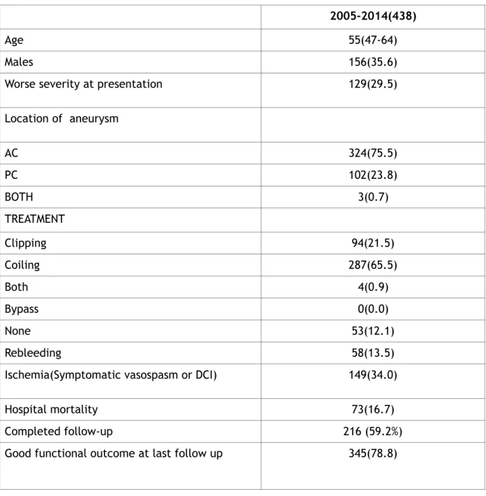
Our research identified 438 patients suffering of aSAH admitted to our department from January 2005 to December 2014. Patients demographics, worse severity at presentation, aneurysm characteristics, treatment modalities, complications as rebleeding, ischemia or symptomatic vasospasm or DCI, hospital mortality and functional outcome at last follow up are summarized in the table 3.
Table 3: Demographics are showed. Severity at presentation: H&H scale of 1-3 (good) vs 4-5(worse) WFNS scale of 1-3 (good) vs 4-5(worse) Ischemia: either symptomatic vasospasm or DCI
2005-2014(438)
Age 55(47-64)
Males 156(35.6)
Worse severity at presentation 129(29.5)
Location of aneurysm AC 324(75.5) PC 102(23.8) BOTH 3(0.7) TREATMENT Clipping 94(21.5) Coiling 287(65.5) Both 4(0.9) Bypass 0(0.0) None 53(12.1) Rebleeding 58(13.5)
Ischemia(Symptomatic vasospasm or DCI) 149(34.0)
Hospital mortality 73(16.7)
Completed follow-up 216 (59.2%)
Good functional outcome: modified rankin scale: 0-3/Glasgow outcome scale: 4-5
The mean age at presentation was 55 years (47-64 years). As typical for aSAH, the majority of patients were female (64.4%), with male representing only the 35.6% of series. Interestingly, 129 patients (29.5%) were admitted with poor clinical grade (that represents a progressive increase if compared to a previous series). 23 Location of the aneurysm reflects the general prevalence of the anterior and posterior circulation aneurysm: 75.5% were found to be anterior locating aneurysms, 23.8% were posterior located aneurysms, while 3 patients (0.7%) had both anterior and posterior circulation aneurysms. Reebleeding was observed in 58 patients (13.5% of our series).
Treatment modalities used during this period were: clipping, coiling, both clipping and coiling, none. No procedure involved the by-pass. The most used technique was coiling: 65.5% (287 patients, according to the general increasing use and development of coiling since the 90s); 94 patients (21.5%) underwent surgical clipping), while 4 patients (0.9%) received coiling and clipping procedures; 53 patients were not treated at all (12.1%).
Cerebral ischemia (or symptomatic vasospasm or DCI) occurred in 149 patients (34.0%). 73 patients (16.7%) died during hospital staying. Among patients who survived hospitalization, 216 patients completed the follow-up at 3 and 6 months. Good functional outcome at last follow-up was observed in 345 patients (78.8%).
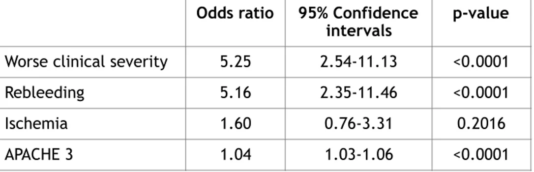
4.1 Multivariate analysis for mortality
Having these datas, we conducted a multivariate analysis for mortality and a multivariate analysis for poor functional outcome, after controlling for comorbidities, considering worse clinical severity, rebleeding, ischemia and APACHE 3 for mortality rate analysis, while age, worse clinical severity, ischemia, rebleeding and APACHE 3 for poor functional outcome multivariate analysis. We observed that worse clinical severity and rebleeding were the most important factors influencing the mortality, 5.25 (2.54-11.13 confidence interval CI with p value < 0.0001) and 5.16 odds ratio OR (2.35-11.46 confidence interval with p value < 0.0001) respectively. Ischemia and APACHE 3 were not statistically relevant for mortality occurrence (1.60 OR, 0.76-3.31 CI and 1.04 OR; 1.03-1.06 CI respectively). See Table 4 below.
Table 4 Multivariate analysis for mortality after controlling for comorbidities. Odds ratio 95% Confidence
intervals p-value
Worse clinical severity 5.25 2.54-11.13 <0.0001
Rebleeding 5.16 2.35-11.46 <0.0001
Ischemia 1.60 0.76-3.31 0.2016
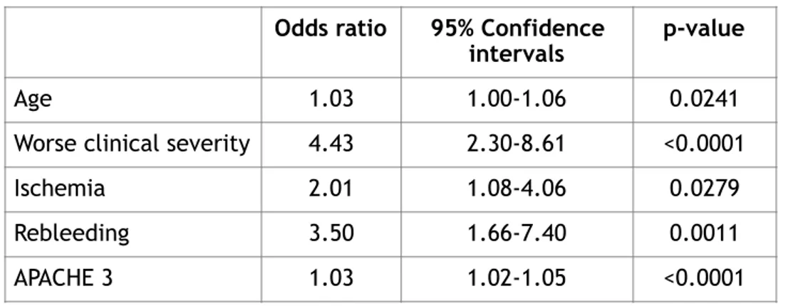
4.2 Multivariate analysis for poor functional outcome
When considering the functional outcome, the most important factors related to a poor clinical conditions were worse clinical severity at presentation (4.43 OR, 2.30-8.61 CI) and rebleeding (3.50 OR, 1.66-7.40 CI). In this case, cerebral ischemia had a little increasing role compared to the multivariate analysis for mortality (2.01 OR, 1.08-4.06 compared to 1.60 OR, 0.76-3.31 CI), but with no reaching statistical significance. See Table 5 below.
Table 5 Multivariate analysis for poor functional outcome after controlling for comorbidities. Odds ratio 95% Confidence
intervals p-value
Age 1.03 1.00-1.06 0.0241
Worse clinical severity 4.43 2.30-8.61 <0.0001
Ischemia 2.01 1.08-4.06 0.0279
Rebleeding 3.50 1.66-7.40 0.0011
5. Discussion
Nothing is more troublesome of dealing with aneurysmal subarachnoid hemorrhage in neurosurgical practice. Patients’ lives are significantly affected by the severity of the initial hemorrhage and of the potential neurological complications such as rebleeding, vasospasm, and hydrocephalus.14
Moreover, during their hospital staying, patients are exposed to the risks of primary and secondary injuries and of any critically systemic disease including pulmonary embolism, deep venous thrombosis (DVT), electrolyte disorders, arrhythmias, and infectious complications in addition to the associated complications of SAH.
Although the mortality rate has decreased in the past 30 decades in our institution, 23 from 22.6% (in the pre-coiling era from 1985 to 1995) to 16.7% in 2005-2014, we investigated if there is also a better functional outcome in those survivors patients, because patients are often young persons with many years of productive life ahead of them. In our previous paper, good functional outcome within 3 to 6 months after aSAH improved consistently, from 64.8% (173) in 1985 to 1994 to 72% (332) in 1995 to 2004 and 78.8% (345) in 2005 to 2014. This considerable improvement in functional outcome is even more remarkable considering that it occurred despite a substantial increase in the percentage of patients admitted in poor neurological condition, from 22.3% (61) in 1985 to 1994 to 24.1% (111) in 1995 to 2004 and 29.5% (129) in 2005 to 2014.
In this thesis we wanted to investigate which factors are ascribable to these results. In our multivariate analysis of 438 patients, admitted to our hospital with a diagnosis of aSAH, in the period from 2005 to 2014, we find out that worse clinical severity at presentation and rebleeding are the most important factors influencing mortality and a poor functional outcome.
Udy et al,13 have reported a total of 11,327 patients with aSAH in the Australian and New Zeland Intensive Care Society Center for Outcome and Resource Evaluation Adult Patient Database. They found that the overall case fatality rate was 29.2%, which declined from 35.4% in 2000 to 27.2% in 2015 (p value 0.01). In their multivariate analysis (p value < 0.05), older age, non operative admission, mechanical ventilation, higher Acute Physiology and Chronic Health Evaluation III score (APACHE III), lower Glasgow Coma Scale, and admission prior to 2004, were all associated with lower hospital survival.
Vergouwen et al15observed a significant decrease in 90-day case-fatality rate, from 39% in 1999 to 2002 to 30% in 2009 to 2012, which was ascribed in part to a decrease in the incidence of in-hospital rebleeding from 24% to 17% in the 2 periods. This decrease in the incidence of rebleeding in their study, coincided with earlier treatment of aneurysms (median day decreased from day 4 to day 1). Specifically the authors compared causes of in-hospital death after aneurysmal subarachnoid hemorrhage (aSAH) in 2 time periods (1999-2002 and 2009-2012) within the same institution. Ninety-day case-fatality declined from 150/381 patients (39%) in 1999–2002 to 140/463 (30%) in 2009–2012 (aRR 0.74 [95% CI 0.62–0.88]). Compared with 1999–2002, the aRR for specific cause of in-hospital death in 2009–2012 was 1.06 (95% CI 0.72–1.56) for death from the initial bleeding, 0.47 (95% CI 0.31–0.71) for death from rebleeding, and 0.91 (95% CI 0.50–1.65) for death from delayed cerebral ischemia. Over time, the proportion of patients with in-hospital rebleeding declined from 90/381 (24%) to 78/463 (17%) (aRR 0.68 [95% CI 0.52–0.90]), median day of rebleeding from day 5 (IQR 1–10) to day 0 (IQR 0–1), and median day of aneurysm treatment from day 4 (IQR 2–13) to day 1 (IQR 1–2).
As described by Nieuwkamp et al, 24 in a population based-study meta-analysis of 9403 patients, the decrease of mortality is not related to the age or gender. Similarly we found no relation between age and poor functional outcome in our study population series.
Lord et al25 identified 120 patients with rebleeding and 359 control subjects from 1996 to 2011. The rebleeding rate was 8.6%. In both the primary and secondary analyses, there was no difference in the incidence of DCI or its time course (29% vs. 27%, p = 0.6; 7 ± 5 vs. 7 ± 6 days, p = 0.9 for primary analysis; 39% vs. 31%, p = 0.1, 7 ± 5 vs. 7 ± 6 days, p = 0.6 for the secondary analysis). In a multivariate logistic regression model, rebleeding was associated with the complications of hyponatremia, respiratory failure, and hydrocephalus. Patients with rebleeding had higher rates of mortality, brain death, and poor outcomes. Zhao et al reported in their study including 297 patients, 30 patients (10.1%) experienced rebleeding.26 Most rebleeding occurred within 24h after ictus. 22 (73.3%) patients died at discharge. Aneurysm rebleeding was independently associated with poor outcome (odds ratio [OR] 36.37, p<0.001) and associated with mortality (OR 25.03, p<0.001) at 12 months. The multivariate analysis showed that a lower Fisher grade (OR 0.49, 95% CI 0.31-0.77; p=0.002), ruptured anterior cerebral artery aneurysms (OR 4.26, 95% CI 1.07-16.90; p=0.039), external ventricular drainage (OR 4.62, 95% CI 1.46-14.59; p=0.009) were independently associated with aneurysm rebleeding.
In another case study conducted by our institution27 and analyzing functional outcome in case of occurrence of rebleeding, we found that rebleeding and DCI after aSAH are highly morbid and potentially deadly events after aSAH, which appear to have independent negative impacts on overall functional outcome. Rebleeding was associated with significantly worse functional outcome, longer intensive care unit length of stay (LOS), and GCE (all p < 0.01); treatment modality, overall LOS, DCI, DCI with infarction, and SV were nonsignificant. In the PSM analysis of 43 matched re-bled and 43 matched nonrebleed cases, only poor functional outcome and GCE remained significantly associated with rebleeding (p < 0.01 and p = 0.02, respectively). Multivariate regression identified that both rebleeding (HR 21.5, p < 0.01) and DCI (HR 10.1, p = 0.01) independently predicted poor functional outcome.
5.1 What definitely has changed in the management of aSAH
Thanks to a better knowledge of the pathophysiological mechanisms underlying the aSAH and the improvement in neurocritical care, the management of aSAH has undergone some impressive changes leading to a better functional outcome and lowering the mortality rate.
It si already known that very few diseases in medicine test the resilience of a team like aneurysmal subarachnoid hemorrhage. Patients are exposed to primary and secondary injuries from this complicated pathology: they are significantly affected by the severity of the initial hemorrhage and of the potential neurological complications such as rebleeding, vasospasm, and hydrocephalus. Moreover, patients are exposed to the risks of clinical disorders, as pulmonary embolism, deep venous thrombosis (DVT), electrolyte disorders, arrhythmias, and infectious complications in addition to the associated complications of SAH.28
After a diagnoses of SAH is done at the emergency room, neurolo-intensivists, neurosurgeons, and neuro-radiologists are notified. The patient is admitted to the neurosciences intensive care unit and his clinical parameters stabilized, before further management of the SAH. We try to maintain oxygen saturations 95% using no-invasive measures as a first line. We routinely obtain baseline chest X-ray for comparison in case acute pulmonary decompensation occurs later (due to neurogenic pulmonary edema, aspiration events, or pneumonia) and a baseline electrocardiogram for evaluation of cardiac status. We also evaluate systemic blood pressure, which is often elevated. We prefer, if possible, to maintain the systolic blood pressure below 160 mmHg, with extreme caution to avoid overcorrecting the systolic blood pressure below 100 mmHg. We prefer to use a short-acting intravenous beta-blocking agent such as Labetolol.
There is no clear evidence that increased systemic blood pressure can increase the risk of re-rupture, but hypotension must be avoided because the injured brain is very sensitive to any secondary injury.
All these precautions are the results of our intensive studies, clinical observation and progression in neurocritical care. We have also noticed that laboratory evaluation including blood count and electrolytes are necessary to maintain a balance in such a compromised system.
Initial laboratory evaluation includes a complete blood count to evaluate hemoglobin and platelets. Our goals during the acute phase are hemoglobin .8 g/dl if the patient is at minimal risk for cerebral ischemia and .10 g/ dl if at increased risk, and platelets .1006106/l. Baseline electrolytes are also obtained and renal function is evaluated with creatine clearance and glomerular filtration rate estimates. If the patient has poor kidney function, n-acetylcysteine is administered with a fluid bolus for its potential renal protection. 29
Once all these medical parameters are fixed, the main focus is prevention of secondary injuries, as rebleeding, vasospasm and hydrocephalus.
We initially had a bad reputation of antifibrinolytic drugs in aneurysmal SAH. This bad reputation was due to some large randomized trials done in the late 1970s and early 1980s. In those studies, antifibrinolytic drugs were used in a continuous intravenous infusion form in very high doses and were administered for several days. They can prevent rebleeding but at the same time, caused a higher incidence of hydrocephalus and cerebral ischemia due to vasospasm. Therefore, their use was abandoned. In more recent years, as Hillman et al described in their randomized trial in Sweden, antifibrinolytic drugs have been proposed again30 . Among 254 patients in the study group who received tranexamic acid, a reduction in rebleeding rate from 10.8 to 2.4% was shown compared to the control group. The protection afforded by tranexamic acid treatment was also suggested by a 3.4% decrease in the overall mortality rate and a 4.3% increase in favorable overall management outcome, indicating that patients move through the outcome scale without adding poor outcomes. Hence, the use of antifibrinolytics in the pre-neurosurgical phase of management was
proposed to effectively reduce the number of very early rebleeding episodes.
We routinely administer tranexamic acid (Cyklokapron, Pfizer, New York, USA) 1 g every 6 hours until the aneurysm is fully protected.
Once the patient is stabilized, the next step, in addition to prevention of rebleeding, is prevention of secondary injury. Patients with aneurysmal SAH have compromised microcirculation ongoing from the acute hemorrhage.31-32 Sehba et al. induced SAH in rats using an endovascular perforation model and documented the widespread aggregation of platelets in cerebral microvessels following an episode of SAH. This aggregation peaks 24 hours later and contributes to microvascular perfusion deficits directly by mechanically obstructing the lumen and indirectly by releasing compounds that cause vasoconstriction and also by releasing enzymes that initiate cascades leading to destabilization of microvascular structure.33
In addition, intracranial pressure is often significantly high, even in those patients who are fully awake but complain of a severe headache. Traditionally, the decision of whether or not to proceed with external ventricular drainage in these patients has been based on either an interval increase in the size of the ventricles or based on the presence of a depressed and compromised level of consciousness using the Glasgow coma scale. As we already stated in the previous paragraph, (see paragraph 2.5) we are very aggressive in the management of intracranial pressure in patients with SAH. That is based on the belief that an increased intracranial pressure further limits the nutritive exchanges at the level of the microcirculation and may increase the occurrence of a delayed cerebral infarction secondary to vasospasm. Our institution has also anecdotally described a clinical case of patient where the placement of an external ventricular drain improved the systemic hypertension and his headaches.34
We usually maintain the external ventricular drainage (EVD) at 5 cm up to the meatus and continuously open in order to avoid spikes in the intracranial pressure seen with drainage at higher pressures (e.g. 15 cm above the meatus) or with intermittent drainage and also to allow for continuous flow through the external
ventricular drain in order to prevent clotting of the drain itself. This aggressive cerebrospinal fluid (CSF) diversion strategy is not without complications, including overdrainage and the formation of subdural hygromas. The rate of infectious complications and ventriculitis is lowered thanks to the use antibiotic-impregnated ventricular catheters.35-36
The next step after medical stabilization and administration of antifibrinolytics is diagnosis of the target aneurysm and definitive treatment.
It is important that up to 15% of patients who present with SAH are not found to have intracranial aneurysms. Patients with typical non-aneurysmal perimesencephalic pattern are fast tracked and after one negative angiography, they are transferred to a regular floor and usually discharged after a few days with the recommendation of avoiding dehydration. No repeat imaging studies are recommended in patients with the typical perimesencephalic pattern after a first negative high- quality six-vessel catheter angiography.
5.2 What we can improve in the management of aSAH
The two reasons identified in our multivariate analysis leading to worse clinical outcome and engraving on mortality are worse clinical conditions at presentation and rebleeding. It’s very difficult to improve the clinical conditions of patients at admission. Still we can’t say exactly if and when an aneurysm will rupture, besides all the clinical studies that tried to predict it. We still have to face with the ISUIA study, published on Lancet in 2003,37 that analyzed the risk of rupture of anterior and posterior circulation aneurysms. Among the 1692 patients observed, they found that the cumulative risk of rupture for anterior circulation aneurysms smaller than 7 mm was 0.1% in 5 years, while 2.5% for the same dimensioned aneurysms of the posterior circulation. These percentages dramatically increase in case of bigger aneurysms: 40% for anterior circulation aneurysms > 25 mm, and 50% for the posterior ones.
For this reason, risk factors scores have been elaborated to predict if an aneurysm will rupture or not, as the PHASES score38 and to rule out if treatment of the aneurysm is safer than to keep it unfixed, as the UIATS score39 . Although these scores efforts, no general consensus and general guidelines of treatment are found to decide if fixing an incidental aneurysm is better than keep it unfixed. Some recent studies are trying to select the aneurysms at risk of rupture through the computational fluid dynamics (CFD).40 It utilizes mechanical engineering principles to explicate what occurs in tubes (vessels) and bulges (aneurysms). Hemodynamic parameters of wall shear stress (WSS), WSS gradient, inflow jet, impingement zone, and aneurysm inflow-angle are considered in papers from 1970 to 2010, but a patient-specific CFD model can predict the rupture risk of unruptured intracranial aneurysms remains to be determined in future studies incorporating multivariate analysis.
What we strongly believe is that keeping an aneurysm unfixed, potentially will affect the patient neurological outcome when the rupture occurs. Thus, when facing with an unruptured aneurysm, keeping in mind that no
real correlation with the dimension has been clearly postulated and that its rupture can lead to a worse severity outcome till death, we recommend treatment if we feel perceptive that in our hands that aneurysm
can be fixed safely. Of course a better knowledge of the pathology itself and identifying those aneurysms that once ruptured, will have a worse neurological condition at presentation, can improve the neurological outcome and the mortality rate.
As we have previously stated in this thesis, rebleeding is an important risk factor both for patient’s neurological and clinical outcome and for death. Various tricks are used by neurologists, neurosurgeons, neuroradiologists and neuro-anesthesiologists, to prevent it. In a recent publication, Duangthongphon et al.41 stabilized a protocol for preventing rebleeding which consisted of absolute bed rest, adequate pain control, avoiding stimuli, keeping euvolemia, preoperative systolic blood pressure <160 mmHg and within 140-180 mmHg after definite treatment, a short course (<72 h) of intravenous transaminic acid, and aneurysm treatment as early as possible. They compared, over a 3-years period, among 208 patients with aSAH, the two cohorts between the group of patients before initiating the protocol (n = 104) and after initiating the protocol (n = 104). They found that there was a reduction in the incidence of in-hospital rebleeding from 6.7% to 2.8% (P = 0.20, odds ratio [OR] = 0.4, 95% confidence interval [CI] = 0.10-1.63) and in the proportion of patients who presented with good WFNS grades (1-3) with unfavorable clinical outcomes at 12 months from 27.0% to 12.8% (P = 0.03, OR = 0.40, 95% CI = 0.17-0.95). The DCI experienced a significant reduction from 44.2% to 7.7% (P < 0.001, OR = 0.10, 95% CI = 0.04-0.23), and their 180-day mortality rate in good WFNS grades patients decreased from 16.3% to 8.8% (hazard ratio 0.80, 95% CI = 0.28-2.28).
Besides all these clinical management tricks that lowered the rebleeding percentage and all the related complications, we believe that more than any other factor, what really prevent rebleeding is the early treatment of the ruptured aneurysm.1 The advent of coiling embolization had offered a great contribution to decrease mortality rate, as it was reserved primarily for patients at surgical high risk, with medical
comorbidities and/or in poor neurological conditions. Nowadays endovascular treatment is used also as a valide alternative to open surgery, but long-term results regarding the definitive closure of the aneurysm are still scant and controversial. Meanwhile, the introduction of intraoperative microvascular Doppler ultra- sonography and intraoperative indocyanine green angiography has allowed refinement of open surgical techniques. These developments have generated a new paradigm using which case-by-case decisions are made by a team of specialists with endovascular and open surgical expertise to select the best treatment option (balancing efficacy against risk of each therapeutic modality) for each patient.42
We believe that further improvements both in surgical and endovascular field and a better selection of which patient is candidate for one treatment or the other, will lead us to a near to zero worse clinical outcome and a lower rate of mortality.
Despite in our multivariate analysis the cerebral ischemia, as a consequence of severe vasospasm, has no the same statistical importance of the above-mentioned factors, we feel perceptive that also the vasospasm has an important role in the clinical outcome, more than for mortality. Several studies have been conducted to prevent the vasospasm, most of them focused on the intravenous or oral administration of nimodipine . More recent paper have focused on the intrathecal administration of vasodilators and sympatholytic or thrombolytic therapies.43-44
Impressive but interim results are published in a recent paper on effects of intrathecal verapamil on cerebral vasospasm in rats.45 A total of 24 Sprague-Dawley rats were randomly divided into the following 3 groups: group 1 (sham), group 2 (subarachnoid hemorrhage), and group 3 (verapamil). A double hemorrhage method was used. Group 2 did not receive any treatment. Verapamil (Eporon, Dem Ilac, Turkey) at a dose of 1000 µg/kg was given intrathecally to group 3 rats. Arterial wall thickness and lumen diameter in the basilar arterial cross-sectional areas, endothelin-1 serum level, oxidative stress index, and apoptosis were measured in all groups. In the verapamil group, wall thickness, endothelin-1 level, oxidative stress index, and apoptosis were found to be significantly lower than the subarachnoid hemorrhage group, but the lumen diameter was found to be greater. Intrathecal verapamil was found to decrease vasospasm parameters and apoptosis and
increase the antioxidant and antiapoptotic pathways. Also in this field we strongly believe that more improvements can be obtained since all the ongoing studies will be enriched and completed.
5.3 Study limitations
Our study has limitations inherent to its retrospective design. Our analysis did not include other relevant factors affecting mortality and worse clinical outcome such as radiological grade to assess the risk of vasospasm, comorbidity’s conditions, and acute systemic complications. Furthermore, for the multivariate analysis purpose, we have arbitrarily decided to consider only the worse clinical severity at presentation, the rebleeding, the ischemia and the APACHE3, and added the age for the poor clinical outcome analysis.
However, these deficiencies should not affect our main results. Our findings may be affected by referral bias, but we tried to limit this by considering only patients admitted within 48 hours from their ictus.
Unlike several other studies on this topic in which the diagnosis of aSAH could not be confirmed, or in which patients with other causes of subarachnoid hemorrhage were included, our study included a population that was homogeneous and included only patients with ruptured aneurysms. The availability of data on post-discharge functional outcome in our cohort also represents an improvement over previous studies, although the functional outcome is restricted to 3- to 6- months of follow-up.
6. Conclusions
Our results clearly indicate that outcome in patients with aSAH is strongly influenced by rebleeding and worse clinical at presentation, with a lower influence from cerebral ischemia/vasospasm. Furthermore, rebleeding and worse clinical at presentation are intimately connected to the in hospital mortality or at 3- to 6- months follow-up.
Although these results are clear and satisfactory, we believe the a more detailed investigation is necessary to determine whether this or other factors may directly explain the trends in survival and functional recovery over time in patients affected by aSAH.
References
1. Rabinstein AA, Lanzino G, Wijdicks EF. Multidisciplinary management and emerging therapeutic strategies in aneurysmal subarachnoid haemorrhage. Lancet Neurol. 2010;9(5):504-519.
2. Lanzino G, Murad MH, d’Urso PI, Rabinstein AA. Coil embolization versus clipping for ruptured intracranial aneurysms: a meta-analysis of prospective controlled published studies. AJNR Am J Neuroradiol. 2013;34(9):1764-1768.
3. Sorenson T, Lanzino G. Trials and tribulations: an evidence- based approach to aneurysm treatment. J Neurosurg Sci. 2016; 60(1):22-26.
4. Samuels O, Webb A, Cullers, Martin K, Barrow D Impact of a dedicated neurocritical care team treating patients with aneurysmal subarachnoid hemorrhage. Neurocrit care 2011; 14(3):334-340.
5. Johnston SC, Selvin S, Gress DR. The burden, trends, and demographics of mortality from subarachnoid hemorrhage. Neurology. 1998;50(5):1413-1418.
6. Lovelock CE, Rinkel GJ, Rothwell PM. Time trends in outcome of subarachnoid hemorrhage: population-based study and systematic review. Neurology. 2010;74(19):1494-1501.
7. Mackey J, Khoury JC, Alwell K, et al. Stable incidence but declining case-fatality rates of subarachnoid hemorrhage in a population. Neurology. 2016;87(21):2192-2197.
8. Macpherson KJ, Lewsey JD, Jhund PS, et al. Trends in incidence and in short term survival following a subarachnoid haemorrhage in Scotland, 1986-2005: a retrospective cohort study. BMC Neurol. 2011;11:38.
9. Mukhtar TK, Molyneux AJ, Hall N, et al. The falling rates of hospital admission, case fatality, and population-based mortality for subarachnoid hemorrhage in England, 1999-2010. J Neurosurg. 2016;125(3):698-704.
10. Nieuwkamp DJ, Setz LE, Algra A, Linn FH, de Rooij NK, Rinkel GJ. Changes in case fatality of aneurysmal subarachnoid haemorrhage over time, according to age, sex, and region: a meta-analysis. Lancet Neurol. 2009;8(7):635-642.
11. Sandvei MS, Mathiesen EB, Vatten LJ, et al. Incidence and mortality of aneurysmal subarachnoid hemorrhage in two Norwegian cohorts, 1984-2007. Neurology. 2011;77(20):1833-1839.
12. Worthington JM, Goumas C, Jalaludin B, Gattellari M. Decreasing risk of fatal subarachnoid hemorrhage and other epidemiological trends in the era of coiling implementation in Australia. Front Neurol. 2017;8:424.
13. Udy AA, Vladic C, Saxby ER, et al; Australian and New Zealand Intensive Care Society Centre for Outcome and Resource Evaluation. Subarachnoid hemorrhage patients admitted to intensive care in Australia and New Zealand: a multicenter cohort analysis of in-hospital mortality over 15 years. Crit Care Med. 2017;45(2):e138-e145.
14. Connolly ES Jr, Rabinstein AA, Carhuapoma JR, Guidelines for the management of aneurysmal subarachnoid hemorrhage: a guideline for healthcare professionals from the American Heart Association/ american Stroke Association. Stroke. 2012 Jun;43(6):1711-37.
15. Kammen MSV, Greving JP, Kuroda S, et al. External Validation of the ELAPSS Score for Prediction of Unruptured Intracranial Aneurysm Growth Risk. J Stroke. 2019 Sep;21(3):340-346
16. Rehman S, Sahle BW, Chandra RV et al. Sex differences in risk factors for aneurysmal subarachnoid haemorrhage: Systematic review and meta-analysis. J Neurol Sci. 2019 Aug 31;406:116446.
17. D'Souza S, Aneurysmal Subarachnoid Hemorrhage J Neurosurg Anesthesiol. 2015 Jul; 27(3):222-40 18. Macdonald RL, Schweizer TA. Spontaneous subarachnoid haemorrhage. Lancet 2017 Feb 11;
389(10069):655-666
19. D u b o s h N M , B e l l o l i o M F, R a b i n s t e i n A A , E d l o w J A . S e n s i t i v i t y o f E a r l y Brain Computed Tomography to Exclude Aneurysmal Subarachnoid Hemorrhage: A Systematic Review and Meta-Analysis. Stroke. 2016 Mar;47(3):750-5.
20. Grossi G, Romanzi F, Macchia G, Ruffinengo U, Calia S. Angio-CT. A Proposal for Emergency Diagnosis in Subarachnoid Hemorrhage as a Preliminary to Therapeutic Choices. Interv Neuroradiol. 1995 Nov 30;1(1):43-57. Epub 2001 May 15
21. Gasparotti R, Bonetti M, Crispino M, Pavia M, Chiesa A, Galli G. [Subarachnoid hemorrhage: assessment in the acute phase with angiography, with high-resolution magnetic resonance (angio-MR)]. Radiol Med. 1994 Mar;87(3):219-28
22. Gangloff A, Nadeau L, Perry JJ, Baril P, Émond M Ruptured aneurysmal subarachnoid hemorrhage in the emergency department: Clinical outcome of patients having a lumbar puncture for red blood cell count, visual and spectrophotometric xanthochromia after a negative computed tomography. Clin Biochem. 2015 Jul;48(10-11):634-9)