0
Università degli Studi "ROMA TRE”
Scuola Dottorale in Biologia (XXV ciclo) Sezione “Scienze biomolecolari e cellulari”
Malattie mitocondriali dovute a mutazioni in SDHAF1 e TMEM70: fattori di assemblaggio del Complesso II e Complesso V della Catena Respiaratoria mitocondriale umana
Mitochondrial disorders due to mutations in SDHAF1and TMEM70: assembly factors of human mitochondrial Respiratory Chain Complexes II and V
Tutors:
Prof. Paolo Mariottini (Università Roma Tre) Dott. ssa Rosalba Carrozzo (Ospedale Bambino Gesù) INDEX
Dottoranda:
1
ABSTRACTS 3
1. INTRODUCTION 11
1.1 MITOCHONDRIA 11
1.1.1 Structure and function 11
1.1.2 Mitochondrial genetics 13
1.2 RESPIRATORY CHAIN COMPLEXES 16
1.3 COMPLEX II: STRUCTURE AND ASSEMBLY 18
1.3.1 SDHAF1: function and human associeted disease 20
1.4 COMPLEX V: STRUCTURE AND ASSEMBLY 21
1.4.1 TMEM70: function and human associate disease
24
2. RESULTS 26
2.1 CII DEFICENCY PATIENT 26
2.1.1 Clincal phenotype in the patient 26 2.1.2 Molecular, biochemical, and structural studies 29
2.2 CV DEFICENCY PATIENT 33
2.2.1 Clincal phenotype patients 33
2.2.2 Molecular, biochemical, and structural studies 33
3. DISCUSSION
3.1 CII PATIENT 46 46
2
3.2 CV PATIENTS 48 4. CONCLUSION 4.1 CII PATIENT 4.2 CV PATIENT 51 51 51 5. REFERENCES 533
ABSTRACT
Le malattie mitocondriali sono sindromi cliniche associate con anomalie del sistema della fosforilazione ossiadtiva, il principale responsabile della produzione di energia nella cellula. Le subunità che costituiscono questi complessi multimerici hanno una duplice origine, mitocondriale e nucleare.Ciò implica che le sindromi mitocnodriali possono essere dovute a mutazioni sia del DNA mitocondriale che a difetti in geni nucleari. La biogenesi dei complessi della catena respiratoria mitocondriale è un processo intricato e finemente armonicizzato. La maggior parte delle malattia midocondirali ereditarie è dovuta a geni nucleari e molti di loro codificano per fattori ancillari: proteine necessarie al corretto assemblaggio e al mantenimento della stabilità dei complessi della catena respiaratoria mitocondriale, anche se non entreranno a far parte del complesso finale. I meccanismi dettagliati di questi processi non sono pienamente compresi e l’esatta funzione di molti di questi fattori rimane oscura. Durante il periodo del mo dottorato ho focalizzato la mia attenzione sul ruolo di due nuovo geni nucleari SDHAF1 e TMEM70 necessari rispettivamente per il corretto assemblaggio del Complesso II e del Complesso V.
La Succinico deidrogenasi o Complesso II partecipa al trasferimento di elettroni all’interno della catena respiaratoria e al catabolismo del succinato all’interno del ciclo Krebs, dove funziona come Succinico deidrogenasi, catalizzando l’ossidazione e la disidratazione del succianto a fumarato. Il CII prende parti alla catena respiaratoria mitocondriale accoppiando tale reazione alla riduzione dell’ubichinone a ubichinolo, che fa fluire gli eletrroni al CIII (Ernster and Dallner 1995). La Succinico deidrogenasi consiste di 4 subunità, tutte codificate dal genoma nucleare (Bugiani M. et al., 2006). Le due subunità piu grandi SDHA e SDHB sono cataliche e sono legate ad SDHC e ad SDHD, due piccoli polipeptidi idrofobici che contengono un gruppo eme b e che ancorano il complesso alla membrana mitocondriale interna (Sun F. et al., 2005).
Difetti isolati del Complesso II sono una rara causa di malattie mitocondriali e abbracciano dall 2-8% dei casi di difetti della fosoforilazione ossidativa (Munnich A. and Rustin P. 2005. Ghezzi D. et al., 2009). Fenotipicamente si conoscono due manifestazioni cliniche : l’encefalopatia mitocondriale e i paraganglioma familiari (tumori delle cellule cromaffini). Sono state descritte molte proteine che assistono il processo di assemblaggio del CII (Rutter j. et al., 2010), ma solo due fattori di recente scopeerta SDHAF1 (Ghezzi D. et al., 2009) e SDHAF2 promuovono direttamente e specificamente l’assemblaggio del CII ( (Hao
4
HX. et al., 2009)nell’ultimo anno del mio dottorato ho studiato gli aspetti biochimici e molecolari di una bambina di 7 annni che manifestava gli aspetti clinici di una leucoencafalopatia simile a quella descritta per pazienti con mutazioni in SDHAF1. Questa ragazza, figlia unica di due cuginidi primo grado originari del Bangladeshi è nata al termine di una gravidanza senza complicazioni. Le tappe del suo sviluppo sono state normali fino a 13 mesi , quando è stata sottoposta ad una visita medica per episodi della durata di 10 giorni di un grave ritardo psicomotorio, di un gattonare goffo, di una parziale perdiata della capacità di stare seduta autonomamente e ipotonia. Studi istochimici su una biopsia di muscolo fatta a 13 mesi mostrava una marcata e diffusa riduzione dell’SDH in molte fibre muscolari. All’età di 7 anni il paziente mostrava una grave tetraparesi spastica-distonica, un moderato ritardo mentale, una lieve disfagia, una scarsa crescita e episodi frequenti durante il giorno di crisi miocoliniche e tonico-cloniche e scarsa risposta a farmaci antiepilettici. La terapia con al riboflavina ha permesso di migliorare lo stato di viglia e di allerta, una diminuizione della frequenza degli episodi epilettici e qualche progresso nello sviluppo.
Abbiamo cosi di attuare una misurazione spettrofotometrica dell’attività degli enzimi della catena respiaratoria, osservando un difetto profondo e isolato del CII (attività residuale del 30% sulo piu basso dei controlli dopo normalizzazione con la citrico sintetasi) e una grave compromissione (<70% dei valori normali) dell’attività del CV ,nel senso della sintesi dellATP, qunado veniva fornito come substrato il succinato; mentre valri normali sono stati osservati in presenza di malato. Dopo analisi mediante Western blotting, abbiamo notato che, la riduzione dell’attività del CII nei mitocondri di muscolo e di fibroblasti era accompagnata da una diminuità quantità delle subuintà SDH70 ed SDH3; mentre le sub unità di altri complessi come il CI, il CIV e il CV avevano normali livelli di espressione. In più il BN-PAGE in prima dimensione e l’SDS-page in seconda dimensione seguito da Western blotting, mostrava un decremento drammatico dell’intero Complesso II con normali livelli di CI e CIV. Cosi, a seguito di questi risultati abbiamo deciso di di effettuare uno screening gentico dei geni strutturali e di assemblaggio del CII e abbiamo identificato una nuova mutazione in omozigosi c.103G>T nel gene SDHAF1, in grado di produrre una proteina tronca di soli 35 amminocidi (p.E35X). la mutazione è risultata eterozigote nei genitori e non è stata trovata in 200 cromosomi di controllo. Sono molte le motivazioni che ci hanno spinto a considerare la mutazione c.103G>T patogenetica. Primo la mutazione segrega con la malattia nella familiglia, essendo eterozigote nei
5
genitori portatori sani ed essendo invece omozigote nella figlia affetta. Secondo , la mutazione non è stata trovata in 200 alleli di controllo e terzo, colpisce un sito altamente conservato della proteina (p.E35X), trasformandolo in un codone di STOP prematuro che genera una proteina tronca di soli 35 amminoacidi. In fine, l’attività ridotta del CII nei mitocondri di muscolo e di fibrobalsti e la drastica riduzione del complesso II in toto , supporta l’idea che SDHAF1 potrebbe essere necessario per il corretto assemblaggio della Succinico deidrogenasi. Infatti SDHAF1, che sta per “fattore di assemblaggio 1 dell’SDH”, è una piccola proteina contenente una sequenza tripeptidica LYR che è considerata come un tag per le proteine coinvolte nel matobolismo dei centri Fe-S. Perciò, SDHAF1 potrebbe giocare un ruolo nell’inserzione e/o nel mantenimento dei cluster Fe-S all’interno dello scheletro del CII. Così , possiamo affermare che il nostro studio ha contribuito ad espandere lo spettro mutazionale del gene SDHAF1e ha avvalorato l’idea che, nonstante SDHAF1 sia una proteina della matrice mentre il CII è ancorato alla membrana mitocondriale interna, mutazioni in questa proteina possono causare un drastico decremento dell’attività del CII accompagnato da una diminuzione quantitativa dell’enzima in toto sia nel muscolo che nei fibroblasti. In più, abbiamo sottolineato mutazioni in questo fattore di assemblaggio possono portare, insieme a difetti in SDHA, a presentazioni cliniche riferibili ad un’encefalopatia mitocondriale. Infatti, ad oggi nessuna mutazioni in SDHAF1 è stata mai associata a paraganglioma.
Tra i numerosi casi che mi si sono presentati durante il mio dottorato , ho anche analizzato due pazienti con peculiari caratteristiche cliniche : cardiomiopatia ipertrofica, 3-metilglutagonico aciduria di tipo IV, ipotonia e un isolato difetto del CV. Entrambi i pazienti sono risultati mutati nel gene TMEM70 con il Pt1 omozigote per una mutazione di splicng ampiamente descritta in letteratura c.317-2A>G e il Pt2 eterozigote composto per la medesima mutazione e per la nuova mutazione missenso c.628A>C. Molte linee di evidenza supportano il ruolo patogenito di questa nuova variante p.209T>R. primo la mutazione segrega con la malattia nella familgia essendo eterozigote nei genitori sani e in combinazione con la c.317-2A>G nei due figli affetti. Secondo, la mutazione non è stata trovata in 200 alleli di controllo e colpisce un sito altamente conservato nella proteina (p.209Thr>Arg). Terzo, la quantità di TMEM70 non è misurabile ne l Pt1 ed è dimunuita di circa il 90% nel Pt2. In fine, nel Pt2 il livello di molte subunità del CV è diminuito cosi come la quantità totale del CV in toto. I miei studi supportano l’ipotesi che TMEM70 giochi un ruolo essenziale nell’assemblaggio/stabilità dell’ATP-sintetasi. Entrambi i pazienti infatti,
6
mostrano una riduzione nella quantità complessiva del CV assemblato e l’accumolo di subcomplessi corrispondenti alla prozione F1, come mostrato attraverso l’esperimento di BNGE. Per poter confermare questi risultati abbimao deciso di analizzare gli effetti dell’oligomicina sull’attività del CV e come variava la sensiblità a questo antibiotico nel corso del tempo a seguito della sonicazione. Infatti è noto che l’ologmicina inibisce la sintesi del ATP e l’idrolisi con meccanismi differenti. L’ologomicina si lega all’ATPasi6 bloccando la traslocazione protonica. L’F1 isolato non riesce a sintetizzare ATP e non è sensibile all’oligomicina, e mostra livelli piu elavati di idrolisi dell’ATP rispetto al complesso F1F0. L’attività idrolitica del CV senza e con l’oligomicina era ridotta del 30% nel Pt1 in entrambe le condizioni; mentre , nel Pt2 risultava simile ai valori normali dei controlli. Quando i mitocondri di emtrambi i pazienti sono stati sottoposti a sonicazione, si è messa in evidenza una rapida e signifiacativa riduzione della sensibilità all’oligomicina dopo appena 20 secondi. Dall’altra parte, le cellule dei controlli non hanno mostrato alcun cambiamento significativo. Perciò, la riduzione della quantità del CV assemblato e l’accumulo dei subcomplssi F1 mostrati nel BNGE sono stati corroborati dagli esperiementi di idrolisi nei queli entrambi i pazienti perdono precocemente la sensiblità all’oligomicina, rispetto ai controlli, quando sono sottoposti a stress meccanici coma la sonicazione, evidenziando un debole legame del settore F1 alla membrana.
In studi precedenti TMEM70 è stato descirtto come una chaperonina in grado di dirigere l’assemblaggio del CV, ma ogni tentativo di mostrare un’interzione di questa proteina con il complesso è fallita (Houstek J. et al., 2010). Noi invece per la prima volta, attraverso esperimenti di BNGE, abbiamo dimostrato la presenza di TMEM70 all’interno di complessi ad alto peso molecolare di circa 470 Kda e 550 Kda a seguito della trasfezione di cellule Hela con una forma HA-taggata della proteina. L’immunoprecipitazione del CV dimostra chiaramente che l’interezione della proteina con lìATP-sintetasi è specifica, sebbene ogni tentativo di identificare le specie intereagenti sia stato vano. Studi precedenti hanno mostrato che mutazioni nelle subunità mitocondriali (a, A6L) causano la riduzione dell’ATP-sintetasi in toto e l’accumolo di sub complessi comparabili con quelli osservati nelle cellule rho zero. Alla luce di ciò abbiamo speculato che TMEM70 potrebbe stabilizzare il settore F1, legandosi ad esso e assistendo l’inserzione delle subunità codifiacate dal DNA mitocondriale. Tuttavia molte domande rimangono senza risposta , è lecito infatti chidersi perché questa proteina è presente solo negli eucarioti superiori e come può intervenire nell’assemblaggio/stabilità del CV essendo
7
una proteina idrofobica che può alterare il contorno lipidico. Ulteriori studi si vedono necessari per chiarire il ruolo funzionale di questa proteina.
Mitochondrial disorders are clinical syndromes associated with abnormalities of the oxidative phosphorylation (OXPHOS) system, the main responsible for the production of energy in the cell. The subunits constituting these multimeric complexes have a dual genetic origin, mitochondrial or nuclear. Hence, mitochondrial syndromes can be due to mutations of mitochondrial DNA or to abnormalities in nuclear genes. The biogenesis of the mitochondrial respiratory chain complexes (MRC) is an intricate and finely tuned process. The majority of the inherited mitochondrial disorders are due to nuclear genes, and many of them encode
ancillary factors: proteins necessary for the proper assembly/stability of the
MRC even if they are not incorporated in the final complexes. The detailed mechanisms of these processes are not fully understood and the exact function of many such factors remains obscure.
During the period of my PhD I focused my attention on the role of two new nuclear genes SDHAF1 and TMEM70 necessary for the correct assembly of Complex II and Complex V, respectively.
Succinate dehydrogenase or Complex II participates in the electron transfer, in the respiratory chain and in succinate catabolism in the Krebs cycle where it functions as a succinate dehydrogenase (SDH), catalyzing the oxidation and dehydration of succinate to fumarate. CII takes part in the MRC by coupling this reaction to the reduction of ubiquinone to ubiquinol, that in turn funnels electrons to CIII (Ernster and Dallner 1995). Succinate dehydrogenase consists of four subunits, all encoded by the nuclear genome (Bugiani M. et al., 2006). The two larger subunits, SDHA and SDHB, are catalytic. and are linked to tSDHC and SDHD, two small hydrophobic polypeptides that contain a heme b moiety and anchor the complex to the inner mitochondrial membrane (Sun F. et al. 2005).
Isolated complex II deficiency is a relatively rare cause of mitochondrial disease encompassing 2–8% of OXPHOS defective cases (Munnich and Rustin, 2001; Ghezzi D.et al., 2009). Fenotipically two main clinical presentations are known: mitochondrial encephalomyopathy and familial paragangliomas (tumors of chromaffin cells). Several proteins assisting assembly of CII have been described (Rutter et al., 2010), but only the two recently discovered factors SDHAF1 (Ghezzi D. et al., 2009) and SDHAF2 ( Hao HX et al., 2009) directly and specifically promote CII assembly.
8
In the last year of my PhD I studied the biochemical and molecular features of a 7 years old girl born from an inbred family originating from Bangladeshi who manifested clinical aspects of leukencephalopathy similar to the ones described for patients with mutations in SDHAF1. This girl was born at term without complications. Developmental milestones were normal until 13-months of age, when she was referred to medical evaluation for a 10 days history of acute psychomotor regression, clumsy crawling, partial loss of voluntary sitting, and hypotonia. Histochemical studies of a muscle biopsy performed at age 13 months showed a markedly-diffuse reduction of SDH in all muscle fibers. At the age of 7 years, the patient showed a severe spastic-dystonic tetraparesis, moderate mental retardation, mild dysphagia, poor growth and showed clonic, myoclonic and tonic-clonic seizures several times a day, scarcely responsive to antiepileptic drugs. Therapy with Riboflavine led to improved vigilance and alertness, lower frequency of epileptic seizures and some developmental progess.
We performed a spectrophotometric determination of respiratory chain enzymes activity and we observe a profound, isolated defect of complex II (residual activity was 30% of the lowest control value after normalization with citrate synthase), and a severe impairment (<70% of normal values) of complex V activity in the direction of ATP synsthesis when succinate was used as substrate; whereas, normal values in the presence of malate. After Western blotting analysis we noticed that the reduced complex II activity in muscle and fibroblasts mitochondria was associated with a decreased amount of complex II subunits SDH70 and SDH30; whereas, complex I, IV, and V subunits were normally expressed. Moreover, BN-PAGE in the first dimension and Tricine/SDS-PAGE in the second dimension followed by Western blotting revealed a drastic decrease of the holocomplex II with normal amount of holocomplexes I and IV.
So after these results we decided to perform a genetically screening of CII structural and assemblatory factors and we identified a novel, homozygous c.103G>T mutation in SDHAF1, predicting a premature protein truncation at amino acid residue 35 (p.E35X). The mutation was heterozygous in the parents and it was not found in 200 control chromosomes. Several lines of evidence support the pathogenic role of the new c.103G>T variant. First, the mutation segregates with disease in the family being heterozygous in the healthy father and mother and homozygous in the affected child. Second, the mutation was not detected in 200 control alleles and affects a highly conserved residue of the protein (p.E35) transformed in a premature stop codon, that generate a truncated polypeptide of only 35 amminoacids.
9
Finally, the reduced complex II activity in muscle and fibroblasts mitochondria and the drastic decrease of the holocomplex II amount support the idea that SDHAF1 could be necessary for the correct assembly of SDH complex.
In fact SDHAF1, stands for SDH Assembly Factor 1, is a small protein containing a tripeptide sequence LYR, a proposed signature for proteins involved in Fe–S metabolism. Hence, SDHAF1 could play a role in the insertion or retention of the Fe–S clusters within the protein backbone of CII.
So overall our studies contribute to expand the array of mutational spectrum of SDHAF1and also corroborate the idea that although SDHAF1 resides in the mitochondrial matrix while CII is membrane bound, mutation in this protein cause a drastic decrease of CII activity and the corrispond amount in human muscles and fibroblast. Furthermore, we underline how mutations in this assembly factor could bear, together with SDHA genetic defects, to a clinical presentation of mitochondrial encephalomyopathy. In fact, to date no mutation in SDHAF1 has been reported in patients with paragaglioma. Among the huge number of cases I’ve analyzed during my Phd work , I’ve also collected two patients displaing peculiar clinical features characterized by hypertrophyc cardiomiopathy, 3-methyl glutaconic aciduria, ipotonia and isolated complex V defect. Both patients resulted mutated in TMEM70 gene, with patient Pt1 being homozygote for the widely described c.317-2A>G splice site mutation and patient Pt2 compound heterozygote for c.317-2A>G and a new c.628A>C missense mutation. Several lines of evidence support the pathogenic role of the new p.209T>R variant. First, the mutation segregates with disease in the family being heterozygous in the healthy father and in combination with the c.317-2A>G in the two affected siblings. Second, the mutation was not detected in 200 control alleles and affects a highly conserved residue of the protein (p.209Thr>Arg). Third, the amount of TMEM70 was undetectable in patient Pt1 and decreased up to 90% in patient Pt2. Finally, in patient Pt2 the level of several complex V subunits was decreased as well as the total amount of complex V. My studies further support the essential role that TMEM70 has in the assembly/stability of ATPsynthase. Both patients, in fact, show a reduction in the amount of fully assembled complex V and accumulation of F1 subcomplexes, as shown by BNGE analysis. In order to confirm these results I decided to analyze the effect of oligomycin or conditions altering the sensitivity to oligomycin, like sonication, towards complex V activity. In fact, it is well known that oligomycin inhibits ATP synthesis and hydrolysis by different mechanisms. Oligomycin binds to the ATPase 6
10
subunit by blocking proton translocation. Isolated F1 does not make ATP, it is not sensitive to oligomycin, and shows a higher ATP hydrolysis than the F1F0 complex. The hydrolytic activity of complex V without or with oligomycin [namely ATPase and oligomycin sensitive-ATPase (OS-ATPase), respectively] was 30% reduced in patient 1 in both conditions; whereas, in patient 2 was similar to the controls mean values. When mitochondria of both patients were submitted to sonication, a rapid and significant loss of the sensitivity to oligomycin was evident at 20 seconds of sonications. Contrariwise, cells of controls displayed no significant changes. Therefore, the reduction in the amount of fully assembled complex V and the accumulation of F1 subcomplexes shown by BNGE analysis is further supported by hydrolysis experiments in which both patients lose earlier the sensitivity to oligomycin than controls when subjected to mechanical stress, underling a weak bound of the F1 sector to the membrane.
In previous studies TMEM70 has been described as a chaperon directing the assembly of complex V, but any attempt to show a direct interaction of this protein with the complex has failed (Houstek J., 2010). We show, for the first time, the presence of TMEM70 in high molecular complexes of about 470 and 550 kDa when Hela cells, expressing a HA tagged form of the protein, were when subjected to BNGE. The immunoprecipitation of complex V clearly demonstrates that the interaction of the protein with ATP synthase is specific, although any attempt to identify the specific interacting protein was unsuccessful. Previous studies have shown that mutations in the mitochondrially encoded subunits (a, A6L) cause reduction of ATP synthase holoenzyme and accumulation of subcomplexes (Jonckheere A.I, 2011). Our patients display a pattern of subassemblies overlapping with the one observed in rho zero cells. In this light is tempting to speculate that TMEM70 may stabilize F1 sector by binding it and assisting the insertion of the mitochondrially encoded subunits. Nevertheless many questions remain to be answered for example why this protein is present only in the higher Eukaryotes, how it can affect complex V activity/assembly, whether it can affect the lipid milieu since it is hydrophobic protein. Further studies are needed to clarify the functional role of this protein.
11
1. INTRODUCTION
1.1 Mitochondria
Mitochondria are membrane-enclosed organelle found in most eukaryotic cells, where they generate the majority of the cell supply of adenosine triphosphate (ATP), used as a source of chemical energy. Mitochondria are called the 'powerhouse of the cell'. They contain a number of enzymes and proteins that help to process carbohydrates and fats obtained from the food we eat to release energy. This energy is stored in ATP molecules that are produced in the mitochondria by the process of oxidative phosphorylation. Though the primary function of mitochondria is to
produce energy, they are involved in a range of other processes, such as signalling, cellular differentiation, cell death, as well as the control of the cell cycle and cell growth (Marazziti D. et al., 2011). Mitochondria are thought to be derived from aerobic bacteria that invaded the protoeukatyotic cell more than a billion years ago and lived in a symbiotic relationship with it, exchanging energy in the form of ATP for residence. However, this “endosymbiotic hypothesis” is not universally accepted and has been challenged (Muller M. et al., 1998).
1.1.1 Structure and function
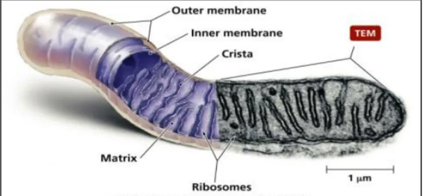
Mitochondria are about 0.5–1 mm in diameter and up to 7 mm long. Their shape and number per cell depends on the particular tissue. They may appear as spheres, rods or filamentous bodies, but the general architecture is the same. The number of mitochondria per cells varies depending on the energy requirements: tissues with a high capacity to perform aerobic metabolic functions such as skeletal muscle or kidney will have a larger number of mitochondria.
Mitochondria have two membranes, each composed of a phospholipid bilayer, quite distinct in appearance and in physico-chemical properties, thus determining the biochemical function of each membrane and shaping four main compartments (Figure 1)
The outer membrane is widely permeable to ions and large molecules The inner membrane encloses and convolutes into the mitochondrial matrix, and forms the cristae. This serves to increase the surface of the inner membrane, which carries the main enzymatic machinery of oxidative phosphorylation.
12
The matrix, or the inner part of mitochondria, is the place where they perform multiple metabolic function including pyruvate oxidation, the Krebs cycle, fatty acid oxidation and aminoacid metabolism. It’s a complex mixture of enzymes, important for the synthesis of ATP molecules, of special mitochondrial ribosomes, of tRNAs and of mitochondrial DNA molecules. Besides these, it has oxygen carbon dioxide and other recyclable intermediates.
The intermembrane space has a composition similar to that of citosol. The inner and outer membranes are characterized by different phospholipid compositions and protein-to-lipid ratios. For the outer membrane, this ratio is about 50:50, and it is thought that the protein has very little enzymatic or transport function. In the inner membrane, the protein-to-lipid ratio is 80:20. The inner mitochondrial membrane is much less permeable to ions and small molecules than the outer membrane, therefore providing compartmentalization through separation of the matrix from the cytosolic environment. This compartmentalization is a central feature of the conversion of free energy derived from oxidizable substrates. The inner mitochondrial membrane is in fact, an electrical insulator and a chemical barrier. Sophisticated ion transporters exist to allow specific molecules to cross this barrier. There are several antiport systems embedded in the inner membrane, allowing exchange of anions between the cytosol and the mitochondrial matrix.
13
1.1.2 Mitochondrial genetic.Mitochondria are under the dual control of nuclear DNA and
mitochondrial DNA (mtDNA). The existence of a separate mitochondrial
DNA genome is explained by the widely accepted endosymbiotic theory according to which the mitochondrion developed from an α-protobacterium (Gray MW et al., 1999)during the course of time, ancestral genes have been transferred from the mitochondrial to the nuclear genome, as is evident from the presence orthologous genes in the mitochondrial genome in some species and in the nuclear genome of other species (Andersson SG. et al., 2003).
Fig2: A map of the human mitochondrial genome. The genes that encode the subunits of complex I (ND1–ND6 and ND4L) are shown in blue; cytochrome c oxidase (COI–COIII) is shown in red; cytochrome b of complex III is shown in green; and the subunits of the ATP synthase (ATPase 6 and 8) are shown in yellow. The two ribosomal RNAs (rRNAs; 12S and 16S, shown in purple) and 22 tRNAs, indicated by black lines and denoted by their single letter code, which are required for mitochondrial protein synthesis are also shown. The displacement loop (D-loop), or non-coding control region, contains sequences that are vital for the initiation of both mtDNA replication and transcription, including the proposed origin of heavy-strand replication (shown as OH). The origin of light-strand replication is shown as OL
14
Gene transfer explain why all proteins necessary for mtDNA replication, as well as transcription and translation of mtDNA-encoded genes, are encoded in the nucleus.
While the nuclear genes is diploid, harboring only two homologous copies of each chromosome, the mitochondrial genome is polyploidy, containing 1 to 10 identical molecules of mitochondrial DNA within its matrix. A somatic mammalian cell contains 1000-10.000 copies of mtDNA but mammalian oocyte could reach even 100.000 copies per cell probably derived by replication of just a few mtDNA copies ina precursor cell (Falkenberg M. et al., 2007). This variable copy number, combined with the variable number of mitochondria in each cell, has important implication for the phenotypic expression of mutation.
Mitochondrial DNA is a double strand closed circular molecule, composed of 16.569 base pair that codes for 13 polypeptide units all of which are components of the respiratory chain. However, each mitochondrion has approximately 900 genes, including 85 to 90 respiratory chain proteins.Thus the great majority of these are encoded by nuclear DNA. The mitochondria have their own machinery for DNA transcription and the RNA translation. In addition to 13 polypeptides genes, mitochondrial DNA encodes for 22 tRNAs and 2 ribosomal RNAs, giving a total of 37 mitochondrial genes ( Figure 2).
The genetic code of mitochondrial DNA differs slightly from the universal genetic code. In fact certain mitochondrial trinucleotides encode different aminoacids or stop codons than those in the universal genetic code
In addition mitochondrial DNA have several characteristic that allow to distinguish it from its nuclear counterpart;
1. Maternal inheritance: a fertilized egg possesses mitochondria derived predominantly from the mother, so that mitochondrial genotype is essentially only transmitted through the mother (Sutovsky P. and Schatten G. 1999);
2. Heteroplasmy: each mitochondria has several DNA molecules and each cell has several hundred mitochondria. In a normal state, all this molecules are identical (homoplasmy). When a pathogenic mutation ensues, it is generally present in some but not all of mtDNA copies (heteroplasmy) (Figure 3). Once it was believed that all pathogenetic mutations are heteroplasmic. This view is no longer accepted as rare homoplasmic mutation have been found to cause also disease (McFarland R. et al ., 2002);
3. Threshold effect: given variable heteroplasmy, not all cells in a tissue are abnormal. As a consequence, a minimal number of mutated DNAs must be
15
present before respiratory chain failure and cellular dysfunction occur. Clinical signs do not become apparent until enough cells are affected. This is know as threshold effect and varies between different tissues (Chinnery PF. et al.,1998);
4. Mitotic segragation: organells are randomly distribuited at the time of cell division, which can lead to a change in the amount of mutant DNA in a cell;
5. Postmitotic replication: mitochondrial DNA replication is not linked to the cell cycle. This allows post-mitotic mitochondrial DNA replication even in terminally differentiated cells such as neurons or muscle in response to specific stimuli (exercise, increased metabolic demand).
Fig.3 The mitochondrial genetic bottleneck. During the production of primary oocytes, a selected number of mitochondria DNA (mtDNA) molecules are transferred into each oocyte. Oocyte maturation is associated with the rapid replication of this mtDNA population. This restriction-amplification event can lead to a random shift of mtDNA mutational load between generations and is responsible for the variable levels of mutated mtDNA observed in affected offspring from mothers with pathogenic mtDNA mutations. Mitochondria that contain mutated mtDNA are shown in red, those with normal mtDNA are shown in green.
16
1.2 RESPIRATORY CHAIN COMPLEXES
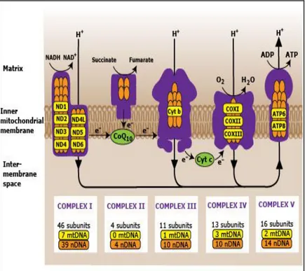
The mitochondrial respiratory chain (MRC) is composed of five multiheteromeric complexes (complex I, CI; complex II, CII; complex III, CIII; complex IV, CIV or cytochrome c oxidase, COX; complex V, CV, or ATP synthase), all embedded in the inner mitochondrial membrane, and two mobile electron shuttles, ubiquinone (Coenzyme Q, CoQ), a lipoidal quinone, and cytochrome c (cyt c), a heme-containing small polypeptide. Electron donors includ reduced nicotinamide-adenine dinucleotide, NADH+ (via CI), and reduced flavin adenine dinucleotide, FADH2(via CII) and other flavoproteins, including electron transfer flavoprotein ubiquinone reductase or ETF-dehydrogenase (EC 1.5.5.1), the terminal component of fatty acid -oxidation and ketogenic aminoacid oxidation pathways, supply electrons to CoQ, which donates them to CIII. CIII transfers one electron at a time to cyt c, which passes it to COX; COX eventually fixes four electrons to molecular oxygen with the formation of two molecules of water. This process, known as respiration, liberates energy that is partly converted by the proton pumping activity of CI, CIII, and CIV into an electrochemical potential ( H) composed of an electrical gradient ( Ψ ) and a pH gradient, across the inner mitochondrial membrane. H constitutes the proton driving force for the phosphorilation of ADP to ATP, operated by CV, and it’s also used for a number of other process such as, heat production, Ca++ import inside mitochondria, and protein translocation (Ghezzi D. et al., 2012)
From a genetic standpoint, the MRC is unique, as it is formed through the complementation of two separate genetic systems: the nuclear and the mitochondrial genomes. Four of the five MRC complexes, namely CI, CIII, CIV, and CV, contain subunits encoded by the mtDNA, and synthesized in situ by the organelle-specific translation machinery.
In humans, seven (ND1, 2, 3, 4, 4L, 5, 6) are components of CI, one (cytochrome b ) of CIII, three (COI, II, III) of CIV and two (ATPase 6 and 8) of CV; only CII is enterly codified by nulear genome (Figure 4) (Ghezzi D. et al ., 2012)
Specific pathways are required for the assembly of each MRC complex, including the insertion of mtDNA-encoded subunits into the inner membrane of mitochondria, in concert with >80 nuclear DNA encoded subunits; the synthesis and incorporation of several prosthetic groups that form the catalytic redox cores of CI, CII, CIII, and CIV; and the ultimate formation of functionally active holocomplexes. Individual holocomplexes can also organize themselves in respiratory supercomplexes.
17
Additional systems warrant the quality control of protein and non-protein components of the MRC complexes, thus contributing to the maintenance of their structural integrity, functional activity and turnover. Thus, a highly regulated, extremely complex process is at work in mitochondria to control the formation, stability, interactions, function, and plasticity of the MRC. Defects in genes encoding the components of these control and execution systems can compromise the function of the MRC, thus leading to faulty OXPHOS and disease (Ghezzi D. et al., 2011).
18
1.3 COMPLEX II : STRUCUTURE AND ASSEMBLY
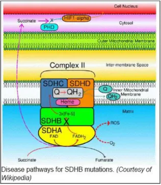
Succinate dehydrogenase or complex II participates in the electron transfer in the respiratory chain and in succinate catabolism in the Krebs cycle where it functions as a succinate dehydrogenase (SDH), catalyzing the oxidation and dehydration of succinate to fumarate. CII takes part also in the MRC by coupling this reaction to the reduction of ubiquinone to ubiquinol, that in turn funnels electrons to CIII (Ernster L. and Dallner G. 1995) and consists of four subunits all encoded by the nuclear genome (Bugiani M. et al., 2004). The two larger subunits, SDHA and SDHB, are catalytic. Dehydration of succinate to fumarate is accomplished by SDHA through reduction of a flavin-mononucleotide (FMN) molecule bound to its
19
protein moiety. These two hydrophilic subunits are linked to SDHC and SDHD, two small, hydrophobic polypeptides that contain a heme b moiety and anchor the complex to the inner mitochondrial membrane (Sun F. et al., 2005)(Figure 5). This reaction is measured as succinate dehydrogenase (SDH) activity. Electrons are then passed to three Fe-S centers bound to SDHB, which eventually transfers them to ubiquinone (coenzymeQ, coQ). The latter reaction is measured as succinate-CoQ reductase (SCoQR) activity. The crystal structure of porcine heart CII consists of a hydrophilic head protruding into the matrix, a hydrophobic tail embedded in the inner membrane, and a short segment projecting into the intermembrane space (Yankovskaya V. et al., 2003 ; Sun F. et al., 2005). Mitochondrial CII shows close homology with a number of bacterial succinate ubiquinone reductases (SQRs), especially those of -proteobacteria, from which mitochondria are supposed to derive (Andersson SG. et al. 1998)
The low frequency of human conditions associated with CII deficiency and the absence of a proton pumping activity and mtDNA-encoded subunits may explain the scarcity of information on CII assembly, in spite of the fact that CII is the smallest and simplest MRC complex.
In E.coli, an active, soluble SDH is composed of SDHA and SDHB ortholog subunits (SdhA,B), independent of the SDHC and SDHD orthologs (SdhC, D) (Nakamura K. et al., 1996 ) . However, there are no data in eukaryotes suggesting that the CII hydrophilic module, composed of SDHA and SDHB, builds up independent of the membrane-embedded module, formed by SDHC and SDHD. SDHB seems to play a central role in the stabilization of the human holocomplex, since mutations in each and every SDH subunits cause the loss of SDHB (Van Nederveen FH. et al., 2009). CII also contains several prosthetic groups, including one heme b moiety, one FAD, and three Fe–S clusters. The function of the single heme b moiety is unknown. In B. subtilis SQR , the absence of heme b prevents the hydrophilic subunits to assemble with the membrane-bound subunits and the site-directed mutagenesis of E. coli residues involved in heme b binding induces the catalytic subunits to dissociate from the membrane-bound subunits (Hagerhall C. 1997 ; Hederstedt L. 2002). Taken together, these observations support a role for heme b in assembly and stability of the complex. So at least the current model for CII assembly provide for an intial step where heme b prosteic group binds to SdhD, followed by linking with SdhC (Lenaz G. and Genova ML. 2010), to form the membrane-bound module. Heme b is also essential to link the hydrophilic module (SdhA + SdhB) to the membrane anchor subunits (SdhC + SdhD).
20
Whereas an increasing number of assembly factors have been identified for complex I, III, and cytochrome oxidase, little is known concerning the assembly of complex II. Recently two genes involved in this process were detected in humans, SDHAF1 (Ghezzi D. et al., 2009 ) and SDHAF2 (Hao HX. et al., 2009).
1.3.1 SDHAF1: function and human associated diseases
Isolated complex II deficiency is a relatively rare cause of mitochondrial disease encompassing 2–8% of OXPHOS defective cases (Munnich A. and Rustin P. 2001; Ghezzi D. et al., 2009). Fenotipically two main clinical presentations are known: mitochondrial encephalomyopathy and familial paragangliomas (tumors of chromaffin cells). Mutations in either CII proteins or chaperones may cause LS(SDHA), infantile leukencephalopathy (SDAHF1,SDHB) or familial paraganglioma syndrome (SDHB, C, D, SDHAF2) whose pathogenicity remains to be explained (Diaz F. et al., 2010) Several proteins assisting assembly of CII have been described ( Rutter J. et al., 2010) but only the two recently discovered factors SDHAF1( Ghezzi D. et al., 2009) and SDHAF2 (Hao HX et al., 2009) directly and specifically aid CII assembly.
SDHAF1 was first described in 2009 by Ghezzi D. et al.,in two affected families with infantile leukencephalopathy and severe CII deficiency (Ghezzi D. et al .,2009). SDHAF1, standing for SDH Assembly Factor 1, is a small protein containing a tri-peptide sequence, LYR, a proposed signature for proteins involved in Fe–S metabolism. Hence, SDHAF1 could play a role in the insertion or retention of the Fe–S clusters within the protein backbone of CII, but this hypothesis needs further experimental evidence.
Although SDHAF1 resides in the mitochondrial matrix, CII is membrane bound and mutations in this protein are associated with a drastic decrease of CII activity. A homozygous missense mutation in SDHAF1, the c.164G<C, (p.R55P) has been identified in a large multi consanguineus kindred of a Turkish origin and a 169G>C mutation, corresponding to p.G57R, affected the genome of three affected Italian children, two of whom were second degree cousin from a small alpin vallay in Lombardy. The clinical features of the patients were vary similar consisting of leukoencephalopathy with accumulation of lactate and succinate in the white matter, and severe reduction of CII activity and amount in muscle and fi broblasts (Ghezzi D. et al., 2009).
21
Complementation assays in both human cells and a yeast model have con firmed the pathogenicity of the mutations.
The new gene was discovered thanks to genome wide linkage analysis in Turkish family and SNP-based mapping of the italian subjects was identified a single exon anonymous gene located in chromosome 19 predicted to translated for a little 115 aminoacid protein sequences (NP_001036096), which scores high when analyzed by mitochondrial targeting prediction programs. SDHAF1 contains a LYR tripeptide motif, which is present in the terminal region of several protein sequences in different species.There are at least eight LYR-motif (LYRM) proteins in humans, including SDHAF1. LYRM-4 is the human ortholog of yeast ISD11, protein that has an essential role in the mitochondrial biosynthesis of Fe-S centers. LYRM-6 is the 14 kDa NDUFA6 subunit of complex I; a second cI subunit, the 22 kDa NDUFB9 is also a LYR, iron-responsive protein. These data suggest that the LYR motif is a signature for proteins involved in Fe-S metabolism. In particular, NDUFA6, NDUFB9 and possibly SDHAF1 as well could be important for the insertion or retention of the Fe-S centers within the protein backbones of CI and CII, respectively. Failure of the Fe-S centers to be incorporated within CII may eventually inhibit the formation of the holocomplex or destabilize its structure. Although there are other examples of low cII content and activity associated with mutations in mitochondrial chaperonins such as yeast Tcm62.p14, or proteins involved in Fe-S biosynthesis such as human and yeast frataxin or IscU15, SDHAF1 is the only protein so far identified with a specific role for cII, as other Fe-S–dependent activities were normal in SDHAF1-defective organisms, including cI in humans and complex III in both humans and yeast (Ghezzi D. et al., 2009)
1.4 COMPLEX V : STRUCUTURE AND ASSEMBLY
Complex V (F1F0-ATP synthase or ATPase) couples proton flow from the intermembrane space back to the matrix for the conversion of ADP and inorganic phosphate to ATP. Mammalian complex V consists of 16 different polypeptides: two subunits, ATP6 and ATP8, are encoded by mitochondrial DNA (mtDNA) the remaining by nuclear DNA (nDNA). The enzyme comprise an integral membrane cylindrical rotor-like structure, the F0 particle, and a peripheral matrix-facing F1 particle, the catalytic ATP synthase domain (Boyer PD. et al., 1997). The hydrophilic F1 contains catalytic sites for ATP synthesis and hydrolysis and has a bulb-like shape, formed by six “cloves”. F0 is the hydrophobic, membrane-embedded
22
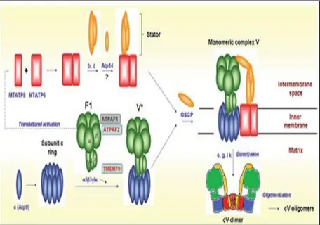
portion and functions as a rotor harboring a proton channel : F0 and F1 are physically connected to each other by two additional structures: a centrally located, asymmetrical stalk and an externally tethered stator. Following the electrochemical gradient, protons flow through the channel guided by the stator, impressing a rotary motion to F0, which is transmitted to the catalytic head (F1) by the centrally projecting stalk. The sequential tilt of the cloves, impressed by the asymmetrical stalk during each F1 rotation cycle, drives the condensation of ADP and Pi to form three ATP molecules for each cycle (Devenish RJ et al., 2008). All five subunits of F1 ( , , , , ), and most of the F0 subunits (b c, d, e, f, g, OSCP and F6) are nuclear encoded (Collinson JR et al.,1996 ). Only two proteins (MTATP6 and 8) are encoded by mtDNA (Boyer PD. et al., 1993). Both MTATP6 and ATP8 are part of the F0, and connect the latter to the stator (Figure 6). Dimeric and higher oligomeric forms of ATP synthase (Arnold S. et al.,1998; Paumard P. et al., 2002) seem critical to maintain the shape of mitochondria by promoting the formation of the inner membrane cristae.
Fig. 6: Schematic pattern of CV assembly. Assembly factors associated with human disease are boxed in red, whereas the other are in gray (Ghezzi D. et al., 2012)
23
The biosynthesis of individual subunits and formation of the ATP synthase holoenzyme depend on several specific helper proteins that are partly common to, and partly unique to, higher and lower eukaryote. The only two mammalian assembling factors that are found in yeast (Pickova A. et al., 2005; Wang ZG. et al., 2001) having identical function are the F1 chaperones, ATPAF1 and ATPAF2, interacting with F1 subunits β and α. Both are essential for assembly of the functional α3β3 heterooligomer.
Currently it has been delineated the following model for CV assembly (Figure 6): the process starts with the formation of the F1 catalytic core, carried out by specific assembly factors ATPAF1 and ATPAF2 in mammalian mitochondria (corresponding to Atp11 and Atp12 in yeast). Next, the initial F1 intermediate interacts with subunit c (Atp9 in yeast) and the other F0 subunits, forming an assembly intermediate named V*. The two mtDNA-encoded subunits, MTATP6 and MTATP8, are added during the last assembly stage, at least in mammalian cells (Nijtmans LG et al. 1995 ), since the V* intermediate and lower order subassemblies build up in MTATP6 or MTATP8 mutant mitochondria (Houstek J. et al.,2006). In yeast, two distinct assembly intermediates have been characterized, one formed by Atp6, Atp8, at least two stator subunits, and the Atp10 chaperone, the second by the F1 ATPase particle and the Atp9/subunit c ring. This recent result indicates that the assembly process is not a linear addition of single subunits one next to the other, but consists of at least two separate, coordinately regulated pathways eventually converging together at the end stage (Rak M. et al., 2011). This is in agreement with the notion that the F1 and F0 components seem to derive from functionally unrelated ancestral proteins (Mulkidjanian AY et al., 2007) that follow independent assembly pathways (Schatz G. 1968 ; Tzagoloff A. 1969) . The very final steps in mammalian CV biogenesis include the formation of dimers, coincidental with the addition of subunits e and g (Schagger and Pfeiffer 2000), and the formation of higher order oligomers (V1-V4) (Krause F. et
al. 2005 ) .
Another essential ancillary assembling factor found in mammals, the TMEM70 protein, has recently been found in fungi (Schizosaccharomyces pombe), and plants (Arabidopsis thaliana); however, it is absent from many species that do have complex V, like Saccharomyces cerevisiae (Cizkova A. et al., 2008, J. Houstek J.et al., 2009; Jonckheere AI et al., 2011). TMEM70 protein seems to play an important role in the ATP synthase biogenesis although its molecular function is still unknown (Torraco A. et al., 2012)
24
1.4.1 TMEM70: function and human associated diseases.
Most cases of isolated ATP synthase deficiency are caused by mutations in the mitochondrially encoded MTATP6 and MTATP8 genes and result in well defined clinical phenotypes; the most common being neuropathy, ataxia, and retinitis pigmentosa (NARP syndrome) (Holt IJ et al.,1990), and maternally inherited Leigh syndrome (Rahman S. et al.,1996) for patients mutated in MTATP6; hypertrophic cardiomyopathy and neuropathy for the only case having mutation in MTATP8 ( Jonckheere AI. et al., 2007). A nuclear defect in ATP synthase was first described in 1999 (Houstek J. et al.,1999), and recently an increasing number of cases have been identified (Houstek J. et al., 2004). De Meirleir and co-authors firstly associated the before-mentioned enzymatic defect with mutation in the nuclear-encoded ATP12 gene (De Meirleir L. et al., 2004).
Three disease-causing nuclear genes have been identified so far, two encoding assembly factors (ATPAF2, TMEM70), whereas the third (ATP5E) encodes the epsilon subunit of the F1 domain (Mayr JA. et al. 2010 ). In other CV deficient cases the genetic cause is still unknown (Sperl W. et al., 2006 ) .
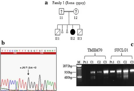
Mutations in the assembly factor TMEM70 have been described in a group of Gipsy patients presenting similar biochemical defect and clinical phenotype of the ones mutated in ATPAF2, characterized by lactic acidosis, cardiomyopathy, 3-methylgutaconic aciduria (3-MGA) and/or variable central nervous system involvement (Cizkova A. et al., 2008). Since the first report, additional cases have been reported (Jonckheere AI et al., 2012; Shchelochkov OA et al., 2010; Spiegel R. et al., 2010) having mutations in TMEM70 gene, leading to consider this gene the most common cause of mendelian inherited isolated ATP synthase deficiency. Finally, Houstek and co-authors have recently described the first mutation in a nuclear encoded structural subunit of ATP synthase in a patient with neonatal onset, lactic acidosis, 3-methylglutaconic aciduria, mild mental retardation and peripheral neuropathy (Mayr JA. et al., 2010)
Since I work as researcher in the Unit of Molecular Medicine for Neuromuscular and Neurodegenerative Diseases of Bambino Gesù Children’s Hospital I’ve always dealt with genetical defects that cause mitochondrial disorders: clinical syndromes associated with abnormalities of the oxidative phosphorilation. The subunits constituting complex of the
25
mitochondrial respiratory chain have dual origin, mitochondrial or nuclear: hence mitochondrial syndromes can due to mutation of mtDNA or to abnormalities in nuclear genes. So through these years I screened either mitochondrial genes or nuclear genes responsible of mtDNA integrity /stability or necessary for the proper assembly/stability of the MRC complex.
In particular during these three years of my PhD I have focalized my attention studying the role of two new nuclear genes TMEM70 and SDHAF1 respectively necessary for the correct assembly of Complex V and Complex II respectively, and consecutively I presents my results in this thesis work
26
2. RESULTS
2.1 CII DEFICENCY PATIENT
2.1.1 Clinical phenotype in the patient
A girl of Bangladeshi first-degree cousins, was born at term without complications after a pregnancy characterized by maternal controlled diabetes. Birth weight was 2,100 g (<3rd percentile) which was remarkable for intrauterine growth retardation. Developmental milestones were normal until 13-months of age, when she was referred to medical evaluation for a 10 day history of acute psychomotor regression, clumsy crawling, partial loss of voluntary sitting, and hypotonia. Physical examination showed an alert girl with no facial dysmorphisms, and bilateral palpebral ptosis, anterior fontanel of 0.5x0.5 cm with fibrotic consistence. Cardiopulmonary and abdominal evaluations were normal. Neurological examination showed truncal hypotonia, spasticity of upper and lower limbs, polyphasic osteotendinous reflexes, and bilateral Babinski sign. Fundoscopy was unremarkable. ECG and echocardiogram were also normal. An awake Video-EEG showed focal spikes localized at the frontocentral regions of the right hemisphere, in absence of epileptic seizures. Electroretinogram and
27
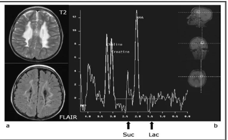
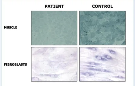
brainstem auditory evoked potentials were normal, whereas bilateral visual evoked potentials showed a marked delay of responses, with normal amplitude and morphology. Nerve conduction velocity studies were normal. Somatosensory evoked potential were compatible with central conduction abnormalities. Brain MRI showed abnormal symmetric T2 hyperintense signaling in periventricular white matter, corpus callosum, cerebellar medial peduncles and brain stem with sparing of U fibers. Magnetic resonance spectroscopy (MRS) evidenced lactate and succinate peaks (Figure 7). Routine blood tests showed normal ammonia, plasma amino acids, acylcarnitines, lysosomal enzymes, and very long-chain fatty acids. Urinary levels of fumaric, glutaric, 2-ketoglutaric, and citric acids were mildly elevated. No evidence was found for an infectious disease. Serum lactate level was 14.8 mmol/L (normal <2 mmol/L). Histochemical studies of a muscle biopsy performed at age 1314 months showed a markedly-diffuse reduction of SDH in all muscle fibers (Figure 8).
The patient was put on steroids but without improvement. During the following years she had a relapsing remitting course. At the age of 7 years,
28
the patient showed a severe spastic-dystonic tetraparesis, moderate mental retardation, mild dysphagia, poor growth (height and weight below 3° percentile), and showed clonic, myoclonic and tonic-clonic seizures several times a day, scarcely responsive to antiepileptic drugs. A new brain MR spectroscopy MRS documented only moderate reduction of NAA peak. Newcastle Paediatric Mithocondrial Disease Scale (NPMDS) at age 7 years was 34 (section I-III) and 15 (section IV, QOL). Therapy with Riboflavine (3,3 mg/kg/die) led to improved vigilance and alertness lower frequency of epileptic seizures and some developmental progess. A new evaluation with NPMDS at six months of follow–up confirmed mild improvement resulting in 28 for section I-III and 7 for section IV (QOL). Follow-up at six months showed a NPMDS of 28 (sections I-III) and 7 (section IV, QoL).
29
2.1.2 Molecular, biochemical, and structural studies
In a patient with clinical and neuroimaging (Figure7) suggestive of leukoencephalopathy, histochemistry showed a profound defect of complex II in all fibres confirmed also in cultured skin fibroblasts (Figure 8). Spectrophotometric determination of respiratory chain enzymes showed a profound, isolated defect of complex II (residual activity was 30% of the lowest control value after normalization with citrate synthase).
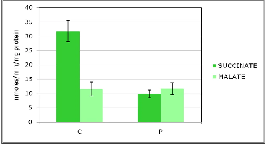
A severe impairment of complex V activity (<70% of normal values,
Figure 9) when succinate was used as substrate has been observed in
fibroblasts mitochondria, confirming the defect observed in muscle biopsy.
Total ATP content in cultured fibroblasts was slightly (p>0.05) reduced in regular medium (-10%), whereas when fibroblasts were growth in galactose-supplemented medium, a stress condition that force the cells to get ATP through the OXPHOS system, we observed a further significant reduction of ATP (-20%) compared to control fibroblasts
Fig. 9: Spettrofotometric determination of ATP-synthase acitvity in controls and Patient’s skin fibroblast mitochondria
30
a
c
b
Fig.10: Familial pedigree of patient 1 (a) with black symbol that denote homozygous individual while half black symbols identify the heterozygous ones. Sequence analysis of the DNA region in patient 1 (b) that encompass the homozygous mutation C.103G>T in SDHAF1.(c) Conservation of the alterade amminoacid p.E35X showen in ClustalW-2.0 at the European Bioinformatic s institute (http//www2.ebi ac.uk/clustalw)
31
Molecular studies identified a novel, homozygous c.103G>T mutation (Figure 10b) in a highly conserved residue of the gene SDHAF1 (Figure
10c), predicting a premature protein truncation at amino acid residue 35
(p.E35X). The mutation just segregate with the disease in the family indeed it was heterozygous in the parents (Figure 10a,) and it was not found in 200 control chromosomes.
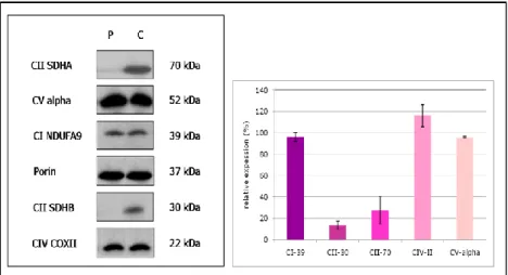
The histochemestry assay show a reduced complex II activity both in muscle and in fibroblasts mitochondria and was associated with a decreased amount of complex II subunits SDH70 and SDH30 (Figure 11 left panel) as shown by Western blotting experiment performed on patient skin fibroblasts mitochondria. Complex I, IV and V subunits were normally expressed, as also demonstrated by densitometric analysis (Figure 11 right panel)
Fig. 11: Fibroblast mitochondria proteins were separeted by SDS-PAGE (left panel) and a specific antibodies against SDHAF1 protein , subunits of complex V (a,b,OSCP,d) and porina were usede. The mitochondrial protien porin was used as a control for egual loading . The amount of SDHA (SDH70) and SDHB (SDH30) proteins was severly decreased in patient 1 against control whereas normal levels of complex V subunits are observable both from SDS-PAGE and from the densitometric analysis (right panel)
32
To better analyesd the overall structure of CII we performed a BN-PAGE in the first dimension (Figure 12 left panel) and a denatureting Tricine/SDS-PAGE in the second dimension (Figure 12 right panel) followed by Western blotting assay. Both these experiments revealed a drastic decrease of the holocomplex II with normal amount of holocomplexes I and IV .In fact, the ratio between complex II/ I and II/IV is 1 in both controls. Conversely, the ratios II/I and II/IV in the patient show a reduction of complex II amount of about 80%. Further, accordingly to literature (Ghezzi et al., 2009) we don’t observe the presence of any CII sub-complexes (Figure 12 right panel)
Fig.12: BNGE performed on mitochondria of control and patient’s fibroblast (left pannel) shows a decreased amount of holocomplex II in patient while antibodies against SDHB, COXII and NDUFS3 identify normal level of CII,CIV and CI rispectivly both in control and patient mitochondria. In right pannel we show a second-dimension SDS-PAGE gel elettrophoresis protein blot analysis from control and patients fibroblast mitochondria. Antibodies against SDHA (70kDa),SDHB (30kDa) and NDUFS9 (39kDa) are used for the immunodetection of subunits of CII and CI. No evidence of complex II subcomplexes are present
33
2.2 CV DEFICENCY PATIENTS
2.2.1 Clinical phenotype in patients
Patient 1: this patient’s clinical and biochemical features have already been described in detail elsewhere (patient 2 in 30). Briefly, Patient 1, a girl born at term after a normal pregnancy and delivery, is the second child of consanguineous parents descended from a Yugoslavian gypsy family (Figure 13a). Two male siblings of this patient died at birth. One of these had neonatal unresponsive lactic acidosis and 3-MGA (1001mmol/mol creatinine); he also presented with hypospadias, club foot and facial dysmorphisms. He was not tested for cataract, and post-mortem examination did not reveal cardiomyopathy. Patient 1 presented with congenital microcephaly, dysmorphic features, status epilepticus and metabolic acidosis at the age of 1 month. At 8 months she showed HCMP, cataract and mild developmental delay. At 3 years of age, she had an acute episode of severe metabolic acidosis with coma. At 6 years she was admitted to the Bambino Gesù Children’s Hospital in Rome for systemic arterial hypertension and severe lactic acidosis. Brain MRI showed only mild brain atrophy. MR spectroscopy showed a slightly reduced N-acetyl aspartate peak and no lactate peak. Abdominal ultrasound was unremarkable. Ecocardiography documented concentric left ventricular hypertrophy, with slight mitral and aortic valves insufficiency. Hyperlactacidemia (2.9-12.3 micromol/L; normal values <2,1), metabolic acidosis (PH 7,07, ABE -19) and hyperalaninemia (588 micromol/l; nv: 150-400) were also detected. The urinary organic acids profile showed increased levels of methylglutaconic (375-400 mM/Mcreatinine; nv <11) and 3-methylglutaric acid (94-123 mM/Mcreatinine; nv <0,1). In addition, variably increased excretion of lactate, pyruvate, and Krebs cycle intermediates was observed. The blood acylcarnitine profile, activity of 3-methylglutaconyl-CoA hydratase (3-MGH) and cardiolipin levels were all normal. At the age of 6 years muscle and skin biopsies were performed. Spectrophotometric analysis of respiratory chain enzyme activities (from complex I to IV) in muscle homogenate was normal. In the course of her follow-up the patient was admitted to hospital on numerous occasions for metabolic acidosis following respiratory infections. At the most recent evaluation, at the age of 9 years, she showed moderate mental retardation and stable HCMP under propranolol therapy. Serial biochemical analysis confirmed persistent lactic acidosis and 3-MGA.
34
Patients 2 and 3 were born to non-consanguineous parents of Italian origin (Figure 14 a). Patient 2 the couple’s first female child, was delivered by caesarean delivery because of breech presentation at 39 weeks’ gestation after an uneventful pregnancy. Her birth weight was 2.880 kg. On the 3rd day of life she presented with acute onset of respiratory distress. Echocardiography, performed when she was one week old, showed hypertrophy of the interventricular septum and a patent foramen ovale. Brain ultrasonography showed no abnormalities. Metabolic work-up showed persistent mild hyperlactacidemia (2.5-5.8 mmol/L; nv<2.1), and hyperalaninemia (551 micromol/l; nv 150-400). Urinary gas chromatography-mass spectrometry (GC/MS) showed increased methylglutaconic (116-332 mM/Mcreatinine; nv <11) and 3-methylglutaric acid (29-35 mM/Mcreatinine; nv <0.1) levels and variably increased excretion of lactate, pyruvate, and Krebs cycle intermediates. At 20 day, skin biopsy was performed. Treatment with carnitine, biotine and sodium bicarbonate was started. Metabolic lactic acidosis responded to intravenous sodium bicarbonate; the hypertrophy of the interventricular septum was found to be improved at 20 days of life and normalized by the age of 1 year. The child was discharged at the age of 1 month following normalization of blood lactate levels. During the subsequent follow-up, she showed normal cognitive development and walked unaided around 14 months of age. Her somatic growth was slightly delayed and now, at the age of 4 years, she is at the 10th percentile. Serial metabolic follow-up revealed mildly elevated blood lactate and the urinary organic acid profile showed persistent 3-MGA. Brain MRI performed during the follow-up was unremarkable. Patient 3: the younger sister of patient 2 was born at 40 weeks’ gestation by urgent caesarean delivery performed because of meconium-stained fluid and increasing signs of foetal bradycardia. The child’s birth weight was 2.700 kg. She had severe respiratory distress and lactic acidosis at birth. Following the appearance of a pneumothorax, intubation and invasive ventilation were started and arterial pulmonary hypertension was discovered. GC/MS showed 3-methylglutaconic and 3-methylglutaric aciduria. Ecocardiography showed hypertrophy of the interventricular septum. The pulmonary hypertension improved after 5 days of nitric oxide inhalation therapy but respiratory failure persisted and at the age of 2 months tracheotomy was performed. In addition, gastrostomy was performed at the age of 5 months because of severe swallowing impairment. The child was discharged from the intensive care unit at the age of 7 months breathing air with the aid of a tracheotomy and under treatment with coenzyme Q, thiamine, sodium bicarbonate and enalapril. She is now 14
35
months old. Her cognitive development is normal and is able to walk with aids. She has normal somatic growth for her age (7.300 kg), persistent metabolic acidosis, with mildly elevated plasma lactate and persistent 3-MGA. Electrocardiography documented a Wolf-Parkinson-White pre-excitation syndrome, while echocardiography was unremarkable. No muscle biopsy was available from patients 2 and 3. This study was performed after receiving written informed consent from the parents of the children involved, and in accordance with the guidelines of the Bambino Gesù Children’s Hospital ethics committee.
2.2.2 Molecular, biochemical, and structural studies
Fig. 13: (a)Familial pedigree of patient 1 (II-2). Familial pedigree of patients 2 (II-1) and 3 (II-2). Blacksymbols denote homozygous or compound heterozygous individuals; half-black symbols denote heterozygous individuals. (b)Sequence analysis of the DNA region, in patient 1, encompassing the homozygousc.317-2A>G mutation inTMEM70. (c) Analysis of the c.317-2A > G mutation at thecDNA level in culturedfibroblasts from patient 1 displays the absence of thecorresponding transcript. SUCLG1 cDNA was used as the control gene. The position of the mutations is shown (arrow). M, DNA molecular marker size; C1, C2, C3, controls; Pt.1, patient 1
a
36
All the three patients, coming from two unrelated families, show 3-MGA, HCMP, and hypotonia; one of them also showed PAH.
Fig. 14: (a)Familial pedigree of patients 2 (II-1) and 3 (II-2). Blacksymbols denote homozygous or compound heterozygous individuals; half-black symbols denote heterozygous individuals. (b) Sequence analysis of the DNA region, in patient 2, encompassing the heterozygous c.317-2A>G and c.628A>C mutations in TMEM70. (c)Sequence analysis, using a reverse oligonucleotide primer,of TMEM70 from fibroblast cDNA of patient 2 revealed the c.628A>C mutation in homozygosity, confirming the loss of TMEM70 transcript caused by the c.317-2A>G splice sitemutation. (d)Conservation of the altered aminoacid (p.T210P) is shown in ClustalW-2.0 at the European Bioinformatics Institute (http://www2.ebi.ac.uk/clustalw). The position of the mutations is shown (arrow)