Università degli Studi della Calabria
Facoltà di Farmacia e Scienze della Nutrizione e della Salute
Dipartimento Farmaco-Biologico
(MED/04 PATOLOGIA GENERALE)
Dottorato di Ricerca in “Biochimica Cellulare ed Attività
dei Farmaci in Oncologia” (XIX ciclo)
Insulin Receptor Substrate 1 modulates the
transcriptional activity and turnover of Androgen
Receptor in breast cancer cells.
Docente Tutor Dottoranda
Ch.mo Prof. Sebastiano ANDO’
Dott.ssa Cecilia GAROFALO
Coordinatore
Index
INDEX
Summary ………. 4
Introduction ……….... 5
Materials and Methods ……….. 8
9 Cell culture and treatments... 8
9 Immunoprecipitation and Western blotting... 8
9 Plasmids, transfections and luciferase reporter assays...8
9 Chromatin immunoprecipitation (ChIP)... 9
9 RT–PCR... 10
Results………. 12
9 DHT regulates cellular localization of IRS-1 in breast cancer cells ... 12
9 IRS-1 is a transcriptional regulator of AR ... 13
9 IRS-1 knockdown decreases AR transcriptional activity ...15
9 IRS-1 knockdown induces ubiquitylation of AR ... 19
Discussion………... 21
References………... 25
Scientific Publications Performed during the PhD Program……….... 30
9 Expression of nuclear insulin receptor substrate 1 (IRS-1) in breast cancer. J
Clin Pathol. 2006 Sep 22; [Epub ahead of print]
9 Increased expression of leptin and the leptin receptor as a marker of breast
cancer progression: possible role of obesity-related stimuli. Clin Cancer Res.
Index
9 RNAi-mediated silencing of insulin receptor substrate 1 (IRS-1) enhances
tamoxifen-induced cell death in MCF-7 breast cancer cells. J Cell Biochem.
2006 May 15;98(2):440-50.
9 Leptin and cancer. J Cell Physiol. 2006 Apr;207(1):12-22. Review.
9 Functional significance of type 1 insulin-like growth factor-mediated nuclear
translocation of the insulin receptor substrate-1 and beta-catenin. J Biol Chem.
2005 Aug 19;280(33):29912-20. Epub 2005 Jun 20.
9 Leptin interferes with the effects of the antiestrogen ICI 182,780 in MCF-7
breast cancer cells. Clin Cancer Res. 2004 Oct 1;10(19):6466-75.
9 Nuclear insulin receptor substrate 1 interacts with estrogen receptor alpha at
ERE promoters. Oncogene. 2004 Sep 30;23(45):7517-26.
9 Leptin expression in breast nipple aspirate fluid (NAF) and serum is
influenced by body mass index (BMI) but not by the presence of breast
cancer. Horm Metab Res. 2004 May;36(5):336-40.
9 Low calcium intake is associated with decreased adrenal androgens and
reduced bone age in premenarcheal girls in the last pubertal stages. J Bone
Summary
Summary
Breast cancer growth is responsive to various growth factors and steroid hormones
and also reflects the interplay between the respective signaling pathways. Even
though the majority of human breast cancers expresses androgen receptor, the
existence and/or the mechanism of a crosstalk between androgen receptor (AR) and
Insulin-like Growth Factors (IGFs) in breast has not been clearly defined. To gain
insight into this functional interplay we investigated whether Insulin Receptor
Substrate-1 (IRS-1), the major IGF-IR signaling molecule, affects AR function. In
MCF-7 breast cancer cells, upon 5-α-Dihydrotestosterone stimulation, IRS-1
associates with AR and is recruited to Androgen Responsive Elements of the
androgen target genes PSA and p21 and it appear to be required for AR
transcriptional activity. Indeed, the ectopic expression of IRS-1 enhances AR
transcriptional activity in a dose dependent manner, while the silencing of IRS-1
significantly represses PSA and p21 mRNA and protein levels. Moreover, IRS-1
knockdown experiments suggest that IRS-1/AR interaction decreases the
ubiquitin/proteasome dependent degradation of AR, reducing AR turnover. Thus,
our data provide novel insights into AR/IGF crosstalk and suggest that IRS-1 might
represent a novel AR regulator.
Introduction
Introduction
Breast cancer development and progression depend on a complex crosstalk
between steroid hormones and growth factors(Aronica and Katzenellenbogen, 1993;
Ignar-Trowbridge et al., 1993; Reddy et al., 1994; Ruohola et al., 1999). Several
authors have reported that the majority of human breast cancers express androgen
receptor (AR) (Kuenen-Boumeester et al., 1992; Lea et al., 1989; Wilson and
McPhaul, 1996), and that many metastatic breast tumors, which are estrogen
receptor (ER) and progesterone receptor negative, still express a significant amount
of AR (Bayer-Garner and Smoller, 2000). Androgens have been demonstrated to
exert differential effects on breast cancer progression, and may either stimulate or
inhibit the growth of AR-positive breast cancer cell lines in vitro (Birrell et al., 1995;
Hackenberg et al., 1991; Yeap et al., 1999). Patients with AR-negative breast tumors
had a significantly poorer response rate to hormone therapy and shorter overall
survival than those with AR-positive ones (Bryan et al., 1984). Androgens have been
found to negatively regulate the growth of mammary epithelial and breast cancer
cells in vitro (Birrell et al., 1995; Lanzino et al., 2005; Szelei et al., 1997). In breast
cancer cells, 5α-dihydrotestosterone (DHT) induces post-transcriptional
destabilization and downregulation of AR mRNA, while AR expression is increased
(Yeap et al., 1999). These observations suggest that the role of androgens and their
receptor in breast cancer growth and progression needs to be clarified (Birrell et al.,
1995; Kuenen-Boumeester et al., 1996; Lea et al., 1989).
Introduction
but also with basal transcription factors responsible for modulating the expression of
androgen-regulated genes (McEwan and Gustafsson, 1997).
In addition to this
classical pathway, a second mechanism of AR action consists of the receptor
interaction with proteins involved in various trnsductional signals (Bubulya et al.,
1996; Castoria et al., 2003; Lobaccaro et al., 1999; Oettgen et al., 2000);. A
substantial body of literature suggests that AR is directly or indirectly regulated by
growth factor in the absence of androgens (Culig et al., 1995; Nazareth and Weigel,
1996).
A major substrate for Insulin-like Growth Factors (IGFs) is Insulin Receptor
Substrate 1 (IRS-1). IRS-1 is generally a cytoplasmic/membrane localized signaling
protein. In addition to its conventional role, IRS-1 has been found in the nuclear
compartment in several cell types, including breast cancer cells and breast tumors,
(Lassak et al., 2002; Morelli et al., 2004; Prisco et al., 2002; Sisci et al., 2006; Sun et
al., 2003). Experimental data suggest that nuclear IRS-1 might function as
transcriptional co-regulator for polymerase I and II (Morelli et al., 2004; Tu et al.,
2002). Recently, we demonstrated that in breast cancer cells IRS-1 can bind ER and
that the IRS-1/ER complex can be translocated to the nucleus upon estradiol (E2)
stimulation. In the nucleus, IRS-1 is recruited to Estrogen Responsive
Element-containing promoters and negatively modulates ER-dependent transcription (Morelli
et al., 2004).
Here we demonstrate that androgen stimulation of breast cancer cells
promotes the binding of IRS-1 with AR and increases nuclear translocation of IRS-1.
We also show that nuclear IRS-1 is recruited to ARE regions of androgen responsive
genes, stimulating AR-mediated transcription in response to DHT. Our results
Introduction
suggest that IRS-1 modulates AR mediated transcription through two different
mechanisms, by participating in AR-recruited transcriptional machinery and by
decreasing AR turnover. Thus, we provide novel information on the regulation of
AR activity and its crosstalk with the IGF system in breast cancer cells.
Materials and Methods
Materials and Methods
Cell culture and treatments
A breast cancer epithelial cell line MCF-7 and a human embryonic kidney cell line
HEK-293 were grown in DMEM/F12 (Gibco, USA) supplemented with 5% calf
serum (CS, Gibco, USA) and in DMEM plus 10% fetal calf serum, respectively.
5α-Dihydrotestosterone (DHT, Sigma, USA) and hydroxyflutamide (OHFl, Sigma,
USA) were used at a concentration of 10
-7M. Before each experiment, cells were
serum starved for 24 h in phenol red-free DMEM (PRF), and than shifted to PRF
containing 5% charcoal-treated fetal calf serum (PRF-CT).
Immunoprecipitation and Western blotting
Total cell proteins and the cytoplasmic and nuclear fractions were obtained from
70% confluent cell cultures, as previously described (Garofalo et al., 2004). The
following monoclonal (m) and policlonal (p) antibodies (Abs) were used: anti-AR
441 mAb (Santa Cruz, USA); anti-IRS-1 pAb (Upstate, USA); anti-c-jun mAb
(Santa Cruz, USA); anti-GAP-DH mAb (Santa Cruz, USA); anti ubiquitin P4D1
mAb (Santa Cruz, USA); p21 pAb
(Santa Cruz, USA).
Plasmids, transfections and luciferase reporter assays
A full-length androgen AR expression plasmid pcDNA3 AR (AR) was
described
by Tilley et al. (Tilley et al., 1990). Firefly luciferase reporter plasmid
containing ARE, pARE2-tk-LUC (ARE-Luc), was a gift from Dr O. Janne. The
IRS-1 knockdown cells were obtained using pSilencer-IRS-IRS-1 plasmid (shIRSIRS-1), as
described by Cesarone et al. (Cesarone et al., 2006), with a scrabled shRNA plasmid
used as a control (Scrambled). The plasmid WWP-Luc containing human
Materials and Methods
p21
WAF1/Cip1promoter (p21-Luc) and p5.3PSAp-Luc encoding the PSA promoter
(PSA-Luc) were gifts from Dr. W. El-Deiry and Dr. Kakizuka, respectively.
For transient transfection assay, MCF-7 cells were grown in 24-well plates.
At 70% confluence, the cultures were transfected for 6 h using Fugene 6
(DNA:Fugene 3:1; Roche, CH). In addition, to assess transfection efficiency, each
DNA mixture contained 5ng of pRL-TK-Luc, a plasmid encoding Renilla Luciferase
(Promega, USA). Empty vectors were used to ensure
that DNA concentrations were
constant in each transfection. At 6 h after transfection cocktail addition, cells were
shifted to PRF for 24 h and then treated with 10
-7M DHT or left untreated in
PRF-CT for 24 h. Luciferase activity was measured using Dual luciferase assay System
(Promega, USA). The firefly luciferase data for each sample were normalized on the
basis of transfection efficiency measured by renilla luciferase activity.
To monitor the influence of IRS-1 on AR activity, ARE-Luc was
co-transfected with AR expression vector alone or in combination with IRS-1
expression vector pCMV-IRS-1 (IRS-1). The experiment was done using an AR-
and IRS1-negative cell line HEK-293.
To asses mRNA and protein levels, MCF-7 cells were plated on 60mm dishes and
transfected with an appropriate amount of various plasmids, as indicated in the
Figure legends. Upon transfection, cells were shifted to PRF for 24 h and then
treated with 10
-7M DHT or left untreated in PRF-CT for 24 h.
Chromatin immunoprecipitation (ChIP).
MCF-7 cells were grown in 10 cm plates. Confluent cultures (90%) were shifted to
PRF for 24 h and then treated with 10
-7M DHT or left untreated in PRF-CT for 2 h.
Materials and Methods
(Morelli et al., 2004). The precleared chromatin was precipitated with anti-AR mAb
(Santa Cruz, USA) for AR, IRS-1 pAb (Upstate, USA) for IRS-1 and
anti-Polymerase II pAb (Santa Cruz, USA) for Pol II. A 4 µl volume of each sample was
used as template for PCR with specific primers.
The following pairs of primers were used to amplify 296 bp of the
ARE-containing p21 promoter CAGCGCACCAACGCAGGCG-3’ (forward);
5’-CAGCTCCGGCTCCACAAGGA-3’ (reverse), and 233 bp of the PSA promoter
including ARE-sequence in proximal region
GATCTAGGCACGTGAGGCTTTGTA-3’ (forward) and
5’-CATGCTGCTGGAGGCTGGAC-3’ (reverse). The PCR conditions for p21
promoter were: 1min at 94 C, 1 min at 65 C, and 2 min at 72 C; for PSA promoter:
40 s at 94 C, 40 s at 57 C, and 1 min 72 C. The amplification products obtained in 28
and 30 cycles were analyzed in a 2% agarose gel and visualized by ethidium
bromide staining.
RT–PCR
MCF-7 cells were transfected with the scrambled and shIRS1 plasmid for 24 h, as
described in the transactivation assays methodology than treated with 10
-7M DHT or
left untreated in PRF-CT for 24 h. Total RNA was isolated using TRIzol reagent
(Invitrogen, USA) according to the manufacturer’s instructions. 2 µg of total RNA
were reverse transcribed using M-MLV reverse transcriptase (Promega, USA) and 2
µl of RT products were then amplified. The following primers were used: IRS-1
forward primer TCCACTGTGACACCAGAATAAT-3’, IRS-1 reverse primer
CGCCAACATTGTTCATTCCAA-3’ (762 bp); PSA forward
5’-TGCGCAAGTTCACCCTCA-3’, PSA reverse 5’-CCCTCTCCTTACTTCATCC-3’
Materials and Methods
(754 bp); AR forward CACAGGCACCTGGTCCTGG-3’, AR reverse
CTGCCTTACACAACTCCTTGGC-3’ (416 bp); p21 forward
GCTTCATGCCAGCTACTTCC-3’, p21 reverse
5’-CTGTGCTCACTTCAGGGTCA-3’ (270 bp). For the internal control gene 36B4,
the primers were: 36B4 forward 5’-CTCAACATCTCCCCCTTCTC-3’, 36B4
reverse 5’-CAAATCCCATATCCTCGTCC-3’ (408 bp). PCR was performed using
the following conditions: for IRS-1, 1 min at 94 C, 1 min at 50 C, 2 min at 72 C; for
PSA, 30 s at 94 C, 30 s at 58 C, 30 s at 72 C; for p21, 1 min at 94 C, 1 min at 58 C,
1min 72 C; for AR, 45 s at 94 C, 45 s at 60 C, 1 min 72 C; for 36B4 1min at 94 C, 1
min at 60 C, 2 min at 72 C. The amplification products were analyzed in a 1,5%
agarose gel
.
Results
Results
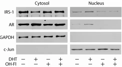
DHT regulates cellular localization of IRS-1 in breast cancer cells.
To determine whether androgens may regulate the expression and
localization of IRS-1 in breast cancer cells, we analyzed IRS-1 abundance in
cytoplasmic and nuclear protein fractions obtained from MCF-7 cells stimulated or
not with 10
-7M DHT. In absence of DHT, IRS-1 was mainly present in the
cytoplasmic compartment, while upon DHT treatment, the nuclear abundance of
IRS-1 significantly increased, with a parallel decrease of cytoplasmic IRS-1
expression. Moreover, the increased localization of IRS-1 in the nuclear fraction
appeared to be specifically mediated by AR, since it is inhibited by the addition of
the androgen antagonist hydroxy-flutamide (OH-Fl) (Figure 1).
Figure 1. IRS-1 colocalizes with AR. MCF-7 cells synchronized in SFM were treated with 10-7 M DHT or 10-7 M OHFl in PRF-CT for 24 h. The expression of IRS-1 and AR was determined by Western blotting in 50 µg of cytoplasmic or in 100 µg of nuclear proteins using specifics Abs, as described in Materials and Methods. The expression of GAPDH and c-jun was assessed as control of protein loading and purity of lysate fractions. The results were obtained after repetitive stripping and reprobing of the same filter. The results are representative of three independent experiments.
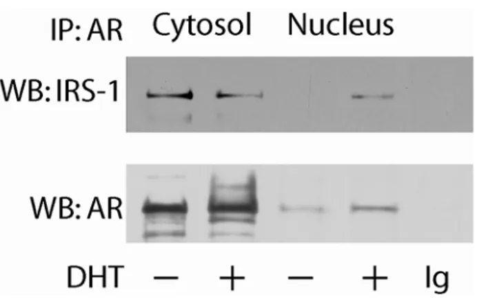
To assess whether the DHT-regulated intracellular localization of IRS-1 could
involve a physical interaction between AR and IRS-1, a coimmunoprecipitation assay
Results
was carried out on nuclear and cytoplasmic protein fractions from MCF-7 cells.
Under basal conditions, a constitutive association between AR and IRS-1 was
observed in the cytoplasm, but not in the nuclear fraction. DHT treatment induced the
translocation of AR/IRS-1 complex in the nucleus with a consequent decrease of its
abundance in the cytosol (Figure 2).
Figure 2. IRS-1 coprecipitates with AR. Cytoplasmic and nuclear fractions were immunoprecipitated (IP) with an anti-AR antibody and immunoblotted (WB) with an anti-IRS-1 pAb and an anti-AR mAb. In control samples, the primary Abs were substituted with non-immune IgGs (rabbit or mouse, depending on the source of the primary Abs). The results are representative of three independent experiments.
These observations suggest that DHT could promote direct interaction
between AR and IRS-1 thereby inducing nuclear translocation of IRS-1.
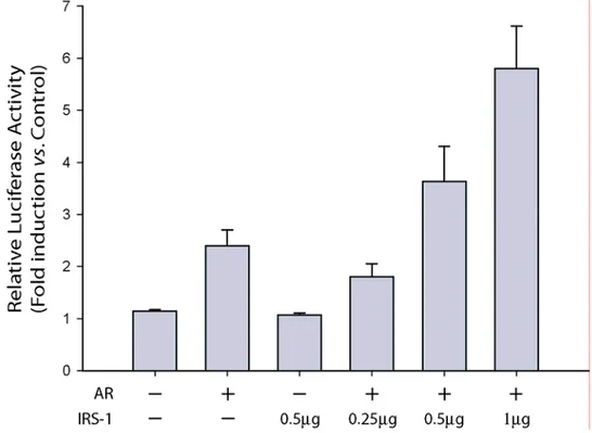
IRS-1 is a transcriptional regulator of AR.
The existence of the nuclear AR/IRS-1 complex lead us to investigate the
role of nuclear IRS-1 on the transcriptional activity of AR. We evaluated the effects
of ectopic IRS-1 expression on the transcriptional activity of AR using the
androgen-response reporter plasmid 2X-ARE-Luc. HEK-293 cells that are IRS-1- and
AR-negative were cotransfected with an AR expression vector and increasing amounts of
Results
transcriptional response to DHT stimulation in a dose-dependent manner, with a
maximal two-fold enhancement above the level observed in the absence of IRS-1
(Figure 3). This stimulatory effect of IRS-1 on AR was blocked by the addition of
OH-Fl (data not shown), suggesting a direct involvement of AR in this process.
Figure 3.Transcriptional activity of AR on ARE promoter in the presence or absence of IRS-1. HEK 293 cells were transfected with 0.5 µg ARE luciferase reporter construct pARE2-tk-LUC plus increasing quantities of pCMV-IRS-1 (0, 0.25, 0.5, and 1 µg) together with 0.1 µg pcDNA3-AR. Upon transfection, the cells were shifted to SFM for 16 h and than treated with 10-7 M DHT, or left untreated in PRF-CT for 18 h. Firefly luciferase activity was internally normalized to Renilla luciferase and expressed as Relative Luciferase Activity (RLA) with respect to the untreated samples. Results represent the mean±s.d. from several experiments.
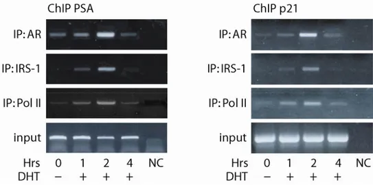
To determine whether the involvement of IRS-1 in AR-mediated
transcription was related to IRS-1 recruitment to ARE-containing regions of
androgen target gene promoters, we used chromatin immunoprecipitation assays
(ChIPs). We focused our attention on the prostatic specific antigen (PSA) and
p21
CIP/WAF(p21) genes that are known to be regulated by androgens in breast cancer
cells (Lu et al., 1999). As illustrated in Figure 4, basal occupancy of AR on PSA and
Results
p21 promoters occurs in untreated cells. The recruitment of AR to both promoters is
increased in response to DHT treatment, reaching the maximum at 2 h (4-fold
increase vs. untreated). IRS-1 is recruited to the same promoter regions in response
to DHT treatment only, reaching the maximal association after 2 h of hormonal
stimulation. The association of AR and IRS-1 with the PSA and p21 promoters
coincided with the loading of RNA polymerase II (Pol II) on the same regions.
Figure 4. IRS-1 is a transcriptional co-regulator of AR. ChIPs were carried out over a 4 h time-course after stimulation with DHT using AR, IRS-1 and Pol II antibodies , as described in Materials and Methods . The PSA and p21 promoter sequences containing androgen responsive element were detected by PCR with specific primers listed in Materials and Methods (fragments of 233 and 296 bp respectively). To control input DNA, PSA and p21 promoter was amplified from 30 µl of initial preparations of soluble chromatin (before IP). In control samples (NC), non-immune IgG (rabbit for IRS-1 Ab and mouse for AR Ab) was used instead of the primary Abs.
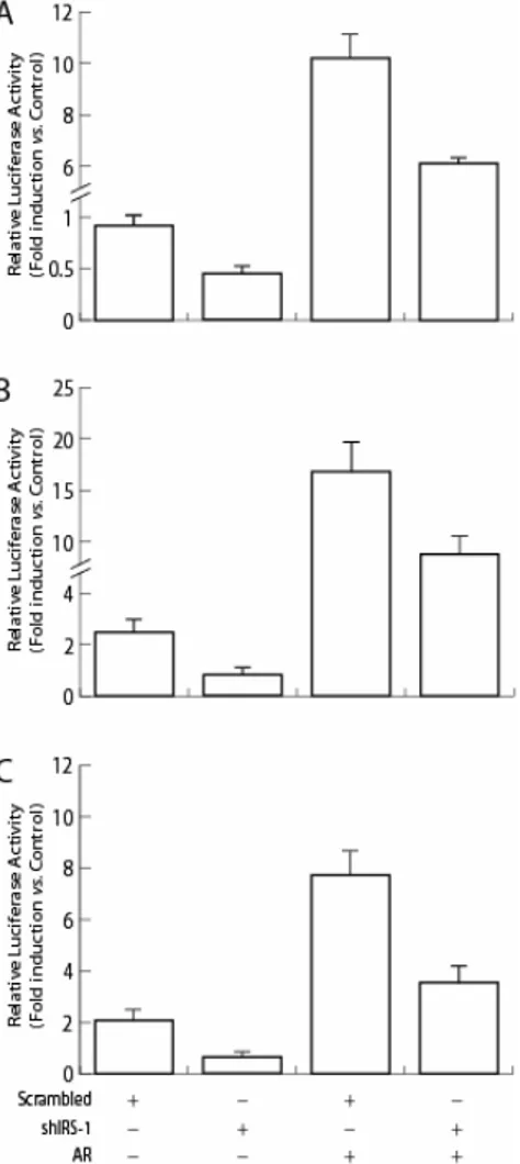
IRS-1 knockdown decreases AR transcriptional activity.
To delineate whether modulation of the AR/IRS-1 interaction could influence
AR function in response to DHT, a silencing RNA technology was used to
knockdown the expression of IRS-1 in MCF-7. A 70% reduction of IRS-1 levels
achieved with anti-IRS-1 shRNA expression in MCF-7 cells correlated with a 40%
decrease of IRS-1 mRNA levels (Figure 4C and B).
Results
Luciferase assays, demonstrated that IRS-1 knockdown with shRNA resulted
in a 40% decrease in transcriptional activation of ARE-containing promoters in both
wild-type and in AR overexpressing MCF-7 (Figure 5A). Similarly, downregulation
of IRS-1 impeded activation of the PSA and p21 promoters by 50% (Figure 5B and
C).
Figure 5. IRS-1 knockdown reduces AR regulated transcription. MCF-7 cells were transiently cotransfected, as described in Materials and Methods, with 0.5 µg IRS-1 shRNA (shIRS-1) or 0.5 µg scrambled control shRNA (Scrambled) either in the absence or presence of 0.1 µg pcDNA3-AR (pcDNA3-AR) together with the following specific reporter constructs: 0.5 µg ppcDNA3-ARE2-tk-LUC (A); 0.5 µg p5.3PSAp-Luc (B); 0.25 µg WWP-Luc containing human p21WAF1/Cip1
promoter (C). Upon
transfection, cells were serum starved overnight, then stimulated with 10-7 M DHT, or left untreated in PRF-CT for 24 h, and than luciferase activities were determined. Firefly luciferase activity was internally normalized to Renilla luciferase and expressed as Relative Luciferase Activity (RLA) with respect to the untreated samples. Results represent the mean±s.d. of at least five independent
Results
These results were confirmed by ChIP analysis demonstrating that IRS-1
knockdown resulted in diminished recruitment of AR and Pol II on the p21 and PSA
promoters (Figure 6).
Figure 6. IRS-1 knockdown decreases AR loading on the promoters of AR-regulated genes. Chromatin immunoprecipitation was carried out on MCF-7 cells transiently transfected with 3 µg IRS-1 shRNA (shIRS-1) or 3 µg scrambled control shRNA (Scrambled), as described in Materials and Methods. Cells were treated with 10-7M DHT for 2 h and DNA-associated proteins were precipitated using AR, and Pol II antibodies. The PSA and p21 promoter sequences containing ARE were detected by PCR with specific primers listed in Materials and Methods (fragments of 233 and 296 bp respectively). To control input DNA, PSA and p21 promoters were amplified from 30 µl of initial preparations of soluble chromatin (before IP). In control samples (N), non-immune IgG (rabbit for IRS-1 Ab and mouse for AR Ab) was used instead of the primary Abs.
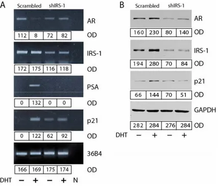
Furthermore, down-regulation of IRS-1 resulted in reduced PSA and p21
mRNA and protein levels (Figure 4B and C). Most importantly, all the above effects
were paralleled by decreased expression of AR protein (Figure 4C), which evidenced
a role for IRS-1 in regulating AR expression. Interestingly, inhibition of AR protein
expression in IRS-1-shRNA MCF-7 cells was not related to decreased AR mRNA
levels (Figure 4B), suggesting that regulation of AR by IRS-1 occurs on the
posttranscriptional level.
Results
Figure 7. IRS-1 knockdown reduces the expression of AR regulated genes. Total cellular RNA (A) and cytoplamic proteins (B) were isolated from transiently transfected MCF-7 cells treated for 24 h with 10-7 M DHT or left untreated in PRF-CT. The expression of AR, IRS-1, PSA, p21, and 36B4 (control of RNA input) mRNA was evaluated by RT–PCR as described in Materials and Methods. The PCR products corresponding to AR, IRS-1, PSA, p21 and 36B4 cDNA fragments (416, 762, 754, 270, and 408 bp respectively) were obtained at 25 and 30 PCR cycles. To control the purity of RNA, the RT step was omitted before the samples were amplified by PCR. 50 µg of protein lysates were probed by WB for the expression of AR, p21, IRS-1, and GAP-DH, as described in Materials and methods. The results, representative of three independent experiments, were quantified and reported as optical density (OD).
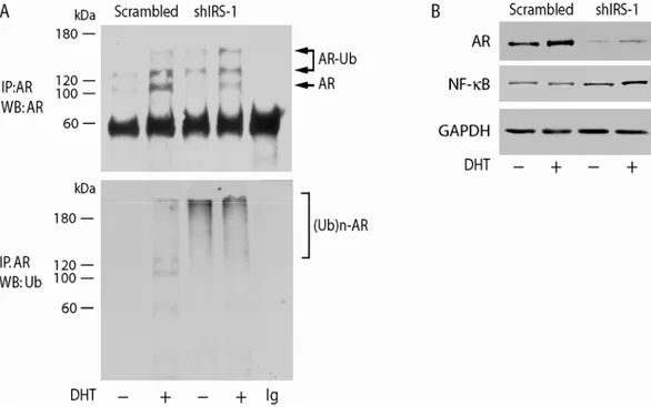
IRS-1 knockdown induces ubiquitylation of AR
Ubiquitin-dependent proteolysis represents an important mechanism for
controlling protein turnover and is pertinent to the regulation of numerous
transcription factors, including AR (Freiman and Tjian, 2003; Nawaz and O'Malley,
2004). Given that polyubiquitylation, in general, constitutes a destructive signal
recognized by the proteasome, we investigated the ubiquitylation status of AR
following downregulation of IRS-1 with shRNA.
MCF-7 and IRS-1-shRNA MCF-7 cells were either left untreated or treated
with 10
-7M DHT for 6 h. Ubiquitylated AR (Ub-AR) was detected by
Results
immunoprecipitation and Western blotting. Three forms of AR were detected, the
regular form of 110 kDa, a form of an apparent molecular weight of 120-130 kDa
consistent with the mono-ubiquitylated receptor, and the poly-ubiquitylated form
ranging from 140-170 kDa. In untransfected MCF-7 cells, mono-ubiquitylation of
AR was ligand dependent, since in the absence of DHT the upper bands were almost
absent and only marginal Ub-AR was observed (Figure 8A).
Figure 8. Effects of IRS-1 on AR ubiquitylation. MCF-7 cells, transiently transfected with 3 µg shIRS-1 or 3 µg scrambled control shRNA (Scrambled) were treated for 6 h with 10-7
M DHT or left untreated in PRF CT. AR was immunoprecipitated (IP) from 500 µg of total protein lysates, and probed with AR mAb (A). The same filter was stripped and re-probed with anti-Ub mAb. The expression of AR, NF-kB, and GAP-DH was assessed by WB on 50 µg of protein lysates (B). The results are representative of three independent experiments.
In IRS-1-shRNA MCF-7 cells, mono-ubiquitylated AR is still present. It is
worth noting that in the same experimental conditions a dramatic increase in the
poly-ubiquitylation status of AR was observed in both DHT-treated and -untreated
samples (Figure 8A). Thus, poly-ubiquitylation of AR may explain its reduced
Results
treatment with the proteasomal inhibitor MG132, evidencing a dramatic increase in
Ub-AR level (Figure 9).
Figure 9. Proteasomal inhibitor MG132 increase Ub-AR level. MCF-7 cells, transfected as before, were treated with or without 1µM MG132 for 8 h. Total protein lysate were immunoprecipitated with an AR mAb and probed with Ub mAb. AR content is shown in the bottom panel. The results are representative of three independent experiments.
Interestingly, IRS-1 knockdown in MCF-7 cells appears to enhance basal
proteosomal activity, as indicated by the increased levels of the DNA binding
subunit of NF-κB, that is produced from a precursor via ubiquitin-proteosomal
processing (Figure 8B). These data suggest that IRS-1 interferes with proteasomal
pathways of different proteins, particularly, the presence of IRS-1 protects AR from
ubiquitin-mediated degradation.
Discussion
Discussion
In addition to the conventional hormone-dependent regulation of steroid
receptor activity, many studies have demonstrated the existence of crosstalk between
growth factor signal transduction pathways and steroid receptors. The modulation of
kinase/phosphatase activity in cells leads either to the direct activation of steroid
receptors in the absence of hormone (Weigel and Zhang, 1998) or to the activation
and recruitment of transcription regulators.
In this study, we report on new aspects of the interaction between AR and an
IGF-I signaling molecule, IRS-1. We demonstrated that IRS-1 associates with AR
and can be translocated to the nucleus in response to DHT. The nuclear trafficking of
IRS-1 seems to be strictly mediated by AR, since the addition of an androgen
antagonist (OH-Fl) inhibits this process. Nuclear localization of IRS-1 has already
been documented in breast cancer cells (Morelli et al., 2004), and many other
cellular systems (Lassak et al., 2002; Prisco et al., 2002; Sun et al., 2003), suggesting
that the protein can function not only as signal transducer but also as transcriptional
regulator (Morelli et al., 2004; Tu et al., 2002).
The possibility that nuclear IRS-1 modulates AR-dependent transcription was
addressed with transient transfection reporter assays in IRS-1 negative cells. The
transcriptional response to DHT stimulation was enhanced by IRS-1 expression in a
dose dependent manner (Figure 3). Moreover, nuclear IRS-1 was co-recruited with
AR on ARE-containing regions of the PSA and p21 promoters (Figure 4A and B),
which are regulated by androgens in breast cancer cells (Lu et al., 1999). The
requirement of IRS-1 expression for AR-dependent transcription was also assessed
Discussion
markedly reduced AR transcriptional response and inhibited the expression of
AR-regulated genes in several experimental models (Figure 5, 7A and B).
Unoccupied AR is labile and its expression can be regulated by
posttranscriptional events in tissues and cell culture models, independently from
changes in receptor mRNA levels (Krongrad et al., 1991; Wolf et al., 1993; Yeap et
al., 1999; Zhou et al., 1995). The binding of ligand to AR, alters receptor interactions
with chaperone proteins, decreasing the turnover and enhancing its ability to activate
androgen-responsive gene expression (Brinkmann et al., 1999; Wang et al., 1999;
Wilson and McPhaul, 1996; Zhou et al., 1995). Since IRS-1 knockdown produced a
reduction in basal AR protein levels (Figure 7B), we investigated whether IRS-1
could regulate AR turnover.
Previous findings demonstrated that proteasome-mediated degradation of
transcriptional activators are coupled to the transcription process (Conaway et al.,
2002; Ferdous et al., 2001; Salghetti et al., 2001) and that hormone binding increases
ubiquitylation and degradation of nuclear receptors (Lange et al., 2000; Nawaz et al.,
1999; Wallace and Cidlowski, 2001). Accumulating evidence indicates that AR may
be targeted for degradation via ubiquitin-proteasome pathways and that this
mechanism is required for AR transactivation in response to DHT (Gaughan et al.,
2005; Lin et al., 2002; Pajonk et al., 2005; Reddy et al., 2006). AR contains an
highly conserved PEST (proline, glutamate, serine and threonine-rich) sequence,
located in the hinge region, which targets proteins for ubiquitylation and degradation
through proteasomal pathways (Sheflin et al., 2000).
The cellular abundance of IRS-1 and AR are both regulated through the 26S
proteasome (Lee et al., 2000; Pederson et al., 2001; Sun et al., 1999; Zhang et al.,
Discussion
2000), Thus, we hypothesized that IRS-1 knockdown could increase the degradation
of AR by desegregating proteins involved in common proteolytic processes. Indeed,
our findings demonstrate that IRS-1 knockdown dramatically enhances the basal
poly-ubiquitylation of AR (Figure 8A).
Consistently, poly ubiquitylation of AR targets it for rapid proteosomal degradation,
while the mono ubiquitylated form represents the trascriptional active form of the
receptor (Burgdorf et al., 2004; Reddy et al., 2006)
Moreover, basic enhancement in cellular proteasomal activity was observed
in IRS-1 knockdown MCF-7 cells since an increase of the NF-κB DNA binding
subunit, which is released from a precursor by the ubiquitin/proteosome-dependent
process, was detected (Figure 8B).
This is in agreement with the increased AR gene transcription (Figure 7B),
since NF-κB DNA binding subunit have been shown to stimulate the AR promoter
activity (Delfino et al., 2003; Zhang et al., 2004).
In summary, our data provide novel insights into the role of IRS-1 as nuclear
regulator of AR function in breast cancer cells. IRS-1 stabilizes AR protein levels
reducing the ubiquitin-dependent proteolysis of AR and optimizes AR-mediated
transcription by participating in the transcriptional machinery on AR regulated
promoters. Further study should help to better understand the mechanisms by which
IRS-1 triggers the ubiquitylation and proteosomal degradation of AR.
In conclusion, taking into account that androgens are potent growth inhibitors
in breast cancer cells (Ando et al., 2002), and that IRS-1 negatively modulates
estradiol-dependent transcription in MCF-7 cells (Morelli et al., 2004), the results
Discussion
reported in this work underline the critical role of IRS-1 in modulating breast cancer
cell proliferation in response to different steroid hormone stimulation.
References
References
Ando, S., De Amicis, F., Rago, V., Carpino, A., Maggiolini, M., Panno, M.L. and
Lanzino, M. (2002) Breast cancer: from estrogen to androgen receptor. Mol
Cell Endocrinol, 193, 121-128.
Aronica, S.M. and Katzenellenbogen, B.S. (1993) Stimulation of estrogen
receptor-mediated transcription and alteration in the phosphorylation state of the rat
uterine estrogen receptor by estrogen, cyclic adenosine monophosphate, and
insulin-like growth factor-I. Mol Endocrinol, 7, 743-752.
Bayer-Garner, I.B. and Smoller, B. (2000) Androgen receptors: a marker to increase
sensitivity for identifying breast cancer in skin metastasis of unknown
primary site. Mod Pathol, 13, 119-122.
Birrell, S.N., Bentel, J.M., Hickey, T.E., Ricciardelli, C., Weger, M.A., Horsfall, D.J.
and Tilley, W.D. (1995) Androgens induce divergent proliferative responses
in human breast cancer cell lines. J Steroid Biochem Mol Biol, 52, 459-467.
Birrell, S.N., Roder, D.M., Horsfall, D.J., Bentel, J.M. and Tilley, W.D. (1995)
Medroxyprogesterone acetate therapy in advanced breast cancer: the
predictive value of androgen receptor expression. J Clin Oncol, 13,
1572-1577.
Brinkmann, A.O., Blok, L.J., de Ruiter, P.E., Doesburg, P., Steketee, K., Berrevoets,
C.A. and Trapman, J. (1999) Mechanisms of androgen receptor activation
and function. J Steroid Biochem Mol Biol, 69, 307-313.
Bryan, R.M., Mercer, R.J., Bennett, R.C., Rennie, G.C., Lie, T.H. and Morgan, F.J.
(1984) Androgen receptors in breast cancer. Cancer, 54, 2436-2440.
Bubulya, A., Wise, S.C., Shen, X.Q., Burmeister, L.A. and Shemshedini, L. (1996)
c-Jun can mediate androgen receptor-induced transactivation. J Biol Chem,
271, 24583-24589.
Burgdorf, S., Leister, P. and Scheidtmann, K.H. (2004) TSG101 interacts with
apoptosis-antagonizing transcription factor and enhances androgen
receptor-mediated transcription by promoting its monoubiquitination. J Biol Chem,
279, 17524-17534.
Castoria, G., Lombardi, M., Barone, M.V., Bilancio, A., Di Domenico, M., Bottero,
D., Vitale, F., Migliaccio, A. and Auricchio, F. (2003) Androgen-stimulated
DNA synthesis and cytoskeletal changes in fibroblasts by a
nontranscriptional receptor action. J Cell Biol, 161, 547-556.
Cesarone, G., Garofalo, C., Abrams, M.T., Igoucheva, O., Alexeev, V., Yoon, K.,
Surmacz, E. and Wickstrom, E. (2006) RNAi-mediated silencing of insulin
receptor substrate 1 (IRS-1) enhances tamoxifen-induced cell death in
MCF-7 breast cancer cells. J Cell Biochem, 98, 440-450.
Conaway, R.C., Brower, C.S. and Conaway, J.W. (2002) Emerging roles of
ubiquitin in transcription regulation. Science, 296, 1254-1258.
Culig, Z., Hobisch, A., Cronauer, M.V., Radmayr, C., Trapman, J., Hittmair, A.,
Bartsch, G. and Klocker, H. (1995) Androgen receptor activation in prostatic
tumor cell lines by insulin-like growth factor-I, keratinocyte growth factor
and epidermal growth factor. Eur Urol, 27 Suppl 2, 45-47.
References
Delfino, F.J., Boustead, J.N., Fix, C. and Walker, W.H. (2003) NF-kappaB and
TNF-alpha stimulate androgen receptor expression in Sertoli cells. Mol Cell
Endocrinol, 201, 1-12.
Ferdous, A., Gonzalez, F., Sun, L., Kodadek, T. and Johnston, S.A. (2001) The 19S
regulatory particle of the proteasome is required for efficient transcription
elongation by RNA polymerase II. Mol Cell, 7, 981-991.
Freiman, R.N. and Tjian, R. (2003) Regulating the regulators: lysine modifications
make their mark. Cell, 112, 11-17.
Garofalo, C., Sisci, D. and Surmacz, E. (2004) Leptin interferes with the effects of
the antiestrogen ICI 182,780 in MCF-7 breast cancer cells. Clin Cancer Res,
10, 6466-6475.
Gaughan, L., Logan, I.R., Neal, D.E. and Robson, C.N. (2005) Regulation of
androgen receptor and histone deacetylase 1 by Mdm2-mediated
ubiquitylation. Nucleic Acids Res, 33, 13-26.
Hackenberg, R., Luttchens, S., Hofmann, J., Kunzmann, R., Holzel, F. and Schulz,
K.D. (1991) Androgen sensitivity of the new human breast cancer cell line
MFM-223. Cancer Res, 51, 5722-5727.
Ignar-Trowbridge, D.M., Teng, C.T., Ross, K.A., Parker, M.G., Korach, K.S. and
McLachlan, J.A. (1993) Peptide growth factors elicit estrogen
receptor-dependent transcriptional activation of an estrogen-responsive element. Mol
Endocrinol, 7, 992-998.
Krongrad, A., Wilson, C.M., Wilson, J.D., Allman, D.R. and McPhaul, M.J. (1991)
Androgen increases androgen receptor protein while decreasing receptor
mRNA in LNCaP cells. Mol Cell Endocrinol, 76, 79-88.
Kuenen-Boumeester, V., Van der Kwast, T.H., Claassen, C.C., Look, M.P., Liem,
G.S., Klijn, J.G. and Henzen-Logmans, S.C. (1996) The clinical significance
of androgen receptors in breast cancer and their relation to histological and
cell biological parameters. Eur J Cancer, 32A, 1560-1565.
Kuenen-Boumeester, V., Van der Kwast, T.H., van Putten, W.L., Claassen, C., van
Ooijen, B. and Henzen-Logmans, S.C. (1992) Immunohistochemical
determination of androgen receptors in relation to oestrogen and progesterone
receptors in female breast cancer. Int J Cancer, 52, 581-584.
Lange, C.A., Shen, T. and Horwitz, K.B. (2000) Phosphorylation of human
progesterone receptors at serine-294 by mitogen-activated protein kinase
signals their degradation by the 26S proteasome. Proc Natl Acad Sci U S A,
97, 1032-1037.
Lanzino, M., De Amicis, F., McPhaul, M.J., Marsico, S., Panno, M.L. and Ando, S.
(2005) Endogenous coactivator ARA70 interacts with estrogen receptor
alpha (ERalpha) and modulates the functional ERalpha/androgen receptor
interplay in MCF-7 cells. J Biol Chem, 280, 20421-20430.
Lassak, A., Del Valle, L., Peruzzi, F., Wang, J.Y., Enam, S., Croul, S., Khalili, K.
and Reiss, K. (2002) Insulin receptor substrate 1 translocation to the nucleus
by the human JC virus T-antigen. J Biol Chem, 277, 17231-17238.
Lea, O.A., Kvinnsland, S. and Thorsen, T. (1989) Improved measurement of
androgen receptors in human breast cancer. Cancer Res, 49, 7162-7167.
Lee, A.V., Gooch, J.L., Oesterreich, S., Guler, R.L. and Yee, D. (2000) Insulin-like
References
mediated by the 26S proteasome and blocked by phosphatidylinositol
3'-kinase inhibition. Mol Cell Biol, 20, 1489-1496.
Lin, H.K., Wang, L., Hu, Y.C., Altuwaijri, S. and Chang, C. (2002)
Phosphorylation-dependent ubiquitylation and degradation of androgen
receptor by Akt require Mdm2 E3 ligase. Embo J, 21, 4037-4048.
Lobaccaro, J.M., Poujol, N., Terouanne, B., Georget, V., Fabre, S., Lumbroso, S.
and Sultan, C. (1999) Transcriptional interferences between normal or mutant
androgen receptors and the activator protein 1--dissection of the androgen
receptor functional domains. Endocrinology, 140, 350-357.
Lu, S., Liu, M., Epner, D.E., Tsai, S.Y. and Tsai, M.J. (1999) Androgen regulation
of the cyclin-dependent kinase inhibitor p21 gene through an androgen
response element in the proximal promoter. Mol Endocrinol, 13, 376-384.
McEwan, I.J. and Gustafsson, J. (1997) Interaction of the human androgen receptor
transactivation function with the general transcription factor TFIIF. Proc Natl
Acad Sci U S A, 94, 8485-8490.
Morelli, C., Garofalo, C., Sisci, D., del Rincon, S., Cascio, S., Tu, X., Vecchione, A.,
Sauter, E.R., Miller, W.H., Jr. and Surmacz, E. (2004) Nuclear insulin
receptor substrate 1 interacts with estrogen receptor alpha at ERE promoters.
Oncogene, 23, 7517-7526.
Nawaz, Z., Lonard, D.M., Dennis, A.P., Smith, C.L. and O'Malley, B.W. (1999)
Proteasome-dependent degradation of the human estrogen receptor. Proc
Natl Acad Sci U S A, 96, 1858-1862.
Nawaz, Z. and O'Malley, B.W. (2004) Urban renewal in the nucleus: is protein
turnover by proteasomes absolutely required for nuclear receptor-regulated
transcription? Mol Endocrinol, 18, 493-499.
Nazareth, L.V. and Weigel, N.L. (1996) Activation of the human androgen receptor
through a protein kinase A signaling pathway. J Biol Chem, 271,
19900-19907.
Oettgen, P., Finger, E., Sun, Z., Akbarali, Y., Thamrongsak, U., Boltax, J., Grall, F.,
Dube, A., Weiss, A., Brown, L., Quinn, G., Kas, K., Endress, G., Kunsch, C.
and Libermann, T.A. (2000) PDEF, a novel prostate epithelium-specific ets
transcription factor, interacts with the androgen receptor and activates
prostate-specific antigen gene expression. J Biol Chem, 275, 1216-1225.
Pajonk, F., van Ophoven, A. and McBride, W.H. (2005) Hyperthermia-induced
proteasome inhibition and loss of androgen receptor expression in human
prostate cancer cells. Cancer Res, 65, 4836-4843.
Pederson, T.M., Kramer, D.L. and Rondinone, C.M. (2001) Serine/threonine
phosphorylation of IRS-1 triggers its degradation: possible regulation by
tyrosine phosphorylation. Diabetes, 50, 24-31.
Prisco, M., Santini, F., Baffa, R., Liu, M., Drakas, R., Wu, A. and Baserga, R. (2002)
Nuclear translocation of insulin receptor substrate-1 by the simian virus 40 T
antigen and the activated type 1 insulin-like growth factor receptor. J Biol
Chem, 277, 32078-32085.
Reddy, G.P., Barrack, E.R., Dou, Q.P., Menon, M., Pelley, R., Sarkar, F.H. and
Sheng, S. (2006) Regulatory processes affecting androgen receptor
References
Reddy, K.B., Yee, D., Hilsenbeck, S.G., Coffey, R.J. and Osborne, C.K. (1994)
Inhibition of estrogen-induced breast cancer cell proliferation by reduction in
autocrine transforming growth factor alpha expression. Cell Growth Differ, 5,
1275-1282.
Ruohola, J.K., Valve, E.M., Karkkainen, M.J., Joukov, V., Alitalo, K. and Harkonen,
P.L. (1999) Vascular endothelial growth factors are differentially regulated
by steroid hormones and antiestrogens in breast cancer cells. Mol Cell
Endocrinol, 149, 29-40.
Salghetti, S.E., Caudy, A.A., Chenoweth, J.G. and Tansey, W.P. (2001) Regulation
of transcriptional activation domain function by ubiquitin. Science, 293,
1651-1653.
Sheflin, L., Keegan, B., Zhang, W. and Spaulding, S.W. (2000) Inhibiting
proteasomes in human HepG2 and LNCaP cells increases endogenous
androgen receptor levels. Biochem Biophys Res Commun, 276, 144-150.
Sisci, D., Morelli, C., Garofalo, C., Romeo, F., Morabito, L., Casaburi, F., Middea,
E., Cascio, S., Brunelli, E., Ando, S. and Surmacz, E. (2006) Expression of
nuclear insulin receptor substrate 1 (IRS-1) in breast cancer. J Clin Pathol.
Sun, H., Tu, X., Prisco, M., Wu, A., Casiburi, I. and Baserga, R. (2003) Insulin-like
growth factor I receptor signaling and nuclear translocation of insulin
receptor substrates 1 and 2. Mol Endocrinol, 17, 472-486.
Sun, X.J., Goldberg, J.L., Qiao, L.Y. and Mitchell, J.J. (1999) Insulin-induced
insulin receptor substrate-1 degradation is mediated by the proteasome
degradation pathway. Diabetes, 48, 1359-1364.
Szelei, J., Jimenez, J., Soto, A.M., Luizzi, M.F. and Sonnenschein, C. (1997)
Androgen-induced inhibition of proliferation in human breast cancer MCF7
cells transfected with androgen receptor. Endocrinology, 138, 1406-1412.
Tilley, W.D., Marcelli, M. and McPhaul, M.J. (1990) Expression of the human
androgen receptor gene utilizes a common promoter in diverse human tissues
and cell lines. J Biol Chem, 265, 13776-13781.
Tu, X., Batta, P., Innocent, N., Prisco, M., Casaburi, I., Belletti, B. and Baserga, R.
(2002) Nuclear translocation of insulin receptor substrate-1 by oncogenes and
Igf-I. Effect on ribosomal RNA synthesis. J Biol Chem, 277, 44357-44365.
Wallace, A.D. and Cidlowski, J.A. (2001) Proteasome-mediated glucocorticoid
receptor degradation restricts transcriptional signaling by glucocorticoids. J
Biol Chem, 276, 42714-42721.
Wang, L.G., Liu, X.M., Kreis, W. and Budman, D.R. (1999)
Phosphorylation/dephosphorylation of androgen receptor as a determinant of
androgen agonistic or antagonistic activity. Biochem Biophys Res Commun,
259, 21-28.
Weigel, N.L. and Zhang, Y. (1998) Ligand-independent activation of steroid
hormone receptors. J Mol Med, 76, 469-479.
Wilson, C.M. and McPhaul, M.J. (1996) A and B forms of the androgen receptor are
expressed in a variety of human tissues. Mol Cell Endocrinol, 120, 51-57.
Wolf, D.A., Herzinger, T., Hermeking, H., Blaschke, D. and Horz, W. (1993)
Transcriptional and posttranscriptional regulation of human androgen
receptor expression by androgen. Mol Endocrinol, 7, 924-936.
References
Yeap, B.B., Krueger, R.G. and Leedman, P.J. (1999) Differential posttranscriptional
regulation of androgen receptor gene expression by androgen in prostate and
breast cancer cells. Endocrinology, 140, 3282-3291.
Zhang, H., Hoff, H. and Sell, C. (2000) Insulin-like growth factor I-mediated
degradation of insulin receptor substrate-1 is inhibited by epidermal growth
factor in prostate epithelial cells. J Biol Chem, 275, 22558-22562.
Zhang, L., Charron, M., Wright, W.W., Chatterjee, B., Song, C.S., Roy, A.K. and
Brown, T.R. (2004) Nuclear factor-kappaB activates transcription of the
androgen receptor gene in Sertoli cells isolated from testes of adult rats.
Endocrinology, 145, 781-789.
Zhou, Z.X., Lane, M.V., Kemppainen, J.A., French, F.S. and Wilson, E.M. (1995)
Specificity of ligand-dependent androgen receptor stabilization: receptor
domain interactions influence ligand dissociation and receptor stability. Mol
Scientific Publications
Scientific Publications
Performed during the PhD Program
Expression of Nuclear Insulin Receptor Substrate 1 (IRS-1) in Breast Cancer
Diego
Sisci
1§, Catia
Morelli
1§, Cecilia
Garofalo
1, Francesco Romeo
2, Lucio Morabito
2, Filomena
Casaburi
2, Emilia Middea
1, Sandra Cascio
3, Elvira Brunelli
4, Sebastiano
Andò
5and Eva
Surmacz
3* 1Department of Pharmaco-Biology, Faculty of Pharmacy, University of Calabria, Arcavacata di Rende,
Italy;
2Division of Anatomo-Pathology, Annunziata Hospital, Cosenza, Italy;
3Sbarro Institute for
Cancer Research and Molecular Medicine, Temple University, Philadelphia, PA, USA,
4Department of
Natural Science and
5Department of Cellular Biology, Faculty of Pharmacy, University of Calabria,
Arcavacata di Rende, Italy.
§
D.S. and C.M. contributed equally to this work
*Corresponding author:
Eva Surmacz, Ph.D.
Sbarro Institute for cancer Research and Molecular Medicine
College of Science and Technology, Temple University
1900 N 12
thSt. Rm. 446, Philadelphia, PA 19122
Tel. 001/215-204-0306, Fax 001/215-204-0303
e-mail [email protected]
Word count (Abstract included)
: 3475
Structured abstract: 251 words.
Abstract
Aims: Insulin receptor substrate 1 (IRS-1), a cytoplasmic protein transmitting signals from the insulin
and insulin-like growth factor 1 receptors, has been implicated in breast cancer. Previously, we
reported that IRS-1 can be translocated to the nucleus and modulate estrogen receptor
α (ERα) activity
in vitro. However, the expression of nuclear IRS-1 in breast cancer biopsies has never been examined.
Consequently, we assessed whether nuclear IRS-1 is present in breast cancer and non-cancer mammary
epithelium and if it correlates with other markers, especially ER
α. Parallel studies were done for
cytoplasmatic IRS-1.
Methods:
IRS-1 and ERα expression was assessed by immunohistochemistry. Data were evaluated
using Pearson correlation, linear regression, and ROC analysis.
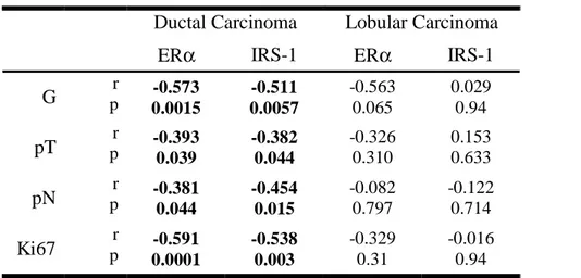
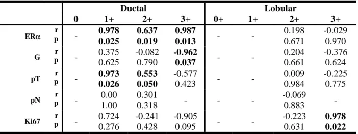
Results: Median nuclear IRS-1 expression was low in normal mammary epithelial cells (1.6%) and
higher in benign tumors (20.5%), ductal grade 2 carcinoma (11.0%), and lobular carcinoma (~30%).
Median ER
α expression in normal epithelium, benign tumors, ductal cancer grade 2 and 3 and lobular
cancer grade 2 and 3 was 10.5, 20.5, 65.0, 0.0, 80, and 15%, respectively. Nuclear IRS-1 and ER
α
positively correlated in ductal cancer (p<0.001) and benign tumors (p< 0.01), but were not associated
in lobular cancer and normal mammary epithelium. In ductal carcinoma, both nuclear IRS-1 and ER
α
negatively correlated with tumor grade, size, mitotic index, and lymph node involvement. Cytoplasmic
IRS-1 was expressed in all specimens and positively correlated with ER
α in ductal cancer.
Conclusions: A positive association between nuclear IRS-1 and ER
α is a characteristic for ductal
breast cancer and marks a more differentiated, non-metastatic phenotype.
Take-home messages
1. This is the first report examining the expression of nuclear IRS-1 in normal mammary tissue,
benign breast tumors and breast cancer in relation to ER
α and clinicopathological features.
2. Nuclear IRS-1 is more prevalent in cancer specimens than in normal mammary tissues.
3. Nuclear IRS-1 and ER
α negatively correlated with tumor grade, size, mitotic index, and lymph
node involvement.
4. A positive association between nuclear IRS-1 and ER
α is a characteristic for ductal breast cancer
and marks a more differentiated, non-metastatic phenotype.
Introduction
Recent experimental and clinical evidence suggests the involvement of the insulin-like growth
factor I (IGF-I) receptor (IGF-IR) in breast cancer development and progression
1-6. The tumorigenic
action of IGF-IR is executed through multiple antiapoptotic, growth promoting, and/or pro-metastatic
pathways
5-9. Many of these pathways stem from IRS-1, a major IGF-I signaling molecule that
becomes phosphorylated on multiple tyrosine residues upon IGF-IR activation. Tyrosine
phosphorylated IRS-1 acts as a scaffolding protein sequestering downstream signaling molecules and
propagating IGF-I signal through the PI-3K/Akt, Ras/Raf/ERK1/2, Jak2/Stat3 and other pathways
10-13.
Overexpression or downregulation of IRS-1 in breast cancer cell models suggested that the
molecule controls several aspects of the neoplastic phenotype, especially anchoragedependent and
-independent cell growth and survival
14 15. In breast cancer cell lines, IRS-1 appears to be expressed at
higher levels in ER
α-positive than in ERα-negative cells and there is evidence supporting the
existence of a crosstalk between IRS-1 and ER
α systems
1 4 6 16-18. Overexpression of IRS-1 in MCF-7
ER
α-positive cells has been shown to induce estrogen-independence and mediate
antiestrogen-resistance
14 19 20. High expression of IRS-1 can be in part attributed to ER
α activity, as 17β-estradiol
(E2) can upregulate IRS-1 expression and function
16 21 22, while antiestrogens reduce IRS-1 mRNA
and protein levels and inhibit IRS-1 signaling
19 20 23. In addition, ER
α can directly interact with IRS-1,
increasing its stability and potentiating its downstream signaling to Akt
24. Notably, increased activity
of IRS-1 is likely to modulate ER
α, via ERK1/2- and Akt-mediated phosphorylation of ERα on
Ser-118 and Ser-167, respectively
25-27.
Recent reports suggested that in addition to its cytoplasmic signaling function, IRS-1 is able to
regulate nuclear processes in different cell models
28-33. For instance, in mouse fibroblasts treated with
IGF-I, a fraction of IRS-1 is translocated from the cytoplasm to the nuclear and nucleolar
compartments where it modulates the expression of genes controlling cell proliferation (i.e., Cyclin
D1) and cell growth in size (i.e., rDNA) by physically interacting with transcriptional complexes of
β-catenin and upstream binding factor 1 (UBF1), respectively
31 32. Our recent work demonstrated that
nuclear IRS-1 is also found in breast cancer cell lines. For instance, in MCF-7 cells treated with E2
nuclear IRS-1 physically interacted with ER
α modulating its transcriptional activity at estrogen
response element (ERE) DNA motifs
33. The exact mechanism of nuclear IRS-1 transport is not clear,
but most likely it involves other proteins containing nuclear localization signals (ER
α, T antigen,
importins).
Despite the evidence that IRS-1 signaling may play a critical role in tumorigenesis, only limited
studies examined the clinical significance of IRS-1 expression in human breast cancer specimens
1834-36