UNIVERSITY OF SASSARI
INTERNATIONAL PHD COURSE IN LIFE SCIENCES AND BIOTECHNOLOGIES
DEPARTMENT OF BIOMEDICAL SCIENCES XXIX Cycle (Curriculum: Microbiology and Immunology)
Director: Prof. Sechi Leonardo Antonio
INTERACTION OF MONOCYTES AND DERIVED
MACROPHAGE SUBSETS WITH AFRICAN SWINE FEVER
VIRUSES OF DIVERSE VIRULENCE
by
Giulia Franzoni
Supervisors:
Prof. Marco Pittau, University of Sassari
Giulia Franzoni
‘Interaction of monocytes and derived macrophage subsets with African swine fever viruses of diverse virulence’ International PhD Course in Life Sciences and Biotechnologies
University of Sassari
Università degli Studi di Sassari Corso di Dottorato di ricerca in Scienze della Vita e Biotecnologie
La presente tesi è stata prodotta durante la frequenza del Corso di Dottorato di ricerca in Scienze della Vita e Biotecnologie dell’Università degli Studi di Sassari, A.A. 2013/2014 - XXIX ciclo, con il sostegno di una borsa di studio finanziata con le risorse dell’INPS – Gestione Ex INPDAP nell’ambito delle Iniziative Accademiche Homo Sapiens Sapiens.
Giulia Franzoni
‘Interaction of monocytes and derived macrophage subsets with African swine fever viruses of diverse virulence’ International PhD Course in Life Sciences and Biotechnologies
University of Sassari
TABLE OF CONTENTS
Abstract
CHAPTER 1. INTRODUCTION
1.1 Monocytes and macrophages 1.2 African Swine Fever (ASF)
1.3 African Swine Fever Virus (ASFV) 1.4 Control of ASF in Sardinia
1.5 ASFV diagnosis in Sardinia 1.6 ASF vaccines and antiviral agents 1.7 Immunology against ASF
1.7.1 ASFV and innate immunity 1.7.2 ASFV and adaptive immunity 1.8 Aims, hypothesis and objectives
CHAPTER 2. MATERIAL AND METHODS
2.1 Animals and blood sampling 2.2 Viruses
2.3 Enrichment of monocytes
2.4 In vitro differentiation of monocyte into moM and activation
2.5 In vitro ASFV infection of monocytes/macrophages subsets and growth curves 2.6 Light microscopy
Giulia Franzoni
‘Interaction of monocytes and derived macrophage subsets with African swine fever viruses of diverse virulence’ International PhD Course in Life Sciences and Biotechnologies
University of Sassari
2.8 Assessment of metabolically active cells 2.9 Flow cytometry
2.10 TNF- release in response to LPS stimulation 2.11 Patterns of cytokine secretion
2.12 DNA extraction and real-time PCR 2.13 Data analysis and statistics
CHAPTER 3. RESULTS: IN VITRO DIFFERENTIATION OF PORCINE MONOCYTES INTO MACROPHAGES
3.1 Introduction
3.2 Phenotype characterization of moMΦ 3.3 Assessment of metabolically active cells 3.4 Susceptibility to ASFV
3.5 LPS-stimulated release of TNF- 3.6 Patterns of cytokine secretion
CHAPTER 4. RESULTS: CHARACTERIZATION OF THE INTERACTION OF ASFV WITH MONOCYTES AND MACROPHAGE SUBSETS
4.1 Introduction
4.2 Generation of monocyte-derived macrophage subsets
4.3 Monocytes and macrophage subsets susceptibility to ASFV infection 4.4 ASFV growth in macrophage subsets
4.5 Effect of ASFV infection on surface marker expression on monocytes and derived macrophage subsets
Giulia Franzoni
‘Interaction of monocytes and derived macrophage subsets with African swine fever viruses of diverse virulence’ International PhD Course in Life Sciences and Biotechnologies
University of Sassari
4.6 Production of cytokines in ASFV-infected monocytes/macrophage subsets
CHAPTER 5. DISCUSSION
Giulia Franzoni
‘Interaction of monocytes and derived macrophage subsets with African swine fever viruses of diverse virulence’ International PhD Course in Life Sciences and Biotechnologies
University of Sassari
ABSTRACT
African swine fever (ASF) is a devastating disease for which there is no vaccine available. The ASF virus (ASFV) primarily infects cells of the myeloid lineage and this tropism is thought to be crucial for disease pathogenesis. A detailed in vitro characterization of the interactions of a virulent (22653/14) and a tissue culture adapted (BA71V) strains of ASFV with porcine monocytes, un-activated (moM), classically (moM1) and alternatively (moM2) activated monocyte-derived macrophages was conducted to better understand this relationship. Low concentration of hM-CSF was selected as the method of choice to generate moM. Using a multiplicity-of-infection (MOI) of 1, both viruses were able to infect monocytes and macrophage subsets, but BA71V presented a reduced ability to infect moM1 compared to 22653/14, with higher expression of early compared to late proteins. Using an MOI of 0.01, only 22653/14 was able to replicate in all the macrophage subsets, with initially lowest in moM1 and moM2 ASFV down-regulated CD16 expression and BA71V-infected but not 22653/14-infected moM and moM2 presented with a reduced expression of MHC class I. Higher levels of IL-18, IL1- and IL-1 were released from moM1 after infection with BA71V. These results revealed differences between these strains, suggesting that virulent isolates have evolved mechanisms to counteract activated macrophages responses, promoting their survival, dissemination in the host and so ASF pathogenesis.
Giulia Franzoni
‘Interaction of monocytes and derived macrophage subsets with African swine fever viruses of diverse virulence’ International PhD Course in Life Sciences and Biotechnologies
University of Sassari
Giulia Franzoni
‘Interaction of monocytes and derived macrophage subsets with African swine fever viruses of diverse virulence’ International PhD Course in Life Sciences and Biotechnologies
University of Sassari
1.1 Monocytes and macrophages
Monocytes and macrophages are the main target of the African Swine Fever Virus (ASFV) (Sánchez-Cordón et al., 2008).
Monocytes are primary immune cells, which are derived from a common myeloid progenitor cells in the bone marrow (haematopoietic stem cell) and there undergo differentiation steps, in response to macrophage colony-stimulating factor, before enter the bloodstream (Volkman and Gowans, 1965, Mosser and Edwards, 2008). In mice, monocytes can also be differentiated into 2 populations: ‘inflammatory’ monocytes, with a GR1+CX
3CR1low phenotype, which rapidly exit the blood, and
‘resident’ monocytes, which do not express GR1− (Geissman et al., 2003). As shown
in fugure 1.1, monocytes circulate in the peripheral blood before migrating to different tissues and replenish the tissue macrophage populations (Volkman and Gowans, 1965; Mosser and Edwards, 2008). Their half life is very short, about one day in mice (Van Furth and Cohn, 1968) and 3 days in humans (Whitelaw, 1972).
Figure 1.1. Monocytes origin and differentiation. Monocytes originate in the bone
marrow from a common haematopoietic stem cell (HSC) and there they undergo differentiation steps during which they commit to the myeloid and then to a monocyte lineage. They differentiate into monoblasts and then pro-monocytes before becoming monocytes, which enter the bloodstream. In mice these cells can differentiated into two populations: inflammatory and resident monocytes. It is unclear if inflammatory monocytes mature into resident monocytes or if they represent distinct populations. Monocytes migrate to different tissues, where they replenish tissue-specific macrophages. CNS, central nervous system; GM-CFU, granulocyte-macrophage colony-forming unit; M-CFU, macrophage colony-forming unit. Figure taken from Mosser and Edwards, 2008.
Giulia Franzoni
‘Interaction of monocytes and derived macrophage subsets with African swine fever viruses of diverse virulence’ International PhD Course in Life Sciences and Biotechnologies
University of Sassari
Monocytes are a heterogeneous population and in human they can be divided into two subsets: CD14+CD16- (90% of circulating monocytes), which are often called
classical monocytes, and CD14+CD16+, which exhibit features of tissue macrophages
(Ziegler-Heitbrock, 1996). CD14+CD16+ express CD32 and higher amount of MHC
II molecules than classical monocytes and they have distinct chemokine–receptor expression profile (Weber et al., 2000). Human monocytes can also be characterised also by their expression of CD64, also known as FcyRI (Grage-Gribenow et al., 2001). CD14+CD16+CD64+ monocytes present a dendritic cells (DC) phenotype,
expressing higher levels of MHC II and CD80/86 than classic monocytes and having an enhanced T-cell-stimulatory activity (Grage-Gribenow et al., 2001). It was speculated that CD14+CD16+CD64+ cells could represent an immunoregulatory
monocyte phenotype or an intermediate phenotype between monocytes and DC (Grage-Gribenow et al., 2001). As previously stated, in mice monocytes can be divided according to their expression of CCR2, CX3C-chemokine receptor 1 (CX3CR1) and GR1 into two subpopulations: inflammatory CCR2+GR1+CX3CR1low
and resident CCR2-GR1-CX3CR1high (Palfrarman, 2001, Geissman et al., 2003). It is
unclear if they represent distinct populations or if inflammatory monocytes are initially released into the circulation and, in the absence of inflammation, they mature into resident macrophages, altering their functional and phenotypic characteristic, passing through an intermediate phenotype (CCR2+GR1low) (Gordon
and Taylor, 2005). Porcine monocytes can instead be characterised based on expression of CD163, which is scavenger receptor of haemoglobin/haptoglobin complexes (Sachez et al., 1999, Kristiansen et al., 2001). CD163+ monocytes
represent a more mature subset, in fact they produce higher amount of TNF-, present higher levels of CD80/CD86 co-stimulatory molecules and SLA DR antigens and have better antigen-presenting capacity to primed T CD4+ lymphocytes
(Chamorro et al., 2004).
As shown in figure 1.1, circulating monocytes migrate into tissue in the steady state or in response to inflammation, where they differentiate into macrophages or into specialised cells such as DC and osteoclasts (Volkman and Gowans 1965, Mosser and Edwards, 2008). Beyond this precursor role, monocytes can carry out specific effector functions during infection (Serbina et al., 2008). In fact in mice
Giulia Franzoni
‘Interaction of monocytes and derived macrophage subsets with African swine fever viruses of diverse virulence’ International PhD Course in Life Sciences and Biotechnologies
University of Sassari
inflammatory monocytes are implicated in defence against bacterial, protozoal, and fungal pathogens. They respond rapidly to microbial stimuli by secreting cytokines and antimicrobial factors (Serbina, et al., 2008). In addition, a recent study reported that in mice monocytes can participate in steady-state surveillance of the lung, in a complementary way to resident macrophages and DC, without differentiating into macrophages (Rodero et al., 2015).
Macrophages as recognised by Metchnikoff are first and foremost professional phagocytes, which express all the genes required to internalize particles and to degrade those particles in lysosomes (Hume, 2015). Tissue macrophages maintain tissue homeostasis though the clearance of senescent cells and the repair/remodelling of tissues after inflammation (Gordon, 1988). They clear the interstitial environment of extraneous cellular materials, independently of immune-cell signalling and in the absence of other immune cells. Macrophages are involved in removal of cellular debris that are generated during tissue remodelling and rapidly clear cells that have undergone apoptosis. In human they also clear approximately 2 x 10 erythrocytes
each day, recovering iron and haemoglobin (Mosser and Edwards, 2008). These cells can rapidly respond to endogenous danger signals generated following injury or infection. They recognize pathogen-associated molecular patterns (PAMPs) through pattern-recognition receptors (PRRs), such as toll like receptors (TLRs), NOD-like receptors (NAPLs) and C-type lectin receptors. Through complement receptors these cells interact with the complement inflammatory cascade, crucial to ward off infection (Martinez et al., 2009). Macrophage are a heterogeneous population and this heterogeneity reflect the specialization of function adopted by macrophages in different anatomical locations. For example osteoclasts are specialised in bone remodelling and instead alveolar macrophages present high expression of pathogen recognition receptors (PRRs), enabling them to clear microorganisms, virus and environmental particles in the lung (Gordon and Taylor, 2005).
In vitro human monocytes can be differentiated into macrophages (MoM with the addition of macrophage colony stimulator factor (M-CSF) to culture media (Gordon and Taylor, 2005). M-CSF, also known as colony-stimulating factor-1 (CSF-1), controls the survival, proliferation, and differentiation of mononuclear phagocytes
Giulia Franzoni
‘Interaction of monocytes and derived macrophage subsets with African swine fever viruses of diverse virulence’ International PhD Course in Life Sciences and Biotechnologies
University of Sassari
(Stanley et al., 1997). It drives the differentiation of monocytes into macrophages acting via a cell surface tyrosine kinase receptor, known as colony stimulating factor receptor 1 (CSF-1R), expressed on cells of the mononuclear phagocyte lineage (Stanley et al., 1997). CSF-1 bind to the CSF receptor (CSF-1R) and it has been reported that treatment of mice with monoclonal antibodies against this receptor depleted most tissue macrophage populations (McDonald et al., 2010).
In pigs monocytes can be differentiated into macrophages through culture with M-CSF for 4-7 days (Singleton et al., 2016, Kyrova et al., 2014) or with different concentration of porcine serum or plasma for 3-4 days (10%: Garcia-Nicolas et al., 2014, Kyrova et al., 2014; 20% Chamorro et al., 2000, Sanchez-Torres et al., 2003; 30%: McCullough et al., 1997, McCullough et al., 1999, Basta et al., 1999, Basta et al., 2001, Tsai et al., 2010). In addition, some authors used the media from the murine fibroblast L929 cells line as a source of M-CSF (Genovesi et al., 1990, Zsak et al., 1998, Wang et al., 2011). In few studies researchers differentiated monocytes into macrophages using media supplemented with just 10% fetal bovine serum (FBS) (10%: Gil et al., 2003, Gil et al., 2008), but the work of McCullough and colleagues (1999) reported that monocytes cultured in media supplemented with FSB remained morphologically closer to monocytes than those cultured in porcine plasma (McCullough et al., 1999). As previously stated, in humans M-CSF is commonly used to in vitro differentiate monocytes into macrophages (Gordon and Taylor, 2005) and treatment of porcine monocytes with M-CSF increases cell viability (Genovesi et al., 1990). Fairbairn et al. (2014) observed that porcine PBMC cultured with hM-CSF (104 U/ml) increased in size and granularity compared with freshly isolated
PBMC and that the CD14+CD172a+ population was selectively expanded, suggesting
that as in humans M-CSF act as a mitogen for pig monocytes (Fairbairn et al., 2014). To date there is not a standardize protocol to in vitro differentiate porcine monocytes into macrophages and in the first part of this thesis we focus on comparing different methods described in the literature, assessing their effect on macrophages morphology, expression of surface markers, cytokine release and susceptibility to monocytropic ASFV (Chapter 3).
Giulia Franzoni
‘Interaction of monocytes and derived macrophage subsets with African swine fever viruses of diverse virulence’ International PhD Course in Life Sciences and Biotechnologies
University of Sassari
Several studies analyzed the interaction of ASFV with monocytes and macrophages, and there are better described in the section 1.7.1. To date, no studies investigated the interaction of ASFV with activated macrophages.
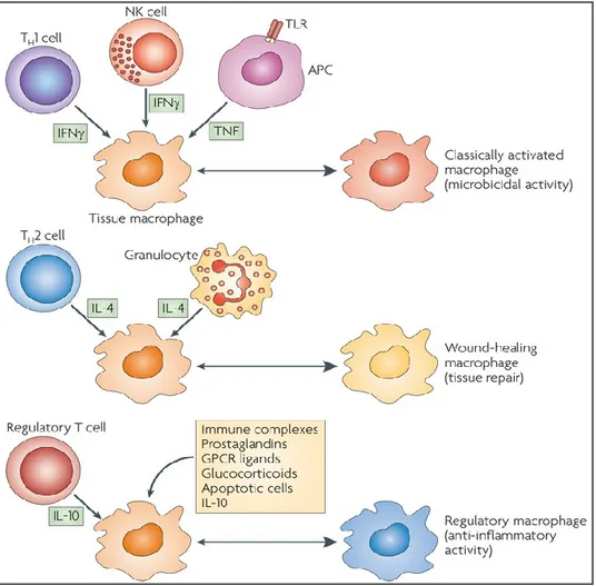
Macrophages present remarkable plasticity, that allow them to respond to environmental signals, produced also by antigen-specific immune cells. Both the innate and the adaptive immune responses affect their phenotype and physiology (Mosser and Edwards, 2008). Macrophage activation results in their polarisation into different functional subsets: classical activated macrophages (M1), alternative activated macrophages (M2) and type II-activated or regulatory macrophages (Mosser, 2003, Mosser and Edwards, 2008), as represented in figure 1.2.
M1 polarization can be reached in vitro by exposure to IFN- and lipopolysaccharide (LPS), which induces TNF production. Classical activation results in secretion of high levels of pro-inflammatory cytokines and increased microbicidal or tumoricidal capacity (Mosser, 2003). Stimulation with IFN- results in increased production of superoxide anions, oxygen and nitrogen radicals by macrophages. In fact the main role of M1 macrophages is in host defence to intracellular pathogens and in driving Th1 cellular immune responses (Mosser and Edwards, 2008). It was reported that mice lacking IFN- expression are more susceptible to various bacterial, protozoal and viral infections, as are humans with genetic mutations in these signalling pathways (Filipe-Santos et al., 2006). Moreover some pathogens have developed mechanisms to interfere with IFN- signalling and to prevent efficient macrophage activation (Mosser and Edwards, 2008). M1 are vital component of the host defence, but their activation might be tightly controlled, in fact they are key mediators of the immunopathology that occurs in several autoimmune diseases (Mosser and Edwards, 2008). Very few studies analysed macrophage classical activation in pigs. It was reported that in pig classical activation led to MHC II up-regulation, suggesting that also in this specie M1 have and enhanced antigen presenting functions (Garcia-Nicolas et al., 2014, Singleton et al., 2016).
Giulia Franzoni
‘Interaction of monocytes and derived macrophage subsets with African swine fever viruses of diverse virulence’ International PhD Course in Life Sciences and Biotechnologies
University of Sassari
Figure 1.2. Cytokines produced by immune cells can give rise to macrophages with distinct physiology. Classical activated macrophages arise in response to
IFN-, which can be produced by T cells or NK cells, and TNF, produced by antigen presenting cells (APCs). Alternative activated macrophages arise in response to IL-4, which can be produced by TH2 cells or by granulocytes. Regulatory macrophages are generated in response to various stimuli, such as immune complexes, prostaglandins, G-protein coupled receptor (GPCR) ligands, glucocorticoids, apoptotic cells or IL-10. Figure taken from Mosser and Edwards, 2008.
Alternative activation of macrophages was discovered later than classical activation. It was found that alternative activation occurred via the stimulation of macrophages with IL-4, which was found to up-regulate expression of the mannose receptor (CD206) (Stein et al., 1992). Activation with this cytokine was clearly distinct from the classic activated macrophage, in fact M2 do not produce NO and have a poor ability to kill intracellular pathogens (Modolell et al., 1995). It was later discovered that alternative activation results also from exposure to IL-13, which shares the same IL-4 receptor alpha (IL-4Rα) chain as IL-4 (Gordon, 2003). M2 macrophages are primarily associated with mechanisms of immunosuppression and wound repair
Giulia Franzoni
‘Interaction of monocytes and derived macrophage subsets with African swine fever viruses of diverse virulence’ International PhD Course in Life Sciences and Biotechnologies
University of Sassari
(Gordon, 2003), in fact they are also called ‘wound-healing’ macrophages (Mosser and Edwards, 2008). They are not efficient at antigen presentation and instead promote cell growth, collagen formation and tissue repair; M2 secrete several fibrogenic factors and insulin-like growth factor 1 and platelet-derived growth factor C, which provide signals for tissue proliferation and repair (Mosser, 2003, Martinez et al., 2009). M2 produce high levels of the pro-inflammatory cytokine IL-10 and mediate repression of pro-inflammatory factors, so are compromised in their ability to kill intracellular pathogens and down-regulate a pro-TH1 response (Mosser, 2003, Martinez et al., 2009). Macrophage alternative activation is implicated in the success of TH2 responses, mainly directed toward extracellular parasites such as helminths and some protozoa. Intracellular pathogens have, as a result, created mechanism to promote macrophage alternative activation and to avoid TH1 immune control (Martinez et al., 2009). Very few studies analysed macrophage alternative activation in pigs. In this specie alternative activation leads to CD163 down-regulation, but has no influence on MHC class I or II expression (Garcia-Nicolas et al., 2014).
Another macrophage category is the type II-activated macrophage, which were recently classified as one M2 subtype. In fact M2 can be subdivided in three subtypes: M2a (after exposure to IL-4 or IL-13), M2b (immune complexes in combination with IL-1beta or LPS) and M2c (IL-10, TGF-beta or glucocorticoids) (Martinez et al., 2008). Macrophage activation with immune complexes and LPS results in inhibition of IL-12 and promotion of IL-10 secretion, up-regulation of antigen presentation and promotion of a TH2 response (Martinez et al., 2008). Glucocorticoids affect monocyte adherence, spreading, phagocytosis and apoptosis, whereas IL-10 is a potent inhibitor of TH1 cells (Martinez et al., 2008).
Nevertheless, macrophage taxonomy and the M1/M2 paradigm is a limited attempt to define the complexity and plasticity of mononuclear phagocytes. Individual macrophage cells differ markedly from each other, and change their functions in response to the subtle and continuous changes in the surrounding environmental signals, so M1/M2 subsets represent the two extreme of diverse functional macrophage activation states (Sica and Matovani, 2012, Italiani and Boraschi, 2014, Hume, 2015).
Giulia Franzoni
‘Interaction of monocytes and derived macrophage subsets with African swine fever viruses of diverse virulence’ International PhD Course in Life Sciences and Biotechnologies
University of Sassari
1.2 African Swine Fever
African swine fever (ASF) is caused by a highly infectious virus that affects domestic and wild pigs and it is considered a notifiable disease by the OIE World Organisation for Animal Health because of its potential for rapid dissemination and significant socio-economical consequences (Sanchez-Vizcaino, 2006).
The aetiological agent is the African swine fever virus (ASFV), the only member of the Asfarviridae family (Dixton et al., 2005).
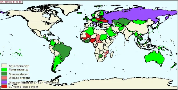
ASF is currently endemic in many sub-Saharan countries of Africa and Sardinia (Italy). The spread of this disease to the Caucasus region in 2007, and from there to Russia and Ukraine in 2012, Belarus in 2013, and European Union countries (Lithuania, Estonia, Latvia and Poland) in 2014-2016, now endangers the pig industry worldwide (Alonso, 2013, OIE WAHIS interface).
In Russia ASFV progressively spread and 18 federal districts reported outbreaks (OIE, WAHIS interface). In this area ASFV has been isolated in wild boars and the persistence of the virus in the sylvatic cycle makes eradication more difficult (Beltran-Alcrudo et al., 2008). In addition, a recent study revealed the existence of long-term infected animals in this area, which could become carriers and persist in the population, leading to the maintenance and future reappearance of the disease (Mur et al., 2016a).
In Poland ASFV was first detected in early 2014 and between 2014 and 2015 65 cases of ASFV infection in wild boar have been recognised, located near the border with Belarus in Sokółka and Białystok counties (Wozniakowski et al., 2016). Recently, in that country cases of ASFV have been described also in domestic pigs (OIE WAHIS interface).
Giulia Franzoni
‘Interaction of monocytes and derived macrophage subsets with African swine fever viruses of diverse virulence’ International PhD Course in Life Sciences and Biotechnologies
University of Sassari
Figure 1.3 Distribution of ASFV from January 2016. Figure taken from http://web.oie.int (disease and distribution maps).
Using retrospective analysis it was established that the first ASFV outbreak occurred in Africa in 1907, but the disease was firstly described in Kenia in 1921 by Montgomery. The first spread of ASF outside Africa was to Lisbon (Portugal) in 1957 and a further outbreak occurred in 1960 in Lisbon. ASF become endemic in all the Iberian peninsula and outbreaks were reported subsequently in a number of other European countries, including Malta, Italy, France, Belgium and The Netherlands (Costard et al., 2013). In Spain and Portugal complete eradication took more than 30 years (Sanchez-Vizcaino, 2006).
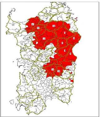
In Sardinia the disease first occurred in 1978 and now is endemic, as a result of extensive pig farming that has been practised for centuries and of the presence of endemically infected wild boar (Firinu et al., 1988; Costard et al., 2009).
There is no vaccine or treatment available and apart from stamping out and movement control, there are no control measures, thereby potentially resulting in extreme losses for producers (Costard et al., 2013).
As previously written, ASF affects both domestic and wild pigs (Sanchez-Vizcaino, 2006).
Giulia Franzoni
‘Interaction of monocytes and derived macrophage subsets with African swine fever viruses of diverse virulence’ International PhD Course in Life Sciences and Biotechnologies
University of Sassari
African wild pigs are resistant to the disease and ASF persists in Africa via a natural cycle of transmission between the warthog (Phacochoerus aethiopicus), bushpig (Potamchoerus porcus) and the soft tick (Ornithodorus moubata). Warthogs are born free from infection and may be infected early in life following the bite of an infected tick. Virus replicates in the warthog and produces a low degree of viraemia for a few weeks, which is sufficient to infect a proportion of ticks that feed on the viraemic young warthogs (Thomson, 1985). Domestic pigs in Africa acquire infection from wildlife reservoirs of the virus primarily by the bite of an infected tick (Plowright, 1977).
In other areas, such as Sardinia, infection can occur either directly (escretions, secretions, dead animals) or indirectly (fomites, such as clothing, equipment and vehicles), in fact ASFV can persist in the environment for several days. The virus can persist in tissues for several months and feeding domestic pigs with uncooked swill can result in infection (Costard et al., 2009). Soft ticks Ornithodoros are absent in Sardinia, so they are not involved in the persistence of the virus in the island (Costard et al., 2013). In a recent study 1767 porcine serum samples collected from all around the island (1261 from domestic and 506 from wild boar) were analysed for antibodies to salivary antigens of Ornithodoros erraticus and only one sample resulted positive. In addition, ticks were directly searched in a number of pig premises with no success, confirming the absence of Ornithodoros Erraticus tick role in ASF cycle in Sardinia (Mur et al., 2016b).
Recovered pigs can remain persistently infected for periods of 6 months or more, representing an obstacle for eradication of the disease from endemic areas (Wilkinson 1984, Oura et al. 2005).
The clinical signs associated with ASF are very varied as they depend on how virulent the viral isolate is and on the breed and physical condition of the pig. African ASFV isolates generally induce peracute or acute disease. European domestic pigs and boars are very susceptible and exhibit a wide range of clinical signs from subacute to chronic. As above stated, wild African pigs are very resistant to infection and do not generally present any lesions (Sánchez-Vizcaíno, 2006).
Giulia Franzoni
‘Interaction of monocytes and derived macrophage subsets with African swine fever viruses of diverse virulence’ International PhD Course in Life Sciences and Biotechnologies
University of Sassari
In Europe, it has been observed that wild boars are susceptible to the disease as domestic pigs (Jori and Bastos, 2009, McVicar et al., 1981). Outbreaks of ASF in wild boars fade out, so the contact with infected domestic pigs or other source of infection is essential for the persistence of the virus in the sylvatic cycle (Laddomada et al., 1994).
In the acute form, the animals show high temperatures (40-42ºC), recumbency and lack of appetite, and suffer respiratory disorders. In some cases there may be nasal haemorrhaging, constipation and vomiting. Exanthemas are very evident (pinkish almost purple skin due to intense hyperaemia), and/or cyanotic foci, which appear as irregular purple-coloured marks on the skin of the extremities, ears, chest, abdomen and perineum. Abortion frequently occurs in gestating females. In acute cases, the disease causes death in 90 to 100% of affected animals. In acute and subacute courses death occurs between 7 and 20 day post infection, the signs develop more slowly. The chronic form is characterised by a large variety of clinical signs which are mainly the result of secondary bacterial complications. Mortality is low, affecting between 2 and 10% of all the sick animals (Sánchez-Vizcaíno, 2006).
Some characteristic ASF lesions are: purplish and megalic spleen, haemorrhaging in the tonsils and lymphatic ganglia, particularly in the gastrohepatic and renal ganglia, and petechial haemorrhaging in the kidneys, bladder mucosa, pharynx and larynx, pleura and heart, endocardium and pericardium, hydropericardium, ascitis, and hydrothorax and hepatic congestion (Figures 1.4, 1.5, 1.6) (Sánchez-Vizcaíno, 2006). The disease is characterised by severe lymphopenia, immunodepression and thrombocytopenia (Gómez-Villamandos et al., 2013).
Giulia Franzoni
‘Interaction of monocytes and derived macrophage subsets with African swine fever viruses of diverse virulence’ International PhD Course in Life Sciences and Biotechnologies
University of Sassari
Figure 1.5 Tonsils with subepithelial haemorrhages (Sánchez-Vizcaíno, 2002).
Figure 1.6 Kidney: petechiae in the entire cortex area (Sánchez-Vizcaíno, 2002). 1.3 African Swine Fever Virus
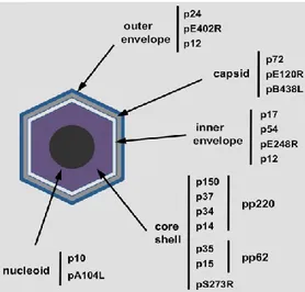
As above stated, the aetiological agent is the African swine fever virus (ASFV), the only member of the Asfarviridae family (Dixon et al., 2005). It is a large, enveloped virus with double-stranded DNA genome of 170 to 190 kbp. ASFV encodes for between 151 and 167 open reading frames (ORFs) (Dixon et al., 2013a).
The ASFV particle has an icosahedral morphology with an average diameter of 200 nm and is composed by several concentric domains, as illustrated in Figure 1.7: 1) an internal core, 2) the core shell, a thick protein layer, 3) an inner lipid envelope, 4) the icosahedral capsid (Carrascosa et al., 1984; Andres et al., 1997). The extracellular virions possess also an external envelope (Breese and DeBoer, 1966).
Thanks to this structure, the virus is highly resistant to PH and temperature variations, in fact it can remain infectious for 18 months at room temperature (Sanchez-Vizcaino, 2006).
Giulia Franzoni
‘Interaction of monocytes and derived macrophage subsets with African swine fever viruses of diverse virulence’ International PhD Course in Life Sciences and Biotechnologies
University of Sassari
ASFV is highly resistant to PH and temperature variations, in fact it remains stable at PH 4-10 and it is inactivated only after cooking for 20 minutes at 60°C. Smoked sausages and matured ham required smoking at 32-49°C for 12 hours and maturing for 25-39 days to inactivate the virus (Plowright et al., 1994).
Figure 1.7 Localization of different ASFV structural proteins inside ASFV
(Figure taken from Salas and Andres, 2013).
ASFV entry involves dynamin-dependent and clathrin-mediated endocytosis (Hernaez and Alonso, 2010) or through macropinocytosis (Sanchez et al., 2012).
During endocytosis, ASFV viral particles undergo disassembly in various compartments, thanks to the acid pH of endosomes. In fact, the inhibition of the endosomal acidification impedes ASFV desencapsidation and so successful infection (Cuesta-Geijo et al., 2012). Then ASFV requires cholesterol to exit the endosome to gain access to the cytoplasm, in order to establish productive replication. In fact a recent study reported that accumulation of cholesterol in endosomes impairs fusion, resulting in retention of ASF virions inside endosomes (Cuesta-Geijo et al., 2015). After exit from the endosome-lysosome, ASFV virions develop a strong association with the microtubular network (Netherton and Wileman, 2013). One of the major structural proteins of ASFV, p54, interacts directly with the 8-kDa light chain of the microtubule motor protein dynein (Alonso et al., 2001) and it has been shown that alteration in the p54-dynein interaction in infected cells results in a marked decrease
Giulia Franzoni
‘Interaction of monocytes and derived macrophage subsets with African swine fever viruses of diverse virulence’ International PhD Course in Life Sciences and Biotechnologies
University of Sassari
in virus infectivity, viral replication and finally virus production (Hernaez et al., 2010).
ASFV DNA replication occurs mainly in perinuclear cytoplasmic viral assembly sites, close to the microtubular organizing centre (MTOC), known as virus factories (Netherton and Wileman, 2013). ASFV factories recruit different cellular components, including cellular chaperones, such as hsp70, and mitochondria (Castelló et al., 2009; Heath et al., 2001; Rojo et al., 1998).
During ASFV infection, all the components of the translation machinery examined (eIF4G, eIF4E, eIF2, eIF3b and the eukaryotic elongation factor 2 [eEF2]) and ribosomes are relocated from a diffused distribution throughout the cytoplasm of infected cells to the viral factories. Also the mitochondria network are mobilized together with ribosomes to the viral factories. The increase in the availability of host sources in the viral factories and simultaneously its depletion in the cytoplasm may results in the shut off of the host mRNA translation (Castello et al., 2009).
ASFV mRNAs are structurally similar to the cellular mRNAs and posses a cap structure in its 5’-UTR and a poly (A) tail of 33 nucleotides in average (Salas et al., 1981). The fact that ASFV mRNAs are capped indicates that they drive translation by a canonical cap-dependent mechanism, as happens with most of cellular mRNAs. Also some studies reported that a part of the genome replication is initiated within the nucleus (Netherton and Wileman, 2013).
ASFV utilises the cytoskeletal network to facilitate its egress from the virus factory to the plasma membrane and out of the infected cell (Netherton and Wileman, 2013). Finally progeny virions are bud out or are propelled away along actin projections to infect new cells (Netherton and Wileman, 2013). In fact, ASFV infection induces long unbranched actin projections, similar to those detected in filopodia, that originate from the plasma membrane (Jouvenet et al., 2006). Interestingly, ASFV infection evolves toward cell lysis at very late times of infection (Breese and DeBoer, 1966), which might represent an alternative mechanism of viral egress.
Giulia Franzoni
‘Interaction of monocytes and derived macrophage subsets with African swine fever viruses of diverse virulence’ International PhD Course in Life Sciences and Biotechnologies
University of Sassari
Figure 1.8 Replication cycle of ASFV. Replication cycle of ASFV, from virus entry
to formation of the early endosome, its maturation, virion desencapsidation in the acidic late endosomal compartments and finally to virions exit along actin projections (Figure taken from Alonso et al., 2013).
The first morphological indication of viral assembly is the accumulation within the factory of viral membranes, which are the precursors of the inner envelope of the viral particle (Salas and Andres, 2013). Then, the capsid is preassembled on the convex face of the viral membranes, which thus become polyhedral forms (Garcia-Escudero et al., 1998). P72 is the major capsid protein, but capsid assembly depends also on protein pB602L, a non-structural protein that acts as a chaperone for the folding of p72 (Epifano et al., 2006a), and on protein pB438L, a minor capsid component probably involved in the formation of the capsid vertices (Epifano et al., 2006b).
Simultaneously to the capsid assembly, the core shell is formed underneath the concave face of the viral envelope (Andres et al., 1997) and its main constituents domain are the proteolytic products of the two virus polyproteins pp220 and pp62 (Andres et al., 2002).
The formation of the nucleoid is likely to be the last step in morphogenesis. Probably the viral DNA is first encapsidated, possibly together with nucleoproteins, and then condensed inside the assembling virus particles to produce the “full” mature virions (Salas and Andres, 2013).
Giulia Franzoni
‘Interaction of monocytes and derived macrophage subsets with African swine fever viruses of diverse virulence’ International PhD Course in Life Sciences and Biotechnologies
University of Sassari
African swine fever virus (ASFV), like other complex DNA viruses, sets up a number of strategies to evade the host’s defense systems, such as apoptosis, inflammation and immune responses (Sanchez et al., 2013). Some viral proteins involved in evading host’s defence are described below.
ASFV proteins involved in modulating host defence, regulating host gene transcription, are:
• A238L protein, which inhibits activation of the transcriptional factors NFkB and NFAT. It interacts with the p65 subunits of NFkB during infection, creating a complex thus inhibiting this transcriptional factor (Revilla et al., 1998). In addition, A238L alters the activity of NFAT, through inhibition of the host calcineurin dependent pathways (Dixon et al., 2004). This protein down-regulates the production of pro-inflammatory cytokines, such as TNF- by ASFV infected macrophages (Powell et al., 1996, Granja et al., 2006a). In fact it has been reported that infection with the mutant ASFV E70, deleted on A238L, is characterized by an increased synthesis of TNF- and other cytokines (Salguero et al., 2008). A238L has effects also on the nitric oxide synthesis. By using a recombinant ASFV lacking of the A238L gene, it has been demonstrated that A238L strongly down regulates inducible nitric oxide synthase (iNOS) activation (Granja et al., 2006b).
• A224L protein, which is an inhibitor of apoptosis (Nogal et al., 2001) and is also involved in the NK-kB activation (Rodriguez et al., 2002).
• ASFVj4R protein, which binds the host -NAC protein (Goatley et al., 2002) and influences its transcription. It has been speculated that the interaction between ASFVj4R and -NAC affects the ability of this cellular factor to act as a transcription co-activator (Sanchez et al., 2013).
• The ASFV ubiquitin conjugating (UBCv) enzyme, which might play a role in regulating host gene transcritpion. In fact it was described that this viral protein binds the NH2-terminal end of a host nuclear protein SMCy, which is involved in gene transcription (Bulimo et al., 2000).
Giulia Franzoni
‘Interaction of monocytes and derived macrophage subsets with African swine fever viruses of diverse virulence’ International PhD Course in Life Sciences and Biotechnologies
University of Sassari
In addition ASFV developed mechanisms to regulate host cell cycle. ASFV strongly induces Myc activation from early times after infection, which promotes cell growing and protein synthesis (Castello et al., 2009). It regulates the expression of the components of eIF4F complex at transcriptional level, in order to guarantee the viral protein synthesis (Castello et al., 2009). Moreover it has been reported that the viral protein A224L inhibits caspase activation, promoting cell survival (Nogal et al., 2001).
This virus monopolizes all the components of the host translation machinery, relocating them to the ‘viral factories’, in order to guarantee viral proteins synthesis. The phosphorylation of the subunit of the eIF2 inhibits its activity and so inhibits translation and protein synthesis, so eIF2 phosphorylation is one of the most important host defense mechanisms against viral infections and ASFV has developed mechanisms to avoid the phosphorylation of this factor (Sanchez et al., 2013). Phosphorilated-eIF2 levels decrease at early times post infection, and remain undetectable throughout the infection (Castello et al., 2009), suggesting a viral mechanism to ensure the availability of this factor for viral protein synthesis.
It has been shown that DP71L induces a decrease of phosphorylated eIF2 and enhances the expression of co-transfected reporters, suggesting that DP71L plays a role in keeping the translation machinery active to allow viral protein synthesis (Zhang et al., 2010). However, ASFV possess multiple mechanisms to avoid eIF2 phosphorylation (Zhang et al., 2010). It has been observed that this virus induces phosphorylation of eIF4E, which takes place after 8 hours post infection (pi) and is associated to an enhancement of the viral replication and protein synthesis (Castello et al., 2009).
Moreover, ASFV encodes several proteins involved in inhibiting apoptosis of infected cells. In fact, ASFV-infected macrophages undergo apoptosis at late time post-infection (24-48 hours pi), suggesting that there are some viral genes that negatively regulate apoptosis (Ramiro-Ibanez et al., 1996), such as:
• The protein encoded by A179L, which represents the viral homolog of the anti-apoptotic protein Bcl2 (vBcl2) and inhibits the action of several
pro-Giulia Franzoni
‘Interaction of monocytes and derived macrophage subsets with African swine fever viruses of diverse virulence’ International PhD Course in Life Sciences and Biotechnologies
University of Sassari
apoptotic protein (Bid, BimL, BimS, BimEL, Bad, Bmf, Bik, Puma and DP5) (Galindo et al., 2008).
• A224L protein, a homolog of inhibitor of apoptosis proteina (IAP), which inhibits caspase 3 (Nogal et al., 2001) and activates the transcription factor NFkB (Rodriguez et al., 2002).
• E153 protein, which acts on the 53 pathway (Hurtado et al., 2004).
• DP71L protein, which induces dephosphorylation of the initiator factor eIF2, playing an important role in keeping the translation machinery active to synthesize new viral proteins (Rivera et al., 2007, Zhang et al., 2010).
Phylogenetic analysis of ASFV is based on the partial sequence of the B646L gene, which encodes for the structural protein p72 (Bastos et al., 2003). This analysis showed that is a strong correlation between isolates from West Africa, South America and Europe collected before 2007, all belonging to genotype I. In contrast, isolates from South and East Africa present more differences and are grouped in other 22 genotypes (Bastos et al., 2003, Lubisi et al., 2005, Boshoff et al., 2007, Achenbach et al., 2016). Genotype XXIII is present in Ethiopia and was only recently discovered (Achenbach et al., 2016).
As previously stated, in June 2007 ASF was confirmed in Georgia, and it has since spread to neighboring countries. Sequence analysis indicated that the Georgia 2007 isolate is closely related to isolates belonging to genotype II, which is circulating in Mozambique, Madagascar, and Zambia (Rowlands et al., 2008). ASFV was first detected in Russia 2007 and all the isolates collected in the Russian Federation from 2007 to 2011 revealed 100% nucleotide identity of B646L gene sequence and formed one genetic cluster within genotype II (Malogolovkin et al., 2012).
Other genes have been used to discriminate between isolates on a regional level: B602L (coding for the J9L protein), CP204 (coding for the P32 protein) and E183L (coding for the envelope protein P54) (Gallardo et al., 2009, Nix et al., 2006, Rowlands et al., 2008). The analysis of this genes is useful to study the epidemiology and evolution of different isolates and can be used to identify the source of contamination in case of new ASFV outbreaks (Nix et al., 2006).
Giulia Franzoni
‘Interaction of monocytes and derived macrophage subsets with African swine fever viruses of diverse virulence’ International PhD Course in Life Sciences and Biotechnologies
University of Sassari
Recently the analysis of the TRS in the intergenic region between the I73R and I329L genes, at the right end of the genome, has been used to investigate the genetic variability among ASFV isolates circulating in eastern Europe. Viruses from Poland and Lithuania presented a TRS insertion identical to that present in ASFV isolates from Belarus and Ukraine, but not in the remaining viruses from eastern Europe, including those obtained in Russia in 2012 and in Georgia in 2007. These molecular data suggested that the ASFV isolstes detected in Poland and Lithuania most probably originated from Belarus (Gallardo et al., 2014).
The phylogenetic analysis, based on the sequence of the gene coding for the protein p72, showed that all the Sardinian isolates belong to the genotype I, along with the isolated from South America, Caribbean, West Africa and Europe (Giammaroli et al., 2011). Between Sardinian isolates there are no differences in the genomic region p54, but there are differences in the B602L gene, which is involved in viral morphogenesis. Sardinian isolated can be divided in two subgroups (X and III) on the basis of the presence/absense of a deletion of 12-13 tetramers in this region. This deletion (subgroup X) is present in almost all the isolates from 1990 and there are no differences between viruses isolated from domestic pigs and wild boar (Giammaroli et al., 2011). In a recent study 44 sardinian isolates collected during 1978-2014 were further characterised, through the analysis of p30, CD2V and I73R/I329L variable regions (Sanna et al., 2016). Researchers showed that Sardinian isolates can be divided into two sub-groups also by the sequences comparison of the CD2v gene: oldest Sardinian isolates (1978-1990) displayed 8 PPPKPC identical hexamers interrupted in the midst by a SPPKPC and a RPPKCP motifs and followed by an hexamer with an aminoacidic substitution (PPSKPC rather than PPPKPC), in contrast viruses isolated in recent years (from 1990 to 2014) show 7 identical repeats with the same central interruption where the last hexamer was not modified (Sanna et al., 2016). These studies suggest that since 1990 the ASF outbreaks in Sardinia are caused by a mutated form of the virus.
Giulia Franzoni
‘Interaction of monocytes and derived macrophage subsets with African swine fever viruses of diverse virulence’ International PhD Course in Life Sciences and Biotechnologies
University of Sassari
1.4 Control of ASF in Sardinia
As above stated, in Sardinia the disease first occurred in 1978 and now is endemic. The disease originated in the south part of Sardinia and it was probably introduced from Spain through food products containing uncooked pork, which were used to feed sardinian pigs (Mannelli et al., 1997). As previously stated, Ornithodoros ticks are not involved in the epizootic cycle of ASF in Sardinia and the control of other risk factors present in the island is necessary for effectively eradicate the disease (Mur et al., 2016b). The persistence of ASFV is probably due to extensive pig farming that has been practised for centuries and of the presence of endemically infected wild boar (Firinu et al., 1988, Costard et al., 2009). In a recent study a Bayesian multivariable logistic regression mixed model was used to assessed the factors associated to the ASFV occurrence in Sardinia. Researchers found that ASFV persistence was associated to particular socio-cultural, productive and economical factors found in the region, particularly to large number of confined farms (most of them backyard), high road density, high mean altitude, large number of open fattening farms, and large number of pigs per commune (Martinez-Lopez et al., 2015).
In the last decades several legislative measures were developed in order to eradicate this disease.
The 14th February 1968 ministerial decree (‘ordinanza ministeriale’) disposed that
the mayor, after notification of ASF presence or suspect, ordered sequestration (‘sequestro di rigore’) of infected, suspected infected and suspected contaminated animals. The ‘provincial veterinarian’ (‘veterinario provinciale’), after mayor’s provisions, issued with urgency decrees of ‘infectious zone’ (‘zona infetta’) and ‘protection zone’ (‘zona di protezione’) and ordered immediate killing of infected, suspected infected and suspected contaminated animals. The decrees of ‘infectious zone’ (‘zona infetta’) and ‘protection zone’ (‘zona di protezione’) were revoked respectively at least 60 and 30 days after the last outbreak (OM 14-2-1968).
In the following years several modification were made to this legislative measure, to more efficiently fight against ASF. The Member States of the European Union
Giulia Franzoni
‘Interaction of monocytes and derived macrophage subsets with African swine fever viruses of diverse virulence’ International PhD Course in Life Sciences and Biotechnologies
University of Sassari
moved from a diversity of national control policies towards a common, community wide approach based on notification of outbreaks, harmonized control measures, uniform diagnostic procedures and contingency plans. In 2002 an EU directive was issued to determine actions to be taken within the EU to fight against ASF, including measures to be applied in case of an outbreak or a suspect or presence of ASF in wild boar (direttiva 2002/60/CE). In 2005 an EU decision was issued focussing on the presence of ASF in Sardinia. This decision established rules about movements, shipments, stampings (‘bollatura’) of sardinian pigs and pig products, to avoid dissemination of the disease in other areas of the EU (decisione 2005/363/CE). Moreover, the decision 2005/362/CE was issued with the aim to eradicate ASF from wild boar in Sardinia (decisione 2005/362/CE). The decision 2005/363/CE has been recently abrogated and it is currently in force the decision of the European Commission of the 27th of March 2014, which will be effective till the 31st of
December 2017. According to this decision, there is a prohibition to export live pigs, porcine sperm, ovules and embryos, pork and any product containing pig meat from Sardinia (and other areas within the EU where ASF cases has been recently reported). There are dispensations to export pork and products containing pig meat from Sardinia if they are made from pigs born and bred outside Sardinia and other areas with ASF. Moreover there are dispensation to export products containing pig meat if they are processed with a treatment that guarantee that there will be no risk related to ASF (decisione di esecuzione 2014/178/UE).
According to the extraordinary ASF eradication plan, since 2014 Sardinian local authorities adopted further measures to eradicate ASFV from Sardinia (Deliberazione 50/17 del 16.12.2014 Regione Autonoma della Sardegna, Legge Regionale n.34 del 22.12.2014, Determinazione 87 del 11.2.2015 Regione Autonoma della Sardegna). According to the current legislation, in case of an ASF outbreak the official veterinarian must contact the ‘Unità di Crisi Locale’, a group of experts which coordinate the execution of the legislative measures.
Further measures were adopted to contrast the contact between domestic pigs and wild boars. In fact in Sardinia wild boar are likely to play an important role in facilitating virus persistence in areas where they lives in continuous contact with free
Giulia Franzoni
‘Interaction of monocytes and derived macrophage subsets with African swine fever viruses of diverse virulence’ International PhD Course in Life Sciences and Biotechnologies
University of Sassari
ranging domestic pigs (Costard et al., 2009). Every pig farm should adopt measure to avoid pigs-wild animals contacts and every pig should be kept on holdings, under human control. If feral pigs are detected, they are going to be killed by official veterinarians, even if they don’t display ASF clinical signs (Legge Regionale n.34 del 22.12.2014, Determinazione 87 del 11.2.2015 Regione Autonoma della Sardegna).
Every pig owner should register animals at the ‘Banca dati nazionale’ and every farm is subjected to official controls. Pigs are examined by official veterinarians, who also assess if in the farm there are effective measures to avoid contact between domestic and feral pigs/wild boars. Official veterinarians collect pig blood, which is sent to the Istituto Zooprofilattico Sperimentale (IZS) of Sardinia, where serological assays to detect antibodies against ASFV are performed. A farm became ‘Azienda certificata per PSA’ if it was inspected at least once in the previous 12 months by an official veterinarian, every pig did not display ASF clinical signs and did not present antibody against ASFV (Legge Regionale n.34 del 22.12.2014, Determinazione 87 del 11.2.2015 Regione Autonoma della Sardegna).
Further measures were taken also to control the spread of the disease in the wild boar population. Hunters must not leave organs or pieces of the carcass in the field, must collect wild boar blood sample for analysis, must inform an official veterinarian in case of finding of dead wild boar Legge Regionale n.34 del 22.12.2014, (Deliberazione 50/17 del 16.12.2014 Regione Autonoma della Sardegna). In some area (‘macroareali’), displayed in Figure 1.9, hunt is forbidden unless special requirements are fulfilled (Determinazione n. 7 del 15.10.2015, Regione Autonoma della Sardegna, Determinazione n. 25 del 20.11.2015, Regione Autonoma della Sardegna).
Giulia Franzoni
‘Interaction of monocytes and derived macrophage subsets with African swine fever viruses of diverse virulence’ International PhD Course in Life Sciences and Biotechnologies
University of Sassari
Figura 1.9. Area (‘macroareali infetti nel selvatico’) of Sardinia where wild boar hunt is forbidden (Determinazione n. 25 del 20.11.2015, Regione Autonoma della
Sardegna).
1.5 ASF diagnosis in Sardinia
Samples of domestic pigs and wild boars, collected by official ASL veterinarians, are sent to the IZS of Sardinia. That institute is responsible for the ASF diagnosis in Sardinia (Legge Regionale n.34 del 22.12.2014, Determinazione 87 del 11.2.2015 Regione Autonoma della Sardegna).
According to the current legislation, ASF should be suspected every time that animals display: fever (more than 40°C), lack of appetite, cutaneous and subcutaneous haemorrhaging, abortion, death of pigs of any ages without identification of other causes. ASF should be suspected every time there is death, infertility or abortion without an identified cause. Samples should be collected in case of outbreaks, epidemiological link with an outbreak, serological positivity, death animal or abortion. Sample suitable for virus isolation are: heparin blood, spleen, kidney, lymph nodes, tonsils, long bone (for bone marrow collection) and foetus (Decisione della Commissione del 26 maggio 2003 recante approvazione di un
Giulia Franzoni
‘Interaction of monocytes and derived macrophage subsets with African swine fever viruses of diverse virulence’ International PhD Course in Life Sciences and Biotechnologies
University of Sassari
manuale di diagnostica della peste suina, OIE Manual of Diagnostic Tests for Aquatic Animals, 2012).
Techniques used to identify ASFV are:
• Direct immunofluorescence, to detect the virus in tissue samples; • Haemoadsorbing test (Malmquist test);
• PCR and RT-PCR, to detect ASFV in any kind of sample, as heparin blood, bone marrow, serum, tissue and organs not well conserved;
• Sequencing and molecular epidemiological analysis, to analyse all the isolates positive at the Malmquist test.
In case of a positive result in one of the previous assays, an ASFV outbreak is opened. Positive samples should be re-tested with a different assay, even on a different animal in the same farm. In case of an ambiguous result or in case of a positive result in the absence of clinical symptoms or pathological lesions of ASF, a test to isolate the virus should be performed. On these samples, one of the following test is performed: PCR, Real Time PCR, Malmquist Test.
Serological assays to detect antibodies against ASFV are performed on blood samples. The first serological assay to be used is the ELISA and in case of a positive result an immunoblotting assay is also performed. Results of the serological assay should be evaluated considering the clinical symptoms and the epidemiological situation, in case of a suspected or an opened ASF outbreak (Decisione della Commissione del 26 maggio 2003 recante approvazione di un manuale di diagnostica della peste suina, OIE Manual of Diagnostic Tests for Aquatic Animals, 2012). During the hunting season, according to the extraordinary ASF eradication plan (Deliberazione 50/17 del 16.12.2014 Regione Autonoma della Sardegna) official veterinarian collect blood samples from dead wild boar, which are sent to the IZS of Sardinia to detect antibodies against ASFV, by ELISA and immunoblotting. In the area displayed in Figure 1.9, also spleens are collected by official veterinarians and are sent to the IZS of Sardinia to detect ASFV, by direct immunofluorescence, haemoadsorbing test (Malmquist test), PCR and RT-PCR.
Giulia Franzoni
‘Interaction of monocytes and derived macrophage subsets with African swine fever viruses of diverse virulence’ International PhD Course in Life Sciences and Biotechnologies
University of Sassari
1.6 ASF vaccines and antiviral agents
Currently there are no vaccine against ASFV. Pigs that survive inoculation with ASFV isolates with reduced virulence can be protected from challenge with the homologous or closely related virulent viruses (Leitão et al., 2001, Oura et al., 2005, King et al., 2011), indicating that anti-ASFV immune response(s), including protective immune response(s), developed in these pigs prior to challenge with the virulent virus (Takamatsu et al., 2013).
Efforts has been done to develop attenuated ASFV strains, by deletion of specific genes from virulent ASFV isolates. Independent deletions of the UK (DP69R) or NL (DP71L) genes from the ASFV E75 (Zsak et al., 1998, Zsak et al., 1996) and deletion of the 9GL (B119) gene from the virulent Malawi Lil-20/1 strains (Lewis et al., 2000) resulted in creation of recombinant deletion viruses with reduced virulence in swine, able to confer protection to challenge with the homologous parental virus (Zsak et al., 1998, Zsak et al., 1996, Lewis et al., 2000). Multigene family (MGF) 360 and 530/550 have been implicated in the modulation of type I interferon (IFN) response, so additional copies of these genes were deleted or interrupted from a virulent genotype I isolate, Benin 97/1. The deleted mutant (BeninMGF) was attenuated in pigs and immunisation and boost with this virus protected against challenge with a lethal dose of Benin 97/1, suggesting that deletion of IFN modulators is a promising route for rational attenuation of virulent ASFV isolates to construct candidate vaccine strains (Reis et al., 2016). Recent studies focused on the creation of a deletion vaccine strain based on the attenuation of the virulent and epidemiologically relevant Georgia2007 isolate. Deletion of 9GL (B119L) resulted in a mutant (ASFV-G-9GL) virus with limited replication in swine macrophages, but able to confer protection to homologous challenge when administered at a lower dose (102-103 HAD
50) 21-28 days before infection with the parental strain (O’Donnel
et al., 2015). Depletion of six genes belonging to MGF 360 and 505 lead to the creation of a vaccine strain (ASFV-G-MGF) able to replicate efficiently in primary macrophage cell cultures, completely attenuated in swine, and vaccination with 102
-104 HAD
50 protected pigs to challenge with the virulent parental strain (O’Donnel et
al., 2015). Despite both vaccine strains (ASFV-G-Δ9GL and ASFV-G-ΔMGF) were efficient in inducing protection, they presented concerns regarding their safety. So a
Giulia Franzoni
‘Interaction of monocytes and derived macrophage subsets with African swine fever viruses of diverse virulence’ International PhD Course in Life Sciences and Biotechnologies
University of Sassari
new mutant, ASFV-G-Δ9GL/ΔMGF, harboring deletions of both 9GL and MGF360/505 genes, was constructed. This virus was highly attenuated in swine, but did not induce protection against challenge with the virulent parental ASFV-G isolate (O’Donnel et al., 2016). In addition, deletion of the thymidine kinase gene was performed in a strain of Georgia adapted to replicate in Vero cells (ASFV-G/V). This mutant (ASFV-G/V-ΔTK) was completely safe in pigs, but unable to provide protection to challenge with the virulent parental strain (Sanford et al., 2016). These results suggest caution towards approaches involving genomic manipulations when developing rationally designed ASFV vaccine strains isolate. In addition, even if attenuated viruses could be used to protect pigs against challenge with virulent strains, the use of this kind of vaccines has some risks, due to the fact that the attenuated virus could revert to virulence in vivo.
The use of vaccine strains which undergo a single cycle of infection but do not produce infectious progeny would avoid this risk. However higher virus load would most likely be required to induce protection (Dixon et al., 2013). In the past it has been shown that inactivated vaccines are not useful to protect pigs against ASFV (Stone et al., 1967; Mebus, 1988) and recently it has been reported that modern adiuvants, like PolygenTM or Emulsigen®-D, do not increase the efficacy of these kind of vaccines (Blome et al., 2014). The construction of vaccines using techniques of molecular biology, not including the whole virus but immunological proteins/peptides, would be useful for the eradication of the disease. Nevertheless, knowledge of protective antigens is essential to create these kind of vaccines (Dixon et al., 2013).
Antibody response against ASFV can be detected even 7 days pi (Parker and Plowright, 1968), but there are controversies about the ability of these antibodies to neutralise the virus (Escribano et al., 2013). Nevertheless, it is not possible to exclude that antibody response does not contribute to protection. Some studies showed that administration of sera of immune animals to infected pigs delay clinical symptoms, reduce viremia and increase the survival probability (Onisk et al., 1994). Instead, several studies reported the importance of the cellular immune response against ASF, especially of the CD8+ T cells (Argilaguet et al., 2012, Argilaguet et al., 2013, Oura et al., 2005).
Giulia Franzoni
‘Interaction of monocytes and derived macrophage subsets with African swine fever viruses of diverse virulence’ International PhD Course in Life Sciences and Biotechnologies
University of Sassari
Recombinant viruses represent an attempt to design a safe and efficient ASFV vaccine. A recombinant Newcastle disease virus (rNDV) expressing the ASFV protein 72 (p72) was recently constructed. The recombinant virus was safe in mice and animals immunized with rNDV/p72 developed high titers of ASFV p72 specific IgG antibody and T cell response (Chen et al., 2016). Futher studies in pigs will evaluate the efficacy of this potential vaccine candidate for preventing ASF. Lokhandwala et al. (2016) recently tested the safety and immunogenicity of an adenovirus-vectored ASFV multi-antigen cocktail. Codon-optimized synthetic genes encoding p32, p54, pp62, and p72 ASFV antigens were used to generate a recombinant adenovirus. Immunization with the cocktail rapidly induced ASFV antigen-specific antibody and cellular immune responses against all the antigens. Importantly, significant antigen-specific IFN-γ responses were detected post-priming and post-boosting (Lokhandwala et al., 2016). A challenge study need to be performed in order to evaluate the relevance of the induced immune responses in regards to protection.
DNA vaccines, where one or more genes encoding immunological proteins are inserted, can be used to induce a stronger cellular response. To date several ASFV proteins have been identified as target of the immunological response, such as p30 and p73 (Leitao et al., 1998, Alonso et al., 1997). Moreover hemagglutinin (HA) showed the ability to confer protetion in the absence of neutralising antibodies (Ruiz-Gonzalvo et al., 1996). Recent studies showed that ASFV vaccines based on the proteins p30, p54 and HA were able to induce a T cell response and partially protected pigs against challenge with a virulent ASFV isolate (Argilaguet et al., 2012, Argilaguet et al., 2013). In fact a DNA vaccine containing p30, p54 and HA fused to ubiquitin was able to protect more than 50% of the vaccinated pigs against challenge with a virulent ASFV strain, in the absence of antibody response (Argilaguet et al., 2012). Protected pigs showed a peak in the number of CD8 T cells 3 days post vaccination and leukopenia was delayed and had lower intensity. In the same study two 9mers target of the T cell response were identified, both located on HA, and immunodominance hierarchy in the T cell response and SLA I restriction were observed (Argilaguet et al., 2012). In addition, in another study the ASFV proteins p54, p30 and HA were inserted in a baculovirus vector (BacMam-sHAPQ),