Abstract
Mouflon (Ovis aries musimon) and sheep (Ovis aries aries) are considered as the wild and domestic subspecies of the same species. A comparative study on the microstructure of mouflon and sheep femoral bone diaphysis is here reported. Bone microstructure is described for the first time in the mouflon. More than 200 secondary osteons from both subspecies were analyzed and qualitative evaluation was followed by quantitative determination of perimeter, area, mini-mum and maximini-mum diameters of secondary osteons and Haversian canals. The basic structural patterns observed in both subspecies can be classified as plexiform and irregular Haversian tissue, in accor-dance with what reported in the literature for most ruminants. The presence of many secondary osteons in the mouflon means that the bone also consists of dense Haversian bone tissue. Statistical analysis
demonstrated that mouflon secondary osteons are larger than in the sheep and made of a greater number of lamellae. Since mouflon and sheep are taxonomically closely related and their body size is very sim-ilar, the qualitative and quantitative differences here reported could be primarily explained on account of their different lifestyle. Indeed, the habits of wildlife typical of mouflons may lead to the presence of wide areas of dense Haversian tissue in that subspecies, as mechanical stresses are known to be related to number and size of secondary osteons. Finally, this analysis could provide a useful tool to recognize bones from different species, in forensic exam and archaeozoological studies as well.
Introduction
Mouflon is a wild sheep native to the Near East. In prehistoric age it was introduced to few Mediterranean islands, such as Sardinia, Corsica, Cyprus and Rhodes.1 In the last centuries, it was widely spread in many mountain regions of Europe. Nowadays mouflon popu-lations are subjected to local restrictions on hunting since it has been listed as vulnerable by the World Conservation Union (IUCN). Hence, any possible difference in bone microstructure between mouflon and sheep may be of interest in the forensic field, especially in those geo-graphic areas where mouflons are legally protected.
In the past, mouflon taxonomy was rather confusing,1until a final classification2 indicated that mouflon can be considered as a wild sheep (Ovis aries musimon) different to the domestic sheep (Ovis aries aries), so they are seen as two subspecies of the same species. Despite some different phenotypic features, such as coat colour and horn morphology, their skeletons are very similar. Thus, distinguish-ing bones between these two subspecies is very hard and depends on different interpretations among osteologists. Regarding bone struc-ture, it is interesting to highlight that despite the fact that mouflon and sheep have the same body size, they live in habitats exposed to dif-ferent biomechanical stresses according to wild or domestic lifestyle, respectively. In Sardinia, mouflons live in rocky hilly regions mainly covered by Mediterranean scrub, frequently jumping, whereas domes-tic sheep are usually less exposed to uneven grounds. Indeed, it is ascertained that the presence, size and morphology of secondary osteons in each bone depend on biomechanical stress.3
The histological structure of bones of ruminants such as the elk (Cervus canadensis), red deer (Cervus elaphus), and roe deer (Capreolus capreolus), where domestic individuals do not exist, was described in previous studies.4-6 In the present work, the results obtained from a wild subspecies are compared for the first time with those from the corresponding domestic subspecies in order to
high-Correspondence: Marco Zedda, Department of Veterinary Medicine, University of Sassari, via Vienna 2, 07100 Sassari, Italy.
Tel. +39.079.229583 - Fax: +39.079.229432. E-mail: [email protected]
Key words: femur, bone histology, mouflon, sheep.
Conflict of interests: the authors declare no potential conflict of interests.
Funding: the work supported by the Bank of Sardinia Foundation.
Contributions: MZ conceived and designed the research; FR and MMN ana-lyzed samples and interpreted the data obtained in two different labs; SG drafted the article; AC, MC and VF revised the text critically and approved the final version.
Received for publication: 7 October 2014. Revision received: 5 December 2014. Accepted for publication: 9 December 2014.
©Copyright S. Giua et al., 2014 Licensee PAGEPress, Italy
Journal of Biological Research 2014; 87:4743 doi:10.4081/jbr.2014.4743
This article is distributed under the terms of the Creative Commons Attribution Noncommercial License (by-nc 3.0) which permits any noncom-mercial use, distribution, and reproduction in any medium, provided the orig-inal author(s) and source are credited.
Comparative histology of the femur between mouflon (
Ovis aries musimon
)
and sheep (
Ovis aries aries
)
Stefano Giua,
1Vittorio Farina,
1Antonio Cacchioli,
2Francesca Ravanetti,
2Marcella Carcupino,
3Miguel Mohadero Novas,
4Marco Zedda
11
Department of Veterinary Medicine, University of Sassari;
2Department of Veterinary Sciences,
University of Parma;
3Department of Natural and Environmental Sciences, University of Sassari,
Italy;
4Faculty of Humanities, University of Cordoba, Spain
[Journal of Biological Research 2014; 87:4743] [page 74]
Journal of Biological Research 2014; volume 87:4743
Non-commercial
[page 75] [Journal of Biological Research 2014; 87:4743] light the role played by the lifestyle on bone structure and secondary
osteon morphology.
Moreover, the femur of sheep brings on some interest because it represents an excellent experimental model to better understand the development and evolution of some human bone diseases, such as osteoporosis and fracture.7-10Finally, the comparison between mouflon and sheep here proposed may represent an example of recognizing bones from different animals, which may be useful in forensic exam and archaeozoological studies.
Materials and Methods
Bone specimens consisted of 4 femoral diaphyses from mouflons and 4 from sheep from both sides. All the femora came from the osteo-logical collection of the Department of Veterinary Medicine, University of Sassari (Italy). Mouflon bones belonged to adult male animals that died of natural causes in the protected natural area of Capo Figari, Isola di Figarolo, in North-Eastern Sardinia, a site of EU Community impor-tance (SIC ITB 01009). Sheep belonged to the Sarda breed and bones came from adult male animals regularly slaughtered in Sardinian abat-toirs. No evidence of skeletal pathology was detected in both subspecies and the age range, estimated on the basis of skeleton ossification and horn morphology,11was 4-6 years in all animals. The femur was chosen as is the longest bone of the skeleton and is subjected to very heavy stress loads. Femora were crosscut at the level of the smallest breadth of their diaphyses (midshaft) using an electrical saw to obtain 2 mm thick sections. The rings were ground and thinned using either a fine sandpapering machine or handily processed by emery paper to obtain 50 µm thick sections. After thorough washing to remove debris, trans-verse sections from periosteal, mesosteal and endosteal zones were mounted onto glass slides with Eukitt (Merck, Darmstadt, Germany) and coverslipped. Afterwards, sections were observed and pho-tographed by means of a Zeiss Axiophoto microscope using ×2.5, ×10 and ×20 objectives. Bone patterns were classified in agreement with the bone tissue classification proposed by Enlow and Brown.12About 200 secondary osteons from each subspecies were examined, that is well-defined osteons surrounded by an evident cement line. The num-ber of lamellae was counted based on the position of osteocyte lacunae. The following parameters were measured by means of Scion Image software (Scion Corporation, Frederick, MD, USA): perimeter, area, minimum and maximum diameters of secondary osteons and Haversian canals. The secondary osteon area included the entire osteon with its Haversian canal. Only intact osteons were considered for statistical analyses. For quantitative comparison, the values obtained for each parameter were compared between mouflon and sheep considering the criterion for statistical significance P<0.05, and variability was expressed as the standard deviation.
Results
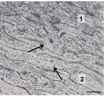
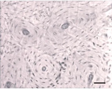
The qualitative observations of the femoral sections belonging to mouflon and sheep indicate that the basic structural patterns can be clas-sified as plexiform and irregular Haversian tissues (Figures 1A, 2), fol-lowing the bone tissue classification proposed by Enlow and Brown.12In the majority of the sections studied, such tissues change one to the other mainly starting from mesosteal zone. The main difference between these two subspecies is related to secondary osteons. In the mouflon, dense Haversian tissue is also present in many areas. Indeed, a high number of secondary osteons are found. They are mainly elliptical in shape and clustered together in small groups (Figures 1B, 3). On the contrary,
sheep secondary osteons are small and isolated (Figure 2). No differ-ences in bone patterns are found among anterior, posterior, medial and lateral sides in both subspecies.
Regarding the morphology of secondary osteons, they are consti-tuted of 5-7 lamellae or more in the mouflon (Figure 3), whereas those of sheep are formed of 4-5 lamellae only (Figure 2).
The quantitative investigation (Table 1) shows that secondary osteons are larger in the mouflon (mean maximum diameter is 214 µm) than in sheep (164 µm), and Haversian canals are wider in mou-flon (mean maximum diameter 27 µm) than in sheep (24 µm). On account of that, values obtained from area and perimeter of secondary osteons and Haversian canals are consequently larger in the mouflon. The comparison between mouflon and sheep demonstrates that all
Article
Figure 1. Different types of bone tissues in the femur of the mou-flon. A) irregular Haversian tissue; B) dense Haversian tissue. Bars=150 µm.
Figure 2. Sheep. Irregular Haversian tissue (1) gradually changes to plexiform tissue (2). The arrows indicate small and isolated sec-ondary osteons formed by 4-5 lamellae. Bar=100 µm.
Non-commercial
morphometrical values (maximum and minimum diameter, perimeter and area) of secondary osteons are significantly different (P<0.01). As to Haversian canals, significant differences are present between the two subspecies for maximum diameter (P<0.01), area (P<0.01) and perimeter (P<0.05). In contrast, values of minimum diameter of Haversian canals do not show significant difference (P>0.05).
Discussion
In this work, the microscopic structure of the femoral diaphysis is described in the mouflon for the first time. A comparison of the mou-flon with the sheep was thought of some interest, especially in forensic and archaeozoological investigations, since it is possible to highlight some differences in histological features. The basic structural pattern observed in both subspecies can be classified as plexiform and irregu-lar Haversian tissues. The literature reports that plexiform tissue, also called fibro lamellar system, consists of primary vascular canals organ-ized into a regular, well-defined plexus. Moreover, as described by Enlow and Brown,12irregular Haversian tissue is characterized by iso-lated secondary osteons, generally having a direction perpendicular to the primary vascular canals.
Our results are mostly in agreement with Skedros and colleagues4
for sheep and deer calcaneus, Metz and colleagues13for sheep ulna, Mori and colleagues14for calf tibia and radius, Skedros5for sheep and elk cal-caneus, Martiniaková and colleagues15,16for sheep femur, Zedda and col-leagues17for bovine femur and humerus, Gudea and Stefan6for sheep, goat and roe deer humerus and metapodial bones. All descriptions made by those authors are rather overlapping, although few differences are present, probably related to the type of bone (depending on main direc-tion and extent of mechanical loads), or species taken into account and are confirmed by the present work. In accordance with Brits and col-leagues18and unlike what reported by Martiniaková and others,16 no dense Haversian bone tissue characterized by a dense concentration of secondary osteons is detected here in sheep femora. Such difference could be related to both diverse age of specimens19and to different sheep breeds. Indeed, Martiniaková and colleagues16 studied Merino sheep, Brits and colleagues18Merino and Dorper breeds, Gudea and Stefan6a Zackel breed, whereas the present work refers to Sarda sheep.
Moreover, it is well-known that bone microstructure and in particu-lar the presence, shape and size of secondary osteons, also depend on the biomechanical stress loading on the bone.20Mouflon secondary osteons are elliptical in shape, which provides better resistance to biomechanical stresses. Indeed, breaking of elliptical osteons at their major axis requires higher strains than those needed for circular osteons.21
Our quantitative data concerning osteons and Haversian canals from sheep are similar to those reported by others, with some differ-ences. Indeed, our values of minimum and maximum diameter of Haversian canals (24±3 and 22±2 µm, respectively) are higher than those reported by Martiniaková and colleagues15,16 in femora (34±9 and 12±3 µm, respectively). The secondary osteons described by these authors in sheep femora are more elliptical in shape as the values of their minimum and maximum diameter are smaller (67±17 µm) and larger (208±70 µm) respectively than those reported here in the same species (min diameter 135±21 µm, max diameter 164±29 µm). In addition, our data are very similar to what reported by Gudea and Stefan6in sheep humerus. Their values often differ from ours by 10% only (Haversian canals: min diameter 19±4 µm, max diameter 25±7µm, perimeter 70±17 µm, area 364±136 µm2; secondary osteons: min diameter 127±22 µm, max diameter 164±26 µm, perimeter 464±77 µm, area 16.514±6542 µm2 ).
Conclusions
In the present investigation, bone microstructure is described for the first time in the mouflon. Since mouflon and sheep belong to the same species and their body size is quite similar, the qualitative and quantitative differences in femoral microstructure here reported could be explained primarily on account of their different lifestyles. The basic structural pattern observed in both subspecies can be classified as
[Journal of Biological Research 2014; 87:4743] [page 76]
Article
Table 1. Mean morphometrical values from secondary osteons and Haversian canals of mouflon and sheep.
Subspecies Osteons Haversian canals
Minimum Maximum Perimeter Area Minimum Maximum Perimeter Area diameter diameter (µm) (µm2) diameter diameter (µm) (µm2)
(µm) (µm) (µm) (µm)
Mouflon
(Ovis aries musimon) 163±37 214±42 597±58 27.382 ±2.945 23±3 27±4 79±8 487±82
Domestic sheep
(Ovis aries aries) 135±21 164±29 472±36 17.380 ±1.540 22±2 24±3 72±9 414±63
Figure 3. Mouflon. A group of well-defined secondary osteons is present in dense Haversian tissue. These secondary osteons are formed by 5-7 lamellae. Bar=50 µm.
Non-commercial
[page 77] [Journal of Biological Research 2014; 87:4743] iform and irregular Haversian tissues. In the mouflon, dense Haversian
tissue is also present in many areas. The main difference between these two subspecies is related to secondary osteons. Indeed, in the mouflon a high number of secondary osteons are found, which are mainly elliptical in shape and clustered together in small groups, whereas in the sheep secondary osteons are small and isolated. The quantitative results show that secondary osteons and Haversian canals are larger in the mouflon than in sheep. Finally, the analysis here reported could provide a useful tool to recognize bones from different species, in forensic exam and archaeozoological studies as well.
References
1. Hiendleder S, Kaupe B, Wassmuth R, Janke A. Molecular analysis of wild and domestic sheep questions current nomenclature and provides evidence for domestication from two different subspecies. P Roy Soc B-Biol Sci 2002;269:893-904.
2. Wilson DE, Reeder DM. Mammal species of the world. A taxonomic and geographic reference. 3rd ed. Baltimore, MD, USA: Johns Hopkins University Press; 2005.
3. Skedros JG, Knight AN, Clark GC, et al. Scaling of Haversian canal surface area to secondary osteon bone volume in ribs and limb bones. Am J Phys Anthropol 2013;151:230-44.
4. Skedros JG, Mason MW, Bloebaum RD. Differences in osteonal micromorphology between tensile and compressive cortices of a bending skeletal system: indications of potential strain-specific dif-ferences in bone microstructure. Anat Rec 1994;239:405-13. 5. Skedros JC. Osteocyte lacuna population densities in sheep, elk
and horse calcanei. Cells Tissues Organs 2005;181:23-37. 6. Gudea AI, Stefan AC. Histomorphometric, fractal and lacunarity
comparative analysis of sheep (Ovis aries), goat (Capra hircus) and roe deer (Capreolus capreolus) compact bone samples. Folia Morphol 2013;72:239-48.
7. Pearce AI, Richards RG, Milz S, et al. Animal models for implant bio-material research in bone: a review. Eur Cells Mater 2007;13:1-10. 8. Oheim R, Amling M, Ignatius A, Pogoda P. Large animal model for
osteoporosis in humans: the ewe. Eur Cells Mater 2012;24:372-85. 9. Zarrinkalam MR, Mulaibrahimovic A, Atkins GJ, Moore RJ. Changes in osteocyte density correspond with changes in osteoblast and osteoclast activity in an osteoporotic sheep model. Osteoporosis Int 2012;23:1329-36.
10. Hernandez-Fernandez A, Vélez R, Soldado F, et al. Effect of admin-istration of platelet-rich plasma in early phases of distraction osteogenesis: an experimental study in an ovine femur model. Injury 2013;44:901-7.
11. Barone R. Anatomie comparée des mammifères domestiques. Tome I, Osteologie. Paris, France: Vigot; 2010.
12. Enlow DH, Brown SO. A comparative histological study of fossil and recent bone tissues. Part I. Tex J Sci 1956;8:405-3.
13. Metz LN, Martin B, Turner S. Histomorphometric analysis of the effects of osteocyte density on osteonal morphology and remodel-ling. Bone 2003;33:753-9.
14. Mori R, Kodaka T, Sano T, et al. Comparative histology of the lami-nar bone between young calves and foals. Cells Tissues Organs 2003;175:43-50.
15. Martiniaková M, Grosskopf B, Omelka R, et al. Histological study of compact bone tissue in some mammals: a method for species determination. Int J Osteoarchaeol 2007;17:82-90.
16. Martiniaková M, Grosskopf B, Omelka R, et al. Histological analysis of ovine compact bone tissue. J Vet Med Sci 2007;69:409-11. 17. Zedda M, Lepore G, Biggio GP, et al. Morphology, morphometry and
spatial distribution of secondary osteons in equine femur. Anat Histol Embryol 2014 (in press).
18. Brits D, Steyn M, L’Abbé EN. A histomorphological analysis of human and non-human femora. Int J Legal Med 2014;128:369-77. 19. Mulhern DM, Ubelaker DH. Differentiating human from
nonhu-man bone microstructure. In: Crowder C, Stout S, eds. Bone histol-ogy, an anthropological perspective. Boca Raton, FL, USA: CRC press; 2012. pp 109-34.
20. Mishra S. Biomechanical aspects of bone microstructure in verte-brates: potential approach to palaeontological investigations. J Bioscience 2009;34:799-809.
21. Zedda M, Lepore G, Manca P, et al. Comparative bone histology of adult horses (Equus caballus) and cows (Bos taurus). Anat Histol Embryol 2008;37:442-5.