Expression of a Truncated Form of the c-Kit Tyrosine
Kinase Receptor and Activation of Src Kinase in
Human Prostatic Cancer
Maria Paola Paronetto,* Donatella Farini,*
Innocenzo Sammarco,* Giovanni Maturo,
†Giuseppe Vespasiani,
†Raffaele Geremia,*
Pellegrino Rossi,* and Claudio Sette*
From the Dipartimento di Sanita` Pubblica e Biologia Cellulare * and the Dipartimento di Biopatologia e Diagnostica per Immagini,†Facolta` di Medicina e Chirurgia, Universita` di Roma “Tor Vergata,” Rome, Italy
A truncated form of the c-Kit tyrosine kinase recep-tor , originally identified in mouse haploid germ cells , is aberrantly expressed in human cancer cell lines of various origin. This alternative transcript originates in the 15th intron of the human c-kit gene. We have previously demonstrated that sperm-carried mouse truncated c-Kit (tr-Kit) is a strong activator of the Src-family tyrosine kinases both in transfected cells and in mouse oocytes. In the present work , we report that human tr-Kit mRNA and protein are expressed in LNCaP prostatic cancer cells. We have identified two regions in the 15th and 16th introns of the human c-kitgene that show homology with sequences in the spermatid-specific tr-Kit promoter within the 16th intron of mouse c-kit. We also show that nuclear factors present in LNCaP cells bind to discrete se-quences of the mouse tr-Kit promoter. Moreover , Western blot analysis of 23 primary prostate cancers indicated that tr-Kit was expressed in ⬃28% of the tumors at less advanced stages (Gleason grade 4 to 6) and in 66% of those at more advanced stages (Gleason grade 7 to 9) , whereas it was not expressed in benign prostatic hypertrophies. Sequencing of the cDNA for the truncated c-Kit , amplified from both LNCaP cells and neoplastic tissues , confirmed the existence in prostate cancer cells of a transcript arising from the 15th intron of human c-kit. We also show that tr-Kit-expressing LNCaP cells and prostatic tumors have higher levels of phosphorylated/activated Src than tr-Kit-negative PC3 cells or prostatic tumors , and that transfection of tr-Kit in PC3 cells caused a dramatic increase in Src activity. Interestingly , we found that Sam68 , a RNA-binding protein phosphorylated by Src in mitosis , is phosphorylated only in prostate tumors expressing tr-Kit. Indeed , both activation of Src and phosphorylation of Sam68 were observed in all of the three grade 7 to 9 tumors analyzed that expressed
tr-Kit. Our data describe for the first time the exis-tence of a truncated c-Kit protein in primary tumors and show a correlation between tr-Kit expression and activation of the Src pathway in the advanced stages of the disease. Thus , these results might pave the way for the elucidation of a novel pathway in neoplastic transformation of prostate cells. (Am J Pathol 2004, 164:1243–1251)
The c-Kit receptor belongs to type III tyrosine kinase receptor, which consists of an extracellular ligand bind-ing domain and an intracellular kinase domain. In the kinase domain, the ATP binding site is separated from the phosphotransferase site by an interkinase sequence re-quired for interaction with signal transduction proteins involved in the c-Kit pathway.1The c-Kit receptor is ex-pressed in a wide variety of normal and neoplastic tis-sues. A positive correlation between misregulation of the
c-kitgene and malignant transformation of cells has been reported.2,3For instance, substitution mutations in exon 11 of the gene, changing amino acids of the juxtamem-brane region of the receptor, are associated with gastro-intestinal stromal tumors, whereas mutations in exon 17 that substitute Asp816, just downstream of the tyrosine kinase signature, are associated with myeloid leukemias and testicular seminomas.2,3These mutations induce li-gand-independent dimerization or autophosphorylation of the receptor and they cause constitutive activation of downstream signaling pathways. Moreover, overexpres-sion of c-Kit and its ligand SCF occurs in several tumors and they probably stimulate proliferation in an autocrine or paracrine manner.2Recently, c-kit expression in vari-ous tumors has been revisited because this receptor is a target of the anti-cancer activity of a well described ty-rosine kinase inhibitor: imatinib (STI1571).2,4Beside mu-tations affecting the activity of the full-length c-Kit, ex-pression of an alternative transcript of human c-kit has been described in several transformed cell lines. The
Supported by MIUR Co-Fin 2002 and by a grant from the Centro di Eccellenza per lo Studio del Rischio Genomico in Patologie Complesse Multifattoriali.
Accepted for publication December 15, 2003.
Address reprint requests to Claudio Sette, Dipartimento di Sanita` Pub-blica e Biologia Cellulare, Facolta` di Medicina e Chirurgia, Universita` di Roma Tor Vergata, Via Montpellier 1, 00133 Rome, Italy. E-mail: [email protected].
alternative transcript encodes for a truncated c-Kit pro-tein that contains only a short sequence of the interkinase segment, the phosphotransferase domain, and the car-boxyl-terminal tail of the receptor.5Originally cloned from the colon cancer cell line Colo201,5 expression of this novel mRNA has been detected in⬃30% of the gastro-intestinal and hematopoietic tumor cell lines examined.6 To date, no function has been ascribed to this truncated c-Kit protein in transformed human cells. Other receptor tyrosine kinases are expressed as truncated forms in malignant cells, but usually these aberrant proteins are constitutively active kinases in which the catalytic domain is freed by negative constraints present in the full-length receptor.7–9By contrast, the truncated c-Kit protein does not contain an ATP binding site and should be catalyti-cally inactive.5Our laboratory has previously described that a mouse homologue of the truncated human c-Kit is physiologically expressed only in the postmeiotic haploid cells of the testis;10 more recently we have detected human tr-Kit also in human mature spermatozoa, indicat-ing a conserved role of this protein in gamete function (Paronetto MP, Geremia R, Rossi P, and Sette C, manu-script in preparation). This alternative c-Kit mRNA en-codes for a truncated protein, named tr-Kit, of the same size and structure as the human homolog characterized in cancer cells.11,12Mouse tr-Kit also lacks the ATP bind-ing site and it is catalytically inactive; however, we have described that it acts as an activator of the soluble ty-rosine kinases Fyn and Src.13The direct interaction of tr-Kit with the SH2 domain of Src-like kinases displaces the autoinhibitory constraint caused by binding of this domain to a phosphotyrosine in the carboxyterminal tail of Src-like kinases.13Tr-kit-induced activation of Fyn or Src triggers cell cycle resumption in metaphase-arrested mouse oocyte, indicating the mitogenic potential of this pathway.13,14 Interestingly, mutations that substitute ty-rosine 530 of Src, which abolishes the autoinhibition of the kinase in the same manner as the interaction with tr-Kit, are associated with colorectal cancer tissues and induce cell transformation when aberrantly expressed in cultured cells.15 More recently, we have demonstrated that activation of Src-kinases by tr-Kit triggers the phos-phorylation of the RNA-binding protein Sam68.16 Func-tioning as a scaffold, Sam68 promotes the recruitment of Src-kinases and PLC␥1, and the phosphorylation/activa-tion of the phospholipase.13,16Because human truncated c-Kit (herein referred as human tr-Kit) contains all of the structural features required for mouse tr-Kit function (see Toyota and colleagues5and Rossi and colleagues10for comparison), it is possible that the two proteins share the same function and that human tr-Kit is able to promote Src activation in the malignant cells where it is expressed. Src activation plays an important role both in prolifer-ation and metastatic transformprolifer-ation of androgen-sensi-tive and -insensiandrogen-sensi-tive prostate cancer cells.17,18 Further-more, Sam68 is highly expressed in prostate epithelial cells and the involvement of this protein in RNA metabo-lism during normal growth and neoplastic transformation of prostatic cells has recently been hypothesized.19 Herein we undertook an analysis of prostate cancer cell lines and primary prostatic tumors for the presence of
human tr-Kit. We found that human LNCaP cells ex-pressed tr-kit, whereas the more undifferentiated PC3 did not. Furthermore, we observed that human tr-Kit was expressed in⬃28% of the less advanced and in 66% of the more advanced primary prostate tumors analyzed. In the latter group, a positive correlation was found between tr-Kit expression in tumors and tyrosine phosphorylation of Src and of the adaptor protein Sam68. Our results suggest that aberrant expression of a truncated c-Kit protein may contribute to cell transformation through the activation of Src kinases.
Materials and Methods
Human Tissue Samples
Hypertrophic prostate samples were obtained, after in-formed consent, from patients who underwent prostatec-tomy for suspected benign prostatic hyperplasia (serum PSA⬍4 ng/ml; negative for cancer to the rectal explora-tion and prostatic ultrasound), confirmed by histological analysis. Prostate cancers were diagnosed by TRU-CUT biopsy. Patients were excluded from the study if they had androgen deprivation therapy or reported a clinical his-tory of repetitive neoplastic lesions at the radionucleotide bone scan and at the abdomino-pelvic computed tomo-graphic scan. Prostate cancers were graded using the Gleason grading system. We selected prostates that were positive for cancer to multiple biopsies in just one peripheric area. Samples of tumor and contralateral pros-tatic tissue (negative to the multiple biopsies analysis and without cancer macroscopy characteristics) from 23 pa-tients, between 58 to 77 years of age, who underwent radical prostatectomy at the Clinical Center located in the Urologic Clinic of University of Rome “Tor Vergata,” were immediately frozen in liquid nitrogen (Table 1).
Immunohistochemistry
Tissues were fixed in formalin and embedded in paraffin immediately after surgery. Antigen retrieval was per-formed on hydrated sections by three microwaving cy-cles (5 minutes each) at high power in 1 mmol/L of ethylenediaminetetraacetic acid, pH 8.0. Slides were in-cubated for 1 hour in blocking solution, phosphate-buff-ered saline (PBS), pH 7.4, supplemented with 5% bovine serum albumin and 1% goat serum. After two washes in PBS, slides were incubated overnight at 4°C with either rabbit anti-human c-Kit (1:100, SC-168; Santa Cruz Bio-technology) or mouse anti-p416Src (1:50, 05-677; Up-state). Controls using nonimmune IgGs were also per-formed and gave no signal (data not shown). Horseradish peroxidase-linked secondary antibody incubation was performed for 1 hour at room temperature. Signals were visualized with diaminobenzidine (Sigma) and analyzed by light microscopy.
Cell Culture and Transfection
LNCaP and PC3 prostatic cancer cells were maintained in Dulbecco’s minimal essential medium supplemented with 5% fetal bovine serum (Gibco BRL). Hek293 cells were maintained in Dulbecco’s minimal essential medium supplemented with 10% fetal bovine serum (Gibco BRL). PC3 cells were transfected with 10 g of pCMV5-tr-Kit using the CaPO4precipitation protocol as described pre-viously.12
Extraction of RNA and Protein from Cultured
Cells and Primary Tissue
Total RNA was extracted by homogenization of samples in TRIzol reagent (catalog no.15596-026; Invitrogen, Life Technologies) and by following the manufacturer’s in-structions. RNA was resuspended in diethyl pyrocarbon-ate wpyrocarbon-ater and immedipyrocarbon-ately frozen at ⫺80°C for further analysis by reverse transcriptase-polymerase chain re-action (RT-PCR). For protein extrre-action, LNCaP cells, PC3 cells, and tissue fragments were homogenized in homogenization buffer (20 mmol/L HEPES, pH 7.5, 120 mmol/L KCl, 0.1 mmol/L ethyleneglycol-bis(-aminoethyl ether)-N,N,N⬘,N⬘-tetraacetic acid, 10 mmol/L -glycero-phosphate, 10g/ml leupeptin, 10 g/ml aprotinin, and 2 mmol/L phenylmethyl sulfonyl fluoride). The extracts were centrifuged for 15 minutes at 12,000⫻ g at 4°C before collecting the supernatant for Western blot or immuno-precipitation experiments.
RT-PCR Analysis
RT-PCR was performed using the RT-PCR kit (catalog no. 28025-021; GibcoBRL, Life Technologies), according to the manufacturer’s instructions. To synthesize comple-mentary DNA (cDNA), 4g of total RNA and 750 ng of random primers were used. Oligonucleotides (0.5 mol/L) used for amplification were: i15A: GCAGTGC-CAATGGTCAATGGCAG (from base 79,786 to 79,808 in the 15th intron of the c-kit gene); i15B: AAATCCTCTCT-TCCTCACAGGCT (from base 80,097 to 80,119 in the 15th intron of c-kit); i16A: CAAGGCTTGGGGTGAAG-CATAGAC (from base 80,606 to 8629 in the 16th intron of
c-kit); i16B: TCCGTGTGTCCTTGGGAGATGTC (from base 80,917 to 80,940 in the 16th intron of c-kit); e18: TGCTTTCAGGTGCCATCCACTTCAC (reverse sequence of c-kit exon 18th from base 84,687 to 84,723). PCR reactions were performed in 25 l, using 1.5 mmol/L MgCl2, 200 mol/L of each dNTP, and 1 l of cDNA reaction. PCR cycles were as follows: 95°C for 5 minutes for denaturation step, followed by 35 cycles of denatur-ation at 95°C for 30 seconds, annealing at 58°C for 30 seconds, and extension at 72°C for 1 minute.
DNA Binding Assays
Nuclear extracts (protein concentration ⬃7 mg/ml) from mouse spermatids, LNCaP and PC3 cells were prepared as previously described.11For each reaction, 10 g of nuclear extracts were used in a final volume of 10l. DNA restriction fragments for electrophoretic mobility shift as-says (EMSAs) were labeled at the 5⬘ end with32P-␥-dATP using sequential alkaline phosphatase and T4 polynucle-otide kinase treatment. A DraI fragment of 200 bp in the mouse tr-Kit promoter within the 16th intron was chosen because it contained the 82-bp region of homology with the human c-kit 16th intron and because it was previously shown to have enhancer-like activity for mouse tr-Kit tran-scription;10a NdeI-StyI fragment of 200 bp in the 16th intron mouse tr-Kit promoter was chosen because it con-tained a 19-bp sequence near the tr-Kit mRNA start site that was essential for transcription and because it showed strong homology with a 15-bp sequence present in the human c-kit 15th intron near the presumptive start site of human tr-Kit.5,6
Immunoprecipitation Assay
Tissue extracts (500 g of total proteins) prepared as described above were incubated with 1 g of the anti-phosphotyrosine antibody (␣-PY20, Santa Cruz Biotech-nology) for 2 hours at 4°C under constant shaking. Pro-tein A-Sepharose or proPro-tein G-Sepharose (Sigma-Aldrich) were preadsorbed with 0.05% bovine serum albumin before the incubation with the immunocom-plexes and added to the extracts together with the anti-body during the last hour of immunoprecipitation. The Sepharose beads were washed three times with homog-enization buffer or lysis buffer. Proteins adsorbed to the
Table 1. Parameters of the 23 Patients Examined for Tr-Kit Expression in Prostatic Tissue
Patient Age Gleason sum Tr-kit expression
1 77 4 ⫺ 2 71 4 ⫹ 3 62 4 ⫺ 4 67 5 ⫹ 5 76 5 ⫺ 6 70 5 ⫹ 7 68 6 ⫺ 8 68 6 ⫺ 9 71 6 ⫺ 10 70 6 ⫺ 11 58 6 ⫺ 12 67 6 ⫹ 13 64 6 ⫺ 14 57 6 ⫺ 15 76 7 ⫹ 16 67 7 ⫹ 17 66 7 ⫹ 18 70 7 ⫺ 19 65 8 ⫺ 20 68 8 ⫹ 21 71 8 ⫺ 22 75 9 ⫹ 23 70 9 ⫹
For each patient is reported the Gleason value and age. Tr-Kit expression in the tumor tissue, as assessed by Western blot analysis (see also Figure 3), is also reported.
antibody-beads complex were eluted in sodium dodecyl sulfate (SDS)-sample buffer for Western blot analysis.
Immunokinase Assay of Src Activity
Extracts (500 g of total proteins) from PC3 or LNCaP cells were incubated with 1g of either monoclonal ␣-Src antibody (Ab-1; Oncogene Research Products) or non-immune IgGs for 2 hours at 4°C under constant shaking. Immune complexes were collected by adsorption onto protein A-Sepharose (Sigma-Aldrich). The Sepharose beads were washed three times with kinase buffer (50 mmol/L Hepes, pH 7.4, 10 mmol/L MgCl2, 10 mmol/L -glycerophosphate, 1 mmol/L dithiothreitol, 0.5 mol/L Na-orthovanadate, 50 mol/L ATP). Half of the beads were eluted in SDS-sample buffer for Western blot anal-ysis, the other half was used for the enzymatic reaction. The kinase reactions were performed by incubating the beads with 10g of tr-kit Y161 peptide, in 25 l of kinase buffer also containing 0.1Ci of32P-␥-ATP for 30 minutes at 30°C and terminated by spotting the reaction mixture onto squares of P-81 phosphocellulose paper (Whatman) as previously described.12 Radioactivity incorporated was determined by scintillation counting of the paper squares or by autoradiography of the dried gels.
Western Blot Analysis
Proteins were separated on 10% SDS-polyacrylamide gel electrophoresis gels and transferred to polyvinylidene fluoride Immobilon-P membranes (Millipore) using a semidry blotting apparatus (Bio-Rad). Western blot anal-ysis was performed as previously reported.13First anti-body (1:1000 dilution) overnight at 4°C: mouse ␣-Src (Ab-1, Oncogene Research Products); mouse anti-p416Src (1:50, 05-677; Upstate); rabbit␣-Sam68 (C-20, Santa Cruz Biotechnology); mouse ␣-phosphotyrosine (PY20, Santa Cruz Biotechnology); rabbit ␣-c-Kit (SC-168, Santa Cruz Biotechnology); goat␣-actin (SC-1616, Santa Cruz Biotechnology). Secondary anti-mouse or an-ti-rabbit IgGs conjugated to horseradish peroxidase (Am-ersham) were incubated with the membranes for 1 hour at room temperature at a 1:10,000 dilution in PBS con-taining 0.1% Tween 20. Immunostained bands were de-tected by chemiluminescent method (Santa Cruz Bio-technology).
Results
Expression of Human tr-Kit in LNCaP Cells
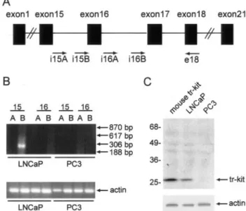
A truncated c-Kit (tr-Kit) transcript and the corresponding protein are expressed in several gastrointestinal and he-matopoietic cell lines.6To determine whether human tr-Kit is expressed also in prostatic tumors, we first ana-lyzed two well-characterized prostate cancer cell lines: the androgen-sensitive LNCaP and the androgen-insen-sitive and more undifferentiated PC3. First we undertook a RT-PCR approach using a battery of forward primers spanning introns 15 and 16 of the c-kit genomic
se-quence, whereas the reverse primer was chosen in exon 18 (Figure 1A). These intron sequences were chosen because the human tr-Kit transcript originates in the 3⬘ region of the 15th intron and is amplified by the i15B oligonucleotide,5whereas the mouse tr-Kit transcript be-gins in the 3⬘ region of the 16th intron10and the homol-ogous region in human c-kit would be amplified by the i16B oligonucleotide. Furthermore, by using an intronic forward primer we could avoid cross-amplification of full-length c-Kit mRNA, whose expression in some prostate tumors has been reported.2 As shown in Figure 1B, a band of the correct size (306 bp) was amplified from LNCaP cells only using the primer combination i15B and exon18; primers i15A, corresponding to the 5⬘ region of intron 15, did not amplify any band as well as primers spanning the 16th intron of human c-kit. Remarkably, regardless of the primers combination used, no amplifi-cation was obtained from the more undifferentiated, an-drogen-insensitive, PC3 cells. Direct sequencing of the band amplified from LNCaP cells with the i15B-exon18 primer combination indicated that prostatic tr-Kit mRNA was identical to the truncated c-Kit transcript described in Colo1 cells.5
Next, we investigated the expression of tr-Kit protein in LNCaP and PC3 cells. In agreement with the RT-PCR data, we found that LNCaP expressed tr-Kit whereas PC3 cells did not (Figure 1C). Human tr-Kit displayed the same apparent molecular weight as recombinant mouse Figure 1. Expression of human tr-Kit in LNCaP cells. A: Schematic
represen-tation of the c-kit genomic structure. The approximate position of the oligo-nucleotides used for RT-PCR analysis of LNCaP and PC3 cells is indicated. B: RT-PCR analysis for the expression of human tr-Kit in prostatic cancer cell lines. Oligonucleotides used as forward primers in the PCR reaction were chosen sequences in the 5⬘ (A) or 3⬘ (B) region of intron 15 and intron 16 of the human c-kit gene; as reverse primer was chosen an anti-sense oligonu-cleotide corresponding to a sequence in the 18th exon of the human c-kit gene. Arrows on the right show the expected size for the bands amplified by i15A (617 bp), i15B (306 bp), i16A (870 bp), or i16B (188 bp). Shown is a representative experiment that was repeated twice with identical results. C: Western blot analysis of cell extracts (40g) from LNCaP or PC3 cells using either an anti-human c-Kit antibody (top) or the anti-actin antibody
(bot-tom). A cell extract from Hek293 cells transfected with a mouse tr-Kit
expression vector (pCMV5-tr-Kit) was run in the first lane as positive control for the Western blot analysis.
tr-Kit, loaded as a control in lane 1 of the SDS-polyacryl-amide gel electrophoresis.
Nuclear Factors Present in LNCaP Recognize
the Mouse tr-Kit Promoter
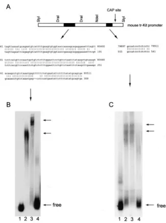
A pairwise BLAST search analysis of the human and mouse c-kit genes shows that, beside the obvious homol-ogy in the exonic sequences, a strong homolhomol-ogy is present in a discrete sequence of the 16th introns (Figure 2A). This region was shown to specifically bind nuclear
factors that recognize a general enhancer element only in mouse spermatids and to be required for maximal activity of mouse tr-Kit promoter during spermiogenesis.11 No apparent homology in other intronic regions, including the 15th intron of the two genes, was observed. However, a small 15-bp sequence present just upstream of the transcriptional start site in the tr-Kit promoter within the 16th intron of mouse c-kit, was found to be present in a reversed orientation, in the presumptive promoter of hu-man tr-Kit within the 15th intron of huhu-man c-kit (Figure 2A) (ie, between the sequences corresponding to oligonucle-otides i15A and i15B). Because we have previously dem-onstrated that this discrete sequence is essential for tran-scription of mouse tr-Kit mRNA,11we set out to determine whether nuclear factors bind to this sequence in LNCaP cells by EMSA. A 200-bp fragment of the mouse 16th intron centered around this sequence bound nuclear fac-tors from both mouse spermatids and LNCaP cells, but not from PC3 cells that do not express tr-Kit (Figure 2C). Moreover, the migration of the complexes formed by nuclear extracts of spermatids and LNCaP cells was identical, suggesting that similar nuclear factors are present in these cells. In addition, LNCaP cells, but not PC3 cells, expressed nuclear factors that bound also the homologous enhancer-like region shared by the mouse and human c-kit genes within the16th introns (Figure 2B), even though the migration of the complexes was different from that observed with mouse spermatid extracts. Over-all, these results suggest that similar genomic elements control the expression of human and mouse tr-Kit and that similar nuclear factors might drive the transcription of tr-Kit mRNA in LNCaP cells and mouse spermatids.
Expression of Human tr-Kit in Primary Prostate
Tumors
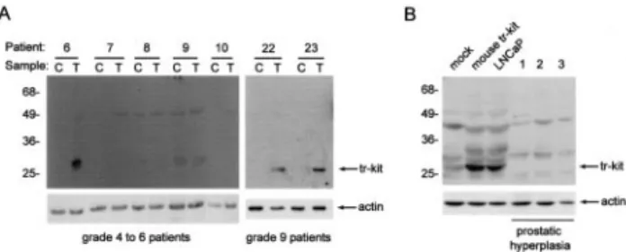
Although expressed in cell lines derived from different tumors (gastrointestinal and hematopoietic),5,6 the ex-pression of human tr-Kit has never been reported in primary tumors. Thus, we tested samples obtained from 26 patients for the expression of human tr-Kit in benign prostatic hyperplasia and prostatic tumors by Western blot analysis (Table 1). Among the 14 patients with low Gleason grade tumors (from 4 to 6), we found that 4 (28%) expressed tr-Kit. Remarkably, when patients with more advanced tumors (Gleason grade from 7 to 9) were examined, we found that 66% (6 of 9) expressed tr-Kit (Table 2). By contrast, none of the three benign prostatic hyperplasia patients examined expressed the truncated form of c-Kit (Table 2 and Figure 3B). For all patients, we examined both the tumor tissue and the contralateral region of prostate, which was not invaded by the tumor. Figure 2. Nuclear factors in LNCaP cells bind to discrete sequences of the
mouse tr-Kit promoter. A: Schematic structure of the mouse tr-Kit promoter within the 16th intron of c-kit. Black boxes represent the two regions previously identified as essential for binding of nuclear factors and for promoter activity in mouse spermatids.11Above are listed the restriction sites
used to isolate these two regions. The homology of these regions with sequences in the human 16th intron (left) or 15th intron (right) are shown below. Nucleotide coordinates refer to accession number U63834 (human
c-kit gene genomic sequence, top lines) and accession number X65998
(mouse tr-Kit promoter, bottom lines). B: EMSA experiment using the 200-bp DraI-DraI fragment containing the homology with the sequence in the human 16th intron. C: EMSA experiment using the 260-bp NdeI-StyI fragment containing the homology with the sequence in the human 15th intron. Free-labeled DNA was loaded in lane 1; 10g of nuclear factors from mouse spermatids (lane 2), LNCaP cells (lane 3), or PC3 cells (lane 4) were used for the EMSA. Arrows on the right show the position of either free DNA or DNA-protein complexes.
Table 2. Tr-Kit Expression Correlates with Phosphorylation of Src and Sam68 in Prostatic Tumors
Patients Number Tr-Kit-positive Phospho-Src-positive Phospho-Sam68-positive
BPH 3 0 (0%) 0 (0%) 0 (0%)
Grade 4–6 14 4/14 (28%) 0/6 (0%) 1/6 (16%)
Grade 7–9 9 6/9 (66%) 3/4 (75%) 3/4 (75%)
We found that tr-Kit was either not expressed or ex-pressed at lower levels in the contralateral regions of the prostate. Representative examples of Western blot anal-ysis of these samples are shown in Figure 3A. Tr-Kit expression was also confirmed by RT-PCR analysis of available RNA samples, followed by DNA sequencing (data not shown). Immunocytochemistry analysis of pa-tient specimens confirmed that tr-Kit expression was re-stricted to the cytoplasm of the epithelial cells of neoplas-tic prostaneoplas-tic glands (Figure 4, B and B⬘), whereas no staining was observed in the contralateral normal glands (Figure 4A). A representative example of a grade 7 pa-tient is shown in Figure 4 (top). These results suggested that expression of tr-Kit correlates with the more ad-vanced stages of prostatic tumors.
Activation of Src and Phosphorylation of Sam68
in Prostatic Tumors
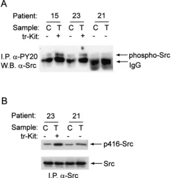
We have previously described that overexpression of mouse tr-Kit triggers the activation of Src-like kinases with the consequent phosphorylation of the adaptor protein Sam68 and PLC␥1.14,16To test whether activation of this signaling pathway was also a feature of prostatic tumors, we immunoprecipitated tyrosine-phosphorylated proteins from extracts of normal or tumorigenic prostate tissue from 10 patients, and analyzed the immunoprecipitated proteins for the presence of Src or Sam68. We found that among the four patients with advanced prostatic tumors (Gleason grade 7 to 9) examined, three showed an in-creased level of phosphorylation of Src in the tumor tis-sue than in the normal tistis-sue (Table 2). Interestingly, the fourth sample was from a patient that did not express tr-Kit (Table 1). A representative Western blot analysis is shown in Figure 5A. To determine whether the phosphor-ylation of Src was activatory, we immunoprecipitated total Src from normal or tumor prostatic tissues and stained immunoprecipitated proteins with the p416Src anti-body, which recognize active Src. The representative example shown in Figure 5B indicates that the signal corresponding to active Src was increased in immuno-precipitates obtained from tr-Kit-expressing tumor pros-tatic tissues. Furthermore, we observed that the anti-active Src antibody stained the membrane of neoplastic prostatic cells from tr-Kit-expressing tumors (representa-tive sample shown in Figure 4, D and D⬘) whereas it did not stain the epithelial cells of the contralateral prostatic gland (Figure 4C). Interestingly, patients that expressed tr-Kit also showed tyrosine phosphorylation of Sam68 Figure 3. Expression of tr-Kit protein in primary prostatic tumors. A: Tissues
from prostatic tumors (T) or the contralateral, morphologically normal, pros-tate (C) (see Materials and Methods) from different patients were lysed and analyzed in Western blot using the anti-c-Kit antibody (top) or the anti-actin antibody (bottom). For each sample were loaded 40g of total protein. The number of the patient corresponds to that in Table 1; Gleason grades for the patients examined are written below the blot. B: Cell extracts (40g) from mock-transfected and mouse tr-Kit-transfected Hek293 cells, or LNCaP cells, and three benign prostatic hyperplasia patients were analyzed in Western blot using the anti-c-Kit antibody (top) or the anti-actin antibody (bottom).
Figure 4. Immunohistochemical analysis of prostate tumors. The analysis of a representative grade 7 sample is shown. Sections obtained from the tumor (B, Bⴕ,
D, and Dⴕ) or the contralateral, morphologically normal, side of the gland (A and C) were stained using either the anti-c-Kit antibody (A, B, and Bⴕ) or anti anti-p416Src antibody (C, D, and Dⴕ). Bⴕ and Dⴕ are enlargements of the neoplastic glands shown in B and D that allow to better appreciate the cytoplasmic staining obtained with the c-Kit antibody and the membrane staining obtained with the anti-p416Src antibody. Scale bars, 100m.
(Figure 6; Table 2), further indicating that Src was acti-vated in these samples. On the other hand, only one among the six patients with low Gleason grade4 – 6 exam-ined displayed phosphorylation of Sam68, but not of Src; this patient also expressed tr-Kit (no. 2 in Table 1). Al-though we found that the expression levels of Sam68 varied from patient to patient, no correlation was found between abundance and phosphorylation of the protein (Figure 6).
Activation of Src by tr-Kit in PC3 Cells
To test whether expression of tr-Kit in prostate cells cor-relates with the activation of Src, we performed a kinase assay on ␣-Src immunoprecipitates from PC3 cells, which do not express tr-Kit, and LNCaP cells, which express tr-Kit. We found that Src was more active in
LNCaP than in PC3 cells (Figure 7B), although similar amounts of the kinase were immunoprecipitated from both cell extracts (Figure 7A), indicating a correlation between tr-Kit expression and Src activity. In agreement with this hypothesis, we found that expression of tr-Kit in PC3 cells induced a dramatic increase in Src activity, as measured by immunokinase assay (Figure 7C). Because similar amounts of Src were immunoprecipitated in both samples (data not shown), this result indicates that ex-pression of tr-Kit is sufficient to trigger Src activation in prostatic cancer cells.
Discussion
Prostate cancer develops as an androgen-dependent hyperproliferation of prostatic gland epithelial cells and it is initially treated with an anti-androgen therapy. How-ever, after initial remission, it often evolves into a highly aggressive and metastatic androgen-independent can-cer, for which a successful therapy has not yet been established.20Little is known about the molecular mech-anisms that render prostatic cancer insensitive to andro-gen deprivation: amplification of the androandro-gen receptor gene and/or mutations that sensitize the receptor to other steroids are among the possible causes; however, acti-vation of pathways that stimulate prostate cell prolifera-tion even in the absence of androgens have also been proposed.20Among these alternative routes, activation of soluble tyrosine kinases of the Src family might play an important role: functional interaction between Src and the estradiol receptor triggers prostate cell proliferation;17 activation of Src is also required for their neuropeptide-induced androgen independence and for their metastatic potential;18,21DOC-2/DAB2, a potent tumor suppressor Figure 5. Phosphorylation of Src in tr-Kit-expressing prostatic tumors. Cell
extracts (500g) from prostatic tumors (T) or contralateral prostate (C) were immunoprecipitated for 2 hours using 1g of either the anti-phosphoty-rosine (␣-PY20) antibody (A) or the anti-Src antibody (B). Immunoprecipi-tated proteins were separated on SDS-polyacrylamide gel electrophoresis and analyzed in Western blot for the presence of Src (A) or active Src (phosphorylated on tyrosine 416, in B). The patient number (as in Table 1) and the positivity for tr-Kit expression are listed above. The Gleason grade of the patients examined was between 7 and 9 (see Table 1).
Figure 6. Phosphorylation of Sam68 in tr-Kit-expressing prostatic tumors.
Samples were processed as described in Figure 5A and the presence of Sam68 among the tyrosine-phosphorylated proteins immunoprecipitated was determined by Western blot analysis.
Figure 7. Assay of Src activity in LNCaP and PC3 cells. Cell extracts (500g) from PC3 and LNCaP cells were immunoprecipitated with 1g of either preimmune IgGs or␣-Src IgGs for 2 hours at 4°C. After washes, samples were divided in two aliquots and either analyzed in Western blot to test the immunoprecipitation of Src (A) or assayed in vitro for kinase activity (B) using a specific peptide.13C: PC3 were either mock- or tr-Kit-transfected and
Src activity was measured by an immunokinase assay as described above. Results in B and C are the mean⫾ SD of three experiments.
in prostate cancer, interacts with and inhibits Src.22 How-ever, activation of this pathway in primary prostatic tu-mors was never tested directly.
Our laboratory has recently described a novel strong activator of Src-kinase pathway: tr-Kit.13,16 Because a homologous truncated c-Kit protein has been detected in several human cancer cell lines,5,6 and given the role played by Src-kinases in prostate cancer cell lines, we set out to determine whether human tr-Kit could also be expressed in this neoplasia. Here we report that expres-sion of human tr-Kit is frequently observed in prostatic tumors, suggesting that activation of the Src-pathway could also play a role in primary tumors. Two observa-tions support the hypothesis that tr-Kit might be involved in prostatic cancer: in most patients examined, tr-Kit was detected only in specimens from the neoplastic lesions and not in the contralateral tissues, in which tumor for-mation was not morphologically diagnosed; and tr-Kit expression was more frequent in patients with an ad-vanced stage of the tumor (66% in patients with Gleason grade from 7 to 9) than in those with less advanced stages (28% in patients with Gleason grade from 4 to 6). Thus, our results indicate that tr-Kit expression correlates with prostatic cancer progression.
Although activation of Src has been positively corre-lated with both cell proliferation and invasiveness of sev-eral prostate cancer cell lines,17,18,21 no data are cur-rently available on the activation of Src in primary prostate tumors. Herein we have investigated activation of Src by assaying the level of tyrosine phosphorylation of the kinase, which is directly correlated to its activity sta-tus.13,18,23 Our results show that Src was activated in 75% (three of four) of the more advanced tumors and in none of the six less advanced tumors tested. Moreover, both tr-Kit and active Src were only observed in the epithelial cells of neoplastic prostate glands by immuno-histochemistry. Remarkably, all samples positive for Src phosphorylation also expressed tr-Kit, whereas the only grade 7 to 9 tumor that did not display phosphorylated Src did not express tr-Kit either. These data, although based on a limited number of patients, show a high correlation between the two events and demonstrate for the first time the presence of phosphorylated/activated Src in prostatic tumor tissue. Interestingly, we observed that the activity of Src was higher in tr-Kit-expressing LNCaP cells than in tr-Kit-negative PC3, and that trans-fection of tr-Kit in PC3 cells dramatically stimulated Src activity. These results further support the correlation be-tween tr-Kit expression and Src activation in prostate cells.
We have previously demonstrated that tr-Kit stimulates Src-kinase activity in vivo, as monitored by the phosphor-ylation status of Src-kinase substrates such as PLC␥113,14and Sam68.13,16Tr-Kit stimulates Src activity by interacting with the SH2 domain of the kinase and displacing the intramolecular interaction of this domain with phosphotyrosine 530 in the carboxyterminus of Src.13,23 Remarkably, mutation and/or deletion of this carboxyterminal phosphotyrosine residue in Src kinases have been detected in human colon cancers.15On the other hand, the strong tumor suppressor DOC-2/DAB2
directly interacts with the SH3 domain of Src and inhibits the activity of the kinase and prostate cell proliferation.22 Thus, taken together these observations suggest that shifting the balance of Src activity toward hyperactive Src may contribute to the advancement of prostate cancer. Whether or not expression of tr-Kit and/or activation of Src is part of the mechanism leading to development of an-drogen insensitivity in prostate cancer cells remains to be studied. The recent finding that the anti-apoptotic action of androgens in several cell lines is mediated by activa-tion of the Src pathway and independent of the transcrip-tional activity of the androgen receptor24suggests that alternative routes activating the Src pathway might be capable of functioning independently of androgen recep-tor activity in target cells.
The best substrate found to be phosphorylated by oncogenic variants of Src-like kinases in transformed cells is the RNA-binding protein Sam68 (substrate of Src in mitosis, 68 kd).25Sam68 belongs to a class of RNA-binding proteins (STAR, signal transduction and RNA metabolism) that appear to link activation of signal trans-duction pathways to translational regulation of target mRNAs.26Indeed, STAR proteins act as translational re-pressors and phosphorylation modifies their subcellular localization and RNA affinity.26In particular, Sam68 was shown to play a scaffold role in Src kinase-activated pathways,16,27and tyrosine phosphorylation of Sam68 by Src kinases triggers the release of bound RNA and might allow translational activation.28 We have previously shown that activation of Src-like kinases by tr-Kit is re-quired for the efficient localization of Sam68 in the nu-cleus and that Sam68 acts as a scaffold protein in the tr-Kit-induced signal transduction pathway.16 A role of Sam68 in the control of cell proliferation was suggested by the transformed phenotype acquired by fibroblast in which the sam68 gene was ablated.29 More recently, Sam68 regulation by the Src-related kinase BRK-Sik in prostate cancer development has been hypothesized. The authors suggested that nonregulated phosphoryla-tion of Sam68 might lead to nonregulated release and translation of mRNAs altering the control of cell cycle progression of prostate cells.19 In agreement with their hypothesis, here we find that tyrosine phosphorylated Sam68 was only immunoprecipitated from tumor tissues derived from the more advanced stages patients (Glea-son grade 7 to 9). Remarkably, all these patients also expressed tr-Kit and phosphorylated Src, and Sam68 was not phosphorylated in the contralateral tissue used as internal control. To our knowledge, this is the first report of phosphorylation of Sam68 in primary tumors.
The strong homology found in the 16th introns of hu-man and mouse c-kit and between the proximal promoter sequences of human tr-Kit (in the 15th intron) and mouse tr-Kit (in the 16th intron) suggest a conserved functional role for these intronic sequences for tr-Kit transcription. Our work also indicates that nuclear factors expressed in LNCaP are capable to bind to these sequences of the mouse tr-Kit promoter. Interestingly, these factors are not expressed in PC3 cells, which do not express tr-Kit pro-tein either. Because we have found a positive correlation between tr-Kit expression and tumor progression, it is
possible that the aberrant expression of nuclear factors able to bind to the human tr-Kit promoter is an early event in prostate cell transformation. Identification of these nu-clear factors, which could be shared by spermatogenic cells, may hence be important to identify novel markers of prostate tumor progression. The observation that the more undifferentiated and aggressive PC3 cells do not express tr-Kit, nor the nuclear factors that bind tr-Kit promoter, might be explained by the fact that PC3 cells are not representative of either normal or transformed prostatic cells.30
In conclusion, our study demonstrates that a truncated form of c-Kit, previously shown to activate Src-like ki-nases,13,16 is frequently expressed in prostate cancer and that its expression correlates with Src activation and phosphorylation of its substrate Sam68. Thus, our work suggests that activation of this pathway might contribute to the transformation of prostate cells. In this regard, it will be interesting to identify the mRNA molecules bound to Sam68 in prostate cells and follow the expression of the corresponding protein during prostate tumor progres-sion.
Acknowledgments
We thank Drs. Paola Grimaldi and Federica Capolunghi for their helpful advice with EMSA experiments and Drs. Luigi Coppola and Antonio Rosario Ricci for help with preparation of the immunohistochemical specimens.
References
1. Besmer P, Manova K, Duttlinger R, Huang EJ, Packer A, Gyssler C, Bachvarova RF: The kit-ligand (steel factor) and its receptor c-kit/W: pleiotropic roles in gametogenesis and melanogenesis. Dev Suppl 1993, 125–137
2. Heinrich MC, Blanke CD, Druker BJ, Corless CL: Inhibition of KIT tyrosine kinase activity: a novel molecular approach to the treatment of KIT-positive malignancies. J Clin Oncol 2002, 20:1692–1703 3. Blume-Jensen P, Hunter T: Oncogenic kinase signalling. Nature
2001, 411:355–365
4. Capdeville R, Buchdunger E, Zimmermann J, Matter A: Glivec (STI571, imatinib), a rationally developed, targeted anticancer drug. Nat Rev Drug Discov 2002, 1:493–502
5. Toyota M, Hinoda Y, Itoh F, Takaoka A, Imai K, Yachi A: Complemen-tary DNA cloning of truncated form of c-kit in human colon carcinoma cells. Cancer Res 1994, 54:272–275
6. Takaoka A, Toyota M, Hinoda Y, Itoh F, Mita H, Kakiuchi H, Adachi M, Imai K: Expression and identification of aberrant c-kit transcripts in human cancer cells. Cancer Lett 1997, 115:257–261
7. Christianson TA, Doherty JK, Lin YJ, Ramsey EE, Holmes R, Keenan EJ, Clinton GM: NH2-terminally truncated HER-2/neu protein: relation-ship with shedding of the extracellular domain and with prognostic factors in breast cancer. Cancer Res 1998, 58:5123–5129 8. Wallenius V, Hisaoka M, Helou K, Levan G, Mandahl N,
Meis-Kind-blom JM, KindMeis-Kind-blom LG, Jansson JO: Overexpression of the hepato-cyte growth factor (HGF) receptor (Met) and presence of a truncated and activated intracellular HGF receptor fragment in locally aggres-sive/malignant human musculoskeletal tumors. Am J Pathol 2000, 156:821– 829
9. Egeblad M, Mortensen OH, Jaattela M: Truncated ErbB2 receptor enhances ErbB1 signaling and induces reversible, ERK-independent loss of epithelial morphology. Int J Cancer 2001, 94:185–191 10. Rossi P, Marziali G, Albanesi C, Charlesworth A, Geremia R,
Sor-rentino V: A novel c-kit transcript, potentially encoding a truncated receptor, originates within a kit gene intron in mouse spermatids. Dev Biol 1992, 152:203–207
11. Albanesi C, Geremia R, Giorgio M, Dolci S, Sette C, Rossi P: A cell-and developmental stage-specific promoter drives the expression of a truncated c-kit protein during mouse spermatid elongation. Devel-opment 1996, 122:1291–1302
12. Sette C, Bevilacqua A, Bianchini A, Mangia F, Geremia R, Rossi P: Parthenogenetic activation of mouse eggs by microinjection of a truncated c-kit tyrosine kinase present in spermatozoa. Development 1997, 124:2267–2274
13. Sette C, Paronetto MP, Barchi M, Bevilacqua A, Geremia R, Rossi P: Tr-kit-induced resumption of the cell cycle in mouse eggs requires activation of a Src-like kinase. EMBO J 2002, 21:5386 –5395 14. Sette C, Bevilacqua A, Geremia R, Rossi P: Involvement of
phospho-lipase Cgamma1 in mouse egg activation induced by a truncated form of the C-kit tyrosine kinase present in spermatozoa. J Cell Biol 1998, 142:1063–1074
15. Irby RB, Mao W, Coppola D, Kang J, Loubeau JM, Trudeau W, Karl R, Fujita DJ, Jove R, Yeatman TJ: Activating SRC mutation in a subset of advanced human colon cancers. Nat Genet 1999, 21:187–190 16. Paronetto MP, Venables J, Elliot DJ, Geremia R, Rossi P, Sette C:
Tr-kit promotes the formation of a multimolecular complex composed of Fyn, PLC␥1 and Sam68. Oncogene 2003, 22:8707–8715 17. Migliaccio A, Castoria G, Di Domenico M, de Falco A, Bilancio A,
Lombardi M, Barone MV, Ametrano D, Zannini MS, Abbondanza C, Auricchio F: Steroid-induced androgen receptor-oestradiol receptor beta-Src complex triggers prostate cancer cell proliferation. EMBO J 2000, 19:5406 –5417
18. Slack JK, Adams RB, Rovin JD, Bissonette EA, Stoker CE, Parsons JT: Alterations in the focal adhesion kinase/Src signal transduction pathway correlate with increased migratory capacity of prostate car-cinoma cells. Oncogene 2001, 20:1152–1163
19. Derry JJ, Prins GS, Ray V, Tyner AL: Altered localization and activity of the intracellular tyrosine kinase BRK/Sik in prostate tumor cells. Oncogene 2003, 22:4212– 4220
20. Grossmann ME, Huang H, Tindall DJ: Androgen receptor signaling in androgen-refractory prostate cancer. J Natl Cancer Inst 2001, 93: 1687–1697
21. Lee LF, Guan J, Qiu Y, Kung HJ: Neuropeptide-induced androgen independence in prostate cancer cells: roles of nonreceptor tyrosine kinases Etk/Bmx, Src, and focal adhesion kinase. Mol Cell Biol 2001, 21:8385– 8397
22. Zhou J, Scholes J, Hsieh JT: Characterization of a novel negative regulator (DOC-2/DAB2) of c-Src in normal prostatic epithelium and cancer. J Biol Chem 2003, 278:6936 – 6941
23. Thomas SM, Brugge JS: Cellular functions regulated by Src family kinases. Annu Rev Cell Dev Biol 1997, 13:513– 609
24. Kousteni S, Bellido T, Plotkin LI, O’Brien CA, Bodenner DL, Han L, Han K, DiGregorio GB, Katzenellenbogen JA, Katzenellenbogen BS, Roberson PK, Weinstein RS, Jilka RL, Manolagas SC: Nongenotropic, sex-nonspecific signaling through the estrogen or androgen receptors: dissociation from transcriptional activity. Cell 2001, 104: 719 –730
25. Taylor SJ, Shalloway D: An RNA-binding protein associated with Src through its SH2 and SH3 domains in mitosis. Nature 1994, 368:867– 871
26. Vernet C, Artzt K: STAR, a gene family involved in signal transduction and activation of RNA. Trends Genet 1997, 13:479 – 484
27. Fusaki N, Iwamatsu A, Iwashima M, Fujisawa J: Interaction between Sam68 and Src family tyrosine kinases, Fyn and Lck, in T cell receptor signaling. J Biol Chem 1997, 272:6214 – 6219
28. Wang LL, Richard S, Shaw AS: P62 association with RNA is regulated by tyrosine phosphorylation. J Biol Chem 1995, 270:2010 –2013 29. Liu K, Li L, Nisson PE, Gruber C, Jessee J, Cohen SN: Neoplastic
transformation and tumorigenesis associated with sam68 protein de-ficiency in cultured murine fibroblasts. J Biol Chem 2000, 275:40195– 40201
30. Webber MM, Bello D, Quader S: Immortalized and tumorigenic adult human prostatic epithelial cell lines: characteristics and applications, part 2. Tumorigenic cell lines. Prostate 1997, 30:58 – 64