Alma Mater Studiorum – Università di Bologna
DOTTORATO DI RICERCA IN
Scienze Mediche Specialistiche: Progetto n°3 Scienze Nefrologiche ed Uroandrologiche Ciclo XXIV
Settore Concorsuale di afferenza: 06/D2 Settore Scientifico disciplinare: MED/14
TITOLO TESI
PREDICTIVE ABILITY OF NGAL AS A
MARKER OF RENAL DAMAGE:
EVALUATION OF MULTIPLE CLINICAL
SETTINGS
Presentata da:
Dr. ssa Katia Nisi
Coordinatore Dottorato Relatore
Prof. S. Mattioli Prof. G. La Manna
TABLE OF CONTENTS
ABSTRACT ... 3
1. INTRODUCTION... 5
1.1 PHYSIOLOGY OF NGAL ... 7
Role in iron metabolism ... 7
Role in innate immunity to bacteria... 9
Role in kidney development ... 11
1.2 NGAL AS A BIOMARKER ... 13
Acute kidney injury ... 14
Renal transplantation ... 16
Chronic kidney disease ... 18
1.3 NGAL IN PATIENT WITH DIABETIC ... 21
NEPHROPATHY ... 21
1.4 SNPs ... 22
2. PURPOSE... 24
3. MATERIALS AND METHODS ... 25
3.1 PATIENTS ... 25
Very low birth weight (VLBW) infants ... 25
Preeclampsia ... 26
Kidney transplantation ... 27
Diabetic nephropathy ... 28
3.2 URINE COLLECTION ... 29
Very low birth weight (VLBW) infants ... 29
Preeclampsia ... 29
Kidney transplantation ... 29
Diabetic nephropathy ... 30
3.3 ELISA TEST ( Enzyme Linked Immunosorbent Assay ) ... 30
3.4 ARCHITECT NGAL assay ... 31
3.5 MOLECULAR BIOLOGY ... 32
Extraction of genomic DNA ... 32
Agarose gel electrophoresis ... 33
Polymerase Chain Reaction (PCR) ... 34
Restriction Fragment Length Polymorphism (RFLP) ... 37
3.6 NGAL/P121S, NGAL/T124M POLYMORPHISM ANALYSIS BY PCR- RFLP ... 40
3.7 STATISTICAL ANALYSIS ... 43
4. RESULTS AND DISCUSSION ... 45
4.1 VLBW INFANTS ... 45 4.2 PREECLAMPSIA ... 47 4.3 KIDNEY TRANSPLANTATION ... 48 4.4 DIABETIC NEPHROPATHY ... 51 5. CONCLUSIONS ... 58 6. BIBLIOGRAPHY ... 59
ABSTRACT
Predictive ability of NGAL as a marker of renal damage: evaluation of multiple clinical settings
Introduction. Neutrophil Gelatinase-Associated Lipocalin (NGAL)
belongs to the family of lipocalins and it is produced by several cell types, including renal tubular epithelium.
In the kidney its production increases during acute damage and this is reflected by the increase in serum and urine levels. In animal studies and clinical trials, NGAL was found to be a sensitive and specific indicator of acute kidney injury (AKI).
Purpose. The aim of this work was to investigate, in a prospective
manner, whether urine NGAL can be used as a marker in preeclampsia, kidney transplantation, VLBI and diabetic nephropathy.
Materials and methods. The study involved 44 consecutive patients
who received renal transplantation; 18 women affected by preeclampsia (PE); a total of 55 infants weighing ≤1500 g and 80 patients with Type 1 diabetes.
Results. A positive correlation was found between urinary NGAL and
24 hours proteinuria within the PE group. The detection of higher uNGAL values in case of severe PE, even in absence of statistical significance, confirms that these women suffer from an initial renal damage. In our population of VLBW infants, we found a positive correlation of uNGAL values at birth with differences in sCreat and eGFR values from birth to day 21, but no correlation was found between uNGAL values at birth and sCreat and eGFR at day 7. systolic an diastolic blood pressure decreased with increasing levels of uNGAL. The patients with uNGAL <25 ng/ml had significantly higher levels of systolic blood pressure compared with the patients with uNGAL >50 ng/ml ( p<0.005). Our results indicate the ability of NGAL to predict the delay in functional recovery of the graft.
Conclusions. In acute renal pathology, urinary NGAL confirms to be a
valuable predictive marker of the progress and status of acute injury.
Keywords: NGAL, Biomarker, Very low birth infant (VLBI), Preeclampsia, Kidney transplantation, Diabetic nephropathy
1. INTRODUCTION
Neutrophil Gelatinase-Associated Lipocalin (NGAL, also known as lipocalin-2, siderocalin, 24p3, or LCN2) is a small molecule belonging to a well-defined superfamily of proteins called lipocalins. Human NGAL consists of a single disulphide-bridged polypeptide chain of 178 amino-acid residues; it is released from neutrophil granules as a 25 kDa monomer, a 46 kDa disulfide-linked homodimer, and a disulfide- disulfide-linked heterodimer with gelatinase B (Matrix Metalloproteinase 9) [Uttenthal et al. 2005].
NGAL was initially found in activated neutrophils. However, many other cells, including kidney tubular cells, have been proven to produce NGAL in response to various insults. Different levels of NGAL gene expression has been demonstrated in several human tissues, such as uterus, prostate, salivary glands, lung, trachea, stomach, colon and kidney [Cowland et al. 1997].
As Dr. Roland Strong, one of the main researchers on these proteins, stated, “Lipocalins are small proteins that cells send out to bind things and carry them back” [Goetz et al. 2000]. This family includes many proteins, such as α1-microglobulin, retinol-binding protein 4, prostaglandin D synthase, and nitrophorines, which are specialized in binding and transporting small hydrophobic molecules (Table 1).
Table 1. Principal members of Lipocalin Protein Superfamily.
Lipocalins are involved in several processes: retinol transport, invertebrate cryptic coloration, olfaction, pheromone transport, and prostaglandin synthesis. Despite limited sequence similarity, the lipocalin fold is remarkably well conserved.
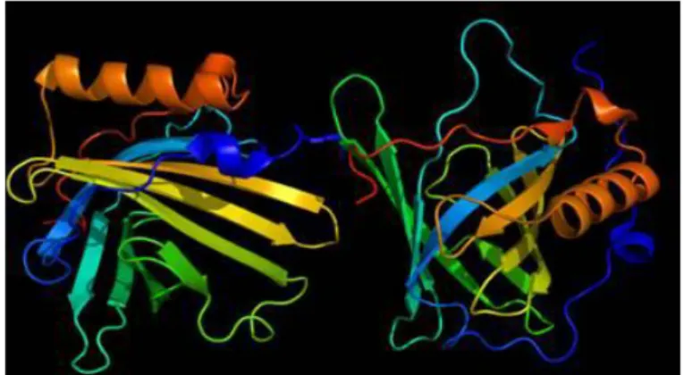
The core structure consists of an eight-stranded, antiparallel, continuously hydrogen-bonded β-barrel which defines a calyx-or cup-shaped structure with an enclosed ligand binding site. The loops linking the strands are typically short β-hairpins, except the first loop, describing a large Ω loop that usually folds back onto the barrel, partially constricting the binding site. There is a short helix at the N-terminus and an α-helix at the C-terminus (Figure 1) [Flower et al. 1996].
Unlike other lipocalins, NGAL shows no affinity for retinoic acid (REA), but is reported to bind the tripeptide N-formyl-Met-Leu-Phe, a potent neutrophil chemoattractant, and possibly other lipophilic mediators of inflammation such as PAF, leukotriene B4 and LPS. Through these
interactions, NGAL is proposed in important immunomodulatory functions. The role of NGAL in tissue remodeling and tumorigenesis is still unknown.
1.1 PHYSIOLOGY OF NGAL
Role in iron metabolism
The delivery of iron to cells is critical for cell growth and development. Iron is located at the active site of a large numbers of proteins including regulators of intermediary metabolism and DNA synthesis, and it stabilizes three-dimensional protein structure [Cooper et al. 1997; Nyholm et al. 1993].
In addition, iron is a unique regulator of gene expression, activating both transcriptional [Yamaguchi et al. 1996] and posttranscriptional mechanisms [Rouault et al. 1997]. For these reasons, the acquisition of iron is central to cell survival, growth and maturation, and its metabolism is tightly regulated by many proteins. Most cells acquire iron by capturing iron-located transferrin. After binding to receptors, transferrin enters an endocytic pathways [van Renswoude et al. 1982].
Within endosomes, iron dissociates from transferrin and is transferred across the vesicle membrane into the cell cytoplasm. In epithelia, this pathway is uniquely located in the basal domain of the cell. However, despite its ubiquity and quantitative importance in adult physiology, the transferrin pathway is not essential for the delivery of iron to many tissues, including epithelia. Hypotransferrinemic micehpx/hpx [Huggenvik et al. 1989; Trenor et al. 2000] and atransferrinemic humans [Hamill et al. 1991] have severe defects in hematopoiesis and central nervous system development, but most epithelial organs are normal.
Likewise, mice lacking the transferrin receptor-1 initiate organogenesis but succumb to the effect of anemia. Given that iron is necessary for all cells, there must be other pathways for iron acquisition in non-hematopoietic cells. These additional pathways might involve the local expression of iron-trafficking proteins, such as lactoferrin, putative low molecular weight siderophores, and cell surface iron transporters like DMT-1 (Divalent Metal Transporter-1) [Ward et al. 1999].
Siderophores are proteins produced by bacteria to scavenge iron from the extracellular space. They trap iron with high affinity and ensure continuous supply of iron necessary for survival and growth of bacteria. Enterochelin is one such siderophore produced by some strains of bacteria. The evidence for role of NGAL in iron metabolism came from crystallographic studies.
To test the hypothesis that NGAL directly interferes with siderophore-mediated iron uptake by bacteria, Goetz et al. investigated the effect of exogenous NGAL on the growth of bacteria under iron-limiting conditions. They noticed that XL1-Blue E.coli expressing NGAL fail to grow in M9 minimal media unless supplemented with 10 μM iron. However, the addiction of exogenous apo-NGAL, at concentration of only 5 μM, resulted in a dramatic 20-fold inhibition of growth. Adding iron just sufficient to saturate the NGAL present rescues growth, demonstrating that NGAL doesn’t have any antibacterial property beyond iron sequestration [Goetz et al. 2002].
Neutrophil gelatinase-associated lipocalin interacts with cells by specific cell-surface receptors. Hitherto, two receptors have been identified: the megalin-cubilin multiscavenger complex found on the brush-border surface of renal tubular epithelial cells [Hvidberg et al. 2005] and the 24p3R (NGAL was originally called 24p3), an organic cation transporter.
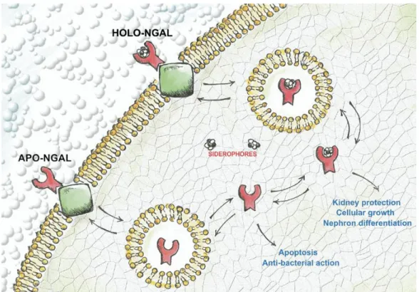
NGAL has also been proposed to interact with some other receptors such as extracellular protein kinases, hepatocyte growth factor, and gelatinase B. These receptors seem to have a role in cellular trafficking and endocytosis of NGAL. Endocytosis occurs as NGAL protein alone (apo-NGAL) or NGAL complexed with iron-binding siderophores (Holo-NGAL).
The cellular effects depend on the form of NGAL endocytosed. When transported as apo-NGAL, it captures intracellular iron and transports it outside leading to depletion of intracellular iron. On the other side, when endocytosed as Holo-NGAL, it releases iron-siderophore complex and contributes to intracellular iron pool. The depletion of iron cellular pools, under particular condition, may even lead to apoptosis (Figure 2) [Devireddy et al. 2005].
Figure 2. Schematic of neutrophil gelatinase-assocaited lipocalin (NGAL) cellular turnover.
Role in innate immunity to bacteria
Goetz et al. showed that the most important ligands of lipocalins are siderophores [Goetz et al. 2000; Goetz et al. 2002], small non-peptidic iron-containing molecules produced in bacteria, plants, and mammals that, through iron transport and supply, involved in cellular growth and survival. Immunohistochemistry revelaed massive staining for NGAL protein in
epithelial cells in both diverticulitis, appendicitis, ulcerative colitis and Crohn's disease, as well as in neoplastic conditions [Nielsen et al. 1996].
All these conditions are characterized by significant inflammation and in some cases also by neutrophil infiltration, but the latter does not explain the presence of NGAL in epithelial cells, and in situ hybridation has demonstrated a marked upregulation of NGAL mRNA in the epithelial cells in the aforementioned situations.
Strong and coll. reported that recombinant NGAL generated by E. coli was deeply red in color in contrast to recombinant NGAL generated by insect cells. It was shown that this red color was due to an iron molecule that was trapped in the siderophore of E. coli, enterochelin, indicating that NGAL is a siderophore binding protein [Strong et al. 1998].
The binding of enterochelin by NGAL is strong (dissociation constant 0.4 nM), and the siderophores are significantly more stable when bound in the lipocalin pocket of NGAL. This means that the ability of NGAL to sequester iron via siderophores is durable.
This evidence was confirmed by experiments where apo-NGAL, NGAL generated without siderophore, was added to E. coli. NGAL was able to completely block growth of E. coli under iron poor condition but NGAL with siderophore iron was not. Besides enterochelin, NGAL has been shown to bind to other siderophores including carboxymycobactin, a soluble protein secreted by mycobacteria. This massive induction of NGAL in epithelial cells made us suspect that NGAL serves a role in innate immunity.
NGAL inhibitory role on bacterial growth by sequestering siderophore iron was investigated in a knock-out mouse model generated by Flow et al. They showed that the resistance of wild-type mice was indeed due to their ability to induce NGAL synthesis and reduce the amount of siderophore iron available to bacteria, since providing the wildtype mice with siderophore iron from a source which NGAL cannot bind, made the mice as vulnerable to infection as knock-out mice [Flo et al. 2004]. This study indicated that NGAL works as a bacteriostatic agent by sequestering siderophore bound iron, but
NGAL is not bactericidal, and prevents bacterial growth only as long as NGAL is able to sequester siderophores. This authors concluded that NGAL participates in the antibacterial iron depletion strategy of the innate immune system.
NGAL is released by neutrophils at sites of infection and inflammation to sequester bacterial ferric siderophores thus participating in the antibacterial iron-depletion strategy of the innate immune system. However, while other components of this system, such as lactoferrin, simply bind to and sequester free iron, NGAL is specific for iron already earmarked for bacterial use as ferric siderophore complexes. NGAL bind siderophores and prevents its uptake by microorganism (Figure 3) [Borregaard et al. 2006].
Figure 3. Epithelial cells synthetize and release NGAL in response to inflammation. Microorganism synthetize and release siderophores that capture iron and provide a mechanism for supplying this essential nutrient to the bacteria. NGAL binds siderophores and prevent their uptake in microorganism and thus deprive them of this essential nutrient.
Role in kidney development
NGAL was originally purified as a protein that promotes the mesenchymal to ephitelial conversion of ureteric bud cell lines. This effect could potentially be related to the ability of NGAL to donate iron to the cells. It
has been suggested that NGAL is involved in kidney development [Gwira et
al. 2005].
Gwira et al. showed that some growth factors such as hepatocyte growth factor (HGF) are able to stimulate epithelial cells to express NGAL which participates in mesenchymal-epithelial transformation via its capability to augment cellular iron uptake.
At concentrations below those found to mediate iron transport, purified NGAL can induce a promigratory and probranching effect that is dependent on ERK activation. The suppression of NGAL expression using short hairpin RNA produces increased cyst formation by tubular cells.
However, the simultaneous addiction of NGAL and HGF leads to direct association of the two proteins, and results in a partial inhibition of HGF-mediated activation of c-Met and the downstream MAPK and phosphatidylinositol 3-Kinase signaling pathways. This inhibitory effect downregulates HGF-stimulated single cell migration, and limits branching morphogenesis at both the single cell and multicellular level. These experiments demonstrate that the local expression of NGAL plays a regulatory role in epithelial morphogenesis by promoting the organisation of cells into tubular structures while simultaneous negatively modulating the branching effects of HGF.
Yang et al. demonstred that, administering purified NGAL to early epithelial progenitor cells obtained from a specific peripheral niche of murine metanephric mesenchyme, a clear proliferative effect was observed, followed by epithelial differentiation of these elements with the subsequent generation of nephron-shape formations expressing glomerular, proximal, and distal tubular surface cellular markers. However, this mechanism does not seem to be essential for in vivo embryo kidney development because genetic model of NGAL inactivation do not give rise to a blockade of renal maturation or complete agenesis of this organ, probably because of the presence of other more redundant pathways. At present, the exact molecular mechanism through which NGAL exert its growth effect on renal cells has not yet been clarified. A
fundamental role seems to be played by the association between NGAL and iron-binding siderophores and its subsequent interaction with specific surface receptors (24p3R; megalin).
Conversely, NGAL alone (apo-NGAL), as well as NGAL associated with an iron-free or gallium-binding siderophore, appears to have a proapoptotic effect very similar to that on which the antibacterial properties of this protein depend, as a result of depletion of intracellular iron reserves [Yang
et al. 2002].
Considered together, these observations confirm the importance of iron capture for the development of embryonic kidneys, but also suggest the presence of an alternative iron-delivery pathway, distinct from the main systems currently recognized in mammals (which is mediated by transferrin). However, it cannot be excluded that other mechanisms, for instance the activation of extracellular kinases or binding with matrix metalloproteinase 9, may be involved in determining growth/differentiation properties of NGAL [Bolignano et al. 2008].
1.2 NGAL AS A BIOMARKER
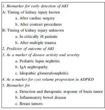
NGAL has been proven to be a promising biomarker in a variety of other renal and non-renal conditions, as shown in Table 2. NGAL is a useful clinical biomarker for early diagnosis, for predicting disease severity, for therapeutic monitoring, and for predicting clinical outcomes. The following section discusses the potential role of NGAL in different renal and systemic diseases.
Table 2. Potential use of NGAL as a biomarker.
Acute kidney injury
Acute kidney injury (AKI) formerly named acute renal failure (ARF) of any origin is associated with a high mortality in the critically ill patient, despite significant technical advances in therapeutics including renal replacement therapy (RRT) such as dialysis [Brar et al. 2008].
AKI is defined as the abrupt (e.g. within 48 hours) and sustained decrease in renal function resulting in retention of nitrogenous (urea and creatinine) and non-nitrogenous waste products as well as in dysregulation of cellular volume and electrolyte handling. In clinical practice, the diagnosis of AKI relies on a decreased glomerular filtration rate (GFR), increased serum creatinine with or without oliguria, classified in RIFLE (Risk, Injury, Failure, Loss, and End-stage kidney disease) [Bellomo et al. 2004b] and AKIN (Acute Kidney Injury Network) criteria [Mehta et al. 2007].
The classification for AKI is defined by three stages of increasing severity, which correspond to the RIFLE criteria for risk (stage 1), injury (stage
2), and failure (stage 3). Loss and end-stage kidney disease were removed and redefined as outcomes.
However, there are major limitations to the use of creatinine for estimating GFR. Serum creatinine does not accurately reflect GFR during the non-steady state of AKI by probably overestimating GFR. Thus, minor changes of creatinine, as typically seen earlier in AKI, do already reflect substantial declines in GFR [Bellomo et al. 2004a]. To overcome these obstacles, an extensive search for more suitable laboratory markers monitoring impaired renal function is required.
NGAL appears to be the most promising molecule among the many novel molecules [Ronco et al. 2008].
Misha et al. first proposed NGAL as a new early biomarker. In mouse model of ischemia-reperfusion injury (IRI) subjected to 30 minutes of bilateral renal artery occlusion, NGAL was found to be one of the seven genes which were highly upregulated. NGAL was easily detected in the urine within 2 hours following ischemia. In the same study, separate sets of mice were subjected to 5, 10, and 20 minutes of ischemia. Urinary NGAL was detected even in these mice, appearing after 6 hours in mice with 5 minutes of ischemia and after 4 hours in mice with 10 and 20 minutes of ischemia. Thus, NGAL was found to be a very sensitive marker of ischemic AKI and its levels correlated to the dose and duration of renal ischemia [Mishra et al. 2003].
Additionally, the same authors reported that uNGAL was detectable after 1 day of cisplatin administration in a mouse model of nephrotoxic AKI, suggesting its sensitivity in other models of tubular injury [Mishra et al. 2008]. NGAL has been widely investigated across a range of different clinical settings of AKI.
However, with accumulating evidence, conflicting observations raised some concerns about the robustness of NGAL as a biomarker. Haase et al. performed a meta-analisys of data from 19 studies including 2500 patients from observational studies to estimate the diagnostic and prognostic accuracy of NGAL and its value in AKI. The population included both adults and
children, studied in several clinical conditions: most frequently investigated AKI after cardiac surgery, followed by AKI in critically ill patients and after exposure to contrast media for coronary angiography.
In conclusion, NGAL was found to be a useful early predictor of AKI, with urine or plasma/serum NGAL levels functioning as well. NGAL levels also revealed a prognostic value for clinical endpoints, such as the initiation of dialysis and mortality. Unfortunately, substantial extrarenal NGAL generation in response to systemic stress can increase urinary NGAL excretion in the absence of AKI as well, and this may also arise from chronic and not just acute, renal disease [Haase et al. 2009].
Renal transplantation
Kidney allograft function after transplantation varies from a rapid increase in GFR, causing brisk reductions in serum creatinine to primary allograft failure.
Delayed graft function (DGF) is defined as the need for dialysis within 1 week of transplantation, and occurs in 20% to 33% of deceased donor kidney transplant (DDKTs) [Halloran et al. 2001; Mikhalski et al 2008].Recent strategies for increasing the donor pool include using “extended-criteria donor” (ECD) and “donation after cardiac death “ (DCD) kidneys. The deleterious effects of DGF in the immediate post-transplant period include increased lengths of stay and total hospital costs, mainly due to the need for dialysis [Rosenthal et al. 1991].
Hall et al. examined the long-term role that DGF plays in patient and graft survival in a recent meta-analysis and demonstrated more than 40% increased risk of graft loss at 1 year with DGF [Yarlagadda et al. 2008]. Even in patients not dialyzed after transplant, some studies have shown poorer long-term outcomes with “slow graft function”(SGF) compared with “immediate
graft function” (IGF) [Rodrigo et al. 2004; Humar et al. 2002; Johnston et al. 2006].
Hall et al. carried out a multicenter, prospective cohort study of patients receiving DDKTs to evaluate the timing and efficacy of using urinary NGAL for predicting recovery of graft function and the need for dialysis within 1 week after transplantation. The study revealed that urinary NGAL is an early, non-invasive, and accurate predictor of the need for dialysis within the first week of kidney transplantation.
After transplantation, local NGAL production within DGF kidneys contributes to the high circulating level, reflecting the ischemia-reperfusion stress applied to the transplanted kidney before organ withdrawal, during the storage period or during reperfusion. In DGF kidneys, in situ expression of NGAL by the tubular epithelium has been demonstrated by a proteomic study performed 1 hour after arterial clamp release. This suggested that the high plasma NGAL value in DGF patients throughout the study period may partly be related to transplanted kidney production [Hall et al. 2010].
Compared with previous studies that assessed plasma NGAL as an early and sensitive marker of the onset of AKI with normal baseline kidney function, it is relevant to consider renal transplantation as a specific model. Starting with a high plasma level, fast plasma NGAL level decrease appeared to be a very sensitive marker of renal function, suggesting less post-ischemic tissue damage. This decline may be initiated by less renal transplant NGAL production or a negative cumulative marker balance related to extra-renal production decrease, renal washout of the marker and modification of renal uptake.
NGAL is physiologically filtered by the glomerulus and almost reabsorbed within the proximal tubule. After ischemia-reperfusion aggression in transplanted kidneys, tubular reabsorption may still be impaired despite glomerular filtration rate restoration and early graft function (EGF).
Since urine output is strictly compensated by intravenous saline infusion during the first postoperative day, there was a wide distribution of urine output
values among patients. Because the proportion of patient with urine output less than 1 L at day 1 is significantly higher in DGF patient than in EGF, still 13% of the patients with immediate graft function had a urine output less than 1 L at day 1. As in the present study, it appears that immediate urine output is not a useful tool to discriminate EGF from DGF patients [Bataille et al. 2011].
Biomarker validation could be included as an integral part of ongoing intervention trials for DGF. Although animal studies have suggested effective therapies in IRI, human trials of DGF to date have been less promising, likely because of late detection [Perico et al. 2004].
If biomarker levels in the immediate post-transplant period can be shown to accurately predict duration of injury and longer term graft function, they could be very useful as surrogate markers. Ultimately, insight into this area of research may provide support for the early detection and stratification of IRI, which will facilitate future DGF/AKI intervention trials [Parikh et al. 2008].
Chronic kidney disease
Recent studies aimed at defining whether NGAL may have a role in renal and systemic adaptions to chronic kidney disease. The principal hypothesis was that chronic renal damage could influence the physiological balance of this protein in a way similar to that observed for acute injury conditions.
Polycystic kidney disease. NGAL has been shown to be an excellent
indicator of severity of CDK in patients affected by autosomal dominant polycystic kidney disease, in which uNGAL and sNGAL levels correlated well with resisual renal function, serum creatinine, and cyst enlargement in patients with autosomal dominant polycystic kidney disease [Bolignano et al. 2007]. Wei et al. showed that addition of NGAL to the cells from PKD1 mutant mice suppressed the cyst development [Wei et al. 2008].
Glomerular disease. Brunner et al. observed a cohort of 35 cases of
childhood-onset systemic lupus erythematosus (SLE) and found a strong correlation of uNGAL levels with renal disease activity. In addition, uNGAL levels correlated with the activity and chronicity score on renal histology [Brunner et al. 2006]. In a similar study, Suzuki et al. found strong correlation of uNGAL levels with SLE disease activity. However, plasma NGAL levels did not show such correlation in this study [Suzuki et al. 2008].Ding et al. evaluated a cohort of patients affected by immunoglobulin A nephropathy. They compared levels of uNGAL, urinary N-acetyl-β-D-glucosaminidase (uNAG) and urinary creatinine with 40 healthy controls. uNGAL levels were significantly elevated in IgA nephropathy patients compared with healthy controls [Ding et al. 2007].
Bolignano et al. recently reported that patients with proteinuria associated with idiophatic glomerulonephritis had also increased uNGAL levels in comparison with healthy subject, and these values correlated well with GFR and serum creatinine level, as well as daily proteinuria extent [Bolignano et al. 2008].
Other systemic conditions. Poniatowski et al. found serum and urine
NGAL as sensitive early markers of renal impairment in patients with chronic heart failure [Poniatowski et al. 2009].
In another study, uNGAL levels were found to be significantly elevated in patients with chronic heart failure. Its level correlated directly with urine albumin excretion rate and inversely with estimated glomerular filtration. So, uNGAL is a potential biomarker for the cardio-renal syndrome [Damman et al. 2008].
La Manna et al. evaluated the ability of serum and urinary NGAL to predict renal function in the first weeks of life of preterm infants. From September 2008 to July 2009, infants weighing ≤1500g at birth with no major congenital anomalies or sepsis were eligible. They proved that uNGAL levels
at birth may have a predictive role in very LBW (VLBW) infants [La Manna et
al. 2011].
Smith et al. indicated that uNGAL, along with other urine metalloproteinases, as a satisfactory biomarker for the presence of brain tumor [Smith et al. 2008].Urinary NGAL can also potentially serve as a marker to assess the severity of inflammatory bowel diseases. Fernandez et al. found uNGAL-MMP9 complex as a useful biomarker for breast cancer.
In addition to NGAL, many other molecules have been studied and found useful as early biomarkers of AKI when compared with serum creatinine. Performance of NGAL has been comparable or better than other molecules [Fernandez et al. 2005].
Preeclampsia is a leading cause of maternal and fetal/neonatal mortality and morbidity worldwide. The early identification of patients with an increased risk for preeclampsia is therefore one of the most important goals in obstetrics. The availability of effective biomarkers would allow not only the detection of patients at risk but also a correct early disease assessment in asymptomatic pregnant women, a timely intervention and closer surveillance.
Among the potential biomarkers, NGAL has been evaluated through normal pregnancy and pregnancies complicated by preeclampsia syndrome, because of the previous evidences proving its reliability as a marker of renal damage. Considering that the kidney damage is always present in preeclampsic women, NGAL has recently gained interest as a candidate diagnostic biomarker in early diagnosis of this disease. A recent study demonstrated significant increased serum NGAL levels in preeclampsic women compared to the control women and a direct correlation to blood pressure and proteinuria, showing a high sensitivity (75%) and specificity (94.5%) [D’Anna et al. 2008].
1.3 NGAL IN PATIENT WITH DIABETIC NEPHROPATHY
NGAL has been proven to be a very promising marker in diabetes. Diabetic nephropathy is a severe complication that develops in 30-40% of diabetic patients [Parving et al. 2008]. Additionally, diabetic nephropathy is associated with a higher risk of other micro and macrovascular complications, including cardiovascular disease, neuropathy and retinopathy, and with an increased all-cause mortality. To prevent the development of diabetic nephropathy, early screening for albuminuria and early renoprotective interventions are essential.
Albumin excretion is primarily a result of glomerular damage, but lack of tubular reabsorption of albumin also contributes to albuminuria. Thus, the urinary albumin excretion is mainly a product of glomerular but also tubular damage and, as the damage to these sites is not always correlated, a more specific marker of tubular injury is needed.
It has been repeatedly demonstrated that glomerular and tubulointerstitial injury are both important factors in the pathophysiology of diabetic nephropathy [Gilbert et al. 1999]. In this regard, Nielsen et al. reported that levels of the tubular marker urinary liver type fatty acid-binding protein, are increased in Type 1 diabetic patients, even before they develop signs of glomerular damage, i.e. albuminuria. Thus it has been hypothesized that NGAL could complement albuminuria (the established marker of glomerular injury) for the early detection of diabetic nephropathy. These authors observed higher urinary NGAL accompanied by rising levels of albuminuria; moreover the urinary/plasma NGAL ratio was significantly increased in diabetic patients compared with healthy subjects and also increased along with the degree of albuminuria. No significant differences were found in this ratio between the microalbuminuric group and the normoalbuminuric group [Nielsen et al. 2009; Nielsen et al. 2010].
It has also been hypothesized that diabetes, as a hyperglycaemic and proinflammatoric state, induces increased NGAL production in extra-renal
tissues, a hypothesis which has been supported by a study in type 2 diabetic patients. The increase of urinary NGAL might represent a protective strategy against renal damage, an hypothesis that has been primarily explored in acute kidney injury.
Bolignano etal.suggested thatNGAL mayplay animportant rolein the pathophysiology of renal adaptation to diabetes, probably as a defense mechanism to mitigate tubular suffering. In this study, patients with type 2 diabetes mellitus divided into three groups: normoalbuminuria, microalbuminuria and diabetic nephropathy. All the diabetic patients showed increased NGAL values compared with healthy subjects; higher NGAL levels were found in the diabetic patients without early signs of glomerular damage (normoalbuminuric). Bothurinary NGAL and serumNGALrose in parallel with the severity of renal disease, reaching the highest levelsin patients withdiabetic nephropathy. Therefore NGAL measurement might constitute a valuable and noninvasive tool for the evaluation of renal involvement in diabetic patients as well as for the early diagnosis of incipient nephropathy [Bolignano etal. 2009].
1.4 SNPs
In recent years the scientific community has given increasing attention to genetic factors potentially related to individual susceptibility of patients to the development of various multifactorial diseases.
The predisposition to suffer from certain diseases, or the type of response to a given treatment may depend on the presence of polymorphisms (Single Nucleotide Polimorphisms, SNPs), or small variations in the genome. The polymorphisms are not the direct cause of the disease, but their presence can influence (increasing or reducing) the risk [Misefari et al. 2001].
SNPs are DNA sequence variations, due to an alteration of a single nucleotide (A, T, C or G) in the sequence of genome, which can occur either in a coding region (exons), or in a non-coding region (introns, regulatory sequences) of the gene.
SNPs sited in a coding sequence do not necessarily change the aminoacid sequence of the protein, due to degeneracy of the genetic code. A SNP which produces the same polypeptide sequence is called a synonymous polymorphism (or silent mutation). If a different polypeptide sequence is produced the polymorphism is a replacement polymorphism, either missense, which results in a different aminoacid, or nonsense, which results in a premature stop codon.
SNPs that are not in protein-coding regions may still affect gene splicing, transcription factor binding, messenger RNA degradation, or the sequence of non-coding RNA. Gene expression affected by this type of SNP is referred to as an expression SNP and may be located upstream or downstream from the gene.
The nucleotide polymorphisms seem to fulfill the characteristics of an ideal genetic marker: abundant in the genome (in fact represent 90% of all human genetic variation), readily reproducible in different laboratories, and easy to locate. SNPs, in general, can be very useful for genetic studies of population and to detect traces of selection: they are indeed very common in populations and on a case by case basis give rise to small effects on the phenotype of an individual. It is well established that specific SNPs or groups of SNPs defined by a haplotype result influence the individual susceptibility or resistance to many of the most common human diseases, namely diabetes, cancer, cardiovascular disease, Alzheimer's disease, arthritis.
2. PURPOSE
The primary goal of this study was to assess the effectiveness of NGAL as a predictive marker of disease onset and/or disease progression in pathological acute and chronic kidney disease (primary endpoint).
We evaluated its efficacy in different clinical conditions:
Very low birth infant (VLBI) Preeclampsia
Kidney transplantation Diabetic nephropathy
3. MATERIALS AND METHODS
3.1 PATIENTS
Very low birth weight (VLBW) infants
A total of 55 infants weighing ≤1500 g hospitalized in S. Orsola University Hospital Neonatal Intensive Care Unit from September 2008 to July 2009 were eligible for the study. Exclusion criteria were major congenital anomalies, abnormal karyotype, sepsis at birth (C-reactive protein > 1.5 mg/dL or positive microbiological blood culture). Forty-four neonates (80%) showed increased eGFR during the first 3 weeks of life (normal renal function, NRF group), while 11 infants (20%) demonstrated stable or progressively deteriorating renal function (impaired renal function, IRF group).
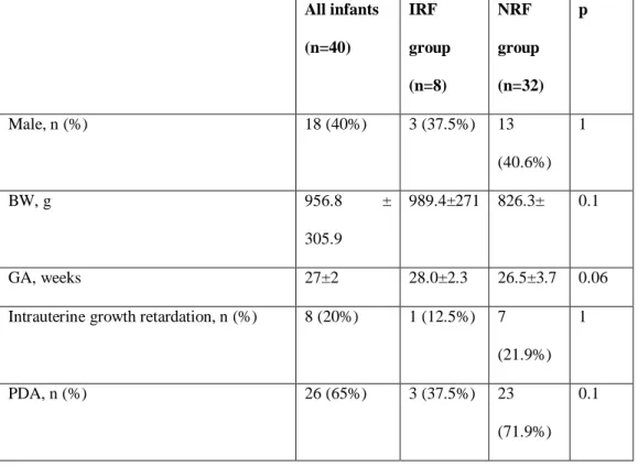
Demographic characteristics, clinical parameters and biomarkers values of the infants are reported in Table 3.
Table 3. Demographic characteristics and clinical parameters of infants. Results are expressed as mean ± SD, unless otherwise stated. RDS, respiratory distress syndrome.
All infants (n=40) IRF group (n=8) NRF group (n=32) p Male, n (%) 18 (40%) 3 (37.5%) 13 (40.6%) 1 BW, g 956.8 ± 305.9 989.4±271 826.3± 0.1 GA, weeks 27±2 28.0±2.3 26.5±3.7 0.06
Intrauterine growth retardation, n (%) 8 (20%) 1 (12.5%) 7 (21.9%)
1
PDA, n (%) 26 (65%) 3 (37.5%) 23
(71.9%) 0.1
Mechanical ventilation and/or surfactant for RDS, n (%)
27 (67.5%) 5 (62.5%) 22 (68.7%)
0.29
Ibuprofen treatment for PDA n (%) 14 (35%) 3 (37.5%) 11 (34.4%)
1
Aminoglycosides, n (%) 31 (77.5%) 6 (75%) 25
(78.1%) 1
Dopamine and/or dobutamine, n (%) 13 (32.5%) 6 (75%) 7 (21.9%)
0.0085
Hypotension at birth, n (%) 2 (5%) 1 (12.5%) 1 (3.1%) 0.36
Preeclampsia
One-year prospective case-control study was performed at the Department of Obstetrics and Prenatal Medicine of the University of Bologna.
Eighteen women affected by preeclampsia (PE) and 22 women with uncomplicated pregnancy matched for gestational age were enrolled. Mild PE was defined as the development of hypertension (systolic blood pressure ≥ 140 mmHg or diastolic blood pressure ≥ 90 mmHg on two occasions at least six hours apart) and proteinuria (> 0.3 g/day) after 20 weeks of gestation. Severe PE was diagnosed on the basis of the presence of one or more of the following evaluation criteria: severe hypertension (systolic blood pressure ≥ 160 mmHg or diastolic blood pressure ≥ 110 mmHg on two occasions at least six hours apart during bed rest), or severe proteinuria (≥ 5 g in a 24-hour urine specimen or 3 + or greater in two random urine specimens collected at least 4 hours apart).
Atypical PE was recognized as the presence of gestational hypertension or proteinuria associated with signs and/or symptoms of organ damage.
Demographic and clinical characteristics of study population are reported in Table 4.
Table 4. Demographic and clinical characteristics of study population. PE (n = 18) Controls (n = 22) p-value
Maternal age (years)* 37±3.9 37±4.3 Ns
Nulliparity ** 50 50 Ns Multiple pregnancies** 16% 9% Ns Preconceptional BMI* 26.5±7.5 23.6±4.4 Ns Smokers** 5.6 4.6 Ns Trhombophlia** 5.6 4.6 Ns Nephropathy** 5.6 0 0.05 Chronic hypertension** 16.7 0 0.0001 *: media +/- SD **: percentage Kidney transplantation
Forty-four consecutive patients who received renal transplantation at the Unit of Nephrology, Dialysis and Transplantation of the S. Orsola University Hospital were enrolled between February 2009 and May 2010.
Urine samples were collected before transplantation (T0) and at 1, 3, 7, 14 and 30 days after transplantation (T1, T2, T3, T4, and T5 respectively).
Demographic and clinical characteristics of the patients, and transplantation-related variables are reported in Table5.
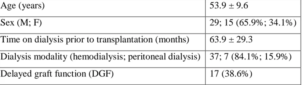
Table 5. Recipients clinical and demographic characteristics. Continuous variables are presented as means ± standard deviation, and categorical variables are presented as n (%).
Age (years) 53.9 ± 9.6
Sex (M; F) 29; 15 (65.9%; 34.1%)
Time on dialysis prior to transplantation (months) 63.9 ± 29.3
Dialysis modality (hemodialysis; peritoneal dialysis) 37; 7 (84.1%; 15.9%)
Early graft function (EGF) 26 (59.1%)
Primary not function 1 (2.3%)
Diabetic nephropathy
The population consisted of 80 patients with Type 1 diabetes recruited from the Diabetes and Pediatrics Clinic of the S. Orsola University Hospital.
Based on urinary NGAL levels the patients were divided into three groups: 49 with urinary NGAL < 25 ng/ml; 15 with urinary NGAL between 25 and 50 ng/ml; and 16 with urinary NGAL> 50 ng/ml.
The main clinical characteristic and biochemical parameters of the patients are presented in Table 6.
Table6. Clinical characteristic and biochemical parameters in the three groups of diabetic patients. uNGAL < 25 ng/ml 25.1-50 ng/ml > 50 ng/ml n 49 15 16 Gender (male/female) 36/13 10/5 4/12 Age (yr) 47 47 43
Systolic blood pressure M1 (mmHg) 126 127 118
Systolic blood pressure M10 (mmHg) 126 124 124
Diastolic blood pressure M1 (mmHg) 78 78 73
Diastolic blood pressure M10 (mmHg) 79 76 76
HbA1c M1(%) 8 8 8 HbA1c M10 (%) 8 8 8 Creatinine M1 (mg/dl) 2 1 1 Creatinine M10 (mg/dl) 1 1 1 Microalbuminuria M1 (mg/24h) 18 11 23 Microalbuminuria M10 (mg/24h) 29 16 13
3.2 URINE COLLECTION
Very low birth weight (VLBW) infants
Urine specimens were collected 24 to 48 hours after birth. A blood specimen was also collected after 7 days and 3 weeks from birth for the determination of serum creatinine (sCreat) and the estimation of eGFR. Blood samples were collected in Vacutainer tubes with clot activator and gel for serum separation. Serum was separated by centrifugation at 2500 rpm for 15 min, and sCreat was assayed immediately.
All urine samples were collected with a urine bag for 3 hours, and urinary creatinine (uCreat) was assayed immediately. For the determination of uNGAL, urine samples were centrifuged for 10 min at 1500 rpm within 24 hours of collection and the supernatant was stored at -20°C until analysis.
Preeclampsia
Urine collection were performed at time of PE diagnosis. Specimens were kept refrigerated at 4°C until they were sent to laboratory, then they were stored within 4 hours of collection and analyzed later. Urine samples were centrifuged at 1500 rpm for 10 minutes, and the supernatants were frozen at -20°C until analysis.
The levels of urinary NGAL was measured using a commercially available ELISA test (BioPorto Diagnostics, Gentafte, Denmark).
Kidney transplantation
Urine samples were collected before transplantation (T0) and at 1, 3, 7, 14 and 30 days after transplantation (T1, T2, T3, T4, and T5 respectively).
Urine samples were centrifuged at 1500 rpm for 10 minutes and the supernatant was frozen at -20°C until analysis.
The levels of urinary NGAL was measured using a commercially available ELISA test (BioPorto Diagnostics, Gentafte, Denmark).
Diabetic nephropathy
Urine specimens were collected at two times: M1 and M10. Urine samples were centrifuged at 1500 rpm for 10 minutes and the supernatant was frozen at -20°C until analysis.
Urine NGAL measurements obtained by ARCHITECT® assay.
3.3 ELISA TEST ( Enzyme Linked Immunosorbent Assay )
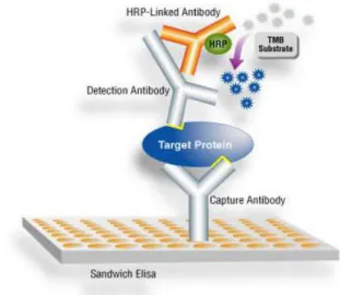
All the samples were analyzed in the laboratory of our Unit. Urinary levels of NGAL were assayed using commercially available ELISA tests (NGAL ELISA Kit 036; BIOPORTO Diagnostic, Grusbakken, Denmark). ELISA (Enzyme Linked Immunosorbent Assay) consists of a solid-phase enzyme immunoassay to detect the presence of an analyte in a liquid sample, i.e. serum, plasma, urine or cell culture medium.
NGAL ELISA is an assay performed in microwells coated with a monoclonal antibody against human NGAL. Bound NGAL is detected with a horseradish peroxidise (HRP)-conjugated monoclonal antibody and the assay is developed by incubation with a color-forming substrate. At first, aliquots of calibrators, diluted samples and any controls were incubated with HRP-conjugated detection antibody in the coated microwells. Only NGAL binds to both coat and detection antibody, while unbound materials are removed by washing. In the second step, a chromogenic peroxidase substrate containing tetramethylbenzidine (TMB) was added to each test well. The HRP linked to the bound detection antibody reacts with the substrate to generate a colored product. The enzymatic reaction was stopped chemically, and the color intensity was read immediately with a reader DV 990 microplate BV5UV (GVD, Rome, Italy) by measuring absorbance at 450 nm (Figure 4).
Figure 4. Enzyme Linked Immunosorbent Assay.
The color intensity (absorbance) is a function of the concentration of NGAL originally added to each well. The results for the calibrators are used to construct a calibration curve from which the concentrations of NGAL in the test specimens are calculated and expressed in pg/ml. The sensitivity limit was 4 pg/ml.
3.4 ARCHITECT NGAL assay
The ARCHITECT NGAL assay utilizes a non-competitive, sandwich format with chemiluminescent signal detection. The assay includes a microparticle reagent prepared by covalently attaching an anti-NGAL antibody to paramagnetic particles and a conjugate reagent prepared by labeling a second anti-NGAL antibody with acridinium. The mouse anti-NGAL antibodies were developed at Abbott Laboratories and are directed against distinct, non-overlapping NGAL epitopes. The calibrators are prepared with recombinant human NGAL expressed and purified at Abbott Laboratories. The recombinant NGAL is a full length protein.
The assay employs an automated sequence consisting of an 18 min incubation step with sample (2.5 μL) and microparticle reagent (50 μL), a solid phase wash step, and a 4 min incubation step with conjugate reagent (50 μL).
Following the immunochemistry steps, the solid phase is washed again and the acridinium label is triggered with peroxide and base to generate the signal. The assay calibrators are at 0, 10, 100, 500, 1000 and 1500 ng/mL and the concentration of NGAL measured is proportional to the signal.
3.5 MOLECULAR BIOLOGY
Extraction of genomic DNA
The patients' genomic DNA was isolated using GenomicPrep ™ Blood DNA Isolation Kit (Amersham Biosciences, Piscataway, NJ, USA). The kit is designed to isolate DNA from nucleated blood cells.
Red blood cells, which lack genomic DNA, were lysed using 900 μl of lysis solution (RBC) which were added to 300 μl of whole blood. The mix was agitated by inversion and incubated at room temperature for 10 minutes. After centrifugation at 13000-16000 g for one minute, the supernatant was removed, leaving the pellet leaving behind the visible white cell pellet and 10–20 μl of residual liquid.
Remaining white cells were then lysed in the presence of a DNA preservative using 300 μl of an anionic detergent which solubilizes the cellular components. The DNA preservative limits the activity of DNases that are present in the cells and elsewhere in the environment. Contaminating RNA was removed by treatment with with 1.5 μl of RNase precipitation solution (at 37°C for 15 minutes). Any cytoplasmic and nuclear proteins are removed by salt precipitation adding 100 μl of precipitation solution
After centrifugation at 13,000–16,000 x g for 3 minutes, the proteins formed a tight, dark brown pellet, well separated from the supernatant; this fraction, containing the DNA, was collected in another tube. Genomic DNA is isolated by precipitation with the addition of 300 μl of isopropyl alcohol at 100%. After two consecutive washes with 300 l of 70% ethanol, the pellet is left to dry for 10-15 minutes. Subsequently, the dried sample is rehydrated with
100 l of sterile distilled water (DNase RNase free H2O Ultra Pure, Invitrogen Life Technologies, Carlsbad, USA) overnight at room temperature.
Agarose gel electrophoresis
Electrophoresis is a procedure which enables the sorting of molecules based on size and charge, using an electric field that allows molecules to migrate toward the pole that has the opposite charge [Westermeier R. et al. 1997]. In biology there are many molecules bearing ionizable groups (such as aminoacids, proteins and nucleic acids) and then, at each pH value, are present in solution as electrically charged species. For example, due to the presence of phosphate groups (PO4
3-), the negatively charged DNA molecules migrate toward the positive pole (anode) when subjected to an electric field.
Agarose gel electrophoresis can be used for the separation of DNA fragments ranging from 50 base pair to several megabases. The distance between DNA bands of a given length is determined by the percent agarose in the gel. The agarose is a linear polysaccharide, normally used at concentrations of 1% and 3%.
Agarose gels are obtained by suspending dry powdered agarose in an aqueous buffer, such as TBE (Tris Borate EDTA), then boiling the mixture until the agarose melts into a clear solution. The solution is then poured onto a gel-tray and allowed to cool to room temperature to form a rigid gel. Upon hardening, the agarose forms a matrix whose density is determined by the initial agarose concentration.
The pore size is a function of agarose concentration: large pores are obtained using low concentrations, while smaller pores are obtained with higher concentrations. Depending on the concentration of agarose (and thus pore size), it is possible to resolve a wide size-range of DNA fragments, usually ranging between 100 kb and 20 kb. In the preparation of the agarose gel ethidium bromide is added to a final concentration of approximately 0.5 mg/ml. The ethidium bromide is one of the most widely used dyes for the detection of nucleic acids: it is a planar aromatic organic molecule capable of intercalation
between the nucleobases and emits orange light at 590 nm when excited by UV radiation with length wave between 254 and 306 nm. DNA bands on the segments of interest are displayed using a BIO-RAD Gel Doc 1000.
In this study, agarose gel electrophoresis was used for different purposes:
- Quantitative estimate of genomic DNA. Genomic DNA isolated from patient’s blood was visualised on agarose gel 1 % in TBE 1X. One l of DNA extract with 4 μl of sterile distilled, and 5 μl of 2X loading buffer (red cresol 1 mg / ml, glycerol 625 mg / ml) were loaded in each well. DNA molecular weight markers VI and VIII (Roche Applied Science, Mannheim, Germany) were used to quantify genomic DNA.
- Assessment of PCR results. Five l of PCR product were analyzed directly by 1.5% agarose gel electrophoresis followed by ultraviolet detection the presence and the yield of the expected bands. DNA molecular weight markers VI and VIII (Roche Applied Science) served for size determination and confirmation of the bands.
- Analysis of the results of RFLP (Restriction Fragment Length Polymorphism). The PCR products digested with restriction enzymes were loaded on 2.5% agarose gel at (+ equal volume of 2X loading buffer). The recognition of the bands according to sizes obtained from digestion was carried out using DNA molecular weight markers VI and VIII (Roche Applied Science).
-
Polymerase Chain Reaction (PCR)
The polymerase chain reaction (PCR) is a technique in molecular biology to amplify a single or a few copies of a piece of DNA across several orders of magnitude, generating thousands to millions of copies of a particular DNA sequence. PCR was developed in 1983 by Kary Mullis who was awarded the Nobel Prize in Chemistry ten years later for his work on PCR.
PCR amplification can be performed in vitro very rapidly, providing the amount of genetic material necessary for subsequent applications.
The method relies on thermal cycling, consisting of cycles of repeated heating and cooling of the reaction for DNA melting and enzymatic replication of the DNA.
Almost all PCR applications employ a heat-stable DNA polymerase, such as Taq polymerase, an enzyme originally isolated from the bacterium
Thermus aquaticus. This DNA polymerase enzymatically assembles a new
DNA strand from DNA building-blocks, the nucleotides, by using single-stranded DNA as a template and primers, which are required for initiation of DNA synthesis. Primers are DNA oligonucleotides containing sequences complementary to the target region. As PCR proceeds, the DNA generated is itself used as a template for replication, initiating a chain reaction in which the DNA template is exponentially amplified.
The sequence to be amplified can be synthesized from a discrete molecule or from a larger molecule. In both cases, the reaction product will be a discrete molecule of dsDNA ends corresponding to the 5' oligomers used. A basic PCR set up requires several components and reagents which include:
- DNA template that contains the DNA region (target) to be amplified. - A pair of primers that hybridize to sequences flanking the target. Each
primer is usually composed of a number of nucleotides ranging from 20 to 30;
- Taq polymerase or another DNA polymerase with a temperature optimum at around 70°C.
- Deoxynucleoside triphosphates (dNTPs; nucleotides containing triphosphate groups).
- Buffer solution, providing a suitable chemical environment for optimum activity and stability of the DNA polymerase.
- MgCl2, a cofactor of DNA polymerase.
- In some cases dimethyl sulfoxide (DMSO), to facilitate the separation between two DNA strands, improving the efficiency of PCR.
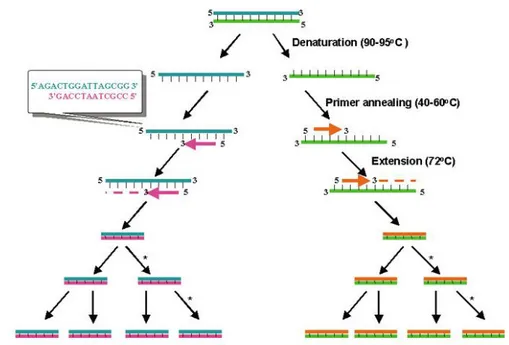
The reaction involves a series of successive steps of denaturation, annealing and extension that are repeated for a large number of cycles (Figure 5).
Figure 5. Schematic drawing of the PCR cycle.
The selected segment of DNA is separated into single strands by heating (denaturation) and then is cooled, allowing the two oligonucleotides serving as primers to hybridize with their complementary sequences on both strands (annealing). In the following phase, the DNA polymerase (Taq polymerase), sets in motion the elongation of the primer by adding nucleotides to the 3' end (extension).
During the first and every subsequent reaction cycle, the extension of each oligonucleotide on the original strand produces a new ssDNA molecule of indefinite length. The long products originated in this way serve as template for the complementary primer during subsequent cycles, and their extension by Taq polymerase molecules makes PCR products of a defined length,
corresponding to that of the target region. Hence a chain reaction proceeds leading to the accumulation of a specific dsDNA (amplicon) in exponentially manner with the number of cycles of reaction. The PCR amplification however is characterized by a plateau effect, in which, beyond a certain number of cycles (25 to 30), the amount of PCR product does not increase exponentially, but it is saturated. The plateau effect is due to several causes:
− the competition between the products of previous cycles and the primers for the hybridization;
− the molar ratio of the concentrations of DNA-polymerase and DNA template, which is reduced up to a critical value;
− the accumulation of pyrophosphates that inhibit the polymerase;
− the progressive decrease in the concentration of one or more components necessary for the reaction;
− the specificity of the sequence to be amplified and the initial value of its concentration.
Restriction Fragment Length Polymorphism (RFLP)
The RFLP technique is based on the ability of enzymes of bacterial origin, called "restriction enzymes", to recognize a specific DNA sequence and cut the phosphodiester bond between two specific nucleotides. Restriction enzymes are divided into two great classes: (1) exonuclease, which catalyze the hydrolysis of single nucleotides present in 5' or 3' and begin to cut in a continuing process towards the opposite end, and (2) endonuclease that instead cut the phosphodiester bonds of both strands in a linear or circular DNA molecule.
All endonucleases recognize specific motives in DNA sequence (restriction site) with variable sizes of 4, 6, 8 or more base pairs. Restriction enzymes belong to the system of restriction and modification (RM) that includes endonucleases and methylases. This system has been observed in microorganisms and is used to eliminate foreign DNA from other species. The enzymes that are part of the MR system can be classified into 3 types (I, II, III)
based on the subunit composition, the need for cofactors, and the mode of action (Pinguod A. et al. 1995).
In type I and III restriction enzymes, both the methylase and restriction activity of the enzyme is carried out by one large enzyme complex; thee enzymes recognize specific sequences in the duplex DNA but cut the DNA far away from the recognition sites.
In contrast type II enzymes cut DNA at a specific and precise site; they are the most widely used for DNA manipulation.
Type II enzymes are homodimers which recognize the palindromic sequence of 4-8 bp and, in the presence of Mg2+, cut the double helix of DNA (Pinguod A. et al. 1995).
Most type II restriction enzymes recognize symmetric sequences, whereas a small minority recognize somewhat non-symmetric sequences, resulting in blunt ends or sticky ends in the 5 'or 3'. The enzyme that runs on the nucleotide sequence and stops when it recognizes the restriction site.
The recognition of the site allows a conformational change that activates the catalytic center allowing the cutting of the phosphodiester bond and consequently the enzyme is detached and available for another restriction enzyme reaction.
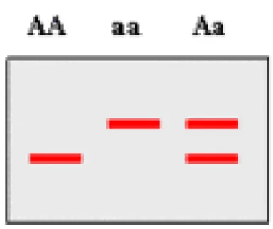
A polymorphism of restriction (Restriction Fragment Length Polymorphism, RFLP) is defined as a SNP located in a short DNA sequence that is recognized and subsequently cleaved by a specific restriction enzyme. The change of one base within the restriction site sequence prevents recognition by the enzyme that, therefore, fails to cut the nucleotide sequence. The enzyme must be chosen so that the recognition site is recognized and cut only if it contains a certain sequence. The identification of alleles can be done by electrophoretic separation on agarose gel according to the size of the fragments produced following by restriction enzyme digestion.
The method consists of an enzymatic digestion of a DNA fragment containing the polymorphic site, previously amplified by PCR. If the individual analyzed carries a homozygous genotype for a SNP that eliminates a restriction
site, the enzyme will not be able to cut and gel electrophoresis will show a single band of the same size of the amplified fragment. On the contrary, if the individual is homozygous for an allele that maintains a cleavage site, two bands will be observed on the gel indicating that the enzyme has cut the amplified fragment. In heterozygous individual in whom both alleles are present, three bands can be observed on the gel, one of the same size of the amplified fragment and two of smaller sizes for the digested fragments (Figure 6).
Figure 6. Possibile RFLP data.
The choice of the primers to amplify the regions containing the different polymorphisms, should be performed so that the restriction site is not exactly in the middle of the amplicon in order to obtain two fragments sufficiently different to be discriminated as two distinct bands on agarose gel.
There are several parameters potentially influencing the restriction enzyme reaction: (1) DNA substrate (quantity, quality), (2) optimum reaction buffer (pH, salts, cofactors), (3) optimum reaction temperature, (4) enzyme (quantity, alterations to the specificity of cut), (5) response time, (6) volume of reaction.
3.6 NGAL/P121S, NGAL/T124M POLYMORPHISM ANALYSIS BY PCR-RFLP
To perform the analysis of polymorphisms of NGAL, the design of primers (synthesized by MWG http://www.mwg-biotech.com/) was performed by use of the program Primer3 Output, referring to the sequences in the public database GenBank..
For each gene the SNP was identified on the nucleotide sequence and has been identified the most suitable pair of primers.
The two SNPs to be analyzed are located upstream of the 5'; they are separated by few basis,so the primers were chosen to amplify a long fragment which contains both polymorphisms (Figure 7).
Figure 7. Long fragment to be amplified with primers highlighted (in blue) and SNPs (in red).
The primers were tested using Oligo primer toolkit. The specificity of the sequence was controlled at http://www.ncbi.nlm.nih.gov:80/blast/Blast.cgi.
To determine the optimum annealing temperature we used a Gradient PCR. All samples were amplified with a Eppendorf Mastercycler Gradient.
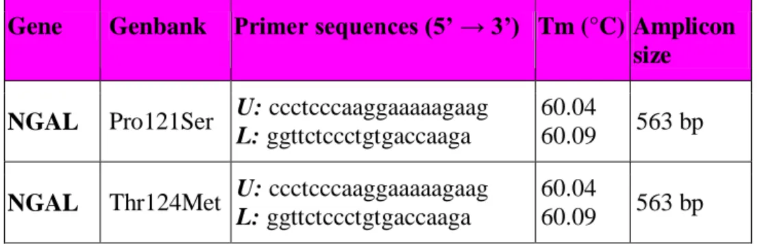
The primer pairs used for each gene and their characteristics are shown in Table 7.
Table 7. Schematic summary of the primers used, the Tm and the size of the amplicon.
Gene Genbank Primer sequences (5’ → 3’) Tm (°C) Amplicon size NGAL Pro121Ser U: ccctcccaaggaaaaagaag
L: ggttctccctgtgaccaaga
60.04
60.09 563 bp NGAL Thr124Met U: ccctcccaaggaaaaagaag
L: ggttctccctgtgaccaaga
60.04
60.09 563 bp
The amplifications were performed in reaction volumes of 25 μL using the enzyme AmpliTaq Gold DNA polymerase (Applied Biosystems).
The reaction mixtures are described in detail below: − 2.5 mL 10x Buffer to a final concentration of 1X; − 0.2 mM dNTPs;
− 6.25 pmol of each primer;
− 0.5 U AmpliTaq Gold DNA Polymerase; − 3.0 mM MgCl2;
− 150 ng of genomic DNA;
− DNase RNase free distilled water to a final volume of 25 mL. The thermal profile consisted of the following steps:
1. Initial denaturation of the templates at 94°C for 7 minutes. 2. Denaturation of the strands at 94°C for 30 seconds.
3. Annealing of the primers at 62°C for 30 seconds. 4. Extension at 72°C for 30 seconds.
Phases 2, 3 and 4 were repeated for a total of 35 cycles 5. Final extension at 72°C for 10 minutes.
6. Block of the reaction at 4°C.
Aliquots of 4.5 μL of the PCR products were loaded on 1.5% agarose gel to estimate the amount of DNA present and its quality (Figure 8).
Figure 8. Agarose gel electrophoresis of amplified NGAL fragment.
Then 5 μL of the amplicons were subjected to digestion with the appropriate restriction enzyme to identify the genotype. Restriction enzymes were chosen with the NEBcutter web-based software tool (http://tools.neb.com/NEBcutter2/index.php) for identifying endonucleases applicable for the genotyping of the 2 SNPs.
Eco 91I (Bste II) (Fermentas Life Sciences) cloned from Escherichia coli RFL91 was used for NGAL Pro121Ser polymorphism, this enzyme works under optimal conditions at 37°C and is inactivated at 65°C.
NGAL Thr124Met polymorphism was analyzed using the Vsp I (Ase I) (Fermentas Life Sciences) cloned from Vibrio species, which has an optimum temperature of 37°C and is inactivated at 65°C.
The restriction sites of enzymes and the dimension of the fragment generated are reported in the Table 8.
Table 8. Schematic summary of the enzymes used, with the corresponding restriction sites and fragments generated.
Gene SNP Amplicon
(bp)
Restriction enzymes
Restriction sites Fragment
size (bp)
NGAL C/T 563 Bste II 5’…G G T N A C C...3’
3’ …C C A N T G G... 5’
T 563 C 284 -279
NGAL C/T 563 Ase I 5’ ...A T T A A T... 3´
3’ ... T A A T T A... 5’
T 563 C 294 -269
The protocols of digestion were as follows: − 5 µL PCR reaction misture
− 1 µL 10 X Buffer 0 − 1 µL Eco91I
− 8 μL Nucleare-free water to a final volume of 15 μl
The reaction was made to be 37°C for 3 hours, followed by 20 minutes at 65°C to inactivate the enzyme.
3.7 STATISTICAL ANALYSIS
Descriptive data are presented as mean ± SD or number and percentage, and biomarker levels are expressed as median and range. The normal distribution of each continuous variable was verified by the Kolmogorov-Smirnov test.
Descriptive analysis was performed by routine tests. The data distribution was represented with Box and Whisker plots and was compared using nonparametric tests (Mann Whitney and Kruskal Wallis test). The level of significance in all cases was set at p <0.05.
Statistical analyses were carried out using SAS 9.1 (SAS Institute, Cary, NC).