R E S E A R C H
Open Access
Elaboration of a nomogram to predict
nonsentinel node status in breast cancer
patients with positive sentinel node,
intraoperatively assessed with one step
nucleic amplification: Retrospective and
validation phase
Franco Di Filippo
1*, Simona Di Filippo
2, Anna Maria Ferrari
3, Raffaele Antonetti
4, Alessandro Battaglia
5,
Francesca Becherini
6, Laia Bernet
7, Renzo Boldorini
8, Catherine Bouteille
9, Simonetta Buglioni
1, Paolo Burelli
6,
Rafael Cano
10, Vincenzo Canzonieri
11, Pierluigi Chiodera
12, Alfredo Cirilli
13, Luigi Coppola
14, Stefano Drago
14,
Luca Di Tommaso
15, Privato Fenaroli
16, Roberto Franchini
17, Andrea Gianatti
16, Diana Giannarelli
1,
Carmela Giardina
18, Florence Godey
19, Massimo M. Grassi
20, Giuseppe B. Grassi
14, Siobhan Laws
21,
Samuele Massarut
11, Giuseppe Naccarato
22, Maria Iole Natalicchio
23, Sergio Orefice
24, Fabrizio Palmieri
25,
Tiziana Perin
11, Manuela Roncella
26, Massimo G. Roncalli
27, Antonio Rulli
28, Angelo Sidoni
28, Corrado Tinterri
27,
Maria C. Truglia
29and Isabella Sperduti
1Abstract
Background: Tumor-positive sentinel lymph node (SLN) biopsy results in a risk of non sentinel node metastases in micro- and macro-metastases ranging from 20 to 50%, respectively. Therefore, most patients underwent unnecessary axillary lymph node dissections. We have previously developed a mathematical model for predicting patient-specific risk of non sentinel node (NSN) metastases based on 2460 patients. The study reports the results of the validation phase where a total of 1945 patients were enrolled, aimed at identifying a tool that gives the possibility to the surgeon to choose intraoperatively whether to perform or not axillary lymph node dissection (ALND).
Methods: The following parameters were recorded: Clinical: hospital, age, medical record number; Bio
pathological: Tumor (T) size stratified in quartiles, grading (G), histologic type, lymphatic/vascular invasion (LVI), ER-PR status, Ki 67, molecular classification (Luminal A, Luminal B, HER-2 Like, Triple negative); Sentinel and non-sentinel node related: Number of NSNs removed, number of positive NSNs, cytokeratin 19 (CK19) mRNA copy number of positive sentinel nodes stratified in quartiles. A total of 1945 patients were included in the database. All patient data were provided by the authors of this paper.
(Continued on next page)
* Correspondence:[email protected];[email protected]
1Regina Elena National Cancer Institute, Via Elio Chianesi 53, 00144 Rome, Italy
Full list of author information is available at the end of the article
© The Author(s). 2016 Open Access This article is distributed under the terms of the Creative Commons Attribution 4.0 International License (http://creativecommons.org/licenses/by/4.0/), which permits unrestricted use, distribution, and reproduction in any medium, provided you give appropriate credit to the original author(s) and the source, provide a link to the Creative Commons license, and indicate if changes were made. The Creative Commons Public Domain Dedication waiver (http://creativecommons.org/publicdomain/zero/1.0/) applies to the data made available in this article, unless otherwise stated.
(Continued from previous page)
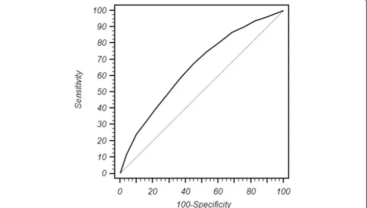
Results: The discrimination of the model quantified with the area under the receiver operating characteristics (ROC) curve (AUC), was 0.65 and 0.71 in the validation and retrospective phase, respectively. The calibration determines the distance between predicted outcome and actual outcome. The mean difference between predicted/observed was 2.3 and 6.3% in the retrospective and in the validation phase, respectively. The two values are quite similar and as a result we can conclude that the nomogram effectiveness was validated. Moreover, the ROC curve identified in the risk category of 31% of positive NSNs, the best compromise between false negative and positive rates i.e. when ALND is unnecessary (<31%) or recommended (>31%).
Conclusions: The results of the study confirm that OSNA nomogram may help surgeons make an intraoperative decision on whether to perform ALND or not in case of positive sentinel nodes, and the patient to accept this decision based on a reliable estimation on the true percentage of NSN involvement. The use of this nomogram achieves two main gools: 1) the choice of the right treatment during the operation, 2) to avoid for the patient a second surgery procedure.
Keywords: Nomogram, Non Sentinel Node status, OSNA method, CK19 mRNA number copies
Background
In the treatment of breast cancer patients sentinel lymph node (SLN) biopsy is a highly accurate predictor of overall axillary status. It has become the standard axillary staging method for the last 15 years in breast cancer (BC) patients who are confirmed clinically negative for lymph node me-tastases [1, 2]. In the case of negative SLN, patients can safely avoid axillary lymph node dissection (ALND), thus preventing associated morbidity [3]. However, approxi-mately 50–70% of patients with positive SLN have no add-itional positive nodes, suggesting that it may be possible to avoid ALND in selected patients [4, 5]. Taking these con-siderations into account, an accurate estimate of the likeli-hood of additional node metastases may be of paramount importance when deciding further treatment. At present, the intra-operative decision on, whether to perform ALND or not, is often only based on the positivity of the SLN. In order to assess the SLN status more rapidly, a semi-automated molecular method called the one step nucleic acid amplification (OSNA) assay has recently been made available [6, 7]. As a matter of fact this method is able to assess the entire SLN in thirty minutes. On the basis of these considerations, the European OSNA Committee de-cided to develop a new nomogram able to predict the non sentinel node (NSN) status, aimed at identifying a tool that gives the possibility to the surgeon to choose intraoperativ-ily whether to perform or not axillary lymph node dissec-tion (ALND). A total of 2460 patients were enrolled in the retrospective phase of the nomogram elaboration. The multivariate analysis demonstrated that only the number of CK19 copies (p < 0.0001) and T size (p < 0.0001) were associated with the NSN metastases. Therefore, a nomo-gram was developed using these two parameters stratified in quartiles. The score of each of the two variables summed and reported in on the total raw score immedi-ately below the percentage of NSN positivity is identified [8]. The aim of the study was to report the results of the
validation phase comprising a total of 1495 enrolled pa-tients (the retrospective phase was already published). The study was conducted with the support of 22 European centers that did not requested any financial support.
Methods
Patients’ population
The European OSNA Users Committee decided to carry out the validation phase of the nomogram project with the following aims: To verify the effectiveness of the nomo-gram to help surgeons in deciding whether to carry out ALND in case of positive SLN; to identify patients at very low risk of positive NSNs in which ALND may be avoided. Our study population only included cases that fulfilled the following criteria: primary invasive cT1-3 BC with clinically and radiological (preoperative sonogram) negative axilla; no prior systemic treatment, or axillary surgery; successful SLN biopsy in which metastatic disease was identified by OSNA; and ALND with at least 10 nodes examined. The following parameters were recorded: Clinical: hospital, age, medical record number; Bio-pathological: tumor size strati-fied in quartiles, grading, multifocality, histological type, LVI, ER-PR status, HER-2, ki67, molecular classification (luminal A, luminal B, HER2 like, triple negative); SLN and NSN related: number of removed SLNs, number of posi-tive and negaposi-tive SLNs, copy number of posiposi-tive SLNs. A total of 2460 patients were included in the database in the retrospective phase. Seventeen European centers contrib-uted in the retrospective enrollment of patients in the val-idation phase up to a total of 1495 patients.
The biopathological parameters and the characteristics of SN and NSN are shown in Tables 1 and 2.
LVI was excluded because the aim of the nomogram is to give the possibility to the surgeon to intra operatively establish whether to perform ALND or not and this par-ameter cannot be assessed reliably in the preoperative breast cancer biopsy.
Sentinel Lymph Node (SLN) sampling method
SLNs were identified using technetium 99 m- labeled, nanosized, human serum albumin colloids. To avoid any contamination during tumor manipulation, SLNs were surgically excised before breast surgery and sent on ice to
the Pathology Department. Each SLN was weighed and measured. SLNs weighing less than 50 mg were excluded from the study. SLNs weighing more than 600 mg were cut in two or more pieces and processed as separate nodes. The weight of lymph node for homogenization should be within a range of 50/600 mg. If the weight of the lymph node is either above or below this specified range accurate results may not been obtained.
One Step Nucleic Acid Amplification (OSNA)
The OSNA assay was performed according to the manu-facturer’s instructions (Sysmex, Kobe, Japan). In short, the SLN was homogenized in 4 ml of the LINORHAG homogenizing buffer (Sysmex) on ice. A small aliquot was used for automated real-time amplification of CK19 mRNA via reverse transcription loop-mediated isother-mal amplification (RT-LAMP) with the ready-to use LYNOAMP reagent kit (Sysmex) on the RD-100i (Sys-mex). It was possible to analyze up to 4 SLNs in one run. The degree of amplification was detected via a bypro-duct of the reaction, i.e. magnesium-pyrophosphate. After use, the excess lysate was stored at minus 80 °C. A lysate with CK19 mRNA copy number/μl less than 250 (a) was regarded as negative (score−); from 250 to 5000 (b) as positive (score +), and greater than 5000 (c) (score ++). The OSNA results were immediately com-municated to the surgeon by telephone within 30– 40 min. For statistical analysis, in case of two or more SLNs, the SLN with the greatest CK19 mRNA copies was chosen. When there was a positive OSNA result, both for micro-metastases (+) and macro-metastases (++), the patients underwent an immediate ALND. ITCs are not detected by the OSNA method. This is not a limita-tion because patients with positive SLNs for ITC are no longer referred to undergo ALND.
Axillary NSNs were routinely examined by H&E.
Statistical method
The outcome of our nomogram was the presence of positive nodes in the axillary dissections following
Table 1 Clinicopathological characteristics of patients
Characteristics N of patients Percent
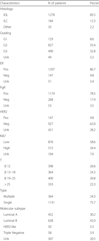
Histology IDL 1278 85.5 ILC 184 12.3 Other 33 2.2 Grading G1 129 8.6 G2 827 55.4 G3 490 32.8 Unk 49 3.2 ER Pos 1297 86.7 Neg 147 9.8 Unk 51 3.4 PgR Pos 1174 78.5 Neg 268 17.9 Unk 53 3.5 HER2 Pos 147 9.8 Neg 927 62.0 Unk 421 28.2 Ki67 Low 876 58.6 High 515 34.4 Unk 104 7.0 T ≤ 12 398 26.6 ≥ 13–18 364 24.3 ≥ 19–25 400 26.8 > 25 333 22.3 Type Multiple 364 24.3 Single 1131 75.7 Molecular subtype Luminal A 452 30.2 Luminal B 628 42.0 HER2-like 50 3.3 Triple Negative 58 3.9 Unk 307 20.5
Table 2 Characteristics of non sentinel node and sentinel node
Number Percent
NSLNs Examined
Median (range) 15 (11–52)
N° of positive NSLNs 610 40.8
Median (range) 2 (1–41)
N° of Copies (Highest copy number)
≤ 1500 305 20.4
> 1500–12,000 329 22.0
> 12,000–111,000 460 30.8
OSNA evaluation in the population defined above. In order to validate the retrospective phase of the nomo-gram we have considered the covariates that predicted this outcome in the previous published paper [8], thus the endpoint was a binary outcome (presence versus ab-sence of at least one positive node other than SLN) and the association with the covariates was analyzed using a logistic linear model. Discrimination ability was assessed by ROC analysis and predictive accuracy was measured by the AUC reported with its 95% confidence interval. Calibration was evaluated by reviewing the plot of pre-dicted probabilities versus the actual probabilities. Well calibrated models have a linear relationship with a slope of 1 and an intercept of 0. Thus, a linear regression coefficient between predicted and observed values was estimated. The resulting model will be validated in a prospective series. All the analyses were performed using IBM SPSS version n. 20 [9].
Ethical consideration
Patient data was anonymously gathered retrospectively with no influence on patient therapy. The Nomogram project was approved by the Ethics Committee of each participating institute.
Results
Table 1 shows the clinical and bio-pathological charac-teristics of the patients. The mean and median ages were
55 and 54, respectively and standard deviation was 13 and range 24–80 years. The vast majority of the patients were affected with infiltrating ductal carcinoma (85.5%). Most of them had an intermediate (55.4%) or high grade tumors (32.8%). Both Estrogen (ER) and Progesterone (PgR) receptors were positive in 86.7 and 78.5%, respect-ively, whereas HER2 was positive only in 9.8% of the pa-tients. Ki67 was high in 34.4% and LVI was present in 24.2% of the patients. These parameters represent the new molecular classifications of breast cancer that not only allow to identify patients at a higher risk of relapse but may also guide postoperative therapies [10, 11]. Tumor size was divided in quartiles, the cut-offs being 12, 18 and 25 mm. The mean and median tumor sizes were 20.5 and 18.2 mm, respectively, ranging between 0.7 and 50 mm. The SLNs and NSLNs characteristics are reported in Table 2. The median number of NSNs removed with ALND is 15 (range11–52). The number of positive NSNs was 610 (40.8%), the median value was 2 (range 1–41). The number of CK19 mRNA was divided in quartiles in order to obtain a better stratification of the patients. In order to validate the nomogram, we eval-uated the discrimination of the model. This parameter which was quantified with the area under receiver oper-ating characteristic (ROC) curve was 0.65 (95% C.I. 0.63–0.69). Figure 1 shows the ROC curve of the valid-ation phase, the values are quite similar being 0.71 in the retrospective phase and 0.65 in the validation phase,
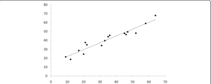
demonstrating a fair level of discrimination. Another parameter that is usually employed to evaluate the reli-ability of a nomogram is the calibration, that is shown in Fig. 2. The calibration determines the distance between predicted outcome and actual outcome. The mean difference between predicted/observed was 2.3 and 6.3% in the retrospective and in the validation phase, respectively. The two values are quite similar, and consequently we can conclude that the nomogram effectiveness was validated [8].
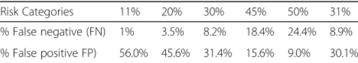
It is well known that SLN micro- and macro-metastases are associated with a mean NSN positivity rate of 20 and 50%, respectively. Consequently, the dilemma for surgeons still persists in how to avoid unnecessary ALND and how to identify patients at high risk of positive NSNs in which ALND is recommended. Given that the validation phase had been evaluated suc-cessfully, all the patients enrolled in the retrospective and validation phase (3955 patients were valuable for the nomogram) were evaluated to develop a tool that allows the surgeon to intraoperatively make a decision on whether to perform ALND in case of positive SLN or not. The nomogram validated the risk percentage of NSN positivity that we recently published [8]. In Table 3, we stratified the patients in risk categories, according the nomogram model, from 11 to 50% and for each risk category we calculated the percentage of false negative and false positive rates in order to identify patients in which ALND is unnecessary and those where ALND is recommended. Moreover, the ROC curve identified in the risk category of 31%, the best compromise between false negative and positive rates. Therefore, in patients below this cut-off ALND may be omitted, for values higher than 31% ALND is recommended.
Discussion
Usually the effectiveness of a nomogram is evaluated with three parameters i.e., discrimination, calibration and the capacity of a nomogram to identify false negatives i.e. pa-tients with a risk of NSN metastases ≤10% in which ALND may be omitted. Discrimination (i.e. whether the relative ranking of individual prediction is in the correct order) was quantified with the area under curve the re-ceiver operating characteristics (ROC) curve (AUC). The AUC is a summary measure of the ROC that reflects the ability of a test to discriminate between a diseased and non-diseased subject across all the possible levels of posi-tivity. AUC ranges from 0 to 1, with 1 indicating perfect concordance, 0.5 indicating no better concordance than “flip the coin”, and indicating perfect discordance.
In our nomogram, the AUCs are 0.71 and 0.65, re-spectively that are considered a fair value of discrimin-ation consistent with the best nomograms published so far. This data has been confirmed by a recent publica-tion by Van Den Hoven who reported a“Head to Head” comparison of nine predictive tools [12]. The majority of nomograms include tumor size, lymphovascular inva-sion, and the size of the SLN metastases. This is very consistent with our predictive tools in which the multi-variate analysis selected T-size, number of mRNA copies in the SLN (i.e.) and tumor load. The comparison of nine predictive tools showed that the MSKCC nomo-gram had best discrimination with an AUC of 0.69, followed by the Stanford, Mayo and MOU models with AUC’s of 0.66, 0.65 and 0.65, respectively. The Stanford model has second best discrimination (AUC 0.66) and the Mayo and MOU models are tied for third (AUC 0.65). These data confirm that the AUC’s of both retro-spective and validation phases are perfectly consistent
Fig. 2 The model performs well and correctly at low and in high risk as shown in calibration plot. The linear regression model has a slope of 0.96 (95% C.I. 0.98/1.40) and a constant of -13.8 between predicted and actual probabilities (95%C.I. -22.9/-4.85)
with the best nomograms published so far. Calibration determines how far the predicted probabilities are from the actual outcomes, that has a higher clinical signifi-cance than discrimination. Recently, Coutant evaluated the AUC and calibration of 9 previously published predicted models [13]. Coutant found that two of the nomograms were well calibrated, whereas the other two showed differences between predicted and observed probabilities. It was also outlined that the difference be-tween the predicted and observed probabilities for these nomograms range from 3 to 25%. In our nomogram, the values were 2.3 and 6.3% for retrospective and validation phase, respectively. Therefore, they belong to the cat-egory of low values of the above mentioned range and can be considered reliable. This information is of clinical utility because it gives clinicians the opportunity to in-form patients about the predicted probability of NSN metastases. As far as the false negative rate is concerned, we stratified our patients in risk categories in a range from 11 to 50% and the percentage of false negative and positive rates was calculated for each risk category (Table 3). It is readily apparent that up to risk category of 30% the false negative rate is 8.2% that is in the range value of the false negative rate reported for SLNB tech-nique. Therefore, up to this category surgeons may feel comfortable in suggesting patients to not undergo ALND and patients accepting this decision. It is worth considering that nomograms safely avoided ALNDs in 1254 (32%). We used the ROC curve analysis to calcu-late best level of risk category in terms of balance be-tween false positive and negative rates. The ROC curve identified a risk category of 31% as the optimal cut-off that the surgeon may employ in the decision-making process on whether ALND may be omitted (<31%) or recommended (>31%). This value was calculated taking into account the sensitivity and specificity of the cut-off. If we would have chosen other cut-offs this would result in a decrease of sensitivity or specificity, therefore this topic has to be discussed when counseling the patients. In fact in the risk category of 30% the false negative rate is 8.2% which is acceptable. These considerations are to be considered valid because in the risk categories >31% we verified patients with a percentage of positive NSNs of 60.2%, 63% (>45%) and 66.5% (>50%). Moreover, in the vast majority of these patients there were more than 3 positive NSNs. In our previous paper we evaluated the capacity of the nomogram in identifying how many false
negative patients were in the risk category of 10% only because this parameter (together wih discrimination and calibration) is employed to evaluate the reliability of the nomogram, as suggested by Coutant [13].
Recently, American Society of Clinical Oncology (ASCO) guidelines for SLNB and ALND have been pub-lished, indicating that patients with micro- and macro-metastases may avoid ALND based on the results of IBCSG 23.01 and Z0011 trials [14, 15]. In the prospect-ive randomized IBCSG23-01 trial, only those patients with micro-metastases were randomized to either ALND or no further treatment in patients with positive SLN. The results of this study showed no differences between the two arms both in terms of disease-free and of overall survival. Some challenges, however, still exist regarding this study. Patients accrual stopped prematurely and only 933 out of 1960 patients were enrolled, therefore the study was underpowered. The patient population had a very good prognosis. In fact, sentinel tumor size ≤1 mm was present in 69% of the patients. As a result, the incidence of additional positive NSN in axillary dis-section group was 13%, very similar to that found in case of ITCs metastases in SLN. This is also because the au-thor included ITCs in the group of micro-metastases. A strict correlation between the size of micro-metastases (less or greater than 1 mm) and positive NSNs was clearly demonstrated by Rahusen and Viale, respectively [16, 17]. Their results confirm that the presence of 69% of patients with SLN micro-metastases ≤1 mm greatly biases the interpretation in clinical practice. The median follow-up of 5-years is too short to assess the long term incidence of axillary recurrence in this study of this good prognosis group. In NSABP-B6, 20% of nodal recur-rences after lumpectomy and 24% of nodal recurrecur-rences after ALND and radiotherapy occurred after 5 years [18]. In the IBCSG 23-01 study, the trialists reported that 6681 patients were registered before surgery of which 934 patients were randomized, indicating that only 14% of eligible breast cancer patients met the inclusion criteria for this study and underwent randomization. We anticipate that this may be due to both node negative patients and also patients with mul-tiple positive nodes and other factors, however the breakdown is unknown. The IBCSG 23-01 data supports omission of ALND for the selected group of patients with small, ER+ tumors undergoing breast conservation with planned whole breast radiation. Omitting ALND in SLN positive mastectomy patients and patients undergo-ing partial breast irradiation requires further investiga-tion. If the primary benefit to these patients primarily through systemic adjuvant therapy and not loco-regional therapy, based on favorable tumor biology, this would seem like the next most logical step. However, data from the NSABP B-32 suggest a statistically significant
Table 3 Percentage of FN and FP rates according to value risk categories are reported
Risk Categories 11% 20% 30% 45% 50% 31%
% False negative (FN) 1% 3.5% 8.2% 18.4% 24.4% 8.9%
survival disadvantage after a median follow-up of 8 years for a subset of 611 women with occult nodal disease [19]. Completion axillary dissection had no bearing on this effect and axillary recurrences were equivalent. The 5 year overall survival was 94.6% versus 95.8%, the 5 year disease-free survival was 86.4% versus 89.2% and the 5 year distant disease-free survival was 89.7% versus 92.5%, respectively, all P < 0.05. The 8 year median follow-up of the B-32 study is longer than that reported for IBCSG 23-01. It is important to counsel patients that the long-term outcomes of SLN biopsy alone for micro-metastases or ITCs disease are unknown. It is conceiv-able that based on the IBCSG 23-01 study, patients with favorable tumor characteristics (low T-size, ER+, post menopausal women with low tumor burden in the SLN are potential candidates for limited axillary surgery. In these patients, SLN biopsy can be regarded as a “super selective therapeutic ALND”.
Recently, the recommendation by the ASCO update committee that ALNDs can be safely avoided in patients with one or two SLN metastases undergoing breast con-serving surgery with conventional whole-breast radio-therapy (RT) is premature as it is based only on the results of the American College of Surgeons Oncology Group Z11 trial [14, 20]. The shortfalls include the following: recruitment rates were poor (50% of original target); patients recruited into the study had generally low-risk cancers; axillary recurrence was not the prede-fined primary trial end point; approximately 50% of patients had micro-metastases; a significant proportion of patients had unknown nodal disease. The two groups had slight inequalities in several prognostic characteristics (T stage, grade, lymph-vascular invasion), all favoring the SLN group. Moreover, micro-metastatic-only node disease was present in a statistically significant higher percentage of patients in the SLN group (44.8% v 37.5%). A high proportion of patients were lost to follow-up (21% in the ALND group and 17% in the SLN group); there was a significant amount of missing data, and there was no prospective RT quality assurance program to mitigate any bias in RT target volume definition. Recently, Goyal outlined that the most critical issue concerning the generalization of trial is that too many patients with can-cers who could have met the eligibility criteria were not represented in the cohort of patients in the trial [21]. Ultimately, the American Society of Clinical Oncology’s recommendation that ALND can be avoided in patients with one or two SLN macrometastases, reflecting the eli-gibility criteria of Z11, is based on a comparison of 228 patients versus 202 patients which falls significantly short of persuasive based evidence [21]. The perception that the Z0011 trial has not completely convinced the oncological community is demonstrated by the fact that additional trials are still ongoing like POSNOC (Positive
Sentinel Node: Adjuvant Therapy Alone Versus Adju-vant Therapy Plus Clearance or Axillary Radiotherapy) trial and the Italian SINODAR-ONE trial that compares SLNB vs. ALND in T1-T2 patients with positive SLN macro-metastases [22]. In regards to the post ACOSOG Z0011 era, another topic still needs to be clarified. Does our new understanding of breast cancer really change clinical practice? Recently, Guth has assessed the poten-tial impact of Z0011 on clinical practice by testing the applicability of its criteria to a European patient popula-tion [23]. The author concluded that “the application of Z0011 led to the omission of completing ALND in less than 10% of all SLNB procedures (<6% of all surgically treated BC patients); therefore, we do not think that the perception of Z0011 as“practice-changing” is justified”. In a recent paper, the Results from the Breast Surg ANZ National Breast Cancer Audit database have been ques-tioned for women treated between 2005 and 2010 who would have met the entry criteria for the Z0011 Trial [24]. A total of 64,883 of breast cancer cases were eli-gible for analysis. 22,731 underwent breast conserving surgery and sentinel node biopsy for invasive breast can-cer. A total of 4482 cases (6.9%) fulfilled the criteria for Z-11 Trial. These data seem to point out that many patients do not fulfill the inclusion criteria for Z0011, therefore the nomogram application is still relevant in clinical practice. Other reports also have shown that many patients evaluated with breast cancer may not meet the defined eligibility criteria for avoidance of ALND in the presence of a positive SLN [25, 26]. Rea-sons may include tumor size, tumor biology, extra-nodal disease, patients undergoing primary chemotherapy, selection of mastectomy, patients treated with PBI or desire to avoid adjuvant breast radiation after breast conserving surgery. Moreover, there are considerations concerning clinical and pathologic subtypes that are less clearly defined in Z0011 trial. For example, patients with lobular histology represented only 7% of the trial popu-lation, consequently limiting an accurate analysis of patients with this histologic subtype. Invasive lobular tumors are more likely to have isolated tumor cells in the SLN, reflecting the non-cohesive cellular characteris-tics that often require IHC detection. [27] and are more likely to have clinical and radiological underestimation of disease burden]. Small-volume nodal disease may have clinical relevance in this patient population unlike those patients with invasive ductal histology. Consider-ation is therefore given to this difference in biology when we are counseling patients with invasive lobular carcin-oma and a positive SLN in performing ALND. Another important factor when making treatment decisions is patient age. Patients older than 18 years of age were eligible to enroll in Z0011. However, the median age of study participants was 54 years in the SLND group and
56 years in the ALND group with more than 62% of pa-tients in each group being older than 50 years. Patient age <50 years was one of only two factors (higher Bloom Richardson grade) associated with local-regional recur-rence on the multivariable analysis. There may have been reluctance from the surgeons towards randomizing younger patients with node-positive disease to the SLND-only group and, as a result fewer patients were in-cluded in the study population. All these considerations lead to the conclusion that most patients may benefit from OSNA nomograms in the decision-making process on whether to perform ALND or not. At this point, we must consider which OSNA nomogram may have an im-pact on clinical practice, in other words how many pa-tients with SLN are assessed with OSNA. To the best of our knowledge, there are more than 6000 patients that undergo OSNA positive SLN assessments each year in Europe. Therefore, this certainly justifies the develop-ment of the OSNA nomogram that has recently been validated. Moreover, nomogram tools have been shown to decrease the rate of completing axillary dissections in a subset of women with more favorable tumor factors with only a marginally higher recurrence rate (2% vs. 0.4% at 23–30 months) [28, 29].
As a surgeon, it is also important to realize that al-though it may be safe to avoid ALND in an ideal setting in which both adjuvant radiation and systemic therapies are given, in reality not all patients do or plan to complete all the recommended adjuvant therapies, including oral therapies such as tamoxifen, due to the perceived or actual side-effects of these treatments. Further study is needed to improve our understanding of breast tumor biology in order to identify those patients for whom less extensive surgery will not compromise long-term oncologic out-comes. In the mean time, patient counseling for options on low volume axillary disease management should address exactly what data we currently have and what re-mains unknown. In this context, OSNA nomograms may help surgeons in counseling patients on whether to per-form ALND or not and aid patients to accept this decision based on the reliable estimation of the percentage of NSN involvement. Therefore the use of this nomogram achieves two main goals: 1) the choice of the right treatment during the operation, 2) to avoid for the patient a second surgery procedure. The above major results have to be validated in a prospective validation study that is already ongoing.
Abbreviations
ALND:Axillary lymph node dissection; ASCO: American Society of Clinical Oncology; BC: Breast cancer; ER: Estrogen receptor; LVI: Lymphatic/vascular invasion; NSN: Non sentinel node; OSNA: One step nucleic acid amplification; PgR: Progesterone receptor; ROC: Receiver operating characteristics; RT-LAMP: Reverse transcription loop-mediated isothermal amplification; SLN: Sentinel lymph node
Acknowledgement
We thank Miss Tania Merlino for English translation and editing and Monika Zaenkert for the logistic support.
Funding Not applicable.
Availability of data and material
All data generated or analysed during this study are included in this published article.
Authors’ contributions
Conceived and designed the study: FDF, DG, RC, LDT, CB, FG; statistical analysis elaboration: IS, DG; collected patients’ information: SDF, AMF, RA, AB, FB, LB, RB, SB, PB, VC, PC, AC, LC, SD, PF, RF, AG, CG, MG, GBG, SM, GN, MIN, SO, FP, TP, MR, MGR, AR, AS, CT, MCT; wrote and revised the manuscript: FDF, RC, LDT and LS. All authors read and approved the final manuscript.
Competing interests
The authors declare that they have no competing interests. Consent for publication
Not applicable.
Ethical approval and consent to participate
Ethical approval was given by the medical Ethics Committees of each participating centers.
Author details
1Regina Elena National Cancer Institute, Via Elio Chianesi 53, 00144 Rome, Italy.2Ospedale di Latina, Latina, Italy.3San Camillo, Milan, Italy.4Az. Ospedaliera Universitaria Foggia, Foggia, Italy.5ASL, Prato, Italy.6ULSS 7 Pieve di Soligo, Pieve di Soligo, Italy.7Hospital Lluís Alcanyís, Xàtiva, Spain. 8Università of Piemonte, Vercelli, Italy.9Hôpitaux de Lyon, Lyon, France. 10
Hospital Universitario de La Ribera, Alzira, Spain.11Centro Regionale Oncologico, Bari, Italy.12San Donato, Italy.13Policlinico of Bari, Bari, Italy. 14
San Filippo Neri, Florence, Italy.15Humanitas Rozzano, Rozzano, Italy.16ASST Papa Giovanni XXIII, Bergamo, Italy.17Azienda Ospedaliera“Maggiore della Carità” di Novara, Novara, Italy.18University of Bari, Bari, Italy.19Eugene Marquis Cancer Center, Rennes, France.20Humanitas Gavazzeni, Bergamo, Italy.21Hampshire Hospitals NHS Foundation Trust, England, UK.22University of Pisa, Pisa, Italy.23Azienda Ospedaliero-Universitaria OO.RR. Foggia, Foggia, Italy.24University of L’Aquila, L’Aquila, Italy.25I.R.C.C.S. L. Spallanzani, Rome, Italy.26Azienda Ospedaliero-Universitaria Pisana, Pisa, Italy.27Humanitas, Pisa, Italy.28University of Perugia, Perugia, Italy.29USL, Prato, Italy.
Received: 4 October 2016 Accepted: 19 November 2016
References
1. Veronesi U, Paganelli G, Viale G, Luini A, Zurrida S, Galimberti V, et al. Sentinel-lymph-node biopsy as a staging procedure in breast cancer: update of a randomised controlled study. Lancet Oncol. 2006;7:983–90. 2. Lyman GH, Giuliano AE, Somerfield MR, Benson 3rd AB, Bodurka DC, et al.
American Society of Clinical Oncology guideline recommendations for sentinel lymph node biopsy in early-stage breast cancer. J Clin Oncol. 2005;23:7703–20.
3. Fleissig A, Fallowfield LJ, Langridge CI, Johnson L, Newcombe RG, Dixon JM, et al. Post-operative arm morbidity and quality of life. Results of the ALMANAC randomised trial comparing sentinel node biopsy with standard axillary treatment in the management of patients with early breast cancer. Breast Cancer Res Treat. 2006;95:279–93.
4. Bolster MJ, Peer PG, Bult P, Thunnissen FB, Schapers RF, Meijer JW, et al. Risk factors for non-sentinel lymph node metastases in patients with breast cancer. The outcome of a multi-institutional study. Ann Surg Oncol. 2007;14:181–9. 5. Fleming FJ, Kavanagh D, Crotty TB, Quinn CM, McDermott EW, O’Higgins N,
et al. Factors affecting metastases to non-sentinel lymph nodes in breast cancer. J Clin Pathol. 2004;57:73–6.
6. Visser M, Jiwa M, Horstman A, Brink AA, Pol RP, van Diest P, et al. Intra-operative rapid diagnostic ethod based on CK19 mRNA expression for the detection of lymph node metastases in breast cancer. Int J Cancer. 2008;122:2562–7.
7. Buglioni S, Di Filippo F, Terrenato I, Casini B, Gallo E, Marandino F, et al. Quantitative molecular analysis of sentinel lymph node may be predictive of axillary node status in breast cancer classified by molecular subtypes. PLoS One. 2013;8:e58823.
8. Di Filippo F, Giannarelli D, Bouteille C, Bernet L, Cano R, Cunnick G, Sapino A. Elaboration of a nomogram to predict non sentinel node status in breast cancer patients with positive sentinel node, intra-operatively assessed with one step nucleic acid amplification method. J Exp Clin Cancer Res. 2015;34:136.
9. Ianosos A, Schrg D, Ray GV, Panageas KS. How to build and interprate a nomogram for cancer program. J Clin Oncol. 2008;16:1364–70.
10. Chen X, Sun L, Cong Y, Zhang T, Lin Q, Meng Q, et al. Baseline staging tests based on molecular subtype is necessary for newly diagnosed breast cancer. J Exp Clin Cancer Res. 2014;33:28.
11. Jiang Z, Guo J, Shen J, Jin M, Xie S, Wang L. The role of estrogen receptor alpha in mediating chemoresistance in breast cancer cells. J Exp Clin Cancer Res. 2012;31:42.
12. van den Hoven I, Kuijt G, Roumen R, Voogd A, Steyerberg EW, Vergouwe Y. A head to head comparison of nine tools predicting non-sentinel lymph node status in sentinel node positive breast cancer women. J Surg Oncol. 2015;112:133–8.
13. Coutant C, Olivier C, Lambaudie E, Fondrinier E, Marchal F, Guillemin F, et al. Comparison of models to predict nonsentinel lymph node status in breast cancer patients with metastatic sentinel lymph nodes: a prospective multicenter study. J Clin Oncol. 2009;27:2800–8.
14. Lyman GH, Temin S, Edge SB, Newman LA, Turner RR, Weaver DL, et al. Sentinel lymph node biopsy for patients with early-stage breast cancer: American Society of Clinical Oncology clinical practice guideline update. J Clin Oncol. 2014;32:1365–83.
15. Galimberti V, Cole BF. Axillary versus sentinel-lymph-node dissection for micrometastatic breast cancer–authors’ reply. Lancet Oncol. 2013;14:e251–2. 16. Rahusen FD, Torrenga H, van Diest PJ, Pijpers R, van der Wall E, Licht J, et al. Predictive factors for metastatic involvement of nonsentinel nodes in patients with breast cancer. Arch Surg. 2001;136:1059e63.
17. Viale G, Maiorano E, Mazzarol G, Zurrida S, Galimberti V, Luini A, et al. Histologic detection and clinical implications of micrometastasis in axillary sentinel lymph nodes for patients with breast carcinoma. Cancer. 2001;92:1378e84. 18. Fisher B, Anderson S, Bryant J, Margolese RG, Deutsch M, Fisher ER, et al.
Twenty-year follow-up of a randomized trial comparing total mastectomy, lumpectomy, and lumpectomy plus irradiation for the treatment of invasive breast cancer. N Engl J Med. 2002;347:1233–41.
19. Weaver DL, Ashikaga T, Krag DN, et al. Effect of occult metastases on survival in node-negative breast cancer. N Engl J Med. 2011;364:412–21.
20. Giuliano AE, Hunt KK, Ballman KV, et al. Axillary dissection vs no axillary dissection in women with invasive breast cancer and sentinel node metastasis: A randomized clinical trial. JAMA. 2011;305:569–75. 21. Goyal A, Dodwell D, Reed MW, Coleman RE. Axillary treatment in women
with one or two sentinel nodes with macrometastases: more evidence is needed to inform practice. J Clin Oncol. 2014;32:3902.
22. ISRCTN Register: A randomized trial of armpit (axilla) treatment for women with early stage breast cancer: POSNOC—POsitive Sentinel NOde: adjuvant therapy alone versus adjuvant therapy plus Clearance or axillary radiotherapy. doi:10.1186/ISRCTN54765244.
23. Güth U, Myrick ME, Viehl CT, Schmid SM, Obermann EC, Weber WP. The post ACOSOG Z0011 era: does our new understanding of breast cancer really change clinical practice? Eur J Surg Oncol. 2012;38:645–50. 24. Ainsworth RK, Kollias J, Le Blanc A, De Silva P. The clinical impact of the
American College of Surgeons Oncology Group Z-0011 trial—results from the BreastSurgANZ National Breast Cancer Audit. Breast. 2013;22:733–5. 25. Delpech Y, Bricou A, Lousquy R, et al. The Exportability of the ACOSOG Z0011 Criteria for Omitting Axillary Lymph Node Dissection After Positive Sentinel Lymph Node Biopsy Findings: A Multicenter Study. Ann Surg Oncol. 2013;20:2556–61.
26. Yi M, Kuerer HM, Mittendorf EA, et al. Impact of the American College of Surgeons Oncology Group Z0011 criteria applied to a contemporary patient population. J Am Coll Surg. 2013;216:105–13.
27. Mittendorf E, et al. Lymphovascular invasion and lobular histology are associated with increased incidence of isolated tumor cells in sentinel lymph nodes from early-stage breast cancer patients. Ann Surg Oncol. 2008;15:3369–77.
28. Park J, Fey JV, Naik AM, et al. A declining rate of completion axillary dissection in sentinel lymphnode positive breast cancer patients is associated with the use of a multivariate nomogram. Ann Surg. 2007;245:462–8.
29. Koca B, Kuru B, Ozen N, Yoruker S, Bek Y. A breast cancer nomogram for prediction of non-sentinel node metastasis– validation of fourteen existing models. Asian Pac J Cancer Prev. 2014;15:1481–9.
• We accept pre-submission inquiries
• Our selector tool helps you to find the most relevant journal
• We provide round the clock customer support
• Convenient online submission
• Thorough peer review
• Inclusion in PubMed and all major indexing services
• Maximum visibility for your research Submit your manuscript at
www.biomedcentral.com/submit