Adult Dorsal Root Ganglia Sensory
Neurons Express the Early Neuronal
Fate Marker Doublecortin
ANNA DELLAROLE AND MARIAGRAZIA GRILLI*
DISCAFF & DBF Center, University of Piemonte Orientale “A. Avogadro”, School of Pharmacy, Novara, 28100 Italy
ABSTRACT
It has been widely accepted that doublecortin (DCX) may represent a neuronal fate marker transiently expressed by immature neurons during development of the central and peripheral nervous tissue and in neurogenic areas of the adult brain. Previous work described the presence of DCX in the developing dorsal root ganglia (DRG), structures of the peripheral nervous system originating from the neural crest, but no information is available on its expression in adulthood. To this purpose, we have performed an immunohistochemical and biochemical analysis for DCX expression in DRG from adult male mice and rats. To our surprise, we demonstrated that the majority of DRG neurons do express DCX, both in somata and in fibers. DCX⫹cells have been characterized morphologically and phenotypically with well-established markers of DRG neuronal subpopulations. A large number of DCX⫹cells belong to the small and medium-sized nociceptive neurons. Additionally, DCX immunoreac-tivity is present in the spinal cord dorsal horns, the projection area of DRG neurons. The novel and unexpected localization for DCX protein opens up new, interesting vistas on the functional role of this protein in mature neurons and in particular in sensory neurons. J. Comp. Neurol. 511:318 –328, 2008. ©2008 Wiley-Liss, Inc.
Indexing terms: doublecortin; dorsal root ganglia; sensory neurons; P2X3; isolectin IB4;
substance P
Doublecortin (DCX) is a microtubule-associated protein that is widely expressed by immature neurons during central and peripheral nervous system development (Des Portes et al., 1998; Francis et al., 1999; Gleeson et al., 1999; Hannan et al., 1999; Couillard-Despres, 2001) and is involved in the regulation of migration (Gleeson et al., 1999; Corbo et al., 2002; Bai et al., 2003). Neuroblasts migrating along radial glial processes during cortex lam-ination express indeed high levels of DCX (Francis et al., 1999; Gleeson et al., 1999), and mutations in the DCX gene are associated with disrupted neuroblast migration in a genetically transmitted disease characterized by a smooth and four-layered cortex and known as
lissenceph-aly (Berg et al., 1998; Des Portes et al., 1998; Gleeson et
al., 1998). In addition to its developmental role, DCX expression in the adult brain is restricted mainly to areas where neurogenesis takes place, such as the subventricu-lar zone (SVZ) and the hippocampal dentate gyrus (Lois and Alvarez- Buylla, 1994; Alvarez-Buylla and Garcia-Verdugo, 2002; Van Praag et al., 2002; Kempermann et al., 2003). Based on these observations, DCX expression is widely utilized as a tool for investigating adult
neurogen-esis (Brown et al., 2003; Rao and Shetty, 2004; Couillard-Despres et al., 2005).
Nonetheless, DCX-immunoreactive cells have also been detected in areas of the adult CNS in the apparent ab-sence of neurogenesis (Nacher et al., 2001), but their func-tional significance is largely unknown. It is of interest that, in the adult brain, some DCX⫹cells display morpho-logical features of mature neurons, whereas others resem-ble migrating neuroblasts. These peculiar features have contributed to the hypothesis that some DCX-expressing cells could represent a reservoir of partially
undifferenti-Grant sponsor: Fondazione Cariplo, undifferenti-Grant number: 2005-0634; undifferenti-Grant sponsor: Ministero della Istruzione della Universita` e della Ricerca; Grant number: PRIN 2005; Grant sponsor: Regione Piemonte, Grant number: CIPE 2004.
*Correspondence to: Mariagrazia Grilli, MD, DISCAFF & DBF Center, Via Bovio 6, 28100 Novara, Italy.
E-mail: [email protected]
Received 6 February 2008; Revised 11 June 2008; Accepted 31 July 2008 DOI 10.1002/cne.21845
Published online September 19, 2008 in Wiley InterScience (www. interscience.wiley.com).
ated neurons that under specific circumstances may com-plete their maturation during adult life (Nacher et al., 2001).
A vast array of experimental works has suggested that new neurons could be added to the dorsal root ganglia (DRG) postnatally (Devor and Govrin-Lippmann, 1985; Devor et al., 1991; Cecchini et al., 1995; Popken and Farel, 1997; Ciaroni et al., 2000; Namaka et al., 2001; Farel, 2002; Li et al., 2007), although adult DRG neurogenesis has not been confirmed in vivo (La Forte 1991; Pover et al., 1994; Geuna et al., 2000; Farel, 2003). Moreover, DRG represent areas subjected to high plasticity, enabling them to respond to and recover from injury (Groves et al., 2003; Kuo et al., 2005; Guseva and Chelyshev, 2006). For these reasons, we have investigated the expression of dou-blecortin in rodent DRG. To this purpose, we have per-formed immunohistochemical and biochemical analysis on DRG from 3-month-old male mice and rats. Surprisingly, we demonstrated that the majority of DRG neurons still retain DCX expression during adulthood, both in somata and in fibers. DCX⫹cells have been further characterized morphologically and phenotypically by immunohisto-chemistry with well-established markers of DRG neuronal subpopulations. Large numbers of DCX⫹cells belong to the small to medium-sized nociceptive neurons. Moreover, we demonstrate that DCX is expressed in the spinal cord (SC) dorsal horns, the projection area of DRG neurons, and that, at this level, DCX expression retains the same pattern of colocalization with the neuronal markers ob-served in the DRG.
MATERIALS AND METHODS
Animals
Adult (3-month-old) male CD1 mice (n ⫽ 6) and Sprague-Dawley rats (n ⫽ 3) purchased from Charles River Laboratories (Wilmington, MA) were utilized. All animals were maintained in high-efficiency particulate air (HEPA)-filtered Thoren units (Thoren Caging System) at the University of Piemonte Orientale animal facility and, kept in numbers of three or four per cage, had unlimited access to water and food. Animal treatments were per-formed in accordance with the NIH guidelines and also were reviewed and approved by the local IACUC.
Tissue preparation
Mice and rats were deeply anesthetized with Avertin (400 mg/kg i.p.) and pentobarbital (75 mg/kg i.p.), respec-tively. The animals were perfused transcardially with sa-line, followed by 4% paraformaldehyde (PFA) in 0.1 M phosphate buffer (PB), pH 7.4, at 4°C. Their brains, spinal cords, and DRG were removed, postfixed in 4% PFA, and then cryoprotected in 30% sucrose/PB 0.1 M. The DRG were embedded in OCT, frozen, and cut longitudinally on a cryostat. Consecutive 10-m-thick sections were thaw-mounted on Superfrost slides (Fisher, Hampton, NH) and stored at –20°C until use. Brain and thoracic spinal cord were cut coronally as 40-m-thick sections, collected seri-ally, and stored at –20°C in a cryoprotectant solution (glycerol, ethylene glycol, and 0.2 mM PB, pH 7.4, 1:1:2 by volume) until processing.
Antibodies
The rabbit polyclonal anti-DCX (catalog No. ab18723; lot No. 155505; Abcam, Cambridge, MA) was raised
against a 16-amino-acid synthetic peptide (sequence: YLPLSLDDSDSLGDSM; manufacturer’s technical infor-mation), which corresponds to amino acids 387– 402 of human DCX, 351–366 of mouse DCX, and 350 –365 of rat DCX. According to the manufacturer, when tested by Western blot, this antiserum detects a 40 – 45-kDa band in mouse brain lysates. The rabbit polyclonal anti-DCX (cat-alog No. 4604; lot No. 1; Cell Signaling Technology, Bev-erly, MA) was raised against two synthetic peptides corrisponding to amino acids 48 – 69 (sequence GHFDER-DKTSRNMRGSRMNGLP) and amino acids 380 – 402 (se-quence LRKHKDLYLPLSLDDSDSLGDSM) of human DCX (manufacturer’s technical information). According to the manufacturer, on Western blot, this antiserum recog-nizes one or two bands at⬃45 kDa. Guinea pig polyclonal anti-P2X3 (catalog No. GP10108; Neuromics, Northfield,
MN) was raised against the peptide VEKQSTDSGAY-SIGH corresponding to residues 383–397 of the carboxy-terminus of rat P2X3. This antiserum recognizes a single
band of 57 kDa in Western blot (Fabbretti et al., 2006). Guinea pig polyclonal anti-substance P (catalog No. AB5892; Chemicon, Temecula, CA; 1:1,000) was raised against a peptide corresponding to amino acids 1–11 of rat substance P. Only cells with the classic distribution and morphology are stained with this antibody in the adult DRG (Hwang et al., 2005).
Immunohistochemistry
Cryosections from T10, T11, and T12 DRGs were uti-lized for this study. After rinsing, sections were blocked for 1 hour with 10% normal goat serum in 0.3% Triton X-100 in 0.05 M TBS, pH 7.4, and then incubated in presence of rabbit polyclonal anti-DCX (1:3,000; Abcam) or rabbit polyclonal anti-DCX (1:200; Cell Signaling Tech-nology) overnight at 4°C. On the next day, after several washes, sections were incubated with Alexa 488-conjugated goat anti-rabbit secondary antibody (1:600; Molecular Probes, Eugene, OR). For double labelling with isolectin B4 (IB4), sections were incubated with biotinyl-ated IB4 (1:300; Sigma, St. Louis, MO) overnight at 4°C. Visualization was achieved by incubation in Texas red-labelled streptavidin (1:250; Vector Laboratories, Burlin-game, CA). Double immunofluorescence was performed by incubating sections overnight at 4°C with rabbit poly-clonal anti-DCX Abcam antibody and one of the following antibodies: guinea pig polyclonal anti-P2X3 (1:600) or
guinea pig polyclonal anti-substance P (1:1,000). After washes, sections were incubated in a mixture of Alexa 488-conjugated goat anti-rabbit IgG (1:600; Molecular Probes) and Alexa 633-conjugated goat anti-guinea pig IgG (1:600; Molecular Probes). TBS containing 3% normal goat serum and 0.1% Triton X-100 served as antibody diluent. Double-immunofluorescence labelling of thoracic spinal cord was performed on 40-m free-floating sections as described above, except that the Abcam anti-DCX an-tibody was utilized at the diluition of 1:1,500. The sections were then mounted on slides and coverslipped with Fluo-rescent Mounting Medium (DakoCytomation, Glostrup, Denmark) as antifading agent. To test the specificity of the immunolabelling, sections were incubated with the anti-DCX antibody previously preadsorbed with 30-fold excess immunizing peptide (Abcam; ab19804) overnight at 4°C. Fluorescent signals were detected by confocal scan-ning laser microscope (Leica TCS-NT; Leica Lasertechnik, Heidelberg, Germany) with a⫻20 objective and ⫻40 PL
Apo oil objective (1.25 NA), using an argon laser (exciting at 488 nm) or a helium-neon laser (exciting at 543– 633 nm). Adobe Photoshop CS (Adobe Systems, Inc., San Jose, CA) was used for digital processing of the images. Only light intensity, brightness, and contrast adjustments were applied to improve information. To exclude false-positive signals from overlying cells, pinhole settings that corre-sponded to an optical thickness of less than 2m were used.
Western blot analysis
Spinal cord, hippocampus, lung, and DRG from adult CD1 mice were used to prepare protein extracts. DRG from three mice were dissected with a stereomicroscope (Leica) and pooled. Tissues were homogenized in 50 mM Tris/HCl pH 8, 100 mM NaCl, 1% Nonidet NP-40, 5 mM dithiothreitol (DTT), 5 mM EGTA, 1 mM sodium or-thovanadate, 25 mM NaF, 1 mM phenylmethylsulfonyl fluoride (PMSF), and protease inhibitor cocktail (Sigma). Extracts were clarified by centrifugation, and protein con-centration was determined by using the Bradford protein assay (Sigma). Proteins were electrophoresed onto 12% sodium dodecyl sulfate (SDS)-polyacrylamide gels and then transferred to nitrocellulose filters, which were blocked with 6% bovine serum albumin and incubated with the rabbit primary antibodies anti-DCX (1:400; Ab-cam; 1:1,000, Cell Signaling Technology) overnight at 4°C. Peroxidase-conjugated goat anti-rabbit IgG (1:10,000; Bio-Rad, Hercules, CA) was used as secondary antibody. Sig-nal was detected by The SuperSigSig-nal WestPico Chemilu-minescent Substrate (Pierce, Rockford, IL) according to the manufacturer’s instructions, and bands were captured digitally by using the Molecular Imager ChemiDoc XRS System (Bio-Rad).
RNA extraction and reverse
transcription-polymerase chain reaction
Total RNA was extracted from DRG, SC, hippocampus, and lung with the SVTotal RNA Isolation System (Pro-mega, Madison, WI) according to the manufacturer’s in-structions. RNA was reverse-transcribed into cDNA with ImProm-II Reverse Transcription System (Promega). For-ward (5⬘-CTTTTGGTTCAGCAGAAGGG-3⬘) and reverse (5⬘-CAAATGTTCTGGGAGGCACT-3⬘) primers corre-sponding to exon 2 of the mouse DCX gene (Santra et al., 2006) were used for the amplification of a 199-bp product. PCR was carried out in a final volume of 50l with 20 ng of cDNA template, 0.4M of forward and reverse primers (Sigma Aldrich), and 1.25 units of GoTaq Flexi DNA Poly-merase (Promega). Initial denaturation for 4 minutes at 94°C was followed by 30 cycles as follows: 30 seconds at 94°C, 30 seconds at 65°C, 30 seconds at 72°C. The final step consisted of 10 minutes of extension at 72°C. PCR product was run onto 1.2% agarose gels and visualized with ethidium bromide with a 50-bp DNA ladder as stan-dard (Sigma). The identity of the DCX RT-PCR product was confirmed by sequencing.
RESULTS
The protein DCX is expressed in the adult
mouse DRG
When the Abcam anti-DCX antibody was utilized for immunohistochemistry, immunoreactivity (IR) was
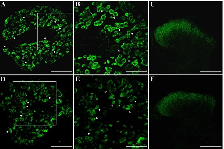
present in most neuronal cells in the adult mouse DRG. The highest level of expression was observed in cells mor-phologically resembling the dark, small to medium-sized type A cells (Lawson, 1992; Lagares and Avendan˜ o, 2000; Fig. 1A,B). Immunoreactivity was mainly in the cytosol, particularly at the periphery of the cell bodies (Fig. 1B), with a pattern overlapping microtubule distribution (Francis et al., 1999; Gleeson et al., 1999). Weakly stained DCX⫹/IB4–medium-sized to large neurons expressing low
levels of DCX were also present. In this case, DCX immu-noreactivity was either spread all over the cytosol or mainly confined at the soma periphery (Fig. 1B). DCX IR was also present along the fibers projecting centrally to the SC dorsal horns. At this level, DCX staining was apparent as a broad band of axon terminals extending across the entire mediolateral extent of the superficial dorsal horns (Fig. 1C). We then tested a rabbit polyclonal antibody (Cell Signaling Technology) recognizing a differ-ent DCX epitope. Again, DRG immunolabelling was local-ized mainly in small neurons, whereas large neurons were weakly stained (Fig. 1D,E). When tested in the spinal cord, the immunoreactivity pattern was very similar to that obtained with the Abcam antibody (Fig. 1F). Antibody specificity for DCX was further assessed using adult mouse brain sections as positive control. Staining of sec-tions through the subgranular zone of the hippocampal dentate gyrus (Fig. 2A) and the SVZ in the lateral wall of the lateral ventricules (Fig. 2B) produced a pattern of immunoreactivity that was typical of DCX⫹neuroblasts in neurogenic regions. Moreover, DRG and SC immunostain-ing was abolished when the Abcam antibody was pread-sorbed with the corresponding immunizing peptide (Fig. 2C,D).
To confirm further that the DCX protein and its mRNA were present in the adult mouse DRG and SC, Western blotting and RT-PCR experiments were performed by us-ing protein and RNA from adult mouse hippocampus and from lung as positive and negative controls, respectively. A single 199-bp band corresponding to the DCX cDNA amplification product was detected in the hippocampus as well as in the SC and DRG cDNAs but not in lung cDNA or in RT(–) samples (Fig. 2E). When tested in Western blotting, the Abcam DCX antibody recognized two distinct bands in hippocampus protein extracts (Fig. 2F): a single band at 40 kDa, possibly corresponding to the native form of DCX (Des Portes et al., 1998; Gleeson et al., 1998), and one at 43 kDa, which has been suggested to correspond to the phosphorylated form of DCX (Francis et al., 1999; Gleeson et al., 1999). Bands of 40 and 43 kDa were present also in DRG and SC extracts, whereas no bands were detected in lung protein extracts (Fig. 2F). Moreover, the intensity of both bands detected in hippocampus and DRG extracts was strongly diminished when the primary anti-body was preincubated with the immunizing peptide (Fig. 2F).
Phenotypic characterization of DCX
ⴙcells
in the adult mouse DRG
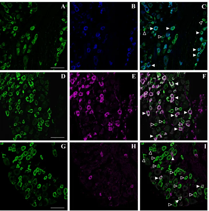
To characterize further the novel DCX-positive cell pop-ulation, we performed double-labelling experiments with markers of DRG neuronal subpopulations. Small neurons can be classified mainly into two populations, identified by the presence or absence of neuropeptides: the nonpepti-dergic P2X3⫹and isolectin IB4⫹ neurons and the
sub-stance P (SP)-immunoreactive population. Confocal microscopy analysis of DRG sections doubly stained for DCX and P2X3 showed that all P2X3-positive neurons
coexpressed DCX (Fig. 3A–C). We also verified whether DCX was expressed in IB4⫹cells. IB4 staining identifies a wider number of small nonpeptidergic neurons, compre-hensive of the P2X3-expressing population (Bradbury et
al., 1998; Vulchanova et al., 1998). Although most IB4-stained neurons were DCX immunoreactive, two popula-tions of cells could be identified in the small DCX⫹cells population: a population of DCX⫹/IB4⫹ neurons and a population of DCX⫹/IB4–cells (Fig. 3D–F). We
hypothe-sized that the small DCX⫹neurons that were negative for P2X3and IB4 may belong to the subpopulation of
pepti-dergic neurons. To evaluate this possibility, we performed double-immunofluorescence experiments for DCX and SP. Confocal microscopy analysis of DRG sections showed that SP-expressing neurons were also immunolabelled for DCX (Fig. 3G–I). Altogether, these data indicate that DCX is expressed by both the peptidergic and the nonpeptidergic subpopulations of nociceptors of adult mouse DRG, con-firming our initial morphological observations.
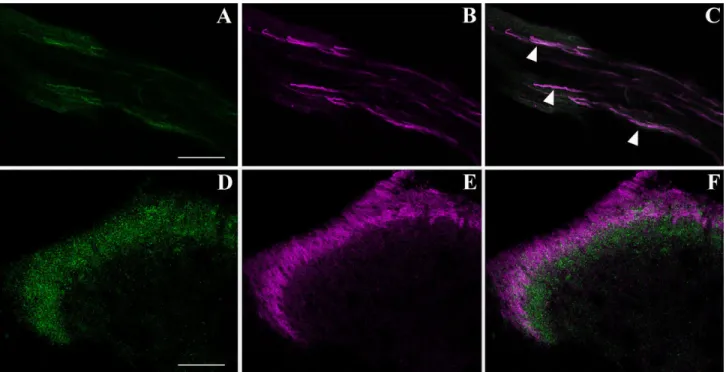
The DRG fibers centrally projecting to the SC showed the same pattern of colocalization observed in the DRG cell somata, with IB4⫹axons also being DCX immunore-active (Fig. 4A–C). In the dorsal horn, DCX IR was present in IB4-labelled central terminals of lamina II (Fig. 4D–F). Nonetheless, the projection area of DCX-positive fibers in the dorsal horns had a wider extension compared with IB4 labelling, possibly representing the axonal terminals of the DCX⫹/IB4–medium-sized to large DRG neurons.
The DCX protein is expressed in DRG and
SC dorsal horns of the adult rat
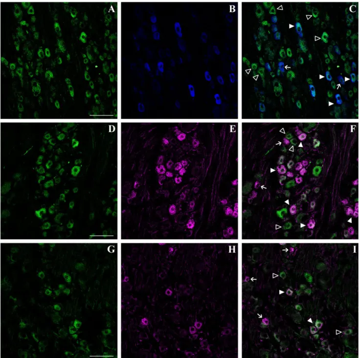
We have characterized the DCX-positive population in adult rat DRG and confirmed that also in this species most DRG neurons were DCX⫹, although staining was gener-ally less intense than in the mouse. DCX immunoreactiv-ity was seen mainly in neuronal cell bodies and in scat-tered fibers. In most cells, staining was widely distributed throughout the cytosol, although a minor cell population showed labelling restricted to part of the cytosol (Fig. 5A). As observed in the mouse, rat DCX immunoreactivity was present mainly in small to medium-sized type A cells (Fig.
Fig. 1. Doublecortin (DCX) immunoreactivity in adult mouse dorsal root ganglia (DRG) and spinal cord (SC) dorsal horns. Rep-resentative immunofluorescent photomicrographs of DRG (A,B,D,E) and SC dorsal horn (C,F) sections from adult mice stained with two antibodies recognizing different epitopes of DCX (Abcam No. ab18723: A–C; Cell Signaling No. 4604: D–F). A,D: DCX immunoreactivity is strongly expressed in a subpopulation of
small to medium-sized DRG neurons (solid arrowheads). B,E: High-power images of the boxed areas in A and D, respectively. Solid arrowheads indicate small to medium-sized DRG neurons stained with DCX antibodies. Large neurons (open arrowheads) are weakly stained. C,F: DCX immunoreactivity in the entire medio-lateral extension of the superficial dorsal horns. Scale bars⫽ 150 m in A,C,D,F; 75 m in B,E.
5A). Confocal microscopic analysis of DCX and P2X3
double-stained sections revealed a partial colocalization of the two proteins in the adult rat, with a subpopulation of P2X3neurons coexpressing DCX (Fig. 5A–C). Similar
re-sults were obtained with IB4 labelling as a marker of
small neurons (Fig. 5D–F). Confocal microscopic analysis of DRG sections doubly stained for DCX and SP demon-strated that SP⫹neurons coexpressed DCX (Fig. 5G–I). We concluded that, as in the mouse, also in the rat DCX is expressed in both nonpeptidergic and peptidergic small to medium-sized neurons. We also found a similar pattern of colocalization at the level of the fibers centrally projecting to the SC dorsal horns (Fig. 6A–C). Some IB4-labelled fibers expressed DCX, whereas others were DCX– (Fig.
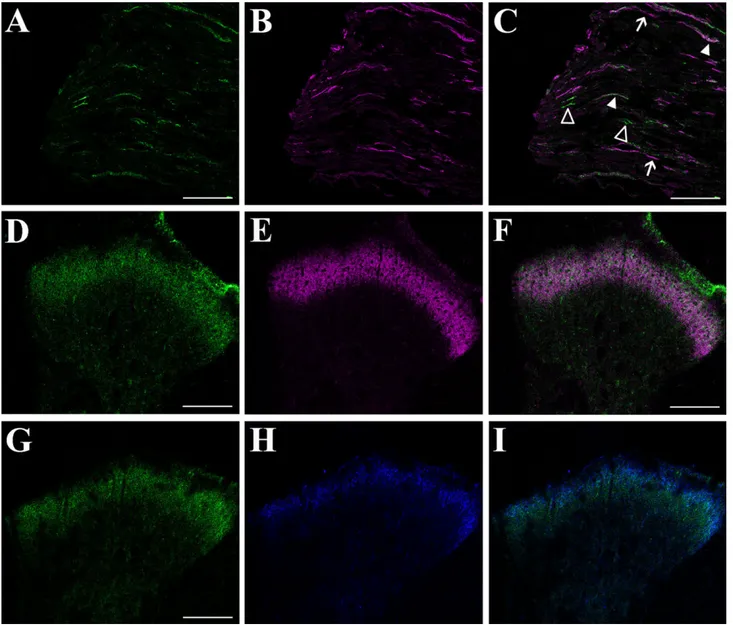
6A–C). In the SC dorsal horns, DCX immunoreactivity colocalized with SP in lamina I and in the outer portion of lamina II (Vulchanova et al., 1998) but also extended to the inner part of lamina II (Fig. 6G–I). Colocalization studies with isolectin IB4 showed a complete overlapping of DCX immunoreactivity with IB4 signal, whose labelling identifies lamina II (Vulchanova et al., 1998). Altogether, our results indicate that, in the rat, DCX is expressed in the cells bodies and in the axon terminals of peptidergic and nonpeptidergic nociceptors projecting to laminae I and II of the SC dorsal horns.
DISCUSSION
Here we describe for the first time the presence of the protein DCX in the adult DRG sensory neurons from both mouse and rat. The protein is expressed mainly by small to medium-sized neurons belonging to the peptidergic, SP⫹, and nonpeptidergic P2X3⫹/IB4-labelled populations.
This novel finding was quite unexpected, insofar as DCX is commonly regarded as a developmentally regulated pro-tein, expressed in immature neurons of both CNS and peripheral nervous system (PNS; Francis et al., 1999; Gleeson et al., 1999) and as an early marker of newly generated neuroblasts in neurogenic areas of the adult brain (Cameron et al., 1993; Doetsch et al., 1997; Seri et al., 2001). In both situations, DCX is attributed mainly a regulatory function in cell migration, based on its role in microtubule reorganization. Now, the presence of DCX in DRG neurons suggests novel functions for this protein in the adult nervous system.
DCX and plasticity
Microtubule reorganization is widely implicated in neu-roblast migration, but it may also subserve plasticity events such as synaptogenesis and axonal outgrowth (Lankford et al., 1990; Gordon-Weeks, 1991). DRG are areas of high plasticity in the nervous system, as sug-gested by their capacity to regenerate sensory fibers in response to injury (Lozeron et al., 2004). In addition, al-though peripheral nerve lesions such as nerve crush can induce DRG neuronal loss within 2–3 months from injury, at later times the number of DRG neurons is fully restored (Groves et al., 1997, 2003). One possible explanation for DCX being expressed in adult DRG neurons is that the protein may be readily available to contribute, through microtubule reorganization, to plasticity events. Nacher et al. (2001) first described DCX⫹ neurons with a mature phenotype in the rat piriform cortex, a cerebral region involved in ongoing structural plasticity events (for review see Brosh and Barkai, 2004; Barkai, 2005) but where neurogenesis has yet to be demonstrated. More recently, the hypothalamic suprachiasmatic nucleus (SCN), an area involved in the control of circadian rhythms, has been identified as an additional CNS region where DCX is expressed through adulthood (Geoghegan and Carter,
Fig. 2. Doublecortin (DCX) expression in adult mouse dorsal root ganglia (DRG) and spinal cord (SC) is confirmed by immunohisto-chemistry, by reverse transcription-polymerase chain reaction (RT-PCR), and by Western blot analysis. A,B: The specificity of the Abcam anti-DCX was confirmed by immunohistochemical analysis on adult mouse brain sections. A: Immunodecoration of cell bodies located in the subgranular zone of dentate gyrus (arrows) and their dendritic arborization (arrowheads) extending throughout the granular cell layer clearly identifies DCX⫹ immature neurons in the well-characterized neurogenic area. B: The Abcam antibody recognizes typical chains of immature neuroblasts in the subventricular zone. C,D: Neutralization of Abcam antibody. Incubation of the primary antibody with the blocking peptide results in a complete loss of stain-ing both in DRG (C) and in SC (D) sections. E: Primers designed for detection of mouse DCX transcript were used to evaluate DCX gene expression in the adult DRG and SC by RT-PCR. RNA was prepared from adult mouse DRG, SC, and from hippocampus (hp) and lung as positive and negative controls, respectively. For each preparation, 400 ng of RNA was either reverse transcribed [RT(⫹) samples] or incu-bated under the same conditions but in absence of the reverse tran-scriptase enzyme [RT(–) samples]. An amplicon of the expected size (199 bp) was obtained by PCR on RT(⫹) samples of hp, DRG, and SC and not from lung cDNA or from RT(–) samples. The 50-bp DNA ladder was utilized as molecular weight standard. F: Western blot analysis on tissue homogenates from adult mouse hp, DRG, SC, and lung with the Abcam antibody. Electrophoresis was performed by using 60g of protein extracts from DRG and SC and 20 g from hp and lung. The antibody recognizes a lower molecular weight band at 40 kDa and one at 43 kDa. Bands of corresponding molecular weight are present in the DRG and SC protein extracts but absent in lung extracts. Incubation of the primary antibody with the corresponding blocking peptide (BP) results in a loss of signal intensity for both bands in hp and DRG protein extracts. Scale bars⫽ 75 m in A (applies to A,B); 75m in C (applies to C,D).
2007). In line with what we observed in the DRG, DCX is expressed by mature, neuropeptide-containing SCN neu-rons. Both in the piriform cortex and in the SCN, the DCX immunoreactivity pattern is similar to that of the polysia-lylated form of the neural cell adhesion molecule
(PSA-NCAM), which is expressed by mature neurons after dam-age (Rutishauser and Landmesser, 1996), suggesting a common functional role for the two proteins. We also eval-uated the presence of PSA-NCAM⫹cells in adult DRG, but for this location, unlike the case for the piriform cortex
Fig. 3. Phenotypic characterization of doublecortin (DCX)-expressing cells in the adult mouse dorsal root ganglia (DRG) by confocal microscopic analysis. A,D,G: DCX (green) is expressed in a subpopulation of DRG neurons morphologically resembling small to medium-sized nociceptors. B: P2X3immunoreactivity (blue,
pseudo-color) in small nociceptive neurons. C: Overlay of the two channels demonstrates that most P2X3⫹ neurons are DCX immunoreactive
(solid arrowheads). Open arrowheads indicate DCX⫹/P2X3 –
neurons. E: IB4-labelled (magenta, pseudocolor) small nociceptive neurons.
F: Overlay of the two channels shows that IB4-stained neurons are DCX immunoreactive (solid arrowheads). A subpopulation of small DCX⫹neurons is not labelled with IB4 (open arrowheads). H: Sub-stance P (SP) immunoreactivity (magenta, pseudocolor) is expressed by a subpopulation of small peptidergic neurons. I: Overlay of the two channels shows that most SP⫹neurons are also DCX immunoreactive (solid arrowheads), but many small DCX⫹neurons do not express SP (open arrowheads). Scale bar⫽ 75 m in A (applies to A–C); 75 m in D (applies to D–F); 75m in G (applies to G–I).
and the SCN, we could not detect any specific signal for the adhesion molecule (data not shown). DCX⫹cells in the piriform cortex and in the SCN coexpress NeuN (neuron-specific nuclear protein), a widely accepted marker of fully differentiated neurons (Mullen et al., 1992). The majority of the DCX⫹neurons in DRG were also expressing NeuN (data not shown), confirming that they share features of mature neurons.
DCX and neuroprotection
A recent finding suggests a direct neuroprotective role for DCX protein (Santra et al., 2006). In vitro, DCX over-expression protects rat SVZ and human glioma cells from oxygen and glucose deprivation, whereas knock down of DCX expression by small interfering RNA increases their vulnerability to damaging insults. Small nociceptors rep-resent a highly vulnerable cellular subpopulation, which rapidly degenerates in response to specific damaging con-ditions (Lisney, 1989; Tandrup et al., 2000; Lozeron et al., 2004; Guseva and Chelyshev, 2006). The presence of DCX in DRG may possibly underlie a potential protective role against insults, which allows neuronal cells to recover from damage. In this regard, it will be interesting to evaluate the consequences of different damaging condi-tions for DRG neuronal subpopulacondi-tions after DCX knock down.
DCX in sensory ganglia and neurogenesis
Although it is still a controversial matter, for decades several groups have collected data suggesting that, in
different animal species, DRG may undergo an age-dependent increase in neuron number, leading to the pro-posal that neurogenesis may occur in this tissue after birth (Devor and Govrin-Lippman, 1985; Devor et al., 1991; Cecchini et al., 1995; Popken and Farel, 1997; Ci-aroni et al., 2000; Farel, 2002). Recently, the production of neurospheres that can differentiate into neurons and glia has been demonstrated in vitro from adult rat DRG and trigeminal ganglia explants (Namaka et al., 2001; Lagares et al., 2007; Li et al., 2007), although DRG neurogenesis in vivo awaits confirmation (La Forte et al., 1991; Ciaroni et al., 2000; Farel, 2002). An alternative hypothesis to ex-plain the possible addition of new DRG neurons after birth has been proposed, namely, the ongoing maturation of preexisting postmitotic immature cells to fully differenti-ated neurons both under physiological conditions and in response to external insults (La Forte et al., 1991; Cec-chini et al., 1993; Ciaroni et al., 2000; Farel, 2002, 2003). At this stage, we cannot exclude that the presence of DCX in DRG may indicate ongoing neurogenesis in this loca-tion, although the fact that most of the cells express the protein would argue against this possibility. Additionally, we never observed BrdU⫹cells in DRG collected from rats that were administered the thymidine analogue (data not shown). In a recent work, Lagares et al. (2007) described the presence of few, sparse DCX⫹cells in the rat trigem-inal ganglia (TG), an area of the PNS that shares, at least in part, a common origin with DRG (D’Amico-Martel and Noden, 1983; Fontaine-Perus et al., 1985). Unlike TG, where very few DCX-immunolabelled cells could be
ob-Fig. 4. Phenotypic characterization of doublecortin (DCX)-positive fibers centrally projecting to the mouse spinal cord (SC) dorsal horns by confocal microscopic analysis. A: Some nerve fibers extending centrally from the DRG neurons to the SC are DCX immunoreactive (green). B: IB4-labelled fibers (magenta, pseudocolor). C: DCX colo-calizes with IB4 at the fiber level (arrowheads). D–F: Confocal micro-scopic analysis of the mouse SC dorsal horns doubly stained for DCX
(green) and IB4 (magenta, pseudocolor). D: DCX immunoreactivity is present at the level of the superficial dorsal horns. E: IB4 labelling of dorsal horns, the target region of DRG nociceptors. F: Overlay of the two channels shows the colocalization of DCX⫹and IB4-stained ter-minals of DRG nociceptive neurons at the SC level. DCX expression extends to a larger dorsal horn area compared with IB4 staining. Scale bars⫽ 75 m in A (applies to A–C); 75 m in D (applies to D–F).
served, in DRG most of the neuronal cells express DCX. Additionally, in rat TG, DCX⫹ cells apparently do not coexpress protein markers of mature neurons, such as the neuron-specific enolase (NSE; Vega et al., 1990). We did
not evaluate NSE immunoreactivity in DRG, but, as pre-viously stated, most DCX⫹cells colocalized with the ma-ture neuronal marker NeuN. Some Nissl-stained cells morphologically resembling mature sensory neurons did
Fig. 5. Phenotypic characterization of doublecortin (DCX)-expressing cells in the adult rat dorsal root ganglia (DRG). A–I: Colocal-ization of DCX with P2X3(A–C), IB4 (D–F), and substance P (SP; G–I)
analyzed by confocal microscopy. A,D,G: DCX (green) immunoreactivity in DRG neurons. Most small to medium-sized neurons are strongly stained. B: P2X3immunoreactivity (blue, pseudocolor) in small DRG
nociceptive neurons. C: Overlay of the two channels shows a partial colocalization between P2X3- and DCX-immunoreactive neurons (solid
arrowheads). Open arrowheads indicate DCX⫹/P2X3
–neurons. Arrows
indicate P2X3⫹neurons that do not express DCX. E: IB4-labelled
(ma-genta, pseudocolor) small nociceptive DRG neurons. F: Overlay of the two channels shows that a subpopulation of small IB4-labelled neurons coexpresses DCX (solid arrowheads). Arrows indicate IB4-labelled neu-rons that do not express DCX. Small DCX⫹neurons that are not labelled with IB4 are also present (open arrowheads). H: SP immunoreactivity (magenta, pseudocolor) in small DRG neurons. I: Overlay of the two channels shows SP⫹/DCX⫹cells (solid arrowheads). Arrows indicate SP⫹
cells that do not express DCX, whereas DCX⫹/SP–neurons are identified
by open arrowheads. Scale bars⫽ 75 m in A (applies to A–C); 75 m in D (applies to D–F); 75m in G (applies to G–I).
not express NeuN, and, as a consequence, few DCX⫹/ NeuN– cells were also present in adult DRG (data not
shown). The absence of NeuN does not necessarily imply a less well-differentiated neuronal phenotype, insofar as NeuN– neurons have been described, among them
Pur-kinje cells, olfactory bulb mitral cells, retinal photorecep-tor cells (Mullen et al., 1992), and many neurons of the dorsal SCN (Geoghegan and Carter, 2007). However, at this stage, we cannot exclude that DCX⫹/NeuN⫹ and DCX⫹/NeuN– cells may represent functionally different
cell populations. Whether or not DCX expression in DRG
underlies neurogenesis occurring in this location, the role of DCX as a selective marker of new neurons generated in the adult brain requires reconsideration. As described by Kempermann et al. (2003), some of the DCX-expressing cells in the dentate gyrus coexpress nestin, which is re-garded as a stem/precursor cell marker (Lendahl et al., 1990; Reynolds et al., 1992). Moreover Walker et al. (2007) showed that DCX⫹cells have different proliferating and differentiating properties, which correlate with protein expression levels, highly expressing cells being committed to the neuronal lineage and low-expressing ones being
Fig. 6. Phenotypic characterization of doublecortin (DCX)-positive fibers centrally projecting to the rat spinal cord (SC) dorsal horns by confocal microscopic analysis. A: Some nerve fibers extending centrally from the DRG neurons to the SC are DCX immunoreactive (green). B: IB4-labelled fibers (magenta, pseudocolor). C: DCX colocalizes with IB4 in some (solid arrowheads) but not all (open arrowheads) fibers. Arrows identify IB4-labelled fibers that do not express DCX. D–I: Con-focal analysis of the mouse SC dorsal horns doubly stained for DCX and IB4 (D–F) and for DCX and substance P (SP; G–I). D,G: DCX immuno-reactivity is present in the superficial layers of dorsal horns. E: IB4
labelling of lamina II in the dorsal horns, the target region of DRG nociceptors. F: Overlay of the two channels shows the colocalization of DCX⫹- and IB4-stained terminals of DRG nociceptive neurons at the SC level. H: SP immunoreactivity in lamina I and the dorsal part of lamina II in the SC dorsal horns (blue, pseudocolor). I: DCX⫹axons terminals
colocalize with SP immunoreactivity in the SC. DCX expression extends to a larger area in the inner laminae of dorsal horns compared with SP, probably representing the IB4-labelled lamina II. Scale bars⫽ 150 m in A (applies to A–C); 150 in D (applies to D–F); 150m in G (applies to G–I).
undifferentiated precursors able to proliferate and give rise to neurospheres. Altogether, these data strongly sug-gest that DCX⫹cells may represent a highly heterogenous cell population.
In conclusion, our data strongly suggest a novel role for DCX in DRG neurons. Additional experimental work must be performed to unravel fully the functional significance of the DCX protein in adult nervous tissues and, in particu-lar, in sensory neurons.
LITERATURE CITED
Alvarez-Buylla A, Garcia-Verdugo JM. 2002. Neurogenesis in adult sub-ventricular zone. J Neurosci 22:629 – 634.
Bai J, Ramos RL, Ackman JB, Thomas AM, Lee RV, LoTurco JJ. 2003. RNAi reveals doublecortin is required for radial migration in rat neo-cortex. Nat Neurosci 6:1277–1283.
Barkai E. 2005. Dynamics of learning-induced cellular modifications in the cortex. Biol Cybernet 92:360 –366.
Berg MJ, Schifitto G, Powers JM, Martinez-Capolino C, Fong CT, Myers GJ, Epstein LG, Walsh CA. 1998. X-linked female band heterotopia-male lissencephaly syndrome. Neurology 50:1143–1146.
Bradbury EJ, Burnstock G, McMahon SB. 1998. The expression of P2X3 purinoreceptors in sensory neurons: effects of axotomy and glial-derived neurotrophic factor. Mol Cell Neurosci 12:256 –268.
Brosh I, Barkai E. 2004. Learning-induced long-term synaptic modifica-tions in the olfactory cortex. Curr Neurovasc Res 1:389 –395. Brown JP, Couillard-Despres S, Cooper-Kuhn CM, Winkler J, Aigner L,
Kuhn HG. 2003. Transient expression of doublecortin during adult neurogenesis. J Comp Neurol 467:1–10.
Cameron HA, Woolley CS, McEwen BS, Gould E. 1993. Differentiation of newly born neurons and glia in the dentate gyrus of the adult rat. Neurosci 56:337–344.
Cecchini T, Cuppini R, Ciaroni S, Del Grande P. 1993. Increased number of DRG neurons in vitamin E-deficient rats. Somatosens Motor Res 10: 433– 443.
Cecchini T, Cuppini R, Ciaroni S, Barili P, De Matteis R, Del Grande P. 1995. Changes in the number of primary sensory neurons in normal and vitamin E-deficient rats during aging. Somatosens Motor Res 12:317–327.
Ciaroni S, Cecchini T, Cuppini R, Ferri P, Ambrogini P, Bruno C, Del Grande P. 2000. Are there proliferating neuronal precursors in adult rat dorsal root ganglia? Neurosci Lett 281:69 –71.
Corbo JC, Deuel TA, Long JM, LaPorte P, Tsai E, Wynshaw-Boris A, Walsh CA. 2002. Doublecortin is required in mice for lamination of the hip-pocampus but not the neocortex. J Neurosci 22:7548 –7557.
Couillard-Despres S. 2001. Molecular mechanisms of neuronal migration disorders, quo vadis? Curr Mol Med 1:677– 688.
Couillard-Despres S, Winner B, Schaubeck S, Aigner R, Vroemen M, Weidner N, Bogdahn U, Winkler J, Kuhn HG, Aigner L. 2005. Dou-blecortin expression levels in adult brain reflect neurogenesis. Eur J Neurosci 21:1–14.
D’Amico-Martel A, Noden DM. 1983. Contributions of placodal and neural crest cells to avian cranial peripheral ganglia. Am J Anat 166:445– 468. Des Portes V, Pinard JM, Billuart P, Vinet MC, Koulakoff A, Carrie A, Gelot A, Dupuis E, Motte J, Berwald-Netter Y, Catala M, Kahn A, Beldjord C, Chelly J. 1998. A novel CNS gene required for neuronal migration and involved in X-linked subcortical laminar heterotopia and lissencephaly syndrome. Cell 92:51– 61.
Devor M, Govrin-Lippmann R. 1985. Neurogenesis in adult rat dorsal root ganglia. Neurosci Lett 61:189 –194.
Devor M, Govrin-Lippman R, Frank I, Raber P. 1991. Neurogenesis in adult rat dorsal root ganglia: on counting and the count. Somatosens Mot Res 3:139 –167.
Doetsch F, Garcia-Verdugo JM, Alvarez-Buylla A. 1997. Cellular composi-tion and three-dimensional organizacomposi-tion of the subventricular germi-nal zone in the adult mammalian brain. J Neurosci 17:5046 –5061. Fabbretti E, D’Arco M, Fabbro A, Simonetti M, Nistri A, Giniatullin R.
2006. Delayed upregulation of ATP P2X3receptors of trigeminal sen-sory neurons by calcitonin gene-related peptide. J Neurosci 26:6163– 6171.
Farel PB. 2002. Sensory neuron addition in juvenile rat: time course and specificity. J Comp Neurol 449:158 –165.
Farel PB. 2003. Late differentiation contributes to the apparent increase in sensory neuron number in juvenile rat. Brain Res Dev Brain Res 144:91–98.
Fontaine-Perus J, Chanconie M, Le Douarin NM. 1985. Embryonic origin of substance P containing neurons in cranial and spinal sensory gan-glia of the avian embryo. Dev Biol 107:227–238.
Francis F, Koulakoff A, Boucher D, Chafey P, Schaar B, Vinet MC, Frio-court G, McDonnell N, Reiner O, Kahn A, McConnell SK, Berwald-Netter Y, Denoulet P, Chelly J. 1999. Doublecortin is a developmen-tally regulated, microtubule-associated protein expressed in migrating and differentiating neurons. Neuron 23:247–256.
Geoghegan D, Carter DA. 2008. A novel site of adult doublecortin expres-sion: neuropeptide neurons within the suprachiasmatic nucleus circa-dian clock. BMC Neurosci 9:2 [E-pub ahead of print].
Geuna S, Borrione P, Fornaro M, Giacobini-Robecchi M. 2000. Neurogen-esis and stem cells in adult mammalian dorsal root ganglia. Anat Rec 261:139 –140.
Gleeson JG, Allen KM, Fox JW, Lamperti ED, Berkovic S, Scheffer I, Cooper EC, Dobyns WB, Minnerath SR, Ross ME, Walsh CA. 1998. Doublecortin, a brain-specific gene mutated in human X-linked lissen-cephaly and double cortex syndrome, encodes a putative signalling protein. Cell 92:63–72.
Gleeson JG, Lin PT, Flanagan LA, Walsh CA. 1999. Doublecortin is a microtubule-associated protein and is expressed widely by migrating neurons. Neuron 23:257–271.
Gordon-Weeks PR. 1991. Control of microtubule assembly in growth cones. J Cell Sci Suppl 15:45– 49.
Groves MJ, Christopherson T, Giometto B, Scaravilli F. 1997. Axotomy-induced apoptosis in adult rat primary sensory neurons. J Neurocytol 26:615– 624.
Groves MJ, Schanzer A, Simpson AJ, An SF, Kuo LT, Scaravilli F. 2003. Profile of adult rat sensory neuron loss, apoptosis and replacement after sciatic nerve crush. J Neurocytol 32:113–122.
Guseva D, Chelyshev Y. 2006. The plasticity of the DRG neurons belonging to different subpopulations after dorsal rhizotomy. Cell Mol Neurobiol 26:1223–1232.
Hannan AJ, Henke RC, Seeto GS, Capes-Davis A, Dunn J, Jeffrey PL. 1999. Expression of doublecortin correlates with neuronal migration and pattern formation in diverse regions of the developing chick brain. J Neurosci Res 55:650 – 657.
Hwang SJ, Oh JM, Valtschanoff JG. 2005. The majority of bladder sensory afferents to the rat lumbosacral spinal cord are both IB4- and CGRP-positive. Brain Res 1062:86 –91.
Kempermann G, Gast D, Kronenberg G, Yamaguchi M, Gage FH. 2003. Early determination and long-term persistence of adult-generated new neurons in the hippocampus of mice. Development 130:391–399. Kuo LT, Simpson A, Schanzer A. 2005. Effects of systemically
adminis-tered NT-3 on sensory neuron loss and nestin expression following axotomy. J Comp Neurol 482:320 –332.
La Forte RA, Melville S, Chung K, Coggeshall RE. 1991. Absence of neurogenesis of adult rat dorsal root ganglion cells. Somatosens Mot Res 8:3–7.
Lagares A, Avendan˜ o C. 2000. Lateral asymmetries in the trigeminal ganglion of the male rat. Brain Res 865:202–210.
Lagares A, Li HU, Zhou XF, Avendan˜ o C. 2007. Primary sensory neuron addition in the adult rat trigeminal ganglion: evidence for neural crest glio-neuronal precursor maturation. J Neurosci 27:7939 –7953. Lankford K, Cypher C, Letourneau P. 1990. Nerve growth cone motility.
Curr Opin Cell Biol 2:80 – 85.
Lawson SN. 1992. Morphological and biochemical cell types of sensory neurons. In: Scott SA, editor. Sensory neurons: diversity, development, and plasticity. New York: Oxford University Press. p 27–59. Lendahl U, Zimmerman LB, McKay RDG. 1990. CNS Stem cells express a
new class of intermediate filament protein. Cell 60:585–595. Li HY, Say EH, Zhou XF. 2007. Isolation and characterization of neural
crest progenitors from adult dorsal root ganglia. Stem Cells 25:2053– 2065.
Lisney SJW. 1989. Regeneration of unmyelinated axons after injury of mammalian peripheral nerve. Q J Exp Physiol 74:757–784.
Lois C, Alvarez-Buylla A. 1994. Long-distance neuronal migration in the adult mammalian brain. Science 264:1145–1148.
Lozeron P, Krarup C, Schmalbruch H. 2004. Regeneration of unmyelinated and myelinated sensory nerve fibers studied by a retrograde tracer method. J Neurosci Methods 138:225–232.
Mullen RJ, Buck CR, Smith AM. 1992. NeuN, a neuronal specific nuclear protein in vertebrates. Development 116:201–211.
Nacher J, Crespo C, McEwen BS. 2001. Doublecortin expression in the adult rat telencephalon. Eur J Neurosci 14:629 – 644.
Namaka MP, Sawchuk M, MacDonald SC, Jordan LM, Hochman S. 2001. Neurogenesis in postnatal mouse dorsal root ganglia. Exp Neurol 172: 60 – 69.
Popken GJ, Farel PB. 1997. Sensory neuron number in neonatal and adult rats estimated by means of stereological and profile-based methods. J Comp Neurol 368:8 –15.
Pover C, Barnes MC, Coggeshall RE. 1994. Do primary afferent cell num-bers change in relation to increasing weight and surface area in adult rats? Somatosens Mot Res 11:163–167.
Rao MS, Shetty AK. 2004. Efficacy of DCX as a marker to analyse the absolute number and the dendritic growth of newly generated neurons in the adult dentate gyrus. Eur J Neurosci 19:234 –246.
Reynolds BA, Tetzlaff W, Weiss S. 1992. A multipotent EGF-responsive striatal embryonic progenitor cell produces neurons and astrocytes. J Neurosci 12:4565– 4574.
Rutishauser U, Landmesser L. 1996. Polysialic acid in the vertebrate nervous system: a promoter of plasticity in cell– cell interactions. Trends Neurosci 19:422– 427.
Santra M, Liu Xs, Santra S, Zhang J, Lan Zhang R, Gang Zhang Z, Chopp M. 2006. Ectopic expression of doublecortin protects adult rat
progen-itor cells and human glioma cells from severe oxygen and glucose deprivation. Neuroscience 142:739 –752.
Seri B, Garcia-Verdugo JM, McEwen BS, Alvarez-Buylla A. 2001. Astro-cytes give rise to new neurons in the adult mammalian hippocampus. J Neurosci 21:7153–7160.
Tandrup T, Woolf C, Coggeshall R. 2000. Delayed loss of small dorsal root ganglion cells after transection of the rat sciatic nerve. J Comp Neurol 422:172–180.
Van Praag H, Schinder AF, Christie BR, Toni N, Palmer TD, Gage FH. 2002. Functional neurogenesis in the adult hippocampus. Nature 415: 1030 –1034.
Vega JA, Rodriguez C, Medina M, del Valle ME. 1990. Neuron-specific enolase (NSE)-like and neurofilament protein (NFP)-like immunoreac-tivities in the rat dorsal root ganglia and sciatic nerve. Cell Mol Biol 36:537–546.
Vulchanova L, Riedl MS, Shuster SJ, Stone LS, Hargreaves KM, Buell G, Surprenant A, North RA, Elde R. 1998. P2X3is expressed by DRG neurons that terminate in inner lamina II. Eur J Neurosci 10:3470 – 3478.
Walker TL, Yasuda T, Adams DJ, Bartlett PF. 2007. The doublecortin-expressing population in the developing and adult brain contains mul-tipotential precursors in addition to neuronal-lineage cells. J Neurosci 27:3734 –3742.