Università degli Studi del Piemonte Orientale
“Amedeo Avogadro”
Dipartimento di Scienze del Farmaco
Dottorato di Ricerca in Biotecnologie Farmaceutiche ed Alimentari
XXVI ciclo a.a. 2010-2013
NANOSYSTEMS FOR MOLECULAR
IMAGING
Università degli Studi del Piemonte Orientale
“Amedeo Avogadro”
Dipartimento di Scienze del Farmaco
Dottorato di Ricerca in Biotecnologie Farmaceutiche ed Alimentari
XXVI ciclo a.a. 2010-2013
NANOSYSTEMS FOR MOLECULAR
IMAGING
Valeria De Biasio
Supervised by:
Prof. Giovanni B. Giovenzana
Contents
Chapter 1
Introduction p. 1
Chapter 2
Outline of the thesis p. 25
Chapter 3
“Supramolecular assemblies based on amphiphilic Mn2+-Complexes as High
Relaxivity MRI probes” p. 27
Chapter 4
“Phosphonated polyethyleneimine (PEI-P): evaluation of a chelating polymer as a unimolecular nanosized MRI contrast agents” p. 51
Chapter 5
“N-Polybenzylated Alicyclic 1,2-Diamines Show Cytotoxicity and G1 Phase
Arrest in Cancer Cell Line” p. 71
Chapter 6
Conclusions p. 95
List of publications
p. 991
Introduction
Molecular Imaging
Molecular Imaging (MI) is a growing research discipline that can be defined as the in vivo characterization and measurement of biologic processes at the cellular and molecular level. Molecular approaches to diagnostics were developed few years ago and they have become available to detect diseases only recently. Molecular Imaging relies on molecular changes because they represent early indicators of pathologies before the latter reach a macroscopic level (Figure 1). In this emergent area of research it is mandatory to anticipate the time to detect molecular or cellular changes, into the body, to predict as soon as possible the disease (1, 2).
Molecular Imaging finds a huge variety of applications, not limited only to detection of a disease in a early stage. Angiogenesis, for example, is a normal process that develops naturally during embryonic development or the female reproductive cycle, etc.; nevertheless, the vascular growth can happen in an unrestrained way determining pathologic conditions. Loss of this regulation is known to occur in more than 30 different diseases, e.g.: cancer, cardiovascular and immunologic disease, diabetes, etc., and can be detected by MI. The latter can be used to locate apoptosis phenomena. Apoptosys is a physiologic process consisting in a programmed cell death. Perturbation of this equilibrium can be related to a huge variety of pathologies. Apoptosys reduction can be related to cancer, autoimmune diseases, and viral infections. An increase in apoptosys can be related to AIDS, neurodegenerative disorders, ischemia, stroke, myelodysplastic syndromes (3).
2
Several instrumental imaging techniques are used to detect molecular changes in living organisms tissues. The most common are:
i) Nuclear techniques, as Positron Emission Tomography (PET), Single Photon Emission Computed Tomography (SPECT),
ii) MR techniques, as Nuclear Magnetic Resonance Imaging (NMRI), Magnetic Resonance Tomography (MRT), Magnetic Resonance Spectroscopy
iii) Optical techniques, as Optical Tomography, Surface Weighted Imaging (reflectance diffuse tomography), Phase-array Detection, Confocal Imaging, Multiphoton Imaging, or Microscopic Imaging
iv) Acoustic techniques: Ultrasonography (US) and Photoacoustic Imaging In addition to the above cited techniques, hyphenated ones are available where a suitable combination of two of them (e.g.: PET-MRI) allow to obtain images endowed with an higher degree of diagnostic information.
Figure 1
Figure 1 shows the different degree of diagnostic information associated with the single imaging technique, ultimately related to their resolution. For example X-Ray Computed Tomography, as Ultrasounds, allow to appreciate the anatomic structure of tissues while Magnetic Resonance Imaging, Nuclear Magnetic Resonance Imaging, Optical Imaging or Nanosensors, having a higher resolution, are able to give diagnostic informations ranging from physiological to the molecular level.
3
Molecular Imaging techniques
Optical Imaging
Optical Imaging encompasses several imaging modalities that can be applied to the diagnostic medical field to detect pathological changes in a non-invasive way, employing different kinds of “light” (UV, visible and IR). Processes can be investigated with Optical Imaging at several levels, at the organ, tissue, cellular and molecular one. The image, that derives from optical techniques, is produced by the interaction of different forms of light, depending on which technique is used. In particular, the image depends on the wavelength of the light that is used, from the instrument optical configuration employed to detect the signal and from auxiliary imaging agents used to give the contrast; the main limitation of OI is just related to the depth of penetration of light, ranging from < 1 mm for UV radiation to some centimeters for IR photons. When light interacts with tissues, the latter are involved in three processes: absorption, photon scattering and the generation of fluorescence emission (4). These three phenomena, on which is based Optical Imaging, are useful to recognize different tissues either functionally, for example to detect hemoglobin concentration, or structurally, e.g.: cellular morphology. The different Optical Imaging techniques are really a lot and generally they are based on the use of visible, ultraviolet and infrared light, absorbed, emitted or scattered by molecules of cells and tissues. Usually the techniques can be classified into Diffuse Optical Tomography (DOT) and Ballistic Optical Imaging (BOI), depending from principles are used to obtain the image (5). A special interest is hold by is Fluorescence Imaging. In this application, biocompatible fluorescent
4
dyes are used and if excitation in the IR region is adopted it is possible to perform in vivo imaging on whole small animals. A current limitation of this technique is the translation from animals to patients, as it means that for patients, for example, bulk tissues should not exceed in size over 10 cm (6). Optical imaging may be combined with different techniques to have more detailed images, as for example OI/MRI (7).
Exogenous imaging agents such as fluorescent dyes are used in Optical Imaging. Thousands of fluorescent dyes are known and some of them currently used in fluorescent imaging applications. Indocyanine green (ICG) is approved for in vivo use. Cyanines represent the largest family of fluorescent dyes and many hydrophilic or conjugated derivatives of cyanines have been synthesized to visualize e.g. tumor cells (8, 9). Rhodamine and fluorescein derivatives have been extensively employed and conjugated with small molecules acting as vectors to detect specifically tumor cells (10). Fluorescent sensors are also available for metal ions, changing their emission propertied in the presence of different concentrations of specific metal ions (for example Cu2+) and being extremely useful for in vivo imaging applications. AlexaFluor® is another class of fluorescent dyes, widely used in virtue of the high intensity of fluorescence of these derivatives. Finally, the acronym BODIPY includes a class of fluorescent dyes where the core structure is represented by 4,4-difluoro-4-bora-3a,4a-diaza-s-indacene; this unusual heteropolycyclic structure is characterized by a very high quantum yield (11).
Positron Emission Tomography (PET)
PET is a diagnostic technique in which images are obtained using the positron emission of radionuclides, used in the form of radiolabeled agents. PET
5
radionuclides, emittes positrons that annihilates with nearby electrons, emitting two collinear opposite γ-rays. An array of detectors collect the radiation, discriminating the γ-rays pair emitted by the radionuclide on the base of their simultaneous detection an reconstructing the emission source point by retroprojecting their trajectories. Several positron emitting radionuclide are available, among them light atoms such carbon and fluorine and radiometals such as copper and gallium. Some of these elements are present into biomolecules into the body, and, this means that the use of positron emitting isotopes does not alter the physicochemical and biochemical properties with respect to the nonlabeled compounds (12). In PET applications, scientists prefer to use 18F, in view of its availability, easy introduction in organic molecules and favourable emission energy. Its half-life, of 110 min, is compatible with 3-4 synthetic steps to be used to prepare labeled organic compounds. One of the most used PET agents is 2-[18F]-fluoro-deoxy-D-glucose, also known like 18 F-FDG; it is used to monitor glucose metabolism and it is really important for clinical diagnosis of tumors, considering that tumoral cells are quite hungry of sugars. 11C is another important positron-emitting isotope and it is less used to prepare organic compounds due to the short half-life of 20.3 min.
In addition to 11C and 19F, several metal isotopes find an application into PET-imaging, as 64Cu, 76Br, 68Ga and that they are most of all used to study biochemical processes connected with slow pharmacokinetics (13).
PET imaging finds many diagnostic applications and it is best suited for detecting some kind of tumors, even if it is becoming a powerful tool in MI applications due to its relevant sensitivity. For example it is used to study the expression of somatostatin receptors, the latter being involved in: CNS, hypothalamopituitary system, gastrointestinal tract, exo- and endocrine pancreas, and immune system. It is used to explore the colinergic system, too,
6
because it can be involved in neurological and psychiatric disorders, as for example Alzheimer’s and Parkinson’s disease (14).
Single-Photon Emission Computed Tomography
(SPECT)
Single-photon emission computed tomography (SPECT), together with PET imaging, was the first imaging modality used in clinical. For SPECT imaging, contrast agents are labelled with specific radionuclides, all able to emit γ rays. The energy arising in the form of radiation from these radionuclides, have to be in the range 100-250 keV to obtain good-quality images. The γ-rays used to create the images, are captured by detectors included into a so called “γ camera”, and sent to an elaborator dedicated to the image reconstruction and editing. Many metal isotopes emit γ-rays useful for SPECT, as 67Cu, 67Ga, 111In,
90Y, 201Tl, and their corresponding ions may be combined with suitable
chelating agents to create γ-emitting complexes, to be conjugated with peptides, antibodies or other targeting vectors (15). Nevertheless, the most important element used for SPECT imaging is 99mTc, because when it emits γ rays, the energy released is of 140 keV (89% abundance), meaning that this element is perfect to be detected in imaging with a gamma camera. A drawback of using
99mTc is its relatively short half-life, about 6 h, hampering long and articulate
preparations, but compensated by the availability of this nuclide through a dedicated generator. Nowadays applications with 99mTc can offer two possibilities to exploit this element, it means that it can be used to label a targeting moiety, as antibody, peptide, hormone, or using the 99mTc-chelate as such.
7
Radiopharmaceuticals containing 99mTc are used to study many diseases: 99m Tc-diphosphonate radiopharmaceuticals are accumulated into bones, and this is important to detect bone metastases or hidden fractures. 99mTc-D,L-HM-PAO (Ceretec) and 99mTc-LL-ECD (Neurolite) for example allow to study blood flow
into the brain, while 99mTc-sestamibi (Cardiolite) gives informations both for myocardial perfusion imaging and also for cancer imaging (16). Cu2+ has a rich
coordination chemistry exploited to in a large variety of complexes with nitrogen-based ligands, including cyclen and cyclam derivatives. Considering that coordination numbers of this element range from 4 to 6, geometries of several Cu2+-complexes can be square planar, square pyramidal, trigonal bipyramidal, and octahedral (17, 18). Several 67Cu2+-complexes were studied for SPECT detection of tumors (19, 20). 67Ga3+ recently received renewed interest for the preparation of chelates for SPECT due to the availability of this nuclide through a dedicated generator. Many derivatives have been reported in the scientific literature, mainly concentrated on hexacoordinated ligands that can match the coordination requirements of this “hard” trivalent ion (21).
SPECT was extensively employed in Molecular Imaging application, such as the sensitive detection of: folate receptors and oxytocin receptor that are generally over-expressed on tumoral cells, or integrins receptors, directly connected to inflammation, lung cancer, teratoma, etc. (22- 24).
8
X-Ray Computed Tomography (x-ray CT)
X-ray-computed tomography (CT) is a technique that allows to obtain three-dimensional images of tissues and screening diseases into a non invasively way. CT is a relatively old technique, having become a diagnostic tool when X-rays were discovered in 1895. Electromagnetic radiation of X-rays has wavelengths within the range of 0.01 and 10 nm. X-rays are generated in a vacuum tube where electrons are accelerated and travel from a catode to a tungsten-alloy anode; colliding with the anode electrons are braked, releasing X-rays with a specific energy dependent from the energy of incident electrons. CT is extremely versatile: gastrointestinal tract, cardiovascular system, renal tract, liver, lungs, bone, cartilage and some tumor tissues can be screened by this technique (25). Diagnostic images are formed by the absorption of X-rays from heavy atoms present in the tissue such as calcium. During CT contrast agents may be used; they generally contain atoms with an high atomic number, as I or Ba as the latter strongly absorb X-rays, and are employed to improve imaging of soft tissues.
Contrast agents based on iodine can be ionic or nonionic molecules usually belonging to the class of 2,4,6-triiodoarylcarboxamides. Ionic compounds are inclined to interact with biological molecules, as peptides or cell membranes, and may increase the plasma osmolality due to the large dose administered (26). For this reason generally nonionic contrast agents are preferred to the charged ones, even if nowadays both of them have been improved thanks to their manipulation, for example adding hydrophilic residues that allows higher solubility in water and lower toxicity (27). Among iodine contrast agents, the most used in CT are iopamidol, iomeprol, iohexol, iopromide. Barium sulfate, is a contrast medium widely used to image the gastrointestinal tract, as are other
9
triiodoaromatics (28, 29). A recently described Ca++-based comtrast agent offers the possibility to study the biochemical state of a tissue, like for example bone or cartilage (30), exemplifying the use of X-ray CT in MI applications.
Ultrasounds echography (US)
Ultrasounds found a broad practical application in clinical diagnostics in virtue to their useful properties. Ultrasound echography is a relatively cheap technique giving the possibility to detect pathologies without relying on ionizing radiations like X-rays and γ-rays; in addition images are obtained in real time. This technique provides defined images of organs, for example hearth chambers and generally images of anatomical structures (e.g. fallopian tubes). Contrast agents, employed in this technique have to be able to reflect and scatter ultrasounds waves and to be necessary biocompatible.
Contrast agents used for ultrasounds are composed of microbubbles with a diameter in average of 1-7 µm. These can be used as tracers to study pathologies into the liver for example, like cirrhosis and metastases (31). The preparation of microbubbles generally is done into a biocompatible liquid, e.g.: saline or viscous dextrose or X-ray contrast solutions. The pulse of ultrasound excites the gas microbubble, and, in this way, thanks to the vibration of the bubble, secondary ultrasound waves are emitted with different intensities. Finally the reflectance of microbubbles provides a bright contrast. US images are exploited for echocardiography, Doppler macrovasculature, and Doppler microvasculature, depending on the area of interest and on the specific diagnostic need (32). The fluid employed to form microbubbles for US can be air, nitrogen, sulfur hexafluoride or perfluorocarbons such as perfluorooctane. Perfluorocarbons and SF6 are chemically inert and can be efficiently closed
10
within microbubbles, and contribute to their stabilization. The first microbubble containing perfluorooctyl bromide (C8F17Br) conferred to emulsions a
significant lipophilicity, so that it was used to study the gastrointestinal tract. SF6 microbubbles, instead, are made of lyophilized phospholipids/polyethylene
glycol/palmitic acid powder stored under SF6 gas (33) and is used to detect liver
lesions otherwise difficult to observe. Nevertheless, microbubbles result useful to detect many pathologies, as diseases of the heart, vascular structures, liver, breast, spleen, pancreas (cystic pancreatic masses with a different vascularization pattern) and gastrointestinal tract (Crohn’s disease with a thickened bowel wall) (34-36). Targeted microbubbles are in development, in which specific molecules are included in the bubble external surface in order to direct them preferentially into tissues of interest, with a focused increase of the echogenic signal.
Magnetic Resonance Imaging (MRI)
Magnetic Resonance Imaging includes different techniques engaged to detect diseases at different levels: anatomical, physiological, metabolic and molecular. Thanks to the development of NMR techniques declined in clinical diagnostic modalities, a new class of pharmaceuticals, the MRI contrast agents (CAs) was developed. CAs are injected to a patient to enhance the image contrast between normal and diseased tissue and/or to indicate the status of organ function or blood flow. The image intensity in 'H NMR imaging, largely composed of the NMR signal of water protons, is dependent on nuclear relaxation times. Complexes of paramagnetic transition and lanthanide ions, which can decrease the relaxation times of nearby nuclei via dipolar interactions, have received the most attention as potential contrast agents.
11
1H-NMR image intensity depends on relaxation times of protons in body
tissues. In this technique, one or more radiofrequency pulses are used to perturbe the net macroscopic magnetization of proton spins, where the latter result aligned in a parallel way along the z axis with the applied field. The component of the magnetization, after being perturbed by the external field, from the z axis ‘relaxes’ back from its equilibrium value in an exponential way. This exponential time constant is called the longitudinal (or spin-lattice) relaxation time and it is indicated with TI. Also the transverse magnetization decays coming back to its equilibrium value of zero, where the time dependence of the magnetization perpendicular to the z axis is called the transverse (or spin-spin) relaxation time, that is T2. Generally, when acquiring NMR image data, many pulses are rapidly repeated and relaxation strongly affect the signal intensity due to its direct influence on magnetization recovery. Moreover, tissues with short T1, give images with a greater intensity if they are compared to tissues with longer T1 values, because the steady-state magnetization along the z axis is better recovered in the tissue with the fastest relaxation. Instead, if we consider T2 constant, short values of this parameter are generally associated with a lower signal intensity, because the net transverse magnetization, that is important to detect the signal, is lower too (37, 38). The main purpose of NMR is to enhance the proton relaxation rate of water, ability known as relaxivity, to increase, finally, in a significant way, the relaxation rate of tissues of interest. This is possible using complexes with paramagnetic metals that exchange rapidly their coordinated water with the bulk water; the proton of coordinated water molecules relax very fast, and the fast exchange allows to spread the relaxation effect to the water molecules of the bulk solution. MRI CAs have to be non toxic, hence an outstanding thermodynamic and kinetic stability of the metal complexes is strictly required. The minimal dose, to produce relevant relaxation rates of tissues should increase l/T1 enough for the tissue to be
12
detected by NMR imaging, and for the currently used Gd-complexes is about 0.1 mmol/Kg.
The diamagnetic and paramagnetic contributions to the relaxation rates of such solutions are additive and given by eq 1, where (l/Ti)obsd is the observed solvent
relaxation rate in the presence of a paramagnetic species, (1/Ti)d is the
(diamagnetic) solvent relaxation rate in the absence of a paramagnetic species, and (1/Ti)p represents the additional paramagnetic contribution.
(1/Ti)obsd = (1/Ti)d + (l/Ti)p i = 1, 2 (1)
If there are no solute-solute interactions, relaxation rates of the solvent become linearly dependent on the concentration of the paramagnetic species ([M]);
relaxivity, ri, is the slope of this dependence and it’s expressed in units of M-l s-l
or, more commonly, mM -l s-l (eq 2).
(l/Ti)obsd = (1/Ti)d + ri[M] i = 1, 2 (2)
The magnetic field right around the paramagnetic center, given its large and fluctuating nature, gives an additional relaxation pathway for solvent nuclei. Far from the paramagnetic center, these fields decrease suddenly, molecules of the solvent and of the complex start to have random translational diffusion and specific chemical interaction; this means that solvent molecules wander around the metal ion, arriving at a distance enough to transmit the paramagnetic effect (e.g.: within 5 Å).
The sum of these contributions gives the total relaxivity of the paramagnetic species. Relevant contributions to water proton relaxivity, may be ascribed to three types of interaction.
13
In the first mechanism (A) the interaction between the water molecule and the metal ion happens in its first coordination sphere, followed by the water molecule exchange with the bulk solvent. This is a relaxation mechanism that is called “inner-sphere relaxation”. This behavior happens in the same way also for the case B, but the interaction, that is with hydrogen-bonded waters, involves the second coordination sphere. The mechanism of the second coordination sphere interactions is complex and not completely clarified; moreover it is not always possible to distinguish this relaxation mechanism (type B) from the type C, i.e.: translational diffusion of the water molecule around the chelate, referring simply to “outer-sphere relaxation”. The total relaxivity of a paramagnetic agent is therefore generally given by eq 3.
(l/Ti)p = (l/Ti)inner sphere + (l/Ti)outer sphere i = 1, 2 (3)
The inner sphere mechanism involves a chemical exchange of the water molecule between the primary coordination sphere of the paramagnetic metal ion and the bulk solvent and follows eq 4.
14
PM is the mole fraction of metal ion, q is the number of water molecules bound
per metal ion, T1M is the relaxation time of the bound water protons, and τM is
the residence lifetime of the bound water.
The correlation times τCand τE, as given by eq 6 and 7, regulate the dipolar and scalar relaxation mechanisms.
T1e, is the longitudinal electron spin relaxation time, τM is the water residence
time and τR is the rotational correlation time of the entire paramagnetic
(metal-water) assembly. The rotational correlation time is a parameter whose variation lead to remarkable effect on the relaxivity; for this reason the research of this PhD has been focused on nanosystems, where the increase in the size of the paramagnetic system is immediately reflected in the reduction of its tumbling rate, with significant increase of τR (39).
15
Nanostructures for Diagnostic Applications
Nanostructures, are systems composed of atoms or molecules of few units till to thousands ones with dimensions of the order of nanometers. Depending on dimensions, nanostructures can be classified in different classes: nanofabrics where the range of the height varies from 0.1 to 100 nm while the depth and width are bigger than nanometer dimensions; nanotubes, like carbon nanotubes, present a nanometric diameter and nanoparticles that have all three dimensions, height, depth and width, of the order of nanometers. To understand the order of magnitude of such particles, it should be noted that the wavelength of photons in the visible spectrum, is much longer than the diameter of the largest nanoparticle. Special instruments can be used to study nanoparticles, such as electron microscopes, or soft and hard X-rays, the use of which, however, is much more complex.
The study of nanosized systems began in the eighties to understand the behavior of matter at the mesoscopic level as it is an important link between the macroscopic matter, whose properties are described by thermodynamics and statistical mechanics, and the microscopic matter (the single atom or molecule). This is important because nanosized systems have some properties that are not showed in macroscopic systems, like transition from van der Waals bonding to metallic bonding in clusters of mercury; transition from paramagnetic to ferromagnetic behavior that has totally disregarded the prior knowledge on the magnetic properties of certain materials; interface between liquid and nanosized solid that presents many interesting aspects about the melting points and the nature of metastable phases, stages and sub-cooled overheated.
Commonly defined nanoparticle vectors include: liposomes, micelles, dendrimers, solid lipid nanoparticles, metallic nanoparticles, semiconductor
16
nanoparticles and polymeric nanoparticles, although the scope of nanoparticle formulations that have been applied to cancer therapy is far more elaborate (40, 41).
Depending on the chemical composition of the nanoparticles, they can carry a wide variety of compounds, making them efficient drug delivery vehicles. Polymers, dendrimers and lipidic nanosystems represent three-dimensional networks that form the matrix system and this one is used to encapsulate active principles. Several kinds of drug vectors have been investigating to increase specificity and activity for drug delivery systems and at the same time to reduce toxicity ensuring maximum treatment safety.
Many active pharmaceuticals ingredients (APIs) are not capable to cross the biological barriers that separate the administration site from the site of action due to physicochemical characteristics, while their inclusion in a nanosized system may represent an alternative pathway to enter the target site.
Sometimes APIs collide with enzymatic barriers, which lead to their degradation and fast metabolization. Therefore these active molecules can difficulty distribute themselves into the diseased target zones and may accumulate themselves in healthy tissues leading to unwanted toxic effects. Inclusion in a nanosized system may efficiently shield the API from the degradative activities of metabolic enzymes.
In addition, installation on the surface of the nanosystem of highly specific vectors lead to the delivery of the API to to the desired action site. Carriers of this type for drug delivery were created in the last twenty years and new medicines are developed to be delivered by matrix or vesicular carriers, such as doxil, where the drug is inside the liposomes is used for cancer treatment, or dendridic vectors used for transfection (42).
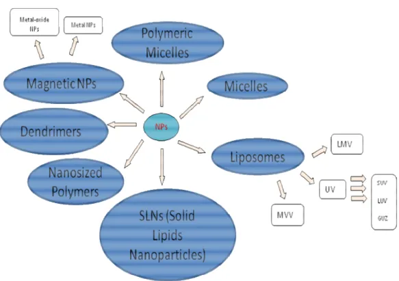
17 Figure 2
Figure 2: Nanosized systems employed in Molecular Imaging.
Micelles are aggregates of amphiphilic molecules in which the polar head groups are in contact with water while the hydrophobic moieties are located in the particle core to minimize their contact with water. In non-polar solvent the orientation is reversed and the micelle is called “reverse micelle”. Micelles can assemble in different shapes, such as spherical, cylindrical, lamellar and disk-shaped. Micelles can form above the critical micelle concentration (CMC) and the size can vary from 1 nm to 100 nm. This system should be thought like a
18
dynamic structure because there is a continuous exchange between micelles and the aqueous solution.
Generally it is possible to modify the surface of the original mixture of micelle-forming surfactants adding new co-surfactants and also size and charge of these systems according always to the molecule that has to be transported (43).
It is really common to use PEG surface modification to avoid uptake by the reticuloendothelial system and mononuclear phagocytes, obtaining in this way stealth-shielded nanoparticles (44). There are also polymeric micelles that are generally more stable than surfactant micelles, and form at markedly lower CMCs. These are less dynamic than those formed from surfactants and it has been demonstrated that if free polymers are separated from polymers forming micelles they are still stable even if CMC decreases (45).
One or several phospholipid bilayers can auto-associate among themselves to form liposomes, leaving an aqueous compartment inside the structure. This structure is strongly similar to cell membranes and has been the subject of study of many research groups in chemistry, biophysics and pharmaceutics.
Liposomes for their composition and also for the preparation method can present different features. Liposomes are usually prepared in simple multi-step operations:
• dissolution of the lipids in an organic solvent • solvent evaporation
• dispersion of the dried lipids in an aqueous solution
The main differences can arise on the method used to disperse the dried lipids: lipid dispersion can be obtained by hydration of the phospholipid film, sonication, microfluidification, extrusion, reverse-phase evaporation, ether infusion, injection of an ethanol solution, freeze-drying/rehydration, freezing/thawing, surfactant removal, or electroformation. The liposome
19
characteristics depend on the preparation technique and in this way the main size of these supramolecular structures can be from tens to a hundred micrometers. The number of lipidic bilayers can vary so that liposomes can be classified into: MLV (Multilamellar Vesicles) present a diameter more than 0.5 µm, UV (Unilamellar Vesicles) with a diameter more than 1 µm and MVV (Multi Vesicular Vesicles) with a diameter more than 1 µm and many vesicles inside the liposome. Unilamellar vesicles can be divided into three subclasses: SUV (Small Unilamellar Vesicles), with a diameter between 20 and 100 nm, LUV (Large Unilamellar Vesicles), with diameters above 100 nm, GUV (Giant Unilamellar Vesicles), with diameters above 1 mm (46).
These lipidic supramolecular systems are endowed with several advantages, and have been used as a vector for drugs. Moreover they are able to include both hydrophobic compounds, anchored into the bilayer, or hydrophilic substances, encapsulated in their cavity.
The passive encapsulation technique consists to rehydrate the lipidic film in the presence of the active substance (47), while the active process is applied after having made liposome using a concentration or pH gradient and it consists to mix hydrophobic drugs with phospholipids before the formation of the lipidic film (48, 49).
Solid lipid nanoparticles (SLN) are made generally of glycerides and present a diameter that can vary from 50 to 1000 nm. These nanoparticles can be obtained by different methods as high pressure homogenization, microemulsion, nanoprecipitation, where crystallinity of lipids can be different and the degree of this depends on the method which is used to product SLNs. The technique chosen to prepare SLNs is important because crystalline polymorphism defines the colloidal stability of these systems (50).
Nanoparticles can be constituted by metals or metal oxides for use as MRI contrast agents. For example there are two types of iron oxide mainly
20
investigated for their properties of superparamagnetism as maghemite (γ-Fe2O3)
and magnetite (Fe3O4). Superparamagnetism is a really important property that
allows for stability and individual dispersion of the particles after that an external magnetic field has been removed (51). Another class of metal nanoparticles are gold-nanoshells where there is an external Au-coating around a silica core, giving them a favourable use as contrast agents in optical coherence tomography, because variations in their size and shape allows for the precise tuning of their resonance wavelength (52).
Dendrimers are molecules composed of monomers with a central plurifunctional core and a tree-like growth process around. Due to their large number of surface groups and monodispersity, dendrimers have the ability to create multivalent interactions leading to their efficient use as vectors (53). Polymers are large molecules composed of many repeated subunits. Polymers, both natural and synthetic, are created via polymerization of many monomers and so their consequently large molecular mass relative to small molecule compounds produces unique physical properties. Moreover incorporating inorganic nanostructures into a polymer matrix, the properties of both materials can be synergistically combined to create new attributes.
Metal-organic frameworks (MOFs) are also known as coordination polymers and they consist into an hybrid system made of metal connecting points and organic bridging ligands (54, 55). In the structure of MOFs bridging ligands can connect either metal ions or small metal clusters. These systems show really interesting properties, mainly due to their wide porosity with tunable pore sizes, shapes and functionalities. High agent loadings through several methods (direct incorporation into the framework or post-synthetic modification), intrinsic biodegradability from labile metal-ligand bonds, high porosity for loading/release of entrapped agents, and versatile functionalization methodologies, making them suitable to be used in biological research.
21
References
1 Weissleder R and Mahmood U (2000), Radiology, 219, 316-333 2 Weissleder R (2006), Science, 312, 1168-1171
3 Harisinghani MG, Barentsz J, Hahn PF, Deserno WM, Tabatabaei S, van de Kaa CH, de la Rosette J, Weissleder R. (2003), N. Engl. J. Med., 348, 2491-2499
4 Hawrysz D J, Sevick-Muraca E M (2000), Neoplasia, 2, 388–417 5 Weissleder R, Ntziachristos V (2003), Nat. Med., 9, 123–128 6 Cutler M, (1929), Surg. Gynecol. Obstet., 48, 721– 729
7 Schoder H, Erdi Y E, Larson S M, Yeung H W (2003), Eur. J. Nucl. Med. Mol. Imaging, 30, 1419– 1437
8 Haglund M M, Hochman D W, Spence A M, Berger M S (1994), Neurosurgery, 35, 930–940
9 Ntziachristos V, Yodh A G, Schnall M, Chance B (2000), Proc. Natl. Acad. Sci. U. S. A., 97, 2767– 2772
10 Guo Z, Park S, Yoon J, Shin I (2014), Chem. Soc. Rev., 43, 16.
11 Nakayama A, Del Monte F, Hajjar R J, Frangioni J V (2002), Mol. Imaging,
1, 365-377
12 Levin C S, (2005), Eur. J. Nucl. Med. Mol. Imaging, 32, 325-345
13 Becherer A, Szabó M, Karanikas G, Wunderbaldinger P, Angelberger P, Raderer M, Kurtaran A, Dudczak R, Kletter K (2004), J. Nucl. Med., 45, 1161-1167
14 Reivick M, Kuhl D, Wolf A, Greenberg J, Phelps M, Ido T, Casella V, Hoffmann E, Alavi A, Sokoloff L (1979), Circ. Res., 44, 127-137
15 Welch M J, Redvanly C S, Eds, Handbook of Radiopharmaceuticals: Radiochemistry and Applications; John Wiley & Sons Inc.: Hoboken, NJ, 2003
22
16 Meares C F, Chen X, Ed (2009), Recent Advances of Bioconjugation Chemistry in Molecular Imaging, 227-241.
17 Kaden T A, Gokel G W, Ed (1993), Advances in Supramolecular Chemistry Elsevier, 3, 65
18 Suchy M, Hudson R H (2008), E. Eur. J. Org. Chem., 29, 4847-4865 19 Fomenko V V, Polynova T N, Porai-Koshits M A, Varlamova G L, Pechurova N I (1973), Zh. Strukt. Khim., 14 (3), 571
20 Broan C J, Cox J P L, Craig A S, Kataky R, Parker D, Harrison A, Randall A, Ferguson G (1991), J. Chem. Soc., Perkin Trans., 2 (1), 87-91
21 Henze M, Schuhmacher J, Hipp P, Kowalski J, Becker D W, Doll J, Macke H R, Hofmann M, Debus J, Haberkorn U (2001), J. Nucl. Med., 42 (7), 1053-1056
22 Sharma V, Beatty A, Wey S P, Dahlheimer J, Pica C M, Crankshaw C L, Bass L, Green M A, Welch M J, Piwnica-Worms D (2000), Chem. Biol., 7 (5), 335-343
23 Sharma V, Prior J L, Belinsky M G, Kruh G D, Piwnica-Worms D (2005), J. Nucl. Med., 46 (2), 354-364
24 Sharma V (2004), Bioconjugate Chem., 15 (6), 1464-1474
25 Smith-Bindman R, Lipson J, Marcus R, Kim K-P, Mahesh M, Gould R, Berrington de Gonzalez A, Miglioretti D L (2009), Arch. Intern. Med., 169, 2078-2086
26 Christiansen C (2005), Toxicology, 209, 185-187
27 Anelli P, Brocchetta M, Fretta R, Lattuada L, Mortillaro A, WO2010105983 (A1), 2010
28 Garrett P R, Meshkov S L, Perlmutter G S (1984), Radiology, 153, 545-546 29 Raptopoulos (1989), V. Radiol. Clin. N. Am., 27, 631-651
23
31 Schutt E, Klein D, Mattrey R, Riess J (2003), Angew Chem Int Ed Engl, 42, 3218-3235
32 Blomley M, Cosgrove D, Albrecht T (1998), Radiology, 224, 124-134 33 Schneider M (1999), Echocardiography 16 (7, Pt 2), 743–746
34 Nanda N C, Wistran D C, Karlsberg R P, Hack T C, Smith W B, Foley D A, Picard M H, Cotter B (2002), Echocardiography, 19(1), 27–36
35 Sidhu P S, Allan P L, Cattin F, Cosgrove D O, Davies A H, Do D D, Karakagil S, Langholz J, Legemate D A, Martegani A, Llull J B, Pezzoli C, Spinazzi A (2006), Br J Radiol, 79 (937), 44–51
36 De Pascale A, Garofalo G, Perna M, Priola S, Fava C (2006), Radiol Med,
111, 539–550
37 Farrar T C, Becker E D, Pulse and Fourier Transform NMR, Academic Press: New York, 1971
38 Greif WL, Buxton RB, Lauffer RB, Saini S, Stark DD, Wedeen VJ, Rosen BR, Brady TJ (1985), Radiology, 157, 461-466
39 Solomon I (1955), Phys. Rev., 99, 559
40 Fonseca C, Simoes S, Gaspar R (2002), J. Control. Release, 83 (2), 273-286 41 Uhrich K E, Cannizzaro S M, Langer R S, Shakesheff K M (1999), Chem. Rev., 99, 3181-3198
42 Mall S, Buckton G, Rawlins D A (1996), J. Pharm. Sci., 85, 75-78 43 Kataoka K (1994), J. Macromol. Sci. Pure Appl. Chem., 31, 1759-1769 44Brannon-Peppas L, Blanchette J O (2004), Adv. Drug Del. Rev., 56, 1649-1659
45 Malmsten M, Surfactants and Polymers in Drug Delivery, Marcel Dekker, New York, 2002
46 Colletier J P, Chaize B, Winterhalter M, Fournier D (2002), BMC Biotechnol., 2, 9-15
24
47 Abraham S A, McKenzie C, Masin D, Ng R, Harasym T O, Mayer L D, Bally M B (2004), Clin. Cancer Res., 10, 728-738
48 Stevens P J, Lee R J (2003), Anticancer Res., 23, 439-442
49 Müller R H, Mäder K, Gohla S (2000), Eur. J. Pharm. Biopharm., 50, 161-178
50 Gupta A K, Gupta M (2005), Biomaterials, 26, 3995-4021
51 Chen J, Saeki F, Wiley B J, Cang H, Cobb M J, Li Z Y, Au L, Zhang H, Kimmey M B, Li X, Xia Y (2005), Nano Lett., 5 (3), 473-477
52 Bromberg L E, Ron E (1998), Adv. Drug Delivery Rev., 31, 197-221 53 Bradshaw D, Warren J E, Rosseinsky M J (2007), Science, 315, 977–980 54 Yaghi O M, O’Keeffe M, Ockwig N W, Chae H K, Eddaodi M, Kim J (2003), Nature, 423, 705–714
55 Kayser O, Lemke A, Hernandez-Trejo N, (2005) Curr Pharm Biotechnol 6, 3-5.
25
Outline of the thesis
The PhD research activity was devoted to explore original nanosystems for Molecular Imaging diagnostic applications. In this wide area of research, the activity of all three years was focused on the design, the preparation and the testing of novel and improved components for nanosized CAs for MRI.
The choice of MRI resides in its enormous potential in Molecular Imaging applications, where the superior resolution attainable led to extensive use of this diagnostic technique. The research group in which this PhD work was carried out has nearly 20 years of experience in MRI CAs, and the collaborations established in this period allow to perform a comprehensive and detailed evaluation of the compounds synthesized.
While resolution of MRI images is already very high, its sensitivity is far from optimal, suffering from the need for a significant concentration of paramagnetic species in order to obtain a suitable contrast efficiency. At least 107-108 molecules are needed to clearly visualize one cell, compared to the 102-103 molecules/cell required for Optical Imaging and the 100-101 molecules/cell needed for visualization with PET/SPECT techniques.
Nanosized systems are especially suited to solve the sensitivity problems, as a single nanometric assembly used as a container can deliver several thousands CA molecules at a time. In addition, the nanosystem can be easily decorated with vector molecules, addressed to its targeting to specific cells or tissues, and with auxiliary components dedicated to the tailoring of its physical, chemical and biologic properties or to modify the nanoparticle behaviour. For example, nanoparticle coating with PEG or other hydrophilic appendages lead to an improvement of both solubility and plasmatic half-life, helping and extending their in vivo application.
26
This thesis reports the results of three different activities carried out during the PhD period and summarized as follows:
1) Design, synthesis and testing of three original amphiphilic MRI CAs, based on Mn2+-complexes of novel lipophilic derivatives of the well know ligand
EDTA. These amphiphilic chelates are included in the formulation of supramolecular lipid-based aggregates (micelles, liposomes, serum-albumin non-covalent conjugates), where they play an active role as contrast agents. 2) A unimolecular nanosized chelating agent is prepared by functionalization of polyethyleneimine with phosphonic groups. The loading of this polymeric chelating agent with Gd3+ ions leads to a paramagnetic nanosized system; relaxometric investigations on this system was undertaken to evaluate its potential as MRI contrast agent. The paramagnetic polynuclear complex is not intended as a component of a larger nanosized Molecular Imaging probe, rather it represents as such a complete MRI CA, containing and transporting several paramagnetic metal ions in a compact molecule of the desired nanometric size. 3) The synthetic activity of the PhD project involved the preparation of several different compounds, the latter being usually represented by polyamine-based chelating agents and their synthetic precursors. In view of the fact that most of these compounds will be employed in vivo, preliminary examination of their toxicity is routinely and periodically performed on the compounds prepared. During this screening, a biological activity was noticed for a family of diamines employed for the synthesis of chelating agents for Mn2+, and found to be related to a block in the G1 phase of cell cycle.
27
Supramolecular
assemblies
based
on
amphiphilic Mn(II)-Complexes as High
Relaxivity MRI probes
Mauro Botta,[a] Valeria De Biasio,[b] Giovanni B. Giovenzana,[b] Gabriele
Rolla[a] and Lorenzo Tei[a]
Submitting to Chemistry - an Asian Journal
Abstract
In the field of MRI contrast agents (CAs) amphiphilic paramagnetic complexes are usually endowed with increased plasmatic half-life and high relaxivity values, but limited examples of amphiphilic Mn(II)-based CAs have been reported to date. In this work the Mn(II)-complexes of three original amphiphilic ligands derived from the well known EDTA and embodying one/two aliphatic chains were evaluated as potential MRI contrast agents and compared. Strong self-association to micelles and binding to serum albumin brought observed relaxivities to significantly high values and confirm the possibility to use supramolecular assemblies based on amphiphilic Mn(II)-complexes as high relaxivity MRI probes.
28
Introduction
MRI contrast agents (CAs) are paramagnetic complexes employed to increase the relaxation rate of the observed nuclei (mainly 1H), allowing better diagnostic images to be acquired in a shorter period (1).Gadolinium based CAs are currently employed in clinical MRI due to the large paramagnetism showed by the f7-lanthanide ion (2). Recent reports on the insurgence of pathologies associated to the release of Gd(III) ions in nephropathic patients, relived the interest in CAs based on different metal ions (3). Mn(II) represents a promising alternative (4): although magnetic properties are lower than those of Gd(III), it is an essential endogenous element and its eventual release in trace in vivo could be more easily managed by the organism. In addition, it is cheap and widely available. In the field of MRI CAs, amphiphilic paramagnetic complexes are usually studied in virtue of their increased plasmatic half-life and high relaxivity values, the latter being usually observed when the CAs are tightly bound to macromolecular or supramolecular systems as lipid-based formulations (5). Suitably designed lipophilic CAs can reach relaxivity values up to 90 mM-1s-1 (6) compared to the values in the range 3-5 mM-1s-1 showed by currently used “hydrophilic” CAs. While a large number of lipophilic Gd-based CAs were studied in the last two decades and summarized in recent reviews (7), scarce reports of lipophilic Mn-based CAs may be found in the scientific literature. Inclusion of the Mn(II)-complex of DTPA bis(stearylamide) in small unilamellar liposomes are the first example, dating back to 1989-90 (8); nevertheless, this system and the cognate EDTA-oleyl ester (9) recent analog represent preliminary approaches where stability issues were not tackled. Large non-hydrophilic appendages were later implanted on the backbone of the EDTA ligand to promote non-covalent interactions with serum albumin, leading to
29
blood-pool CAs with a significant improvement of the observed relaxivities (10, 11).
Almost all chelating agents employed for the formation of Gd(III) or Mn(II) complexes for clinical MRI applications may be classified in two families, i.e. acyclic and macrocyclic polyamino-polycarboxylates (12-17). As Mn-based CAs are usually endowed with lower relaxivity values compared to the corresponding Gd-based CAs, we were prompted to explore the possibility to boost their relaxometric properties by taking advantage of the formation of lipidic aggregates. To this purpose, three different ligands were synthesized, choosing the hexadentate ligands EDTA as preferred scaffold (Figure 1). The Mn(II) complexes of these newly synthesised ligands were prepared and studied by relaxometric techniques in both their aggregated form and in their supramolecular adduct with serum albumin.
Figure 1
Figure 1: Ligands synthesized in the present work.
N HOOC COOH N COOH COOH TDDTA N HOOC COOH N COOH COOH N HOOC COOH N COOH COOH ODDTA HCDTA
30
Results and Discussion
Ligands design
Several examples of Mn(II)-complexes with penta- or hexa-dentate polyaminopolycarboxylate ligands have been recently reported with the aim to form Mn(II)-complexes with the best balance between their thermodynamic and kinetic stabilities and the number of inner-sphere water molecules (4). Among them, Mn(II)-EDTA-like chelates still represent good systems on which build a contrast agent as Mn(II)-EDTA is well tolerated, sufficiently stable and it is has a water molecule coordinated to the metal centre (q = 1) (18). Moreover, the Mn(II)EDTA complex has the great advantage to be endowed with a water exchange rate (kex) close to an optimum value for a contrast agent (4.7 × 10⁸ s-1
at 25°C). The latter property is fundamental to reach high relaxivity values once the Mn(II) complex is in a slowly tumbling form with long τR i.e. when it is in
aggregated form or bound to serum albumin.
Lipophilic EDTA derivatives were designed by placing one (TDDTA and
ODDTA) or two (HCDTA) aliphatic straight chains on the central
ethylenediamine backbone, to reduce any potential steric hindrance with the metal coordination sphere. To this end, we considered the recently reported approach that used Gd-chelates bearing two aliphatic chains on adjacent coordinating arms for the formation of micelles or liposomes with the aim to reduce considerably their local rotational motion (19); with this approach, high relaxivity values up to 40 mM-1s-1 (298 K, 20 MHz) were obtained for liposomes loaded with GdDOTA(GAC12)2. Moreover, it is well known that
lipidic nanoparticles formed by amphiphilic complexes containing one aliphatic chain are not sufficiently stable in vivo giving also a strong haemolytic effect. Thus, it was shown that the presence of two aliphatic chains in the complex
31
allows a higher stability, a long blood half-life and no haemolytic effect. Finally, as also the length of the hydrophobic chain can influence stability, size and clearance time of the nanoparticles, twelve and sixteen carbon chains were chosen in the synthesised ligands. It is noteworthy that amphiphiles bearing C12
chains have been recently reported to accelerate considerably the clearance rate of liposomes embedding them in the membrane bilayer (20).
Ligands synthesis
Single chain EDTA derivatives were prepared through a common synthetic pathway, depicted in Scheme 1. 1-Tetradecene and 1-octadecene, were subjected to bromination with molecular dibromine. The vicinal dibromides were converted to the corresponding diamine through sequential conversion to the vic-diazide (with NaN3 in DMF) and catalytic transfer hydrogenation (CTH)
(HCOONH4, Pd/C in refluxing 2-propanol). Conventional per-alkylation with
t-butylbromoacetate/K2CO3 in acetonitrile and t-butyl esters removal with neat
TFA completes the 5-steps preparation of 1,2-tetradecanediamine-N,N,N’,N’-tetraacetic acid (TDDTA) and 1,2-octadecanediamine-N,N,N’,N’-1,2-tetradecanediamine-N,N,N’,N’-tetraacetic acid (ODDTA).
32 Scheme 1: Synthesis of TDDTA and ODDTA.
The third ligand (13,14-hexacosanediamine-N,N,N’,N’-tetraacetic acid) (HCDTA) was designed to embody two aliphatic straight chains on the vicinal position of the ethylenediamine backbone. For its preparation, a different synthetic strategy was devised, reported in Scheme 2. A multicomponent condensation of 1H-benzotriazol, glyoxal and dibenzylamine led to a mixed diaminal (21), then reacted with 2 equivalents of dodecylmagnesium bromide giving, through a double displacement of the benzotriazole moieties, a tetrabenzylated 1,2-didodecylethylenediamine 6. Removal of N-benzyl groups is accomplished by CTH to give 13,14-hexacosanediamine 7. The primary diamine is tetraalkylated with the combination t-butyl bromoacetate-potassium carbonate in acetonitrile generating the tetraester 8, subsequently converted to the final ligand HCDTA by treatment with neat TFA.
R R Br Br R N3 N3 Br2 1a 1b 2a2b (99%) (99%) CH2Cl2 NaN3 DMF HCOONH4 Pd/C iPrOH R NH2 NH2 Br COOtBu K2CO3 CH3CN R N N COOtBu COOtBu COOtBu tBuOOC TFA R N N COOH COOH COOH HOOC a R = C12H25 b R = C16H33 3a 3b 4a (56%) 4b (61%) 5a (67%) 5b (75%) TDDTA ODDTA
33 Scheme 2
Scheme 2: Synthetic scheme for HCDTA.
(BtH) CHO CHO C12H25MgBr Mg C12H25Br HCOONH4 Pd/C K2CO3 CH3CN TFA N H N N N N Bt Bt Ph Ph Ph Ph N N C12H25 C12H25 Ph Ph Ph Ph NH2 NH2 C12H25 C12H25 BrCH2COOtBu N N C12H25 C12H25 COOtBu COOtBu COOtBu COOtBu N N COOH COOH COOH COOH C12H25 C12H25 84% 61% 82% 67% 6 7 8 9 Ph NH Ph 2 HCDTA
34
Relaxometric characterization
A detailed relaxometric characterization was undertaken on the Mn(II)-complexes prepared on the three ligands described in the previous section, in order to ascertain their potential as MRI CAs.
Determination of the relaxivity of the Mn(II)-complexes was at first accomplished on dilute solution, in order to measure relaxivity values on the monomeric form of amphiphilic complexes, i.e. under the critical micelle concentration above which self-aggregation may occur. The complexes
Mn(II)-TDDTA and Mn(II)-ODDTA showed in these conditions relaxivities of 4.7
mM-1s-1 and 5.9 mM-1s-1, respectively, in good agreement with values expected for monomeric Mn(II)-chelate with one coordinated water molecule (q = 1). On the other side, the complex Mn(II)-HCDTA shows even in dilute conditions evidence of self-association and no cmc values can be determined. The relaxivity value of 18.4 mM-1s-1 measured is compatible with a supramolecular aggregate even at the lowest concentrations used for relaxivity determination. In order to establish the microscopic (supra)molecular parameters that govern the relaxivity of the complexes, their 1H NMRD profiles were measured at different temperatures and concentrations ∼ 1 mM.
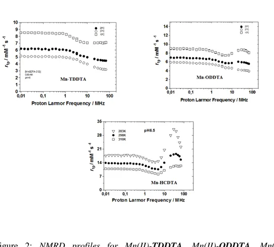
35 Figure 2
Figure 2: NMRD profiles for TDDTA, ODDTA,
Mn(II)-HCDTA.
The results reported in Figures 2 clearly show some difference in the behaviour of the three paramagnetic species. Mn(II)-TDDTA, bearing a single C12
-aliphatic chain on the ethylenediamine backbone, show a NMRD profile quite similar to that observed for the parent EDTA. On the other hand, in the NMR profile of the longer chain analog Mn(II)-ODDTA a distinctive broad hump at higher fields demonstrate the formation of high-molecular weight paramagnetic species, expected in view of the concentration used (>cmc). A sharper and higher peak is observed in the NMRD profile of Mn(II)-HCDTA, indicative of the presence of higher-grade supramolecular aggregate than the former chelate,
36
further supporting the strong propensity of Mn(II)-HCDTA to self-association phenomena.
The NMRD profiles data at three different temperatures allow to extract information on the structural dynamics of the paramagnetic complexes. Fitting the data by the Solomon-Bloembergen-Morgan theory gives access to the parameters that influence the relaxivity, reported in Table 1.
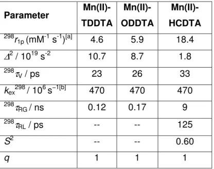
Table 1: Best-fit parameters obtained by analysis of the 1
H NMRD profiles for Mn(II)-TDDTA, Mn(II)-ODDTA, Mn(II)-HCDTA in their aggregated form.
Parameter Mn(II)-TDDTA Mn(II)-ODDTA Mn(II)-HCDTA 298r 1p (mM-1 s-1)[a] 4.6 5.9 18.4
∆
2 / 1019 s-2 10.7 8.7 1.8 298τ
V / ps 23 26 33 kex298 / 106 s−1[b] 470 470 470 298τ
RG / ns 0.12 0.17 9 298τ
RL / ps -- -- 125 S2 -- -- 0.60 q 1 1 1RMnH, a and 298D were fixed in the fitting procedure to 2.83 Å, 3.6 Å and 2.3 ×
10-5 cm2 s-1, respectively. [a] 30 MHz; [b] fixed in the fitting procedure.
The fitting shows that the three complexes share a common EDTA-like behaviour, with q = 1 and fast exchange rate of the coordinated water molecules (to be confirmed by 17O NMR measurements). This is extremely useful in view
37
of the devised application, as the combination of a short lifetime of the coordinated water and the raise in the rotational correlation time related to the increase in the size of the paramagnetic system lead to very high values of relaxivity. Indeed Mn(II)-HCDTA with its significant degree of association in these conditions and shows an interesting relaxivity of 18.4 mM-1s-1, with the beneficial combination of fast water exchange and slow rotation of the paramagnetic system.
Interaction with HSA
The favourable structural and dynamic features of the EDTA-derived amphiphilic complexes pushed us to explore their behaviour in different conditions. In particular, investigation of the complexes in the presence of human serum albumin (HSA) is of great interest because this 64 KDa protein is present in high concentration in blood and its remarkable transport properties was shown to be useful for hosting paramagnetic complexes, boosting their relaxivity (6).
Relaxometric titration was then conducted on the three complexes, exposing them to increasing concentration of HSA, registering the increase in relaxivity consequent to the eventual interaction.
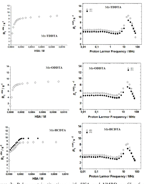
The results of these titrations are reported in Figure 3, along with the NMRD profiles of the complexes in the presence of an excess of HSA registered at two different temperatures (298K and 310K).
38 Figure 3
Figure 3: Relaxometric titrations with HSA and NMRD profiles for
39
The analysis of the combined data from the titrations and the NMRD profiles allowed to extract parameters related to the interactions and to the paramagnetic system formed by the interactions of the complexes with HSA.
The results of the fitting of the data according to the SBM theory are reported in Table 2.
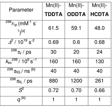
Table 2: Best-fit parameters obtained by analysis of the 1
H NMRD profiles for the interaction of Mn(II)-TDDTA, Mn(II)-ODDTA, Mn(II)-HCDTA with HSA.
RMnH, a and 298D were fixed in the fitting procedure to 2.83 Å, 3.6 Å and 2.3 ×
10-5 cm2 s-1, respectively. [a] 20 MHz; [b] fixed in the fitting procedure.
The results are very interesting as all three paramagnetic complexes efficiently interacts with the protein as can be appreciated by the curves of the relaxometric titrations. Even more astonishing are the relaxivity of the
Parameter Mn(II)-TDDTA Mn(II)-ODDTA Mn(II)-HCDTA 298r 1p (mM-1 s -1)[a] 61.5 59.1 48.0
∆
2 / 1019 s-2 0.69 0.6 0.68 298τ
V / ps 30 20 24 kex298 / 106 s−1 160 160 130 298τ
RG / ns [b] 40 40 40 298τ
RL / ps 880 1200 261 S2 0.72 0.70 0.66 q [b] 1 1 140
paramagnetic systems Mn(II)-complex-HSA, towering in the range 48-61.5 mM-1s-1; these values are extremely high for this family of CAs, and rarely observed even with Gd-complexes.
The parameters of Table 2 clearly evidence that the huge increase in the relaxivity has to be ascribed to the slowed tumbling of the paramagnetic chelate as a consequence of the strong interaction with serum albumin, the latter hosting the former in one of the possible binding sites.
Additional work is in progress in order to shed more light on the nature of this strong interaction and to test these paramagnetic complexes in the presence of other molecular or supramolecular hosts.
Conclusion
In the present work, three amphiphilic ligands derived from the well known EDTA are designed and prepared. Two ligands bear a single aliphatic chain (C12 and C16), placed on the ethylenediamine central moiety; on the same
residue of the third ligand are placed in vicinal positions two identical aliphatic C12 chains. The Mn(II)-complexes prepared from these ligands show relaxivity
values of 4.6-5.9 mM-1s-1 for the single-chain derivatives, compatible with an EDTA-like behaviour. In contrast, the double-chain derivatives shows higher relaxivity values due to self-aggregation even at the lowest concentration employed. The presence of HSA to simulate the blood environment brings a massive increase in the relaxivity, boosting to 48-61.5 mM-1s-1 as a consequence of the strong interaction of the serum protein, hosting the paramagnetic complex in a compact assembly. These results shows that Mn(II)-based CAs may be used to reach high relaxivity, usually ascribed only to Gd-based CAs, and may represent a valid and efficient alternative to the latter.
41
Experimental
General Information
Solvents and starting materials were purchased from Aldrich, Acros, Fluka and Alfa and used without further purification. All water solutions were prepared from ultrapure laboratory grade water (18 MΩ·cm) obtained from Millipore/MilliQ purification system. 1H NMR spectra were recorded at 300 MHz with a Jeol Eclipse ECP300 spectrometer. Chemical shifts are reported in ppm with the protic impurities of the deuterated solvent as the internal reference. Mass spectra were obtained with a Finnigan LCQ-ion trap equipped with an electrospray source. TLC and gravimetric chromatography were performed with silica gel 60 (MN Kieselgel 60).
General procedure for the preparation of 1,2-dibromoalkanes
1-Alkene (1.0eq) was dissolved in dichloromethane (1 M) and bromine (1.05eq) was added dropwise to the solution until a stable red colour was obtained. The reaction mixture was further stirred at room temperature for 1 h. The solvent was removed in vacuo giving 2a-2b as colourless oils, directly used for the following step. 1,2-Dibromododecane (2a) Yield 99%.1H-NMR (CDCl3):4.16 (tt, 1H, J = 9.3, 6.8 Hz) 3.84 (dd, 1H, J = 10.2, 4.4 Hz), 3.61 (t, 1H, J = 9.9 Hz), 2.11-2.06 (m, 1H), 1.88-1.72 (m, 1H), 1.63-1.19 (m, 20H), 0.88 (bt, 3H, J = 6.6 Hz).13C-NMR (CDCl3): 53.2 (CH), 36.4 (CH2), 36.1 (CH2), 36.0 (CH2), 29.76 (4xCH2), 29.66 (CH2), 29.51 (CH2), 29.48 (CH2), 28.9 (CH2), 26.9 (CH2), 22.8 (CH2), 14.2 (CH3).
42 1,2-Dibromooctadecane (2b) Yield 99%. 1H-NMR (CDCl3): 4.15 (m, 1H), 3.83 (dd, 1H, J= 10.1, 4.3 Hz), 3.60 (t, 1H, J= 9.9 Hz), 1.87-1.74 (m, 2H), 1.62-1.16 (m, 28H), 0.89 (bt, 3H, J= 6.7 Hz).13C-NMR (CDCl 3): 53.0 (CH), 36.2 (CH2), 36.1 (CH2), 32.1 (CH2), 29.9 (7xCH2), 29.7 (CH2), 29.6 (2xCH2), 29.0 (CH2), 26.9 (CH2), 22.9 (CH2), 14.3 (CH3).
General procedure for the preparation of 1,2-diazidoalkanes
Compounds2a-2b (1.0 eq) and sodium azide (3.0 eq) were dissolved in dimethylformamide (1M) and the reaction was heated to 130 °C for 2 h. Ice was added to the mixture and the pH adjusted to ∼10 with NaOH. The mixture was extracted with diethyl ether (2×10 mL), and the extracts were concentrated with the help of a N2 flow. Due to the unstable nature of diazides, compounds 3a-3b
were not isolated and characterized and were used as such for the following step.
General procedure for the preparation of 1,2-diaminoalkanes 4a-4b
Compounds 3a-3b (1.0 eq) were slowly added dropwise into a round-bottomed flask containing triphenylphosphine (2.2 eq) and THF/diethyl ether/conc. aq NH3(3:2:1, 0.3M) at room temperature. The reaction mixtures were the strirred
at 60 °C for 3 h, then cooled, diluted with water and basified with NaOH. Extraction with diethyl ether followed by drying with Na2SO4, filtration and
evaporation gave the crude diamines. Purification by silica-gel column chromatography (eluant toluene:2-propanol:ammonia 9:1:0.5) provided pure
4a-4b as light yellow oils. 1,2-Dodecanediamine (4a)
Yield 56%, 1H-NMR (CDCl3): 2.69-2.53 (m, 2H), 2.36 (dd, 1H, J= 11.8, 7.8
Hz), 1.68-1.52 (m, 6H), 1.38-1.09 (m, 20H), 0.79 (bt, 3H, J= 5.5 Hz).13C-NMR
43
(5xCH2), 29.4 (CH2), 26.2 (CH2), 22.7 (CH2), 14.1 (CH3). ESI-MS(+): calcd for
C14H32N2: 228.3; found: 229.3 (MH+); 251.4 (MNa+).
1,2-Octadecanediamine (4b): Yield 61%, 1H-NMR (CDCl3): 2.62 (dd, 1H, J=
12.2, 3.7 Hz), 2.54 (m, 1H), 2.33 (dd, 1H, J= 12.1, 7.5 Hz), 1.33-1.08 (m, 34H), 0.77 (bt, 3H, J= 6.3 Hz). 13C-NMR (CDCl3): 53.6 (CH),48.6 (CH2), 35.7 (CH2),
31.9 (CH2), 29.7-29.5 (10xCH2), 29.3 (CH2), 26.2 (CH2), 22.6 (CH2), 14.1
(CH3). ESI-MS(+): calcd for C18H40N2: 284.3; found: 285.5 (MH+).
General procedure for the preparation of compounds 5a-5b: Diamines 4a-4b (1.0 eq) were dissolved in acetonitrile (1.5 M) at room temperature.
Powdered K2CO3 (7.0 eq) and t-butyl 2-bromoacetate (4.5 eq) were added to the
mixtures.The reaction mixture was stirred at 60 °C for 8 h.Inorganic salts were separated by filtration and the filtrate evaporated in vacuo. Crude compounds
5a-5b were purified by gravimetric chromatography (eluant petroleum ether,
ethyl acetate, 2-propanol9.4:0.4:0.2), obtaining the pure tetraesters as clear light yellow viscous oils.
Tetra-t-butyl 1,2-dodecanediamine-N,N,N’,N’-tetraacetate (5a): Yield 67%,
1H-NMR (CDCl 3): 3.49-3.34 (m, 8H), 2.85 (dd, 1H, J= 13.2, 6.1 Hz), 2.74 (bquint, 1H, J= 6.1 Hz), 2.46 (dd, 1H, J= 13.2, 6.1 Hz), 1.42 (s, 18H), 1.41 (s, 18H), 1.37 (m, 4H), 1.27-1.16 (m, 18H),0.84 (bt, 3H, J= 6.4 Hz). 13C-NMR (CDCl3): 171.7 (C), 170.9 (C), 80.7 (C), 80.4 (C), 61.1 (CH), 56.5 (CH2), 56.3 (CH2), 53.3 (CH2), 32.0 (CH2), 30.9 (CH2), 30.0 (CH2), 29.7 (5xCH2), 29.4 (CH2), 28.22 (CH3), 28.16 (CH3), 27.0 (CH2), 22.7 (CH2), 14.1 (CH3).
ESI-MS(+): calcd for C38H72N2O8: 684.5; found: 685.6 (MH+), 707.6 (MNa+), 723.5
(MK+), 629.5 (MH+-tBu). Tetra-t-butyl 1,2-octadecanediamine-N,N,N’,N’-tetraacetate (5b): Yield75%, 1H-NMR (CDCl 3): 3.96 (d, 2H, J= 17.5 Hz), 3.85 (d, 2H, J= 17.5 Hz), 3.82 (d, 2H, J= 17.2 Hz), 3.67 (d, 2H, J= 17.2 Hz), 3.46-3.33 (m, 2H), 3.05 (bt, 1H), 1.45-1.12 (m, 30 H), 1.28 (s, 36 H), 0.89 (bt, 3H, J = 6.6 Hz).13


![Figure 5: Relaxivity vs pH for meglumine-coated Gd-PEI-P ([Gd-PEI-P]=70 µ M, [meglumine]=100mM, 20 MHz, 298K)](https://thumb-eu.123doks.com/thumbv2/123dokorg/4814536.50046/71.748.156.589.190.524/figure-relaxivity-meglumine-coated-pei-pei-meglumine-mhz.webp)
