Università degli Studi del Piemonte Orientale
“Amedeo Avogadro”
Dipartimento di Scienze del Farmaco
Dottorato di Ricerca in Biotecnologie Farmaceutiche ed Alimentari XXVIII ciclo a.a. 2012-2015
Migraine and its chronicization to medication overuse headache:
a pharmacogenetic-pharmacoepidemiologic approach
Copyright Migraine Action (UK National Charity) 2015, reproduced with permission. Sarah Cargnin
Università degli Studi del Piemonte Orientale
“Amedeo Avogadro”
Dipartimento di Scienze del Farmaco
Dottorato di Ricerca in Biotecnologie Farmaceutiche ed Alimentari
XXVIII ciclo a.a. 2012-2015
Migraine and its chronicization to medication overuse headache:
a pharmacogenetic-pharmacoepidemiologic approach
Sarah Cargnin
Supervised by Prof. Armando Genazzani
Co-Advisor: Dott. Salvatore Terrazzino
Sarah Cargnin was the recipient of a PhD fellowship by
Compagnia San Paolo, Italy, that is deeply acknowledged.
“Variability is the law of life,
and as no two faces are the same, so no two bodies are alike,
and no two individuals react alike and behave alike under the abnormal conditions
which we know as disease.”
Contents
Chapter 1 ... 1 Introduction ... 1 1. Migraine ... 1 1.1 Clinical presentation ... 4 1.2 Pathophysiology ... 81.3 Genetic basis of migraine ... 11
1.4 Pharmacological treatment ... 15
1.5 Pharmacogenetics of migraine ... 23
2. Medication overuse headache ... 27
2.1 Clinical classification ... 28
2.2 Pathophysiology ... 31
2.3 Treatment ... 33
2.4 Genetics and pharmacogenetics of MOH ... 36
2.5 Pharmacoepidemiology of triptan-overuse headache ... 39
References ... 44
Chapter 2 ... 59
Outlines of the thesis ... 61
Chapter 3 ... 65
"Pharmacogenomics of episodic migraine: time has come for a step forward" Chapter 4 ... 91
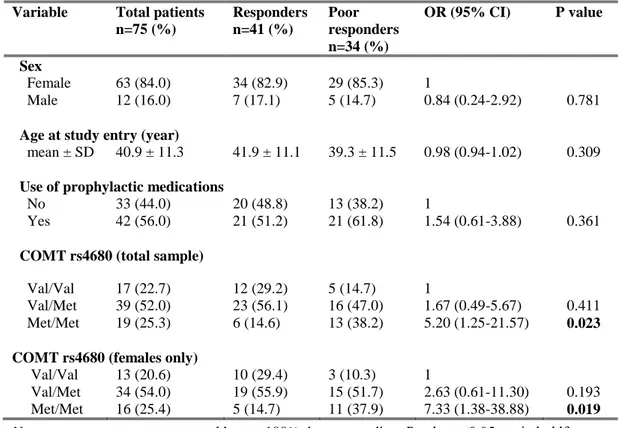
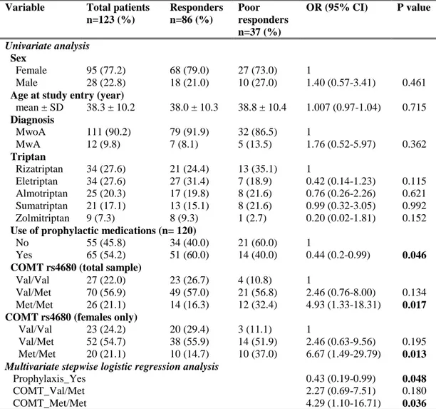
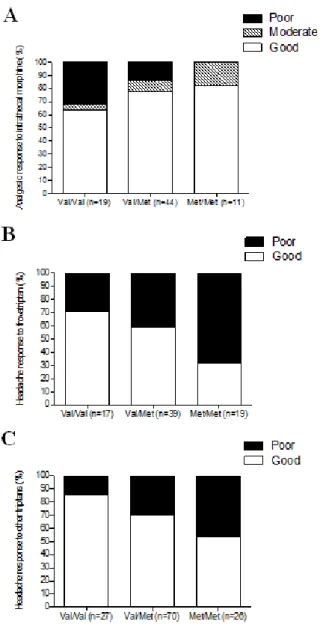
"An opposite-direction modulation of the COMT val158met polymorphism on the clinical response to intrathecal morphine and triptans" Chapter 5 ... 119
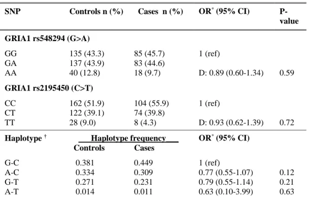
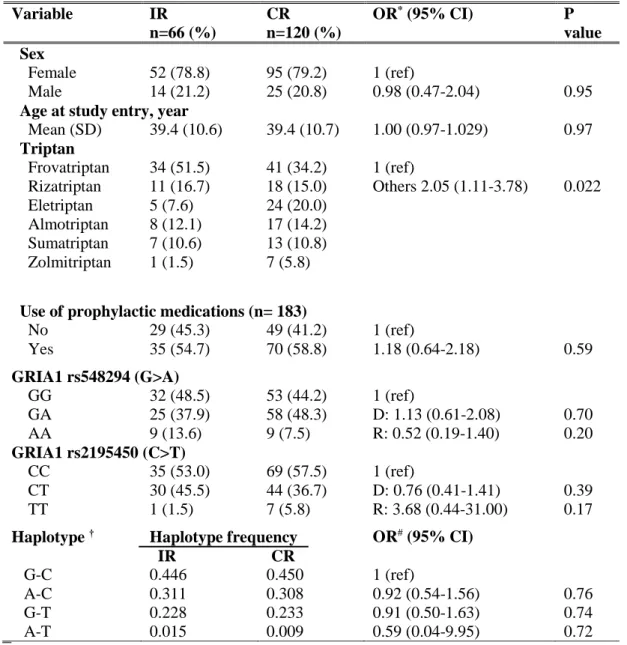
"Lack of association between GRIA1 polymorphisms and haplotypes with migraine without aura or response to triptans."
Chapter 6 ... 139
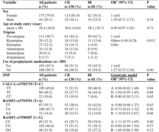
"Association of RAMP1 rs7590387 with the risk of migraine transformation into medication overuse headache” Chapter 7 ... 161
"Combined effect of common gene variants on response to drug withdrawal therapy in medication overuse headache" Chapter 8 ... 189
"Functional polymorphisms in COMT and SLC6A4 genes influence the prognosis of patients with medication overuse headache after withdrawal therapy" Chapter 9 ... 211
"Triptan use in Italy: insights from administrative databases” Chapter 10 ... 233
Discussion ... 235
Chapter 11 ... 247
List of publications... 249
1
Introduction
Headache disorders are ubiquitous, with almost half of the world’s adult population having recently experienced one or more headache types [1]. Migraine, tension-type headache and medication overuse headache (MOH) are currently the most common headache disorders worldwide, leading to significantly reduced patients’ quality of life and higher costs for society [2]. The Global Burden of Disease Study 2013 (GBD2013) reported migraine as the sixth highest cause of disability in the world and, for the first time, highlighted the relevance of MOH as the 18th most disabling disease [3]. Despite the substantial contribute to public-ill-health of such headache types, their burden is still paradoxically ignored.
Migraine as well as medication overuse headache still remain under-recognized, under-diagnosed and under-treated, with more than 50% of patients worldwide estimated to be primarily self-treated and not consulting a specialist [1].
In the light of the aforementioned remarks, healthcare for headache disorders has the need to be still more improved at multiple levels, with the final aim of successfully diagnose and effectively treat these neurological conditions, primarily including migraine and MOH.
1. Migraine
Migraine is a common primary headache disorder that affects around 10% of the worldwide adult population [4] [5], with a male to female ratio of 1:3 [6].
This neurobiological condition is characterized by altered brain sensitivity and its clinical presentation, in terms of pain intensity, attack frequency and associated
2
symptoms, broadly varies among migraineurs [7]. Individual variability in migraine phenotype has emerged as presumably resulting from the interaction between multiple genetic and non-genetic (endogenous or exogenous) risk factors [8]. Supporting this, genetic epidemiologic studies reported migraine as a strongly heritable disorder with a substantial risk of familiar occurrence. Twin studies have revealed that almost 50% of migraine susceptibility risk could be attributable to additive genes while the remainder may be due to shared or unshared environmental factors among twins [9]. Exposure to stress, odours, bright lights and sounds, hypoglycemia, consumption of certain foods (for example chocolate, salami, milk, alcohol and excessive caffeine intake) smoking, and sleep disturbances are now listed among potential exogenous triggers of migraine attacks in predisposed subjects [10].
Gender differences in migraine occurrence were also hypothesized on the basis of epidemiological evidences reporting a major prevalence of migraine in females compared to males. Migraine is more common in women than in men during adulthood, with a prevalence peak in women’s fertile age. However, this imbalance in terms of prevalence between sexes does not occur in childhood and old age, when the disorder tends to affect the same percentages of males and females [11]. Fluctuation of female sex hormones levels have been proposed as the more plausible endogenous factor responsible for variations of migraine prevalence throughout female life span [12]. Consistent with this, stable and low levels of ovarian hormones, characterizing childhood as well as natural menopause, emerged to be correlated with lower migraine frequency. On the contrary, reduced levels of estradiol and progesterone, typical of menstruation, resulted triggering frequent migraine attacks [13]. In addition to hormonal causes, sex differences in pain perception and stress response have been proposed as additional plausible factors explaining gender differences in migraine [14].
3
From a physiological viewpoint, a migraine attack could be defined as a complex sequence of brain events that can last from hours to days. For descriptive purposes, several “phases” can be simplistically identified in a migraine attack, each of them corresponding to specific chemical, biological and anatomical mechanisms partially overlapping in the sequence of migraine phases themselves. These phases, chronologically ordered, are named premonitory, aura (if present), headache and
postdrome phase. It must be highlighted that not all aforementioned phases are
manifested by all migraineurs or present in all attacks.
More than 80% of adults and a slightly lower percentage of children experience
premonitory symptoms, which can occur up to hours before headache. The most
commonly reported premonitory symptoms preceding headache are mood changes, fatigue, irritability, difficulty concentrating, stiff neck, phonophobia and nausea. Other symptoms that have been reported in this phase include change in appetite, bloating, piloerection and change in facial expression or body perception. Some symptoms can resolve before the headache phase, whereas others can increase in intensity in headache phase and furthermore persist during the postdrome phase [15].
A consistent proportion of migraineurs also manifests a set of neurological symptoms, called aura, which usually occur after premonitory signals and before the headache phase. Sometimes, migraine aura begins after the pain phase has commenced or continues into the headache phase. The most common type of aura is the visual one, characterized by visual symptoms ranging from the most common scintillating scotoma to the rarest visual hallucination. Some subjects can also subsequently experience sensory and language aura, respectively characterized by migrating paresthesias and altered language capacity [16].
The migraine headache phase properly named is characterized by headache pain, usually described as of pulsating quality and with unilateral localization. Headache
4
pain tends to occur in a gradual manner till reaching a stable moderate or severe intensity during the remaining part of headache phase. Photophobia, phonophobia, nausea and vomit can frequently accompany headache pain [17].
After headache resolution, symptoms such as tiredness, weakness, cognitive difficulties, mood changes, residual head pain, dizziness and gastrointestinal symptoms are frequent among migraineurs. These symptoms characterize migraine
postdrome phase, which can occur from hours to days after resolution of headache.
The overlapping between premonitory and postdrome symptoms fostered the hypothesis that postdromal signs could be present throughout the attack, probably disguised by headache, nausea or, if present, aura [18].
1.1 Clinical presentation
The International Classification of Headache Disorders, version III beta (ICHD-IIIb) recognizes two major subtypes of migraine, which are migraine without aura and migraine with aura [17]. These migraine types are not exclusive but can co-occur in the same migraineur, either alternating or at different phases of patient’s life [19].
Migraine without aura (MwoA), previously named “common migraine” or
“hemicrania simplex”, is defined as a recurrent headache disorder with headache attacks lasting between 4 and 72 hours and characterized by headache pain having at least two of the following characteristics: typical unilateral location, moderate or severe intensity, pulsating quality and aggravation by routinary physical activity, such as walking or climbing stairs. Nausea, vomiting, photophobia and phonophobia may accompany migraine [17].
5
Table 1: ICHD-IIIb diagnostic criteria for migraine without aura [17].
CRITERIA DESCRIPTION
A: At least five attacks fulfilling criteria B–D
B: Headache attacks lasting 4-72 hours (untreated or unsuccessfully treated)
C: Headache has at least two of the following four characteristics: 1. unilateral location
2. pulsating quality
3. moderate or severe pain intensity
4. aggravation by or causing avoidance of routine physical activity (e.g. walking or climbing stairs)
D: During headache at least one of the following: 1. nausea and/or vomiting
2. photophobia and phonophobia
E: Not better accounted for by another ICHD-3 diagnosis.
In both men and women, migraine without aura is more common than migraine with aura and often seems to correlate with menstrual status in women [11]. On the contrary, the frequency of migraine without aura attacks usually decreases during pregnancy, when the levels of ovarian hormones in serum are stable and high [14]. In children and adolescents aged under 18 years and affected by MwoA, the headache pain is more often bilateral than in adults and unilateral pain usually emerges in late adolescence or in early adult life [17].
Around 20-30% of migraineurs experience an additional complex of transient neurological symptoms, called aura, which can precede or accompany headache pain. [19]. Migraine with aura (Mwa), previously also named “classic or classical
6
migraine” “ophthalmic, hemiparaesthetic, hemiplegic or aphasic migraine” or “complicated migraine”, is defined by ICHD-IIIb as a migraine characterized by “recurrent attacks, lasting minutes, of unilateral fully reversible visual, sensory or other central nervous system symptoms that usually develop gradually and are usually followed by headache and associated migraine symptoms” [17].
More precisely, migraine aura is defined as a fully reversible neurologic dysfunction, characterized by a gradual onset of specific symptoms, which usually last between 5 and 60 minutes. Visual aura is the most common type of aura, occurring in 99% of migraineurs [20]. Migraineurs describe visual impairments symptoms characterized by “a zigzag figure near the point of fixation that may gradually spread right or left and assume a laterally convex shape with an angulated scintillating edge, leaving absolute or variable degrees of relative scotoma in its wake” [17]. Posphenes, white or coloured dot, curved lines or other geometric forms may also be seen by migraine aura patients.
Sensory aura is the next more frequent aura symptoms, affecting around 54% of MwA patients [21], followed by language aura, which is experienced by 32% of MwA subjects [22]. Sensory aura, is often described as migrating paresthesias, with numbness typically occurring in the hands and then affecting arms, face, lip and tongue (cheiro-oral sensory changes). Language aura may consist of impaired language comprehension and speaking, accompanied or not by decreased ability to read or write. It should be noted that sensory aura may lead to stuttering words and should not be confused with language aura [23]. If co-occurring, aura symptoms types usually follow one other in succession, starting from visual and ending with aphasic symptoms [17].
7
Table 2: ICHD-IIIb diagnostic criteria for migraine without aura.
CRITERIA DESCRIPTION
A: At least two attacks fulfilling criteria B and C
B: One or more of the following fully reversible aura symptoms: 1. visual
2. sensory
3. speech and/or language 4. motor
5. brainstem 6. retinal
C: At least two of the following four characteristics:
1. at least one aura symptom spreads gradually over ≥ 5 minutes, and/or two or more symptoms occur in succession
2. each individual aura symptom lasts 5-60 minutes 3. at least one aura symptom is unilateral
4. the aura is accompanied, or followed within 60 minutes, by headache
D: Not better accounted for by another ICHD-3 diagnosis, and transient ischaemic attack has been excluded.
Aura symptoms may worsen or appear for the first time during pregnancy, highlighting the role of high estrogen levels in the development of migraine with aura [11].
Some patients may also experience migraine aura followed by a less distinct headache or even without headache. In the first case, they may suffer from typical
aura with headache, characterized by aura accompanied or followed within 60
8
patients are diagnosed as having had typical aura without headache, in which aura is neither accompanied nor followed by headache of any sort within 60 minutes [17].
1.2 Pathophysiology
Migraine pathophysiology has greatly evolved in the last century till currently describing migraine as a “neurovascular” disorder, resulting from the interaction between vascular and neurological events.
The exact cause of migraine is still not completely understood but some key events characterizing migraine pathophysiology have emerged, such as the phenomenon of cortical spreading depression and the activation of trigeminovascular systems accompanied by neurogenic inflammation [24].
Historically, two independent theories explaining migraine etiology were proposed. The “vascular theory” was initially introduced by Thomas Willis in 1664, who suggested, for the first time, the involvement of vasodilatation of cerebral and meningeal arteries in migraine onset [25]. In the 20th century Graham and Wolff refined the aforementioned theory, highlighting that the vascular event was mediated by an initial intracranial vasoconstriction subsequently followed by a rebound vasodilatation of the extracranial terminal branches of the external carotid artery. Consistent with this hypothesis was the observed pulsating quality of migraine pain, which they attributed to the amplitude of pulsation of the occipital and superficial temporal branches of the external carotid artery [26].
The alternative theory subsequently proposed was the “neurogenic” one, which reported neuronal networks dysfunctions as key mechanisms underlying migraine pathogenesis. Support for the neurogenic theory came from the fact that typical
9
neurological symptoms of migraine aura could not be explained by a vascular pathophysiological model alone. In 1944, Aristides Leão, described for the first time the phenomenon of cortical spreading depression (CSD), defined as “a self-propagating wave of depolarization that begins in the neuronal/glia cells of local areas of the brain and subsequently spread in all directions at a rate of ≈ 3 mm/min” [27]. Milner in 1958 and then Olsen in the 1980s caught, for the first time, similarities between CSD phenomenon and aura symptoms (previously described in 1941 by the psychologist Karl Spencer Lashley) and fostered pathogenic theories changing from primary vascular to primary neuronal mechanism [28].
However, even if aura is a symptom exclusive for a specific migraine subtype, the observation of the possible co-occurrence of the two subtypes of migraine in the same subject suggested that these two conditions could share at least some mechanisms involved in the initiation and resolution of attacks.
In this context, Moskowitz and collegues, in the 1980s, integrated the vascular theory with the neurogenic one and proposed the “trigeminovascular theory”. They hypothesized that probably CSD could depolarize the trigeminocervical nerve terminals innervating meninges which in turn may release pro-inflammatory peptides responsible for “neurogenic inflammation”, consisting in meningeal vessels vasodilatation and plasma protein extravasation. Such neurogenic inflammation was hypothesized to lead to migraine pain [29].
Nowadays, migraine is viewed as a complex neurological disorder that affects multiple cortical, subcortical and brainstem areas that regulate several functions, including autonomic, affective, cognitive and sensory ones. The interaction between neurons, glia and blood vessels still appears to be crucial in migraine pathogenesis, with CSD phenomenon and activation of trigeminovascular system accompanied by neurogenic inflammation being recognized as key events implicated in migraine pathogenesis.
10
In more detail, trigeminovascular system consists of trigeminal nerve and nerve fibers innervating intra- and extra- cranial meningeal blood vessel as well as brainstem [30]. The trigeminovascular system role is to regulate both neurotransmission of pain signals and vascular tone. The transmission pathway originates in trigeminal ganglion neurons whose central axons reach the nociceptive dorsal horn laminae of the spinal trigeminal nucleus. Here, the nociceptors converge on neurons receiving additional inputs from the periorbital skin and precranial muscles. The ascending axons of spinal trigeminal nucleus neurons convey nociceptive signals to brainstem, hypothalamic and basal ganglia nuclei, which overall seem to be crucial in mediating symptoms typical of migraine attacks, such as nausea initiation, vomiting, yawning, loss of appetite, anxiety, irritability and lacrimation [7]. Trigeminal sensory nerves store several vasoactive neuropeptides, including substance P, calcitonin gene-related peptide (CGRP), neurokinin A, nitric oxide (NO) and pituitary adenylate cyclase-activating peptide (PACAP). These neuropeptides, when released, are crucial in provoking an increase in blood flow and vasodilation in meningeal vascular system, where edema and inflammation occur, conceivably causing headache pain. Supporting the hypothesis of trigeminovascular system activation is the evidence of increased levels of CGRP both in external and internal jugular blood of migraineurs during headache attack [31].
Recently Amin and colleagues identified, through a magnetic resonance angiography imaging technique, that the release of neuropeptides and proinflammatory substances (e.g. histamine, bradykinin, serotonin and prostaglandins) from trigeminal afferents in the meningeal vessels, results in an altered molecular environment causing sensitization of both peripheral and central neurons of trigeminovascular system. Once sensitized, peripheral trigeminovascular neurons begin to respond to stimuli to which they showed minimal or no response at baseline [32].
11
Endogenous events that may trigger trigeminovascular system, and so neuropeptides release, are still largely unknown. Several experimental models report CSD as an important upstream trigger of trigeminovascular system activation. Cortical SD seems to activate trigeminovascular pathway, leading to a prolonged neurogenic inflammation around meningeal vessels [33]. Even if it is not clear how CSD begins in human brain, genetic factors are likely to play a substantial role in modulating individual CSD susceptibility by regulating brain excitability.
1.3 Genetic basis of migraine
The current knowledge of genetic basis of migraine has come from different scientific approaches, encompassing linkage studies in family pedigrees and candidate genes or genome-wide association studies.
Much interest of geneticists has been initially focused on the study of genetic basis of familial hemiplegic migraine (FHM), a rare subtype of migraine with aura, characterized by a prolonged visual aura typically accompanied by hemiparesis [24]. FHM is inherited in an autosomal dominant fashion and was identified as the first primary headache disorder with a genetic basis. The substantial overlapping between phenotypes of FHM and migraine with aura fostered FHM to be considered a good model to study the genetic architecture of migraine. In this context, CACNA1A, ATP1A2 and SCNA1A have been identified as causative genes in FHM. More precisely, the proteins encoded by these three genes form channels involved in the regulation of ions flow across neuronal and glial cell membranes. Collectively, these genes regulate glutamate availability in the synaptic cleft by means of a fine tuning of glutamate release and re-uptake as well as the generation of action potentials. It has been hypothesized that mutations in all
12
three genes may result in an increased efflux of glutamate and potassium in the synaptic cleft, in turn leading to an increased susceptibility to cortical spreading depression. Mutations in these genes specifically identify three different forms of FHM, named, respectively, FHM1, FHM2 and FHM3 [7]. However, these identified genetic variants do not account for the totality of FHM cases and it has been suggested that additional genetic variants resulting in altered ions flow may lead individuals to be more susceptible to FHM [34] [35].
Strongly supporting the hypothesis of common genetic basis between FHM and other migraine subtypes is the subsequent evidence provided by Cuenca-Leon and colleagues which, in 2009, reported 14q32 locus as a shared susceptibility locus in a Spanish family affected by FHM, MwoA and MwA [36].
Numerous linkage studies were also performed on families of different ethnic origin. Several migraine susceptibility loci have been identified in a wide range of chromosomes, suggesting how migraine could be substantially considered as a polygenic disease [37]. However, many of the findings in the field have not been replicated in populations of different origin. Heterogeneity of analyzed cohorts, both in terms of phenotype and diagnosis, or the existence of rare high-impact family specific markers may explain, almost in part, inconsistency of results between studies.
Another commonly used approach to identify genetic variants potentially influencing migraine susceptibility is represented by the conduction of candidate-genes association studies performed in a case-control setting. These studies generally attempted to investigate genetic basis of migraine through the analysis of tagging SNPs or selected functional variants located on specific genes plausibly involved in migraine susceptibility. On the basis of available evidences regarding migraine pathophysiological mechanisms, the majority of studied SNPs were located in genes primarily involved in neurological, hormonal and vascular
13
functions. Among neurological candidate genes, genes involved in serotonin neurotransmission have been extensively studied. Even if the exact mechanisms of the serotoninergic system in migraine are still unknown, a deficit on 5-HT descending pain inhibitory system is still probably today the most implicated in migraine pathophysiology [38]. These genes included 5-HT1B, 1D and 2C receptors, SLC6A4 (encoding for SERT, a serotonin transporter), TPH2 (encoding for tryptophan hydroxylase, a rate-limiting enzyme in the synthesis of serotonin), and MAOA (encoding for monoamine oxidase A enzyme which degrade serotonin). Similarly, genes regulating dopaminergic neurotransmission were studied, such as DRD2 (dopamine receptor D2), DRD3 (dopamine receptor D3), SLC6A3 (encoding for the dopamine active transporter), DBH (encoding for dopamine beta hydroxylase, an enzyme converting dopamine into noradrenaline), TH (tyrosine hydroxylase) and COMT (catechol-O-methyltansferase). In the same way, genes implicated in glutamate transmission, such as GRIA1 (glutamate receptor 1) and GRIA3 (glutamate receptor 3) were considered as well good candidate genes [37].
As previously mentioned, fluctuations of hormonal levels, in particular estrogen, seem to trigger migraine attacks. In this context, genetic variations on ESR1 (encoding for estrogen receptor 1) and PGR genes (progesterone receptor gene) emerged as potential predictors of migraine susceptibility [39] [40]. Being vascular events also crucial in migraine pathogenesis, genetic variations in CGRP (calcitonin gene related peptide), ACE (angiotensin I-converting enzyme), MTHFR (methylenetetrahydrofolate reductase) and NOTCH3 were analyzed in correlation with migraine risk [37].
However, even if several replication studies were performed to validate significant associations between SNPs in all aforementioned genes and migraine risk, a consistent part of these results still remains controversial or inconclusive. The small effect size of these genetic variants, the generally small sample size of studies
14
as well as the strong heterogeneity among migraineurs included in the patient sets may explain, almost in part, the difficulty in replicating such results.
A recent advance in the knowledge of genetic basis of migraine has been provided by genome-wide association studies (GWAS). More precisely, two GWAS were performed on clinic based collections of patients affected by MwA [41] or MwoA [42] whilst two other were conducted on population-based collections of migraineurs [43] [44]. A meta-analysis performed by Anttila and colleagues in 2013 quantitatively summarized previously reported results in the four GWAS added together with unreported GWAS findings for migraine susceptibility obtained in additional 9 population-based collections [45]. A total of 12 loci emerged as significantly associated with migraine susceptibility. Among them, 5 loci were new (near AJAP1, near TSPAN2, FHL5, c7orf10, and near MMP16) while 7 confirmed previously found migraine loci (PRDM16 [43], MEF2D [42], TRPM8 [41] [43], near TGFBR2 [42], PHACTR1 [42], ASTN2 [42], and LRP1 [43]). More in detail, LRP1 modulates synaptic transmission through the NMDA receptor while MEF2D is involved in glutamatergic excitatory synapse. AJAP1, TSPAN2 and MMP16 (whose encoded protein cleaves LPR1) seem to regulate activity of metalloproteinase, which are responsible for the breakdown of extracellular matrix in normal physiological processes. ASTN2 and FHL5 are involved in neuronal neurodevelopment and synaptic plasticity.
Overall, these results support the importance of glutamatergic neurotransmission and neuronal development/plasticity in migraine pathogenesis and, in conjunction with all evidences retrieved from other genetic studies, make conceivable the hypothesis of a “generalized” neuronal hyperexcitability in migraine brain.
15
1.4 Pharmacological treatment
The pharmacological treatment of migraine traditionally includes:
i) the acute/symptomatic treatment, able to relieve headache pain and corollary symptoms accompanying migraine;
ii) the prophylactic therapy, aimed to reduce intensity and frequency of headache attacks.
The Italian guidelines for primary headache (revised version 2012) recommend the symptomatic treatment alone when migraine attacks are not-disabling or if they occur < 4 days per month. On the contrary, preventive therapy is suggested when disabling migraine attacks are present for ≥ 4 days per month or in case of poor response to symptomatic drugs [46].
Acute treatment
Nowadays, several drugs are available for symptomatic pharmacological treatment of migraine. Antimigraine drugs can be divided in specific antimigraine drugs, characterized by being effective only for headache pain (i.e. triptans and ergot derivates), and non-specific antimigraine drugs, resulting effective in relieving pain of several origins, including headache (e.g. non-steroidal anti-inflammatory drugs, simple analgesics and antiemetics) [47].
The Italian guidelines for primary headache (revised version 2012) propose a “stratified approach” when the initial pharmacological acute treatment of migraine attacks must be outlined. More precisely, specific drugs are recommended for the treatment of moderate or severe attacks while non-specific drugs should be administered for the therapy of migraine attacks of mild/moderate intensity. Moreover, preparations with only one active principle should be preferred and the
16
most appropriate drug should be taken at the lowest effective dosage and as early as possible from migraine attack onset [46].
As mentioned before, specific antimigraine drugs include triptans and ergot derivates.
Triptans are considered by many specialists as the gold-standard therapy of migraine. However, there’s a lack of knowledge regarding the complexity of their hypothesized multiple mechanisms of action. Their action is primarily attributed to their agonist effect on serotonin 5HT1B/D/F receptors, resulting in the vasoconstriction of meningeal vessels and in the inhibition of the neurogenic inflammation. In addition to this well-known mechanism, recent evidences support the potential action of triptans on modulating calcium and potassium currents in dural-projecting trigeminal neurons in vitro [48].
Nowadays, six triptans are on Italian market: sumatriptan, frovatriptan, eletriptan, rizatriptan, almotriptan, zolmitriptan. All triptans are available in conventional oral formulations. Sumatriptan is also marketed in other several formulations, such as nasal spray, suppository and subcutaneous, the latest one being considered the most effective one [49]. Zolmitriptan has also the nasal spray formulation and, as rizatriptan, is available in rapid-dissolving formulations (RPD), which have an effectiveness similar to that of tablet formulations of the same drugs at the same dosages [50], [51].
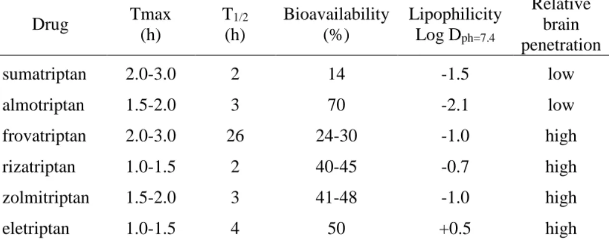
Triptans differ from each other in terms of specific pharmacokinetic and lipophilic properties [52].
17
Table 3: Pharmacokinetic parameters and lipophilicity of oral triptans available on
Italian market [52]. Drug Tmax (h) T1/2 (h) Bioavailability (%) Lipophilicity Log Dph=7.4 Relative brain penetration sumatriptan 2.0-3.0 2 14 -1.5 low almotriptan 1.5-2.0 3 70 -2.1 low frovatriptan 2.0-3.0 26 24-30 -1.0 high rizatriptan 1.0-1.5 2 40-45 -0.7 high zolmitriptan 1.5-2.0 3 41-48 -1.0 high eletriptan 1.0-1.5 4 50 +0.5 high
T max: time to maximum concentration; t1/2: biological half-life; Log D ph=7.4 quantifies triptan
lipophilicity (increasing numbers indicate greater lipid solubility).
They are all effective on relieving migraine pain, associated symptoms and functional disability, without provoking common relevant side effects in patients. Only the 4-5% of migraineurs treated with subcutaneous sumatriptan experience the “triptan syndrome”, characterized by chest pain and chest and neck tightness, while the 2-4% of subject also complaint symptoms such as somnolence, dizziness, paresthesia, asthenia, nausea and facial flush [46]. Myocardial infarction and ictus related to triptan use were rarely reported. Contraindications to triptans are uncontrolled blood hypertension, coronary artery disease, history of ischemic stroke, peripheral artery disease, pregnancy, lactation and age <18 years and > 65years.
Despite the well documented efficacy of triptans, about one third of migraineurs do not respond to a specific triptan [53]: in this case, other triptans can be administered and, if ineffective, non-steroidal anti-inflammatory drugs can be used [54].
18
Ergot derivates have been used for many years in migraine treatment before the advent of triptans. These drugs are partial agonists of α-adrenergic and dopaminergic receptors and interact with 5HT1B,D,F receptors, so resulting in vasoconstriction of meningeal vessels and inhibition of pro-inflammatory neuropeptides release.
Ergotamine (not available in Italy) and dihydroergotamine are the most common used ergot derivates and have emerged to be more effective vs placebo in migraine treatment, but less effective compared to triptans [55]. Their use should be restricted to migraineurs which fail to be responsive to other drugs. Ergot derivates may worsen nausea and vomiting in migraineurs, so contemporary administration of antiemetic drugs is generally indicated. Major side effects include nausea, vomiting, diarrhea and ergotism. Cardiovascular and cerebrovascular diseases, uncontrolled blood hypertension, Raynaud disease, renal failure, pregnancy and lactation represent contraindications for ergot derivates treatment.
Non-specific antimigraine drugs include non-steroidal anti-inflammatory drugs,
simple and combination analgesics, antiemetics, barbiturates, lidocaine and steroids.
Non-steroidal anti-inflammatory drugs (NSAIDs) are indicated for the treatment of migraine attacks of mild or moderate intensity or when triptans resulted ineffective or contraindicated [46]. Acetaminophen, acetylsalicylic acid, ibuprofen, naproxen sodium, and diclofenac potassium have good evidence supporting their use for migraine. Among the latest, acetaminophen is considered less effective than other NSAIDs and is generally administered for the treatment of relatively mild migraine attacks [56]. Acetylsalicylic acid is recommended in subjects with cardio- and cerebrovascular comorbidities. Only ibuprofen (the most commonly used one), ketoprofen and morniflumate can be used in patients under 14 years of age. Due to
19
their well-known mechanisms of action, adverse events mainly consist in gastrointestinal toxicities (e.g. gastric pain, gastric or duodenal ulcer).
Concerning simple analgesics, paracetamol has a good efficacy on mild-moderate migraine pain and corollary symptoms and it is the first-choice drug for migraine treatment during pregnancy.
Analgesics in combination have the same indication of NSAIDs and simple analgesics. Analgesic combinations available in Italy include: acetylsalicylic acid + acethaminophen + propyhenazone, acetylsalicylic acid + acethaminophen + indomethacin (with/without caffeine), and acetaminophen + propyphenazone and acetaminophen + codeine. Side effects and contraindications of combinations are the same as those for each component [46].
Antiemetics are considered adjuvant drugs in migraine therapy, especially during attacks characterized by prominent nausea and vomiting. The association between antiemetics and other classes of antimigraine drugs, such as NSAIDs, ergotamine or triptans, may result in a major absorption of antimigraine drugs and in the effective treatment of nausea and vomiting. Among common used antiemetics, metoclopramide emerged effective in reducing headache pain if intravenously administered alone in migraineurs [57].
Barbiturates are usually prescribed in addition to other antimigraine drugs. However, this class of drugs should in principle be avoided because barbiturates may induce intoxication, addiction and dependence.
Steroids, such as dexamethasone and prednisone, seem also to be effective in migraine treatment but available findings are conflicting. Only one study demonstrated the superiority of the association dexamethasone-rizatriptan compared with rizatriptan alone in women affected by menstrual migraine.
20
New specific drugs for the abortive treatment of migraine: a focus on CGRP as a therapeutic target
In the light of the substantial abundance of non-responders among migraineurs treated with triptans, a digression concerning new developed drugs representing a valid alternative to triptans is mandatory.
The evidence of CGRP involvement in the transmission of pain [58] as well as its release during migraine attack [31], fostered the hypothesis that CGRP could be a potential good target for new drugs. In this context, CGRP receptor antagonists emerged as effective molecules compared to placebo. CGRP receptor antagonists include, telcagepant, olcegepant, rimegepant, 3207, BI 44370 TA and MK-1602. However, despite the promising results in term of efficacy, the drug development programmes of all these new drugs terminated because of different reasons, including toxicity evidences and commercial decision. Only MK-1602 is in phase 2 study [59].
In addition to CGRP receptor antagonist, four CGRP monoclonal antibodies (mAbs) were developed, of which three targeting CGRP ligand (LY2951742, ALD-403 and TEV-48125) and one targeting CGRP receptor (AMG 334). The mAbs against the ligand are thought to remove CGRP that is released at perivascular trigeminal sensory nerve fibers, while the receptor mAbs block CGRP signalling. Preliminary data showed positive results in terms of efficacy and safety for all four mAbs. However, subcutaneous and intravenous administration routes, that are mandatory for mAbs, represent a significant limitation for their everyday use in clinical practice [60].
Prophylactic therapy
As previously mentioned, preventive therapy is recommended when disabling migraine attacks are present for ≥ 4 days per month or in case of poor response to symptomatic drugs. A wide range of drugs have been studied for the purpose of
21
reducing both frequency and intensity of migraine attacks. However, the majority of preventive medications currently available were not specifically design for migraine treatment and their use is often limited by their toxicities or eventual inefficacy.
Preventive drugs must be chosen on the basis of patient’s comorbidities, with a particular attention to drug-drug and food-drug interactions. To minimize side effects, the most appropriate drug should be administered at the lower dose and preferentially as a monotherapy. The preventive therapy will be considered effective if the frequency of migraine attacks will be reduced by at least half and if a significant improvement in quality of life will be reached. Clinical benefit may take some time to be obtained, so preventive treatment should be maintained for at least 3 months. Due to the teratogenic effect of the majority of prophylactic drugs, preventive treatment during pregnancy should be limited to special situations for which the use of drugs with the lowest teratogenic effect should be preferred [46].
Preventive drugs include beta-blockers, calcium channel blockers, antidepressants, antiepileptic drugs, angiotensin inhibitors, botulinum toxin A and other supplements. However, few evidences clearly stated their efficacy and no universally accepted guideline unequivocally suggest the preventive treatment of choice for migraine. Depending on the methodological assessment underlying guidelines development, the levels of recommendation reported for every single active principle broadly vary among national and international guidelines.
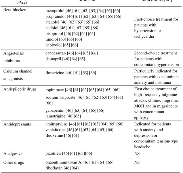
Table 4 simplistically summarized the most commonly recommended preventive drugs on the basis of two Italian [46] [61] and five international guidelines [62] [63] [64] [65] [66] for preventive migraine treatment. No emphasis was herein put on the levels of recommendation for single drugs reported by each guideline. Specific indications for each class of drugs are also reported.
22
Table 4: Prophylactic drugs for migraine.
Prophylactic medications
Pharmacological
class Molecule Indications [46]
Beta-blockers metoprolol [46][61][62][63][64][65][66] propranolol [46][61][62][63][64][65][66] atenolol [46][62][63][65][66] nadolol [46][61][63][65][66] bisoprolol [46][62][64][65] timolol [63][65][66] nebivolol [65][66]
First-choice treatment for patients with hypertension or tachycardia Angiotensin inhibitors candesartan [46][64][65][66] lisinopril [46][64][65] Second-choice treatment for patients with
concomitant hypertension Calcium channel
antagonists
flunarizine [46][61][63][66] Particularly indicated for patients with concomitant anxiety and insomnia Antiepileptic drugs topiramate [46][61][62][63][64][65][66] First choice treatment of
high-frequency migraine attacks, chronic migraine, MOH and in migraineurs with concomitant epilepsy sodium valproate [46][61][62][63][64][65] [66] gabapentin [46][63][64][65][66] lamotrigine [46][65] Antidepressants amitriptyline [46][61][62][63][64][65][66] venlafaxine [46][61][63][64][65][66] fluoxetine [46][61]
Indicated for patients with anxiety and depression or
concomitant tension-type headache
Analgesics pizotifen [46][61][63][66] NS
Other drugs onabutlinum toxin A [46][61][64][65] riboflavin [46][64]
NS
23
1.5 Pharmacogenetics of migraine
Pharmacogenetics is the study of the influence of individual genetic background on drug response, in terms of both therapeutic effect and drug safety. The identification of subpopulations inadequately responding to a specific drug may lead to a personalized pharmacological therapy, resulting in improved patients’ quality of life and optimization of healthcare resources.
Despite rigorous pharmacogenetic studies have been carried out for a multitude of diseases with interesting and clinical-relevant results, surprisingly a very few pharmacogenetic evidences have been reported in migraine. Nevertheless, strong clinical evidences have highlighted a significant individual variability in response to all drugs routinely used for the acute treatment of migraine. A recent network meta-analysis, aimed to compare relative efficacy of triptans with respect to other acute antimigraine drugs, reported that the proportions of responders to abortive antimigraine drugs were 42-76% for triptans, 38% for ergots, 46-52% for NSAIs and paracetamol, and 62-80% for combination therapies [67]. In this context, non- response to acute medications can be hypothesized not to be only influenced by factors such as altered drug absorption, inadequate dosing or incorrect time of drug administration, but also by individual genetic background, plausibly impacting on both pharmacokinetic and pharmacodynamic drug variability. In addition to this, the observation that migraineurs may experience adverse reactions more frequently than other groups of patients [68] further supports the need of improving pharmacogenetic knowledge in migraine, which in turn may allow to unravel subpopulations of migraineurs that, at present, are inadequately treated by antimigraine drugs on the market.
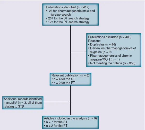
With the aim of collecting all available pharmacogenetic evidences in episodic migraine, we performed in 2012 a systematic review of literature concerning
24
genetic predictors of response to acute or preventive drugs commonly used in migraine treatment 1. We identified only 7 candidate-gene association studies investigating the correlation between genetic polymorphisms and clinical response to acute antimigraine drugs [69] [70] [71] [72] [73] [74] [75]. Despite the wide range of symptomatic drugs available for migraine therapy, all 7 pharmacogenetic studies were performed on patients exclusively treated with triptans. Triptan efficacy was always included as the primary outcome while four out of seven studies also investigated the potential correlation between gene polymorphisms and triptan-induced side effects as secondary endpoint [70] [71] [72] [75]. The polymorphisms studied were related to 5HT1BR (serotonin receptor 5HT1BR) [69] [71] [72] [73] [75], SLC6A4 (serotonin transporter SERT) [69] [73] [74], DRD2 (D2 subtype of dopamine receptor) [69] [73] [74], MAOA (monoamine oxidase A) [69] [73], GNB3 (guanine nucleotide-binding protein G(I)/G(S)/G(T) subunit beta-3) [69] [74], 5HT2A (5HT2A receptor) [69], 5HT1F (5HT1F receptor) [70] , MTHFR (methylene tetrahydrofolate reductase) [69], ACE (angiotensin-converting enzyme) [69], ESR1 (estrogen receptor alpha) and TNF-B (tumor necrosis factor-beta) genes [69]. Among them, only DRD2 rs6275 and SLC6A4 Stin2 VNTR genetic variations have been reported to be potentially associated with triptan response, evaluated in terms of drug efficacy. More precisely, Asuni and colleagues reported that carriers of DRD2 rs6275 C/C genotype showed a better response to triptans compared to other DRD2 rs6275 genotypes [73]. Conversely, Ishii et al found an association between rs6275 C/C genotype and a lack of response to triptan class [69] whereas our group did not confirm any correlation between the aforementioned genetic variant and triptan response [74]. Moreover, we found a significant association between SLC6A4 Stin2 VNTR and response to triptans [74] but Ishii and collegues subsequently fail to replicate our results [69].
25
Four additional pharmacogenetic studies investigating potential genetic predictors of triptan response were published after our database search for the systematic review of literature was performed. More precisely, three out of four studies were performed by our group 2, whereas the remainder was conducted by Christensen and colleagues in 2015 [76]. Christensen et al aimed to test the role of 12 SNPs, previously emerged to be correlated with migraine susceptibility [45], as genetic predictors of response to both acute antimigraine drugs (triptans and ergotamine) and prophylactic medications (beta-blockers, calcium antagonists, angiotensin II receptor antagonists and anti-epileptics) in two large cohorts of migraineurs. A number of tested SNPs showed a trend of association with the efficacy of triptans or some of the prophylactic drugs. Among them, only the genetic variant PRDM16 rs2651899 resulted significantly associated with the efficacy of triptans in the exploratory cohort. However, this correlation failed to be replicated in the validation cohort.
As previously mentioned, pharmacological treatment of migraine includes also preventive therapy. Through our systematic review of the literature, we identified only two pharmacogenetic studies investigating the role of genetic variants as predictors of preventive therapy response [77] [78]. One study did not report any correlation between ACE I/D polymorphism and the response to ACE inhibitors (i.e. lisinopril and candesartan) [77]. The other one found a potential association between non-H mitochondrial DNA haplotypes and better response to riboflavin [78]. No replication cohorts were present in these two studies.
After the publication of our systematic review, only one pharmacogenetic study investigating the role of genetic background in modulating the response to preventive drugs was reported in the literature [79]. This study included 80 migraine patients treated with tricyclic antidepressants (the molecules were not
26
specified). Being nitric oxide involved in migraine pathogenesis, the genetic variant Glu298Asp of NOS3 gene was investigated as genetic predictor of tricyclic antidepressants response. As results, they found that homozygous carries of T allele of the Glu298Asp SNP showed a better response to tricyclic antidepressants compared to other genotypes. However, the absence of a replication cohort as well as the limited sample size of the study do not allow to consider these results as conclusive.
The data reported herein highlight that only little information is still available in the field of migraine pharmacogenetics. Given the high burden of migraine, in terms of high prevalence, disability and healthcare costs, it is surprising that a tool aimed to tailor and optimize antimigraine therapies has not received enough attention. Moreover, the absence of genome-wide association studies in the field emphasizes the immense distance between research in migraine genetics and migraine pharmacogenomics.
27
2. Medication overuse headache
Epidemiological evidences report that each year about 2.5% of people with migraine show a progressive worsening of headache clinical presentation, in terms of both increased frequency and severity of migraine attacks [80]. This “migraine transformation” usually takes place gradually over months to years [81] and leads to the development of chronic migraine, defined by the International Classification of Headache Disorders, version III- beta (ICHD-IIIb) as a “headache occurring on 15 or more days per month for more than 3 months, which has the features of migraine headache on at least 8 days per month” [17].
Chronic migraine has an estimated worldwide prevalence ranging from 1.2% to 2.2%, with a prevalence peak in women aged 18-49 years [82]. It mainly affects people during their most productive years of life, resulting in greater disability and higher direct/indirect costs than episodic migraine [83]. It is often comorbid with fatigue, sleep disorders, cerebrovascular disease, cardiovascular disease, and gastrointestinal problems. Compared to subjects affected by episodic migraine, patients with chronic migraine are twice as likely to have depression, anxiety, other chronic pain, bipolar disorders and respiratory illness, such as asthma and chronic obstructive pulmonary disease [84].
Epidemiological studies have identified several modifiable risk factors associated with chronic migraine, including obesity, depression, female sex, snoring, comorbid pain disorders, stressful life events (e.g. divorce, marriage, or change of employment status) and lower socioeconomic status [85]. However, the ICHD-IIIb reports that the most common risk factor for migraine chronicization is paradoxically represented by the overuse of any one or more antimigraine drugs routinely taken for acute headache pain relief. If some degree of medication
28
overuse co-occurs in chronic migraine patients, their headache disorder is specifically defined by ICHD-IIIb as medication overuse headache.
2.1 Clinical classification
Medication overuse headache (MOH), previously named “rebound headache”, “drug induced headache” or “medication-misuse headache”, is defined by ICHD-IIIb as a secondary chronic “headache occurring on 15 or more days per month developing as a consequence of regular overuse of acute or symptomatic headache medication (on 10 or more, or 15 or more days per month, depending on the medication) for more than 3 months. It usually, but not invariably, resolves after the overuse is stopped.” [17].
Table 5: ICHD-IIIb diagnostic criteria for medication overuse headache [17].
CRITERIA DESCRIPTION
A: Headache occurring on ≥ 15 days per month in a patient with a pre-existing headache disorder
B: Regular overuse for >3 months of one or more drugs that can be taken for acute and/or symptomatic treatment of headache
C: Not better accounted for by another ICHD-3 diagnosis.
It is now accepted that all symptomatic medications may lead to MOH in susceptible patients. The ICHD-IIIb accurately report diagnostic criteria for each MOH “subtypes” according to the specific drug (or drugs) overused [17].
29
Table 6: ICHD-IIIb diagnostic criteria for MOH depending on drug(s) overused
[17].
MOH SUBTYPE DIAGNOSTIC CRITERIA
Ergotamine-overuse headache Regular intake of ergotamine on ≥ 10 days per month for > 3 months.
Triptan-overuse headache Regular intake of one or more triptans, in any formulation, on ≥ 10 days per month for > 3 months.
Simple analgesic-overuse headache
(acetylsalicylic acid or paracetamol)
Regular intake of acetylsalicylic acid on ≥ 15 days per month for >3 months.
Other non-steroidal anti-inflammatory drug (NSAID)-overuse headache
Regular intake of one or more NSAIDs other than acetylsalicylic acid on ≥ 15 days per month for > 3 months. Opioid-overuse headache Regular intake of one or more opioids on ≥ 10 days per
month for > 3 months Combination-analgesic-overuse headache
(intended as formulations combining drugs of two or more classes, each with analgesic effect or acting as adjuvants)
Regular intake of one or more combination analgesic medications on ≥ 10 days/month for > 3 months.
Medication-overuse headache attributed to multiple drug classes not individually overused
Regular intake of any combination of ergotamine, triptans, simple analgesics, NSAIDs and/or opioids on a total of ≥ 10 days per month for > 3 months without overuse of any single drug or drug class alone.
Medication-overuse headache attributed to unverified overuse of multiple drug classes
Regular intake of any combination of ergotamine, triptans, simple analgesics, NSAIDs and/or opioids on ≥10 days per month for > 3 months. The identity, quantity and/or pattern of use or overuse of these classes of drug cannot be reliably established.
Overall, it is estimated that MOH affects around 1% of general population, with a male to female ratio of 1:3-4 [1]. It’s prevalence peaks in the forties and then decrease with older age [86].
30
Chronic analgesic consumption rarely induce MOH in non-headache patients, suggesting that MOH may results from an interaction between the overuse of acute medications and a susceptible patient [87].
The most common headache types affecting patients before MOH development are migraine (65%), tension-type headache (27%) and mixed/other headaches (8%) [88]. The desire to relieve pain and to normally carry out daily activities may lead these subjects to overuse migraine acute medications. The fear of experiencing severe pain and disability may also foster them to preemptively take acute medications at the first weak warning or, even worse, in anticipation of an attack [89]. In this context, it must be underlined that over-the-counter drugs are the most commonly overused antimigraine medications in primary care while triptans and other more potent, centrally acting, drugs emerged as the most overused ones by secondary e tertiary care patients [86]. Among overused drugs, triptans cause MOH faster and with fewer doses compared with ergots and analgesics [90]. In Europe, MOH is rarely cause by opioids overuse cause their prescription is largely discouraged in the medical environment [91].
Clinical presentations of MOH strongly vary among patients in terms of localization, quality and intensity of headache attacks. A plausible explanation underlying this variability in headache characteristics may consist in the fact that more than 90% of patients tend to concomitantly use different painkillers [88]. Common traits in MOH subtypes are morning headaches (possibly due to overnight drug withdrawal) and accompanying symptoms such as neck pain, cutaneous allodynia, rhinorrhea, lacrimation and gastrointestinal symptoms [92].
Risk factor for migraine transformation to MOH other than anti-migraine drug(s) overuse include tobacco smoke, sedentary lifestyle, low socioeconomic status and gastrointestinal complaints [93]. Chronic musculoskeletal pains, anxiety,
31
depression and obsessive compulsive disorder are often comorbid with MOH [94] [95].
2.2 Pathophysiology
The pathophysiological mechanisms underlying the development of MOH are not completely known.
As in migraine, the development of MOH seems to be triggered by cortical spreading depression (CSD). Neurophysiological studies demonstrated that an increased in neuronal excitability, at least in somatosensory and visual cortices, is identifiable in MOH patients. Supporting this, the electrical stimulation on the forehead or limb in MOH patients resulted in increased sensory-evoked cortical potentials that reverted/normalized after withdrawal from acute medications [96]. Moreover, chronic paracetamol or ergot use emerged leading to augmented frequency of CSD in rats [97].
Cortical changes are evident in MOH. More precisely, Coppola and colleagues in 2010 reported that MOH patients, especially those overusing NSAIDs, had an increased sensitization of somatosensory cortex [98]. By measuring glucose metabolism with 18-FDG PET, Fumal et al identified several areas of hypometabolism in MOH patients, including bilateral thalamus, orbifrontal cortex and insula/ventral striatum. All dysmetabolic areas recovered to almost normal glucose uptake after withdrawal of analgesics, except for the orbifrontal cortex [99]. Alterations of gray matter volume in cortical areas were also reported by anatomical studies. More precisely, increased volumes of periacqueductal grey, bilateral thalamus and ventral striatum as well as decreased volumes of orbifrontal cortex, left and right insula and precuneus were identified in 29 MOH patients [100].
32
Another mechanism underlying pathogenesis of MOH is represented by peripheral and central sensitization via the trigeminal pain pathway. De Felice and colleagues demonstrated that rats who overused triptans had cutaneous allodynia (an index of central sensitization in trigeminal nucleus caudalis) and increased CGRP levels [101]. Moreover, chronic ergot and paracetamol administration seem to facilitate trigeminal nociception [102].
MOH may also be considered a bio-behavioral disorder. Patients affected by this condition show dysfunctions in the ventromedial prefrontal cortex of the mesocorticolimbic dopamine circuit, which plays a crucial role in reward circuit and in drug dependence [103].
Serotonin, endocannabinoids, orexin A and corticotrophin-releasing factor neurotransmission systems are altered in MOH patients.
A suppression of serotonin function has been described in MOH. More precisely, MOH patients had lower levels of platelet serotonin (which revert after withdrawal) and higher density of 5-HT2A receptors in platelets [104] [105]. Moreover, Ayzenberg and colleagues in 2008 demonstrated that activity of the platelet serotonin transporter was increased in patients with analgesic- and triptan induced MOH [106]. Serotonin neurotransmission is closely linked to cortical spreading depression and trigeminovascular system. Supporting this, cortical spreading depression emerged leading to an increased 5-HT2A receptors expression in cerebral cortex in rats chronically treated with paracetamol [107]. In addition to this, animals with low levels of serotonin also showed an increase in CGRP expression in trigeminal ganglion as well as an augmented release of CGRP induced by cortical spreading depression [102].
Platelets levels of anandamide and 2-acylglicerol, two endogenous cannabinoids, were decreased in MOH patients compared to controls [108]. The endocannabinoid system seems to inhibit the trigeminovascular system antagonizing the
33
development of neuronal sensitization [92]. Lastly, augmented concentration of orexin A (a protein involved in regulating sleep cycles) and corticotropin-releasing factors were found in cerebrospinal fluid in MOH patients compared to controls [108].
However, because several alterations herein reported are also observed in patients affected by chronic migraine that did not show medication overuse, it is plausible that these anatomical, functional and biochemical changes may reflect headache worsening not specifically induced by drug overuse.
2.3 Treatment
Due to the lack of knowledge concerning MOH pathophysiology, there’s still a lack of mechanism-based therapies aimed to effectively treat patients affected by this pathological condition. Nevertheless, the scientific community agrees that withdrawal from overused drug(s) is the first mandatory approach able to lead to an improvement of headache in the majority of MOH patients, irrespective of the drug(s) previously overused. However, no internationally accepted guidelines for the clinical management of MOH are currently available.
Detoxification protocols broadly vary among centers and include home treatment with simple advice of withdrawal, hospitalization or day hospital. Nevertheless, contrasting results have emerged from clinical studies investigating the efficacy/superiority of each different detoxification settings.
Few evidences suggest that simple advice to withdrawal could be effective [109] [110]. For example, two Italian studies performed on MOH patients showed that the efficacy of simple advice in reverting MOH at 2 months were of 78-92% in MOH patients with low medical needs (previously defined by ICHD-II “simple
34
MOH” ) and 60% in MOH patients presenting also psychological problems or medical comorbidities (defined by ICHD-II “complicated MOH”) [111] [112]. However, several experts are in favour of a more robust support to patients undergoing detoxification program. Indeed, it must be acknowledged that drug detoxification may result in withdrawal symptoms in the majority of MOH patients. Withdrawal symptoms may last 2-10 days and include rebound headache (described as an initial worsening of headache pain), nausea, vomiting, hypotension, anxiety, tachycardia and sleep disturbances [113]. The duration of rebound headaches varies according to the specific drug overused. More in detail, triptan overuse, when interrupted, may lead to withdrawal headache lasting 4.1 days while ergotamine and NSAIDS detoxification may induce rebound headaches lasting, respectively, 6.7 days and 9.5 days [114]. Pharmacological therapies for withdrawal symptoms are so needed and broadly vary among centers. In this context, the most commonly used drugs include intravenous hydration, NSAIDs with a long duration of action (i.e. naproxen and piroxicam, if not previously overused), corticosteroids (prednisone), antiemetics, benzodiazepines and eventual rescue medication (i.e. other analgesics than overused one) [115] [116].
Among robust detoxification programs, the inpatient setting emerged as the most effective approach, with a 70% rate of reported success [113]. Hospitalization may last from 2 days to 2 weeks and should be preferred to other settings in patients with opioids and barbiturate overuse, in subject with psychiatric comorbidities and, in general, in patients having difficulties in stopping the overuse. On the contrary, motivated patients without psychiatric comorbidities or subjects with NSAIDs- or analgesic-overuse headache seem to better benefit from an outpatient setting [91]. A day hospital regimen also emerged as effective in reducing long-term headache frequency and analgesic intake [117] [118].
35
Abrupt vs gradual suspension of the overused drug(s) is an another aspect of detoxification protocols largely debated by specialists. Even if no studies directly compared efficacy of abrupt versus gradual withdrawal, clinical practice suggests that abrupt detoxification may be preferred for triptan-, ergot- and NSAID-overuse headache while a gradual washout is indicated for patients overusing barbiturates, opioids, benzodiazepine or compounds containing caffeine [113].
Preventive therapies are often initiated when MOH treatment begins and include the same classes of drugs administered for migraine prophylaxis (i.e. beta-blockers, calcium channel blockers, anticonvulsants, ACE inhibitors) [115]. The most effective prophylactic drugs include topiramate and onabotulinum toxin A, resulting in significantly reduced headache days per month [119] [120]. However, two studies did not found a superior efficacy of withdrawal combined with prophylactic therapy compared to withdrawal alone, suggesting that a preventive therapy should be initiated in MOH patients who did not previously benefit from detoxification alone [109] [121].
Medication overuse headache usually, but not invariably, resolves after the overuse is stopped. At short term, the withdrawal intervention has good outcomes, with only the 25% of patients not responding to the treatment [116]. However, the long term prognosis of patients initially responding to the withdrawal worsens over time. In fact, a consistent proportion of patients, varying from 22 to 75%, relapse again into medication overuse headache within 1 year from an initially effective withdrawal [122][123][124][125]. The risk of relapse fortunately tends to decrease if medication overuse is avoided for at least 12 months after withdrawal. Risk factors for relapse into medication overuse headache include: male sex, primary tension type headache, higher severity of migraine condition, longer duration of overuse, higher number of prophylactic medication previously administered to the