[Pediatric Reports 2019; 11:7848] [page 31]
Sclerosing angiomatoid nodular
transformation presenting with
abdominal hemorrhage:
First report in infancy
Gloria Pelizzo,1Vincenzo Villanacci,2
Luisa Lorenzi,2Orietta Doria,1
Anna Maria Caruso,1
Vincenza Girgenti,1 Elettra Unti,3
Laura Putignano,4Gabrio Bassotti,5
Valeria Calcaterra6,7
1Pediatric Surgery Department,
Children’s Hospital “G. di Cristina”, ARNAS "Civico-Di
Cristina-Benfratelli", Palermo; 2Pathology Unit,
Spedali Civili di Brescia; 3Pathology
Unit, ARNAS “Civico-Di Cristina-Benfratelli”, Palermo; 4Pediatric
Radiology Unit, Children’s Hospital “G. di Cristina”, ARNAS "Civico-Di Cristina-Benfratelli", Palermo;
5Gastroenterology Section, Department
of Medicine, University of Perugia Medical School, Perugia; 6Pediatrics and
Adolescent Unit, Department of Internal Medicine University of Pavia; 7Pediatric
Unit, Fondazione IRCCS Policlinico San Matteo, Pavia, Italy
Abstract
A limited number of sclerosing Angiomatoid Nodular Transformation (SANT) have been reported in pediatric age. We describe the first case of SANT occurring in a nine-week-old female infant that was admitted to our unit for severe abdominal distension and rectal bleeding. Enlarged spleen was detected on physical examination. Laboratory investigations revealed severe anemia and coagulation abnormalities. Abdominal ultrasound and computed tomography revealed ascites and splenomegaly with a large mass at the lower medial splenic pole. A diagnosis of intra-abdominal hemorrhage was presumed and an exploratory laparotomy was performed. A complete transformation of the giant splenomegaly to bossellated masses and multiple bleeding capsular ruptures without subcapsular hematoma were found and an urgent splenectomy was performed. At histology, a SANT was diagnosed (CD34, CD31, CD8 positivity). The postoperative follow up was uneventful. SANT may also occur in infancy with a potentially life-threatening presentation. Splenectomy may represent the only treatment in severe cases.
Introduction
Sclerosing Angiomatoid Nodular Transformation (SANT) is a rare, benign, entity of the spleen characterized by prominent vascular proliferation associated with sclerosis. Although its etiology remains unknown, SANT predominantly effects females, usually affecting middle-aged adults.1,2Most lesions are found incidentally upon imaging carried out for other causes, whereas some patients may present with abdominal pain. To date, a limited number of pediatric cases have been reported.3-6
We describe the first case of SANT occurring in an infant and presenting with a severely distended abdomen, splenomegaly, anemia, coagulation abnormalities and rectal bleeding. A review of the literature regarding SANT pediatric cases is also reported.
Case Report
A nine-week-old female infant, with an unremarkable birth-medical history, was admitted to the Pediatric Surgery Department for severe abdominal distension and rectal bleeding. Three days prior to admission the mother noted poor feeding, diarrhea and vomiting. The infant’s condition worsened one day before hospital admission, with the appearance of progressive abdominal distention and rectal bleeding. The infant was born at term (weight 3.200 kg) by cesarean section (CS) (due to a previous maternal CS). No antenatal concerns or postnatal medical problems were reported; however, the mother reported a family history of Mediterranean anemia and maternal diabetes.
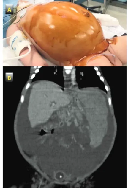
Abdominal distension (Figure 1) and an enlarged spleen (more than 5 cm below the costal arch) were detected upon physical examination. The baby (weight 5.5 kg) looked markedly pale, and was afebrile (36°C); eupneic (54 breaths/minute) with good oxygen saturation at room air (99%), the heart rate was 160 beats/minute with hypertension (105/52 mmHg). Laboratory investigations revealed severe anemia (Hb 4.6 g/dL), thrombocytopenia (54×103/µL) and coagulation abnormalities: fibrinogen 50 mg/dL vn 150-45 and prothrombin time (PT) 15.7 sec vn 9-12.5; PT% 60 vn 70-120. Liver function tests were normal. A plain abdominal X-ray revealed multiple air fluid levels in the distended small intestine. Abdominal ultrasound showed ascites and splenomegaly (11 cm in longitudinal diameter) with a bossellated mass in the lower part of spleen. Computed tomography (CT) revealed
corpuscolated liquid in the abdominal cavity, hepatomegaly and splenomegaly (longitudinal diameter 9.5 cm) with a large isodense mass, without intraparenchymal contrast enhancement, at the lower medial pole of the spleen (Figure 1).
Based on the triad of pallor, anemia and abdominal distension, in addition to the imaging and biochemical findings a diagnosis of intra-abdominal hemorrhage was hypothesized. The infant received several blood transfusions and underwent monitoring for 24 hours in the Pediatric Intensive Care Unit. The subsequent increase in abdominal distension, appearance of respiratory distress, and persistent abdominal hemorrhage un-responsive to blood transfusions were then considered indications for an exploratory laparotomy.7 During surgery, a great amount of bloody ascites (more than 1.5 L) were aspirated. Considering the complete transformation of the spleen to bosselated masses of different dimensions (varying from 1 to 2 cm in
Pediatric Reports 2019; volume 11:7848
Correspondence: Gloria Pelizzo, Pediatric Surgery Unit, Children’s Hospital, Via dei Benedettini 1, 90127 Palermo, Italy.
Tel.: +39.091.6666007
E-mail: [email protected]
Key words: Sclerosing Angiomatoid Nodular Transformation, infant, spleen.
Acknowledgments: The authors thank Dr. Laurene Marguerite Kelly for English revision of the manuscript.
Contributions: GP surgical management of the patient, drafting the article, critical revision of the article; VV, LL, EU histological analysis, drafting the article; OD, AMC, VG surgical man-agement of the patient; ZA neonatal manage-ment of the patient, drafting the article; LP radi-ological management of the patient; GB, VC drafting the article, literature review, critical revision of the article.
Competing interests: The authors declare no potential conflict of interest.
Funding: none.
Received for publication: 23 August 2018. Accepted for publication: 13 December 2018. This work is licensed under a Creative Commons Attribution NonCommercial 4.0 License (CC BY-NC 4.0).
©Copyright G. Pelizzo et al., 2019 Licensee PAGEPress, Italy Pediatric Reports 2019; 11:7848 doi:10.4081/pr.2019.7848
diameter) and the multiple bleeding capsular ruptures without subcapsular hematoma an urgent splenectomy was performed.
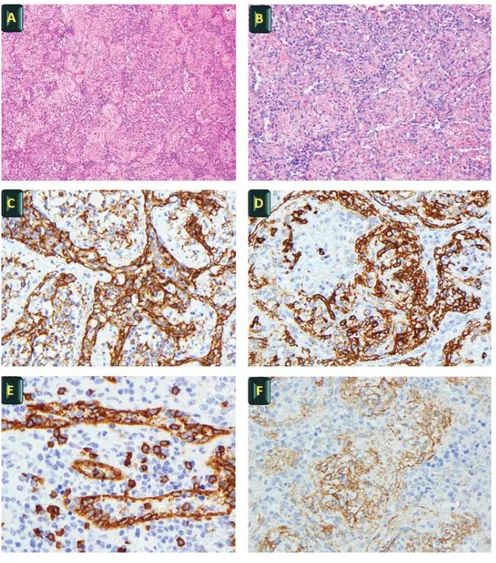
At gross pathologic evaluation the lesion was composed of multiple red-brown nodules diffusely distributed, without a coalescent scar. At histological evaluation the mass showed a multinodular architecture composed of numerous, convoluted, congested vessels without significant atypia of the endothelium. Subtle sclerotic tissue surrounded the vascular structures and an abundant lymphoid infiltrate was intermingled among the vessels. By immunohistochemistry, endothelial cells expressed the vascular markers CD34 and CD31 and positivity for CD8, whereas podoplanin (D2 40), CD21, CD35 e CD 68 were negative. Smooth muscle actin (SMA) stained the perimeter of the vascular structures. No infection by HHV8/ORF73 or EBV/EBER and IGg4 were detected by immunohistochemistry and in situ hybridization, respectively (Figure 2). The lymphoid infiltrate contained both CD20-positive B and CD3-CD20-positive T cell components with no evidence of lymphoproliferative disease.
Elevated polymorphonuclear leukocyte counts were detected after splenectomy; however, no infectious complications occurred and the monthly postoperative follow up has been uneventful. Vaccination against the most common pathogens that can cause post-splenectomy infection and antibiotic prophylaxis were prescribed, together with parental education on the consequences of splenectomy.
Discussion and Conclusions
SANT of the spleen is a benign, non-hematolymphoid tumor that arises in the red pulp of the spleen (Tracher, Abbot).8,9 Since its first description in 2004 by Martel et al.,2 155 cases of SANT have been reported in adults (Cipolla, cao),10,11while only four cases have been reported in pediatric patients (Table 1). Zhang et al.6 described a 3-year-old child injured in a car accident with a postsplenectomy SANT diagnosis; Vyas et al.4presented a case of SANT with inflammatory pseudotumor-like areas in an 11-year-old child with a history of trauma presenting with a rapidly growing splenic lesion; Agrawal et al.3 described SANT in a 12-year-old girl with upper quadrant discomfort which lasted six months; Kuybuli et al.5reported a case of SANT in an 11-year-old girl with growth retardation and increased sedimentation rate, mimicking chronic inflammatory disease.
The present report describes the first SANT case in infancy. Patients with SANT are usually asymptomatic or have non-specific abdominal pain. Most cases are found incidentally on radiographic examination or during surgery for an unrelated condition.12On the contrary, our case displayed life-threatening intra-abdominal bleeding with severe and rapid abdominal distension and coagulation abnormalities. This was a particularly unusual presentation requiring splenectomy as a life-saving treatment.
The pathogenesis of SANT remains unclear.1,2 It has been hypothesized that passive congestion of the splenic pulp secondary to trauma or unknown causes leads to sinus endothelial cell damage, fibrin deposition, and inflammation resulting in
pseudotumor appearance or SANT.2Other authors have proposed that SANT represents a peculiar hamartomatous transformation of the splenic red pulp in response to exaggerated non-neoplastic stromal proliferation.13Finally, SANT has also been reported to be associated with Epstein-Barr virus (EBV) infection,14 and immunoglobulin (Ig)G4-related sclerosing disease.15,16A significantly higher number of IgG4+ plasma cells and an increased IgG4/IgG ratio has also been reported in some studies.15,17,18
In our case, EBV was not found and although we did not measure serum IgG4 levels, immunochemical staining for IgG4 showed only extremely rare plasma cells. Therefore, we ruled out the hypothesis of a SANT IgG4-associated disease.
Case Report
Figure 1. Clinical and imaging features. A) severe abdominal distension at admission; B) large isodense splenic mass and abdominal hemorrhage at computed tomography.
[Pediatric Reports 2019; 11:7848] [page 33] Considering the patient’s age, the role of
genetic or gestational risk factors, such as gestational diabetes, cannot be excluded in the pathogenesis. Considering the early presentation in our case, a long-term post-splenectomy follow-up to detect possible hepatic involvement of SANT over time is recommended.
The differential diagnosis of SANT includes consideration of several other benign as well as malignant vascular lesions of the spleen, such as hemangioma, lymphangioma, littoral cell angioma,
hamartoma, lymphangiomas,
hemangioendotheliomas, angiosarcoma and inflammatory pseudotumor (IPT).19 The preoperative differential diagnosis to exclude other splenic tumors or malignant lesions is difficult.20 A thorough histopathologic examination and immunohistochemical analysis are necessary to make a diagnosis of SANT. The characteristics of SANT with regard to the immunohistochemical profile
include three distinct types of blood vessels and endothelial cells stained with CD34, CD8 or CD31, respectively: i) CD34+/CD31+/CD8- indicative of capillary derivation; ii) CD34-/CD31+/CD8+ indicative of splenic sinusoidal lining cell involvement and iii) CD34-/CD31+/CD8-indicative of small vein involvement.3,19The phenotypic profile of our case suggested a genetic component.
In our case, splenectomy was the only curative option for the management of SANT and is the treatment of choice in symptomatic patients. SANT patients have a good prognosis, with no recurrence after splenectomy.
In conclusion, SANT may also occur in infancy and present with potentially life-threatening conditions. Although splenectomy increases the patient’s risk for infection, particularly in neonates, infants and small children, it represents a life-saving treatment. In early onset SANT in infants,
long term follow-up is recommended to detect further multi-organ involvement.
References
1. Pradhan D, Mohanty SK. Sclerosing angiomatoid nodular transformation of the spleen. Arch Pathol Lab Med 2013;137:1309-12.
2. Martel M, Cheuk W, Lombardi L, et al. Sclerosing angiomatoid nodular transformation (SANT): report of 25 cases of distinctive benign splenic lesion. Am J Surg Pathol 2004;28:1268-79.
3. Agrawal M, Uppin SG, Bh S, et al. Sclerosing Angiomatoid Nodular Transformation of the Spleen: A New Entity or a New Name? Turk Patoloji Derg 2016;32:205-10.
4. Vyas M, Deshmukh M, Shet T, et al. Splenic angiomatoid nodular transformation in child with inflammatory pseudotumor-like areas. Indian J Pathol Microbiol 2011;54:829-31.
5. Kuybulu A, Sipahi T, Topal I, et al. Splenic angiomatoid nodular transformation in a child with increased erythrocyte sedimentation rate. Pediatr Hematol Oncol 2009;26:533-7. 6. Zhang S, Yang W, Hongyan XU, et al.
Sclerosing Angiomatoid Nodular Transformation of Spleen in a 3-year-old Child. Indian Pediatr 2015;52:1081-3. 7. Bickler S, Ramachndran V, Gittes GK,
et al. Nonoperative management of newborn solenic injury: a case report. J Pediatr Surg 2000;35:500-1.
8. Thacker C, Korn R, Millstine J, et al. Sclerosing angiomatoid nodular transformation of the spleen: CT, MR, PET, and ⁹⁹(m)Tc-sulfur colloid SPECT CT findings with gross and histopathological correlation. Abdom Imaging 2010;35:683-89.
9. Abbott RM, Levy AD, Aguilera NS, et al. From the archives of the AFIP: primary vascular neoplasms of the spleen: radiologic-pathologic correlation. Radiographics 2004;24: 1137-63.
10. Cipolla C, Florena AM, Ferrara G, et al. Sclerosing Angiomatoid Nodular Transformation: Laparoscopic Splenectomy as Therapeutic and Diagnostic Approach at the Same Time. Case Rep Surg 2018;2018:7020538. 11. Cao Z, Wang Q, Li J, et al. Multifocal
sclerosing angiomatoid nodular transformation of the spleen: a case report and review of literature. Diagn
Case Report
Figure 2. Histological evaluation. A-B) H&E - typical vascular appearance of SANT (x20, Panel A; and x40, Panel B); C-D) Diffuse positivity of vascular channels for CD 31 (x40, Panel C) and CD34 (x40, Panel D); E-F) Vascular channels positive for CD8 (x40, Panel E) and sclerosis (x40, Panel F)
[page 34] [Pediatric Reports 2019; 11:7848] Pathol 2015;11;10:95.
12. Atas H, Bulus H, Akkurt G. Sclerosing angiomatoid nodular transformation of the spleen: An uncommon cause of abdominal pain. Euroasian J Hepatogastroenterol 2017;7:89-91. 13. Önder S, Kosemehmetoglu K,
Himmetoglu Ç, et al. Sclerosing angiomatoid nodular transformation (SANT) of spleen: a case report describing cytology, histology, immunoprofile and differential diagnosis. Cytology 2012;23:129-32. 14. Weinreb I, Bailey D, Battaglia D, et al.
CD30 and Epstein–Barr virus RNA expression in sclerosing angiomatoid nodular transformation of spleen. Virchows Arch 2007;451:73-9.
15. Kashiwagi S, Kumasaka T, Bunsei N, et al. Detection of Epstein-Barr virus-encoded small RNA-expressed myofibroblasts and IgG4-producing plasma cells in sclerosing angiomatoid nodular transformation of the spleen. Virchows Arch 2008;53:275-82. 16. Kim HH, Hur YH, Koh YS, et al.
Sclerosing angiomatoid nodular transformation of the spleen related to IgG4-associated disease: report of a case. Surg Today 2013;43:930-6. 17. Kuo TT, Chen TC, Lee LY. Sclerosing
angiomatoid nodular transformation of the spleen (SANT): clinicopathological study of 10 cases with or without abdominal disseminated calcifying fibrous tumors, and the presence of a
significant number of IgG4+ plasma cells. Pathol Int 2009;59:844-50. 18. Nagai Y. Hayama N, Kishimoto T, et al.
Predominance of IgG4+ plasma cells and CD68 positivity in sclerosing angiomatoid nodular transformation (SANT) Histopathology 2008;53:495-8. 19. Wang TB, Hu BG, Liu DW, et al.
Sclerosing angiomatoid nodular transformation of the spleen: A case report and literature review. Oncol Lett 2016;12:928-32.
20. Matsubara K, Oshita A, Nishisaka T, et al. A case of sclerosing angiomatoid nodular transformation of the spleen with increased accumulation of fluorodeoxyglucose after 5-year follow-up. Int J Surg Case Rep 2017;39:9-13.