R E V I E W
Open Access
Revised recommendations of the Italian
Society of Pediatrics about the general
management of Kawasaki disease
Alessandra Marchesi
1, Donato Rigante
2,3, Rolando Cimaz
4,5*, Angelo Ravelli
6, Isabella Tarissi de Jacobis
1,
Alessandro Rimini
7, Fabio Cardinale
8, Marco Cattalini
9, Andrea De Zorzi
1, Rosa Maria Dellepiane
10, Patrizia Salice
11,
Aurelio Secinaro
1, Andrea Taddio
12,13, Paolo Palma
1, Maya El Hachem
1, Elisabetta Cortis
14, Maria Cristina Maggio
15,
Giovanni Corsello
15and Alberto Villani
1Abstract
Aim of these revised recommendations for the general management of Kawasaki disease is to encourage its prompter recognition and warrant the most appropriate therapy, based on ascertained scientific data, raising awareness of the complications related to misdiagnosis or delayed treatment. A set of 20 synthetic operative statements is herein provided, including the definition of Kawasaki disease, its protean presentations, clinical course and seminal treatment modalities of all disease phases. The application of these recommendations should improve prognosis of Kawasaki disease and prevent the progression to permanent vascular abnormalities, thereby
diminishing morbidity and mortality.
Keywords: Kawasaki disease, Coronary artery abnormalities, Intravenous immunoglobulin, Aspirin, Children Background
Two years after the diffusion of the Italian Guidelines for diagnosis and treatment of Kawasaki disease (KD), we have updated 20 operative recommendations focus-ing on the general management of this widespread ill-ness of unknown etiology, which mostly occurs in infants and children under 5 years of age with risk of heart implications, especially if the diagnosis is missed or if timely appropriate treatment is overlooked [1, 2]. First described in Japan in l967 by Tomisaku Kawasaki, the disease occurs in children of all ethnicities and is one of the leading cause of acquired cardiovascular dis-ease in the pediatric age [3]. The basic symptom of KD is persistently high and nonresponding fever, despite antipyretic and antibiotic treatments [4]. Japan, Korea
and Taiwan are the countries with the highest frequency of KD, reflecting a potential genetic susceptibility among the Asian populations. In addition, the age distribution of KD, higher in children less than 2 years and lower in those less than 6 months, is consistent with a potentially infectious disease, though KD has been even reported in newborns [5,6].
The identification of the causing mechanisms behind KD would be essential to develop preventive strategies or focused therapies, but the definite etiology remains elusive. A number of theories have linked KD to bac-teria, viruses and different environmental factors, but none has been proved. It is discussed if the trigger of KD might be an RNA virus that normally causes an asymptomatic infection in most children and KD in a subset of genetically predisposed children [7]. The most significant complications in KD are cardiovascular and prominently involve coronary arteries, though the overall incidence of coronary artery abnormalities (CAA) has © The Author(s). 2021 Open Access This article is licensed under a Creative Commons Attribution 4.0 International License, which permits use, sharing, adaptation, distribution and reproduction in any medium or format, as long as you give appropriate credit to the original author(s) and the source, provide a link to the Creative Commons licence, and indicate if changes were made. The images or other third party material in this article are included in the article's Creative Commons licence, unless indicated otherwise in a credit line to the material. If material is not included in the article's Creative Commons licence and your intended use is not permitted by statutory regulation or exceeds the permitted use, you will need to obtain permission directly from the copyright holder. To view a copy of this licence, visithttp://creativecommons.org/licenses/by/4.0/. The Creative Commons Public Domain Dedication waiver (http://creativecommons.org/publicdomain/zero/1.0/) applies to the data made available in this article, unless otherwise stated in a credit line to the data.
* Correspondence:[email protected]
4Pediatric Rheumatology, ASST Gaetano Pini-CTO, Milan, Italy 5University of Milan, Milan, Italy
been reduced by treatment with intravenous immuno-globulin (IVIG) given within 7–10 days after disease on-set [8]. Diagnosis of KD is clinical, supported by the results of labworks and imaging, but no findings or test can be considered highly specific. Diagnostic difficulties depend also on the different presenting times of signs and symptoms, the gruelling identification of other in-fectious and non-inin-fectious illnesses mimicking KD, the occurrence of non-typical manifestations and incomplete patterns of the disease [9,10].
Aim of these synthetically revised recommendations is suggesting the best practice for the management of chil-dren with KD, based on the most recent scientific evidence.
Definitions of Kawasaki disease Typical KD
Typical KD is characterized by fever lasting at least 5 days combined with at least 4 among 5 main clinical fea-tures (clinical criteria):
[a] bilateral non-exudative conjunctivitis,
[b] anomalies of the lips and oral mucosa (redding and cracking of lips, strawberry tongue, pharyngeal hyperemia),
[c] anomalies of the extremities (redding of palms and soles, indurative edema of hands and feet) and perineal region,
[d] polymorphous exanthema,
[e] acute nonpurulent cervical lymphadenopathy (usu-ally unilateral and larger than 1,5 cm).
The suspicion of KD may be initially cumbersome, as clinical features do not appear at the same time [11]. The presence of fever is universal and fever onset is con-sidered the first day of the illness, though the diagnosis of KD should be considered also in children in whom fever has subsided before the 5th day [12].
The 2017 American Heart Association guidelines sug-gest that in the presence of more than 4 main clinical features, particularly when the child shows redness and swelling of hands and feet, the diagnosis can be estab-lished with only 4 days of fever. They also claim that ex-perienced clinicians who have treated many KD patients could establish a diagnosis even with only 3 days of fever in the presence of classic clinical features [13].
The decision to hospitalize or discharge children with fever lasting around 5 days might depend on the educa-tional and socioeconomic status of their families or the availability of community medical services [14]. Several children may have incomplete or atypical forms of KD and discrimination of such forms from infections and other febrile conditions of childhood is an arduous chal-lenge even for an experienced pediatrician: scholastically, incomplete KD should be considered in case of children with unexplained fever for more than 5 days associated
with 2 or 3 classic features, while atypical KD in case of children with unexplained fever for more than 5 days as-sociated with non-classic clinical signs which do not re-spond to a proper therapy. The most fearful complications of KD are CAA, which develop in 15-to-25% of non-treated patients, but only in 5% of those properly treated, and are normally detected by echocar-diography [13].
Recommendation 1
Typical KD is diagnosed when fever lasting more than 5 days is associated with≥ 4 clinical criteria (bilateral non-exudative conjunctivitis, changes of lips and oral mu-cosa, changes of the extremities and perineal region, polymorphous exanthema and cervical lymphadenop-athy); in the case of CAA early demonstrated on echo-cardiography diagnosis of KD is possible also when a 4-day-lasting fever is associated with the known criteria.
Incomplete KD
The medical literature has been recently enriched by in-creasing reports of incomplete KD, mostly in children younger than 12 months, and this should be suspected in every infant showing fever for more than 5 days with docu-mented systemic inflammation in terms of increased C-reactive protein (CRP) and white blood cell count [15–17]. The diagnosis of incomplete KD is suggested if less than 4 main clinical features are found after exclusion of many fe-brile illnesses. In a 2-year nationwide survey (counting more than 23.000 children with incomplete KD) 3, 2 and 1 of the main clinical features were met in 26.6, 6.1 and 0.7% of cases, respectively [18].
Recommendation 2
Incomplete KD can be diagnosed when fever lasting more than 5 days is associated with 2 or 3 clinical cri-teria, with or without CAA.
Atypical KD
Atypical KD occurs in children with a classic fever pat-tern combined with further different non-classic signs, including aseptic meningitis, seizures, peripheral facial nerve palsy, sensorineural hearing loss, gallbladder hydrops, jaundice, acute abdomen, pancreatitis, arthritis, renal injury, orchitis, pneumonia, sterile pyuria, with or without CAA [17,19].
Recommendation 3
Atypical KD is diagnosed when fever lasting more than 5 days, not otherwise explained, is combined with non-classic manifestations nonresponsive to standard treat-ments, with or without CAA.
The clinical course of Kawasaki disease
KD clinical course can be divided into three phases: (a) acute (1st-2nd week), in which diagnosis should be established and treatment started,
(b) subacute (3rd-4th week), characterized by deferves-cence, periungual skin desquamation, thrombocytosis and eventual formation of CAA,
(c) convalescence phase (5th–8th week), in which all KD signs disappear, inflammatory markers subside to normal and nail indentations (Beau’s lines) may appear.
Child’s extreme irritability is characteristic during the acute phase of KD [20].
Investigations in Kawasaki disease
Diagnosis of KD should be considered in all children dis-playing fever for 5 days (or more), as prognosis relies on a prompt treatment. Consequently, in case of suspected KD, it is important to recommend patient’s hospitalization. Unfortunately, laboratory tests are nonspecific for KD, though leukocytosis, anemia and elevation of acute-phase reactants, such as CRP, can support the diagnosis in com-bination with classic features [21, 22]. Hypoalbuminemia is common and associated with more severe and more prolonged acute disease. Urinalysis may show pyuria in up to 80% of children. Additional helpful findings are elevated levels of brain natriuretic peptide (BNP) and N-terminal pro-BNP. Thrombocytosis is characteristic during the sub-acute phase, peaking in the third week and normalizing 4-to-6 weeks after onset. Thrombocytopenia is rare, but may occur in the first 1–2 weeks of illness: it can be a sign of disseminated intravascular coagulation or macrophage ac-tivation syndrome (MAS) and is a risk factor for the devel-opment of CAA [23].
Recommendation 4
Abnormal laboratory parameters (mostly the elevation of C-reactive protein) can only support the diagnosis of KD in febrile patients with evocative clinical features.
Treatment of Kawasaki disease
Intravenous immunoglobulin
The main goal of treatment in the acute phase of KD is suppressing systemic inflammation and minimizing the risk of developing CAA. Administration of IVIG is over-all recognized as the “first-line” treatment for KD both in terms of efficacy and safety [24]. In particular, high-dose IVIG (2 g/kg of body weight in a single infusion) significantly reduces the incidence of CAA and over-throws both inflammation and fever, as shown by a sys-tematic Cochrane review: IVIG is indicated in typical, incomplete and atypical KD [25]. IVIG infusion should be performed in 10–12 h if patient’s cardiac function is normal or in 16–24 h for patients displaying cardiac fail-ure. IVIG should be administered before the 10th day of
illness, best if before the 7th day since onset, as CAA might appear starting from the 8-9th day [26]. Treat-ment before the 5th day of fever should be reserved to exceptional cases of unequivocal diagnosis of KD [27]. IVIG should also be administered to children presenting after the 10th day of illness in case of persistent fever or ongoing systemic inflammation. Defervescence after IVIG occurs usually within 48 h in about 80–85% of cases. A reoccurring fever 36–48 h after a first adminis-tration of IVIG configures the framework of IVIG-resistant KD (this approximately occurs in 15–20% of patients) [28]. Nowadays there is no universally accepted treatment for these patients yet, and therefore the man-agement of these cases should be established individually.
Recommendation 5
IVIG at the dose of 2 g/kg of body weight is the treat-ment of choice for KD, preferably given within the 10th day, better if within the 7th day of illness, but as soon as possible after diagnosis.
Aspirin
Treatment of KD is completed by adding aspirin (acetyl-salicylic acid, ASA): medium-high dosage of ASA (from 80 mg/kg/day, mostly in the USA, to 30–50 mg/kg/day in Japan, UK and Europe, divided into 4 doses) is used for its anti-inflammatory effect during the acute phase, while 48 h after defervescence ASA can be switched to a lower dosage (3–5 mg/kg/day in a single dose) due to its anti-platelet effect. The overall duration of anti-platelet treatment is 6–8 weeks in KD patients without CAA, while it is maintained until the resolution of coronary le-sions in patients with CAA [29]. Recent studies have also suggested that ASA could be started immediately at the anti-platelet dosage in the acute phase, though further randomized clinical trials are required to compare the efficacy of medium-high and low-dose aspirin [30]. Medium-high dose ASA should be replaced in case of concurrent varicella or influenza to avoid the potential development of Reye’s syndrome [31]. Conversely, low-dose ASA has not been associated with the occurrence of this syndrome: however, in case of symptoms or ex-position to influenza or varicella, parents of children with KD are asked to promptly contact their physician [13]. Children may receive dipyridamole (1–5 mg/kg/day divided into 3 doses), ticlopidine (2–7 mg/kg/day divided into 2 doses) or clopidogrel (1 mg/kg/day in a single dose up to a maximum of 75 mg/day in children > 2 years or 0.2 mg/kg of body weight in children < 24 months) in replacement of ASA, even if there is less ex-perience with the use of these drugs. Ibuprofen (like other nonsteroidal anti-inflammatory drugs involving the cyclooxygenase pathway) antagonizes the
anti-platelet effect of low-dose ASA and usually should be avoided in children on anti-platelet ASA [13]. If ibupro-fen or similar drugs are needed, alternative anti-platelet therapies (e.g. clopidogrel) should be considered.
Recommendation 6
Treatment of KD is completed by ASA given at a daily dosage of 30–50 mg/kg in the acute phase of KD until 48 h after the disappearance of fever, then switched to the anti-platelet dose (3–5 mg/kg once daily).
Recommendation 7
Low-dose ASA must be continued until 6-to-8 weeks in children without CAA and continued in children with CAA until the resolution of coronary artery lesions.
Recurrence of Kawasaki disease
Recurrence of KD ranges from 1.4 to 3% of cases, and symptoms are often the same as for the first episode. IVIG resistance, the elevation of liver enzymes and re-duced hemoglobin level are risk factors for the recur-rence of KD. In addition, recurrent forms of KD can be associated with a higher incidence of CAA [32]. Recur-rent KD requires the same treatment as used for the first episode. Hereditary autoinflammatory syndromes need to be considered for a comprehensive differential diag-nosis of recurrent KD in childhood [33–35].
Non-responsiveness of Kawasaki disease
Resistant KD is defined by failure to respond to IVIG therapy and consists in a recrudescent fever 36–48 h after IVIG infusion [36]. It is believed that IVIG-resistant KD reflects severity of the underlying inflam-mation, explaining the increased incidence of CAA in this subset of patients. Severe anemia at disease onset, early development of CAA, and signs of MAS or septic shock should be considered in predicting the risk of IVIG non-responsiveness [37]. The identification of pa-tients with KD having a higher propensity to be IVIG-refractory is challenging: many efforts have been made to discriminate which patients might not respond to IVIG in order to undertake a more aggressive treatment combined with IVIG, but different scores useful in the Eastern Asiatic populations have been unsuccessful in the Caucasians [21,38–40].
Recommendation 8
Resistant KD is defined by failure in the response to IVIG and is revealed by recrudescent fever reoccurring or persisting 36–48 h after IVIG infusion.
Treatment of resistant forms of Kawasaki disease
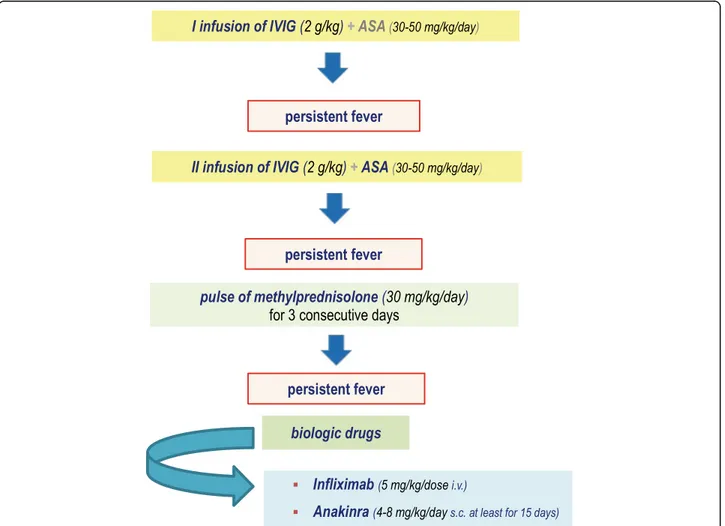
Several second-line treatment options are available for resistant KD, which are represented by an additional
IVIG infusion, intravenous methylprednisolone pulses, infliximab and other biologic drugs. Randomized con-trolled trials evaluating the effectiveness of these differ-ent options apart from the second infusion of IVIG are poor [41].
Corticosteroids
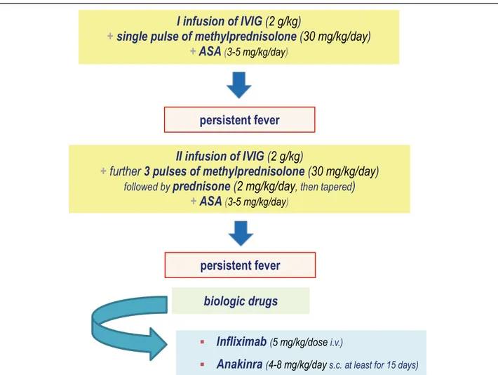
Most vasculitides are treated with corticosteroids due to their immunosuppressive and anti-inflammatory proper-ties [42]. The use of corticosteroids as second-line treat-ment in KD has been hugely debated, because of differences among studies (all patients versus high-risk patients, Eastern Asiatic versus Caucasian populations). Intravenous pulses of methylprednisolone at the dose of 30 mg/kg of body weight can be provided as second-line treatment for resistant KD patients [43, 44]. Indeed, non-responder patients with KD should be managed with a second IVIG cycle and - in case of failure - with 3 pulses of methylprednisolone (30 mg/kg/day), followed by oral prednisone (2 mg/kg/day, then gradually tapered up to the resolution of symptoms and normalization of CRP). Following the observation that methylpredniso-lone added to the standard regimen of IVIG improved outcomes of coronary arteries in Japanese patients with severe KD [45], “high-risk” patients, i.e. children less than 12 months or those having CRP higher than 200 mg/l, severe anemia at disease onset, albumin level below 2.5 g/dl, liver disease, overt coronary artery aneu-rysms, macrophage activation syndrome or septic shock, may require an initial therapy with IVIG (plus ASA) in combination with an intravenous pulse of methylpred-nisolone (30 mg/kg/day). When these KD patients fail to respond to such scheme, a second IVIG cycle combined with further 3 pulses of methylprednisolone (30 mg/kg/ day) plus ASA should be given, followed by prednisone at the initial dose of 2 mg/kg/day, then tapered up to the resolution of symptoms and normalization of CRP.
Recommendation 9
In non-responder patients with KD treatment requires a second infusion of IVIG and - in case of failure - pulses of methylprednisolone (30 mg/kg/day) for 3 consecutive days, followed by oral prednisone (2 mg/kg/day, then gradually tapered).
Recommendation 10
In high-risk patients with KD initial treatment should include:
– IVIG + single intravenous pulse of
methylprednisolone (30 mg/kg/day) + low-dose aspirin (3–5 mg/kg/day).
– In case of failure treatment should be implemented with a further infusion of IVIG and three pulses of
intravenous methylprednisolone (30 mg/kg/day, followed by prednisone: 2 mg/kg/day, then gradually tapered) + low-dose aspirin (3–5 mg/kg/day).
Biological drugs
Biologics may target the presumed key-cytokines in-volved in KD pathogenesis, namely tumor necrosis factor (TNF)-α and interleukin (IL)-1 [46,47]. Many consider-ations about their use are derived from the experience with anti-TNF or anti-IL-1 drugs on small series of KD patients. Current evidence supports the use of inflixi-mab, a chimeric monoclonal antibody against TNF-α, as rescue therapy at a single intravenous dose of 5 mg/kg of body weight (given in 2 h) for IVIG- and corticosteroid-resistant KD patients. However, no trials have evaluated its use as adjunctive therapy in patients with early evi-dence of CAA [48]. There are studies confirming the ef-fectiveness of anakinra, the recombinant IL-1 receptor antagonist blocking the natural biological activity of IL-1, in children with a refractory KD, given subcutaneously at a daily dose of 4–8 mg/kg of body weight for an over-all period of 15 days or for a longer period, depending
on the specific clinical scenery. Anakinra might also pre-vent the development of CAA [49]. The use of canakinu-mab, a high-affinity human monoclonal antibody targeted against IL-1β (using a single subcutaneous injection of 4 mg/kg/dose) may be also a future option for cases of IVIG-resistant and corticosteroid-resistant KD [50,51].
Recommendation 11
Current evidence supports the use of infliximab as res-cue therapy in IVIG- and methylprednisolone-refractory patients with KD; IL-1 blockade with anakinra is highly promising in treating the most dramatically severe multi-refractory patients with KD, with potential benefits also on the cardiovascular complications.
We have summarized the therapeutic recommenda-tions for patients with KD (Fig. 1) and for those who should be defined as“high-risk” patients (Fig.2).
Complications of Kawasaki disease
The most relevant risk for children with KD is the devel-opment of vascular abnormalities, affecting small-to-mid size arteries, but characteristically the coronary arteries.
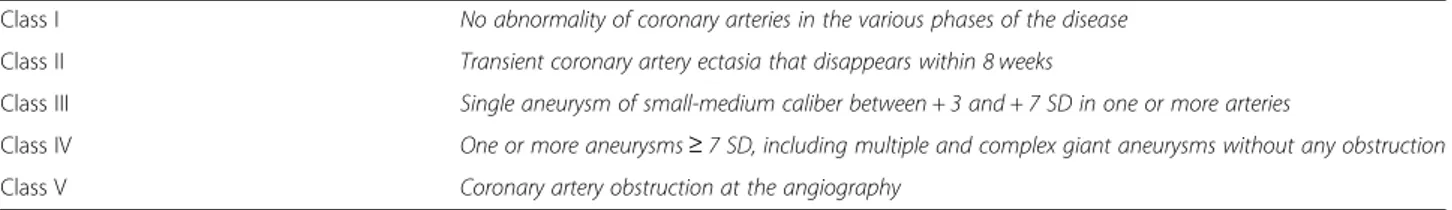
A coronary artery anomaly may start as ectasia, progress to moderate dilatation (5-to-8 mm of diameter) until the formation of large aneurysms (measuring more than 8 mm). A standardized classification to measure coronary arteries and characterize CAA, comparing vessel diame-ters to the body surface area and measuring the standard deviation from the average in z units, is listed in Table1 [52]. This classification is recommended for the right coronary, left anterior descending and left main coron-ary arteries, and z-scores with reference values for pediatric echocardiography are available at the website www.parameterz.com.
Coronary artery involvement is virtually absent if the z-score is < 2 and present if the z-score is beyond 2. Pa-tients with giant aneurysms (having z-scores≥10) display the highest risk of both coronary thrombosis and sten-osis [53]. Other cardiovascular complications include myocarditis, pericarditis, myopericarditis, valvular le-sions, arrhythmias, heart failure in addition to coronary
thrombosis or stenosis and myocardial infarction [54]. Pneumonia, encephalitis, seizures, paralytic ileus, chole-cystitis, liver dysfunction, cholechole-cystitis, anemia, hypoal-buminemia, electrolyte imbalance, diarrhea, vomiting and dehydration might occur in KD, and specific treat-ments may be required [19]. Other severe non-cardiovascular complications are MAS, largely known to occur in a host of disorders characterized by hypercyto-kinemia, heralded by non-remitting fever, impaired liver function, hyperferritinemia, hypertriglyceridemia,
Fig. 2 Treatment of High-Risk patients with KD. (children less than 12 months or children having C-reactive protein higher than 200 mg/l, severe anemia at disease onset, albumin level below 2.5 g/dl, liver disease, overt coronary artery aneurysms, macrophage activation syndrome or septic shock)
Table 1 Classification of coronary artery abnormalities in the acute phase of Kawasaki disease
No coronary artery involvement: z-score < 2
Dilatation of the coronary artery: z-score > 2 to < 2.5 SD Small aneurysm of the coronary artery: z-score≥ 2.5 to < 5 SD Medium aneurysm of the coronary artery: z-score≥ 5 to < 10 SD Giant aneurysm of the coronary artery: z-score≥ 10 SD
hypofibrinogenemia, pancytopenia and frequently hemo-phagocytosis [55, 56], and KD-related shock syndrome, associated with hypotension, platelet consumption and increased risk of CAA [57].
Evaluation of the cardiovascular involvement in Kawasaki disease
KD vasculitis involves a variety of blood vessels and, in particular, small muscular arteries, being caused by dif-fuse neutrophil infiltration with necrotizing features, which might potentially lead to vessel dilatation mostly occurring during the subacute phase [58]. In general terms, CAA may start as ectasia which might regress within 8 weeks in most cases [59]. The regression of small or medium coronary aneurysms might occur within 1–2 years after onset, but large and giant aneu-rysms may persist over a long time due to local fibrosis and calcifications, with the risk of acute coronary syn-drome [60]. Non-responsiveness to IVIG is a crucial sign to define KD severity, as these children have the highest risk of developing CAA [61]. Additionally, CAA are most prevalent in patients having incomplete KD [62].
Recommendation 12
The most relevant complications in KD are cardiovascu-lar sequelae starting as CAA, which are more frequently disclosed during the subacute phase of the disease.
The role of echocardiography
Bidimensional and color-Doppler echocardiography must be performed when KD diagnosis is suspected, then after 2, 4 and 8 weeks in the non-complicated cases by cardiologists experienced in pediatrics [63]. However, it is important to ensure that the timing or results of the echocardiogram should not delay the initial treatment of KD during the acute phase. An initial echocardiogram in the first week of illness is typically normal and does not rule out KD diagnosis. The uncooperative irritable child may require sedation; if this is not possible, a further as-sessment should be repeated as soon as possible within 48 h after diagnosis. The coronary artery imaging should include the left main coronary artery, left anterior de-scending, left circumflex, right coronary artery (prox-imal, middle and distal segments), and posterior descending coronary artery. In patients with evolving CAA, follow-up assessments with echocardiography should be performed more frequently to monitor even-tual increases in vessel dimensions and the potential presence of thrombosis.
Recommendation 13
Echocardiography is pivotal for the diagnosis of coronary artery involvement in children with KD and should be
periodically repeated 2, 4 and 8 weeks after onset in the uncomplicated cases.
The role of other imaging techniques
Advanced techniques for the assessment of KD-related cardiovascular sequelae are computed tomography (CT) and magnetic resonance (MR) angiography [64]. CT scan should be used to confirm CAA, define size and morphology of CAA, identify thrombosis or occlusions, and evaluate other aneurysms, both central or periph-eral, in the whole body, but its use is limited by exten-sive radiation exposure [65]. Cardiovascular MR angiography with gadolinium-based contrast often re-quires the use of general anesthesia in younger patients and can be used to confirm CAA, investigate other cen-tral or peripheral anuerysms, identify biventricular sys-tolic function, detect areas of inducible myocardial ischemia during pharmacological stress and evaluate myocardial scars [66].
Recommendation 14
Cardiovascular CT scan and MR angiography, where available, are important to assess persistent CAA in chil-dren with KD and monitor remodeling of either coron-ary or systemic arteries in the whole body.
Treatment of the cardiovascular complications of Kawasaki disease
Long-term therapy in KD patients with CAA aims at preventing the cardiovascular acute events with reduc-tion of both incidence and severity of acute coronary syndrome. Such therapy depends on the features and ex-tension of CAA. In the absence of“evidence-based” stud-ies, recommendations have been drafted by pediatric retrospective studies. Platelet activation is critical in all KD phases, therefore, in case of persistent CAA, chronic use of anti-platelet low-dose ASA (3–5 mg/kg/day) must be provided, optionally associated with other anti-platelet and/or anticoagulant and/or anti-angina drugs, according to the size of aneurysms, flow characteristics, and presence or absence of myocardial ischemic changes [67].
Supplementary anti-platelet drugs
Persistent structural multiple or complex coronary artery aneurysms may require dual anti-platelet prophylaxis, based on low-dose ASA and clopidogrel. There is still insufficient data on the use of clopidogrel, an ADP-P2Y12 receptor inhibitor, in infants and children, for
whom anti-platelet response might be different. Anti-platelet action of clopidogrel is usually achieved with lower doses (0.2 mg/kg/day) in children aged < 24 months, while older children may require higher doses (up to 1 mg/kg/day): light transmission platelet aggrego-metry is a common method to assess platelet function
in vitro for patients using clopidogrel, though universal standards are lacking [68]. Clopidogrel may be also a valid alternative to ASA in case of ASA intolerance or resistance, varicella (either the wild type disease or vac-cination against varicella) and influenza [69].
Recommendation 15
KD patients with medium-sized coronary artery aneu-rysms or those with multiple and complex aneuaneu-rysms re-quire dual anti-platelet prophylaxis, based on low-dose ASA (at a single dose of 3–5 mg/kg/day) and clopidogrel (at a single dose of 0.2 mg/kg/day in children aged < 24 months and up to 1 mg/kg/day in children aged≥ 24 months).
Anticoagulant drugs
Anticoagulants are indicated in the case of giant aneu-rysms, coronary thrombosis or history of acute myocar-dial ischemia. Warfarin (0.05–0.12 mg/kg/day once daily, carefully adjusting the dose until reaching an inter-national normalized ratio, INR, between 2.0 and 3.0) is the most widely used drug for long-term anticoagulation [70]. The use of low-molecular weight heparin (LMWH) can be considered in infants with KD for whom a regular assessment of INR is troublesome. In particular, enoxa-parin (100 IU/kg/dose every 12 h) is a LMWH with dem-onstrated safety and efficacy, frequently used in pediatrics as a bridging anticoagulant regimen with low risk of thromboembolism and low risk of bleeding com-plications [71]. Fondaparinux, an antithrombin-dependent selective inhibitor of factor Xa, is not ap-proved for use in children. However, in comparison to LMWH, fondaparinux has several properties which make it attractive, including a longer half-life, lack of ef-fects on bone metabolism and reduced risk of heparin-induced thrombocytopenia. Fondaparinux (at a single daily dose of 0.1 mg/kg of body weight) has been admin-istered to children with deep venous thrombosis without severe adverse events [72]. Novel oral anticoagulants do not require routine laboratory assessment, but there are very few data about their employment in pediatrics.
Recommendation 16
It is reasonable to treat KD patients having complex or severe CAA with low-dose ASA associated with warfarin (keeping INR targeted at 2.0–3.0) or LMWH (if regular INR checking is difficult). Triple therapy with ASA, war-farin or LMWH and clopidogrel should be considered in KD patients with a relevant risk of thrombosis.
Non-invasive treatment of coronary artery thrombosis
Patients with giant aneurysms of coronary arteries are at higher risk of presenting thrombosis and developing
acute coronary syndrome. The goal of treatment in KD patients who show coronary thrombosis is to restore flow patency, preserve the myocardial tissue and increase patient’s survival [73]. Thrombolytic treatment with tissue-type plasminogen activator is the most commonly administered therapeutic regimen in treating fresh thrombosis within medium/large or giant coronary an-eurysms. A multidisciplinary approach is needed to optimize outcome of KD patients receiving thrombolysis, that should be performed by experienced interventional radiologists or cardiologists in intensive care setting to allow rapid intervention in the case of bleeding. The ability to quickly obtain coagulation testing results for ongoing adjustments of therapy is critical for managing patients who require thrombolysis and concomitant anticoagulation. The most relevant thrombolytic drug in young patients with KD complicated by coronary artery thrombosis is the intravenous recombinant tissue plas-minogen activator (rtPA), and in recent years“alteplase” has become the most widely used in children. Fresh fro-zen plasma (10–20 ml/kg) given before using rtPA works as a plasminogen source. Various dosing regimens have been compared, but high doses of alteplase (0.5 mg/kg, 10% infused over 1–2 min and the remainder over 60 min) are more commonly used [74]. A careful monitor-ing of coagulation testmonitor-ing every 6–12 h is mandatory to prevent bleeding (maintaining the fibrinogen level > 100 mg/dl and platelet count > 50,000/mm3). Low-dose thrombolytic therapy may be also associated with abcixi-mab, a glycoprotein IIb/IIIa inhibitor, given intraven-ously (as a bolus of 0.25 mg/kg in 30 min, then followed by 0.125μg/kg/minute, max: 10 μg/min, for 12 h) in the case of thrombosis with high risk of forthcoming vessel occlusion [75, 76]. Both rtPA and abciximab require to be associated with low-dose ASA and intravenous hep-arin (10 U/kg/hour). Coronary thrombosis should be reassessed with echocardiographic imaging during and after completion of thrombolysis.
Recommendation 17
Recombinant tissue plasminogen activator (rtPA) is the first-choice thrombolytic drug in children with KD com-plicated by coronary artery thrombosis; the glycoprotein IIb/IIIa inhibitor abciximab may be used in case of thrombosis with high risk of occlusion. Both therapies require a concomitant association with low-dose ASA and intravenous heparin.
Invasive treatment of coronary artery thrombosis
Coronary artery reperfusion by both invasive cardiac interventional procedures and cardiac surgery should be considered after unsuccessful pharmacological thromb-olysis or when stenotic lesions are eventually present in selected patients. It is important to consult an adult
interventional cardiologist and/or an adult cardiothor-acic surgeon with experience in revascularization of pa-tients with KD if revascularization is considered.
Cardiac procedures include intracoronary catheter-directed thrombolysis, percutaneous balloon angioplasty with or without coronary stenting and rotational ather-ectomy with or without coronary stenting [77]. There is no standard protocol or preferred device in pediatrics, and the use of any of these modalities often relies on physician’s experience. In order to determine the most appropriate procedure it is desirable that physicians con-sider patient’s body size, coronary angiography findings and intravascular ultrasound. Invasive treatment should be considered in patients with or without ischemic symptoms caused by coronary artery stenosis (≥75% of the luminal diameter), but with instrumentally demon-strated ischemia (via stress electrocardiogram or stress perfusion imaging) [78]. Coronary angioplasty procedure has a risk of vessel re-stenosis and occlusion, requiring the use of stenting or alternative procedures, such as coronary artery bypass grafting or percutaneous translu-minal rotational ablation [79]. Heart transplantation can be proposed in selected cases with irreversible myocar-dial dysfunction [80].
Recommendation 18
First-choice cardiologic interventional treatment in pa-tients with KD should be chosen in the shortest time based on the specific experience of a pediatric cardiac surgery team.
Follow-up of patients with Kawasaki disease
Patients with KD must undergo regular cardiologic eval-uations with electrocardiogram and echocardiography in the long-term. For patients with large coronary aneu-rysms diagnosed in the first months after disease onset it is recommended to perform echocardiography weekly [81]. Cardiologic evaluations performed in the convales-cence phase allow to subdivide KD patients according to their cardiovascular impairment into different risk clas-ses and establish a personalized follow-up protocol. It is reasonable to consider KD patients worthy of close mon-itoring for their cardiovascular risk, evaluating and con-trolling blood pressure, body mass index, blood lipid profile and promoting correct lifestyles [82]. Cardiologic assessment allows to consider 5 different risk classes ac-cording to the cardiovascular sequelae and establish a personalized follow-up protocol (Table2) [1,2,13].
Recommendation 19
Cardiologic assessments allow to split KD patients into different risk classes according to the cardiovascular se-quelae and establish a personalized follow-up protocol.
Vaccinations in children with Kawasaki disease
Treatment of KD with IVIG does not significantly inter-fere with inactivated vaccines, oral attenuated live virus vaccines, live virus nasal vaccines, bacillus Calmette-Guérin vaccine or yellow fever vaccine. Subsequently, these vaccines can be safely administered at any time after IVIG administration. Conversely, it is recom-mended to wait 10–12 months before administration of vaccines against measles, mumps, rubella (MMR), vari-cella (V) and MMRV, as IVIG administration might re-sult in reduced immunogenicity and interfere specifically with the immune response of these live attenuated vac-cines. A seasonal inactivated influenza vaccine is recom-mended in children with KD receiving ASA [83].
Recommendation 20
All inactivated vaccines can be safely administered at any time after IVIG in KD patients. Attenuated live virus vaccines (MMR, V and MMRV vaccines) should be ad-ministered 10–12 months after the administration of IVIG to avoid a reduced specific immune response in KD patients; influenza vaccination is recommended in KD patients receiving ASA.
A relationship with the SARS-CoV-2 pandemic threat with Kawasaki disease?
During the 2020 pandemic caused by the novel corona-virus (SARS-CoV-2) infection a number of severely ill patients with a systemic inflammatory syndrome, which has been named “pediatric multisystem inflammatory syndrome temporally associated with coronavirus disease (COVID-19) or PIMS-TS” in the UK and “multisystem inflammatory syndrome associated with COVID-19 or MIS-C” in the USA, have been reported. The pathogen-esis of this multisystem syndrome is rather unclear, though some overlapping features suggesting an immune-mediated vasculitis like KD have been de-scribed: the inflammatory response in this condition dif-fers from KD with respect to cell subsets involved and biomarkers associated with vascular damage [84]. There has been a huge debate about the substantial risk that first-line providers encountering febrile patients might miss or delay the diagnosis of KD and proper treatment with IVIG. While there are no definite proofs of a rela-tionship between SARS-CoV2 and KD, it is important to remind a high suspicion level for KD in all children with prolonged fever, mostly those younger than 12 months, and to perform echocardiograms to monitor eventual cardiovascular complications in these little patients even after a confirmed infection by SARS-CoV-2 [85].
Final remarks
This list of 20 revised recommendations, drafted 3 years after the American Heart Association Statement and 2
years after the publication of the Italian Guidelines for diagnosis and treatment of KD, summarizes the most relevant clues to manage KD patients, highlighting rele-vant points for both clinicians and researchers. Their aim is spreading the best practice in general manage-ment of children with such a complex disorder, so that long-term morbidity and potential mortality might be prevented across the life span. We have herein refined the definition of cardiovascular and systemic complica-tions of KD, recurrent forms of KD and treatment mo-dalities with corticosteroids or biological agents in refractory patients. Treatment of cardiovascular compli-cations with anti-platelet drugs and/or anticoagulants and treatment of coronary artery thrombosis with both pharmacological and non-pharmacological approaches have been also discussed. Every clinical decision-making should be specifically tailored on the individual patient with KD.
Abbreviations
KD:Kawasaki disease; CAA: Coronary artery abnormalities; IVIG: Intravenous immunoglobulin; ASA: Acetylsalicylic acid (aspirin); CT: Computed tomography; MR: Magnetic resonance; CRP: C-reactive protein; TNF: Tumor necrosis factor; IL: Interleukin; INR: International normalized ratio; LMWH: low molecular weight heparin; MMR: Mumps, measles, rubella; V: Varicella; MMRV: measles, mumps, rubella, varicella; rtPA: Recombinant tissue plasminogen activator
Authors’ contributions
AM, DR, RC, AR, GC and AV: conception and design of the recommendations, collection of in-depth analyses performed by each coauthor, critical revision, writing and final approval of the manuscript. ITDJ, FC and MEH participated in writing the chapter about presentation and clinical course. EC participated in writing the chapter about disease presentation and clinical course, espe-cially about recurrent forms. MC and AT participated in writing the chapter about acute phase therapy. MCM participated in writing the chapter about high risk patients’ therapy. AR and AD studied application and limits of the echocardiography in KD. RMD worked on the non-cardiovascular complica-tions and patients’ follow-up. AS produced the chapter about instrumental investigations in KD. PS worked to define patients’ follow-up. All authors read and approved the final version of this manuscript.
Funding
No person who participated in the drafting of this set of recommendations has been sponsored.
Availability of data and materials
No datasets were generated or analyzed during the current article. Ethics approval and consent to participate
Not applicable. Consent for publication Not applicable.
Competing interests
The authors declare that they have no competing interests. Author details
1Bambino Gesù Children’s Hospital, Rome, Italy.2Department of Life Sciences
and Public Health, Fondazione Policlinico A. Gemelli IRCCS, Rome, Italy.
3Università Cattolica Sacro Cuore, Rome, Italy.4Pediatric Rheumatology, ASST
Gaetano Pini-CTO, Milan, Italy.5University of Milan, Milan, Italy.6Pediatrics
and Rheumatology Unit, IRCCS Istituto Giannina Gaslini, University of Genoa, Genoa, Italy.7IRCCS Istituto Giannina Gaslini, Genoa, Italy.8Department of Pediatrics, AOU Policlinico Giovanni XXIII, Bari, Italy.9Pediatrics Clinic,
University of Brescia, ASST Spedali Civili di Brescia, Brescia, Italy.10UOC of
Pediatrics, UOS of Pediatric Immunology, Fondazione IRCCS Ca’ Granda Ospedale Maggiore Policlinico, Milan, Italy.11Section of Pediatric Cardiovascular Diseases, Fondazione IRCCS Ca’ Granda Ospedale Maggiore Policlinico, Milan, Italy.12University of Trieste, Trieste, Italy.13Institute of Child
and Maternal Health, IRCCS Burlo Garofolo, Trieste, Italy.14UOC of Pediatrics,
S. Eugenio Hospital, Rome, Italy.15Università degli Studi di Palermo, Palermo, Italy.
Received: 16 October 2020 Accepted: 4 January 2021
References
1. Marchesi A, Tarissi De Jacobis I, Rigante D, Rimini A, Malorni W, Corsello G, et al. Kawasaki disease: guidelines of the Italian Society of Pediatrics, part I -definition, epidemiology, etiopathogenesis, clinical expression and management of the acute phase. Ital J Pediatr. 2018;44:102.
2. Marchesi A, Tarissi De Jacobis I, Rigante D, Rimini A, Malorni W, Corsello G, et al. Kawasaki disease: guidelines of Italian Society of Pediatrics, part II -treatment of resistant forms and cardiovascular complications, follow-up, lifestyle and prevention of cardiovascular risks. Ital J Pediatr. 2018;44:103. 3. Kawasaki T. Acute febrile mucocutaneus syndrome with lymphoid
involvement with specific desquamation of the finger and toes in children. Arerugi. 1967;16:178–222.
4. Yim D, Curtis N, Cheung M, Burgner D. Update on Kawasaki disease: epidemiology, aetiology and pathogenesis. J Paediatr Child Health. 2013;49: 704–8.
5. Rowley AH, Shulman ST. The epidemiology and pathogenesis of Kawasaki disease. Front Pediatr. 2018;6:374.
6. De Rose DU, Coppola M, Gallini F, Maggio L, Vento G, Rigante D. Overview of the rarest causes of fever in newborns: handy hints for the neonatologist. J Perinatol. 2020. Epub ahead of print. https://doi.org/10.1038/s41372-020-0744-8.
7. Rigante D. Kawasaki disease as the immune-mediated echo of a viral infection. Mediterr J Hematol Infect Dis. 2020;12:e2020039.
8. Falcini F, Capannini S, Rigante D. Kawasaki syndrome: an intriguing disease with numerous unsolved dilemmas. Pediatr Rheumatol. 2011;9:17. 9. Newburger JW, Takahashi M, Gerber MA, Gewitz MH, Tani LY, Burns JC, et al.
Diagnosis, treatment, and long-term management in Kawasaki disease: a statement for health professionals from the committee on rheumatic fever, endocarditis and Kawasaki disease, council on cardiovascular disease in the young. Am Heart Assoc Pediatrics. 2004;114:1708–33.
10. Cimaz R, Falcini F. An update on Kawasaki disease. Autoimmun Rev. 2003;2: 258–63.
11. Newburger JW, Fulton DR. Kawasaki Disease. Curr Treat Options Cardiovasc Med. 2007;9:148–58.
12. De Rosa G, Pardeo M, Rigante D. Current recommendations for the pharmacologic therapy in Kawasaki syndrome and management of its cardiovascular complications. Eur Rev Med Pharmacol Sci. 2007;11:301–8.
Table 2 Cardiovascular risk classes in patients with Kawasaki disease
Class I No abnormality of coronary arteries in the various phases of the disease Class II Transient coronary artery ectasia that disappears within 8 weeks
Class III Single aneurysm of small-medium caliber between + 3 and + 7 SD in one or more arteries
Class IV One or more aneurysms≥ 7 SD, including multiple and complex giant aneurysms without any obstruction Class V Coronary artery obstruction at the angiography
13. McCrindle BW, Rowley AH, Newburger JW, Burns JC, Bolger AF, Gewitz M, et al. Diagnosis, treatment, and long-term management of Kawasaki disease, a scientific statement for health professionals from the American Heart Association. Circulation. 2017;135:e927–99.
14. Rigante D. When, how, and why do fevers hold children hostage? J Evid Based Med. 2020;13:85–8.
15. Yu JJ. Diagnosis of incomplete Kawasaki disease. Korean J Pediatr. 2012;55:83–7. 16. Chang FY, Hwang B, Chen SJ, Lee PC, Meng CC, Lu JH. Characteristics of
Kawasaki disease in infants younger than six months of age. Pediatr Infect Dis J. 2006;25:241–4.
17. Cimaz R, Sundel R. Atypical and incomplete Kawasaki disease. Best Pract Res Clin Rheumatol. 2009;23:689–97.
18. Nakamura Y, Yashiro M, Uehara R, Sadakane A, Tsuboi S, Aoyama Y, et al. Epidemiologic features of Kawasaki disease in Japan: results of the 2009-2010 nation-wide survey. J Epidemiol. 2012;22:216–21.
19. Jindal AK, Pilania RK, Prithvi A, Guleria S, Singh S. Kawasaki disease: characteristics, diagnosis, and unusual presentations. Expert Rev Clin Immunol. 2019;15:1089–104.
20. Falcini F. Kawasaki disease. Curr Opin Rheumatol. 2006;18:33–8. 21. Türkuçar S, Yıldız K, Acarı C, Dundar HA, Kır M, Ünsal E. Risk factors of
intravenous immunoglobulin resistance and coronary arterial lesions in Turkish children with Kawasaki disease. Turk J Pediatr. 2020;62:1–9. 22. Rigante D, Valentini P, Rizzo D, Leo A, De Rosa G, Onesimo R, et al.
Responsiveness to intravenous immunoglobulins and occurrence of coronary artery abnormalities in a single-center cohort of Italian patients with Kawasaki syndrome. Rheumatol Int. 2010;30:841–6.
23. Arora K, Guleria S, Jindal AK, Rawat A, Singh S. Platelets in Kawasaki disease: is this only a numbers game or something beyond? Genes Dis. 2019;7:62–6. 24. Furusho K, Kamiya T, Nakano H, Kiyosawa N, Shinomiya K, Hayashidera T, et al.
High-dose intravenous gammaglobulin for Kawasaki disease. Lancet. 1984;2:1055–8. 25. Oates-Whitehead RM, Baumer JH, Haines L, Love S, Maconochie IK, Gupta A,
et al. Intravenous immunoglobulin for the treatment of Kawasaki disease in children. Cochrane Database Syst Rev. 2003;4:CD004000.
26. Muta H, Ishii M, Egami K, Furui J, Sugahara Y, Akagi T, et al. Early intravenous gamma-globulin treatment for Kawasaki disease: the nationwide surveys in Japan. J Pediatr. 2004;144:496–9.
27. Shamim I. Diagnosis and treatment of suspected Kawasaki disease before the fifth day of illness. Ped Inf Dis J. 2013;32:424.
28. Eleftheriou D, Levin M, Shingadia D, Tulloh R, Klein NJ, Brogan PA. Management of Kawasaki disease. Arch Dis Child. 2013;99:74–83. 29. Zheng X, Yue P, Liu L, Tang C, Ma F, Zhang Y, et al. Efficacy between low
and high dose aspirin for the initial treatment of Kawasaki disease: Current evidence based on a meta-analysis. PLoS One. 2019;14:e0217274. 30. Jia X, Du X, Bie S, Li X, Bao Y, Jiang M. What dose of aspirin should be used
in the initial treatment of Kawasaki disease? A meta-analysis. Rheumatology (Oxford). 2020;59:1826–33.
31. Fitzgerald DA. Aspirin and Reye syndrome. Paediatr Drugs. 2007;9:205–6. 32. Yang HM, Du ZD, Fu PP. Clinical features of recurrent Kawasaki disease and
its risk factors. Eur J Pediatr. 2013;172:1641–7.
33. Rigante D. A systematic approach to autoinflammatory syndromes: a spelling booklet for the beginner. Expert Rev Clin Immunol. 2017;13:571–97. 34. Rigante D, Frediani B, Galeazzi M, Cantarini L. From the Mediterranean to
the sea of Japan: the transcontinental odyssey of autoinflammatory diseases. Biomed Res Int. 2013;2013:485103.
35. Rigante D. Phenotype variability of autoinflammatory disorders in the pediatric patient: a pictorial overview. J Evid Based Med. 2020;13:227–45. 36. Newburger JW, Takahashi M, Burns JC, Beiser AS, Chung KJ, Duffy CE, et al.
The treatment of Kawasaki syndrome with intravenous gamma globulin. N Engl J Med. 1986;315:341–7.
37. Piram M, Darce Bello M, Tellier S, Di Filippo S, Boralevi F, Madhi F, et al. Defining the risk of first intravenous immunoglobulin unresponsiveness in non-Asian patients with Kawasaki disease. Sci Rep. 2020;10:3125. 38. Shulman ST, Rowley AH. Kawasaki disease: insights into pathogenesis and
approaches to treatment. Nat Rev Rheumatol. 2015;11:475–82. 39. Sleeper LA, Minich LL, McCrindle BM, Li JS, Mason W, Colan SD, et al.
Evaluation of Kawasaki disease risk-scoring systems for intravenous immunoglobulin resistance. J Pediatr. 2011;158:831–5.
40. Rigante D, Andreozzi L, Fastiggi M, Bracci B, Natale MF, Esposito S. Critical overview of the risk scoring systems to predict non-responsiveness to intravenous immunoglobulin in Kawasaki syndrome. Int J Mol Sci. 2016;17:278.
41. Burns JC, Capparelli EV, Brown JA, Newburger JW, Glode MP. Intravenous gamma-globulin treatment and retreatment in Kawasaki disease. US/Canadian Kawasaki syndrome study group. Pediatr Infect Dis J. 1998;17:1144–8. 42. Rigante D. Clinical overview of vasculitic syndromes in the pediatric age. Eur
Rev Med Pharmacol Sci. 2006;10:337–45.
43. Newburger JW, Sleeper LA, McCrindle BW, Minich LL, Gersony W, Vetter VL, et al. Randomized trial of pulsed corticosteroid therapy for primary treatment of Kawasaki disease. N Engl J Med. 2007;356:663–75.
44. Ogata S, Ogihara Y, Honda T, Kon S, Akiyama K, Ishii M. Corticosteroid pulse combination therapy for refractory Kawasaki disease: a randomized trial. Pediatrics. 2012;129:e17–23.
45. Kobayashi T, Saji T, Otani T, Takeuchi K, Nakamura T, Arakawa H, et al. Efficacy of immunoglobulin plus prednisolone for prevention of coronary artery abnormalities in severe Kawasaki disease (RAISE study): a randomised open-label blinded-endpoints trial. Lancet. 2012;379:1613–20.
46. Hii-Yuen JS, Duong TT, Yeung RSM. TNF-α is necessary for induction of coronary artery inflammation and aneurysm formation in an animal model of Kawasaki disease. J Immunol. 2006;176:6294–301.
47. Leung DY, Cotran RS, Kurt-Jones E, Burns JC, Newburger JW, Pober JS. Endothelial cell activation and high interleukin-1 secretion in the pathogenesis of acute Kawasaki disease. Lancet. 1989;2:1298–302. 48. Burns JC, Best BM, Mejias A, Mahony L, Fixler DE, Jafri HS, et al. Infliximab
treatment of intravenous immunoglobulin-resistant Kawasaki disease. J Pediatr. 2008;153:833–8.
49. Ferrara G, Giani T, Caparello MC, Farella C, Gamalero L, Cimaz R. Anakinra for treatment-resistant Kawasaki disease: evidence from a literature review. Paediatr Drugs. 2020;3. Epub ahead of print. https://doi.org/10.1007/s40272-020-00421-3.
50. Burns JC, Koné-Paut I, Kuijpers T, Shimizu C, Tremoulet A, Arditi M. Found in translation: international initiatives pursuing interleukin-1 blockade for treatment of acute Kawasaki disease. Arthritis Rheumatol. 2017;69:268–76. 51. Vitale A, Insalaco A, Sfriso P, Lopalco G, Emmi G, Cattalini M, et al. A
snapshot on the on-label and off-label use of the interleukin-1 inhibitors in Italy among rheumatologists and pediatric rheumatologists: a nationwide multi-center retrospective observational study. Front Pharmacol. 2016;7:380. 52. de Zorzi A, Colan SD, Gauvreau K, Baker AL, Sundel RP, Newburger JW.
Coronary artery dimensions may be misclassified as normal in Kawasaki disease. J Pediatr. 1998;133:254–8.
53. McCrindle BW, Cifra B. The role of echocardiography in Kawasaki disease. Int J Rheum Dis. 2018;21:50–5.
54. Burns JC. Kawasaki disease. Adv Pediatr Infect Dis. 2001;48:157–77. 55. Stabile A, Bertoni B, Ansuini V, La Torraca I, Sallì A, Rigante D. The clinical
spectrum and treatment options of macrophage activation syndrome in the pediatric age. Eur Rev Med Pharmacol Sci. 2006;10:53–9.
56. Wang W, Gong F, Zhu W, Fu S, Zhang Q. Macrophage activation syndrome in Kawasaki disease: more common than we thought? Semin Arthritis Rheum. 2015;44:405–10.
57. Kanegaye JT, Wilder MS, Molkara D, Frazer JR, Pancheri J, Tremoulet AH, et al. Recognition of a Kawasaki disease shock syndrome. Pediatrics. 2009; 123:e783–9.
58. Takahashi K, Oharaseki T, Yokouchi Y. Histopathological aspects of cardiovascular lesions in Kawasaki disease. Int J Rheum Dis. 2018;21:31–5. 59. Holve TJ, Patel A, Chau Q, Marks AR, Meadows A, Zaroff JG. Long-term
cardiovascular outcomes in survivors of Kawasaki disease. Pediatrics. 2014; 133:e305–11.
60. Sasaguri Y, Kato H. Regression of aneurysms in Kawasaki disease: a pathological study. J Pediatr. 1982;100:225–31.
61. Sudo D, Monobe Y, Yashiro M, Mieno MN, Uehara R, Tsuchiya K, Sonobe T, Nakamura Y. Coronary artery lesions of incomplete Kawasaki disease: a nationwide survey in Japan. Eur J Pediatr. 2012;171:651–6.
62. Takahashi K, Oharaseki T, Yokouchi Y, Hiruta N, Naoe S. Kawasaki disease as a systemic vasculitis in childhood. Ann Vasc Dis. 2010;3:173–81.
63. McCrindle BW, Li JS, Minich LL, Colan SD, Atz AM, Takahashi M, et al. Pediatric Heart Network Investigators. Coronary artery involvement in children with Kawasaki disease: risk factors from analysis of serial normalized measurements. Circulation. 2007;116:174–9.
64. Kim JW, Goo HW. Coronary artery abnormalities in Kawasaki disease: comparison between CT and MR coronary angiography. Acta Radiol. 2013; 54:156–63.
65. Yu Y, Sun K, Wang R, Li Y, Xue H, Yu L, et al. Comparison study of echocardiography and dual-source CT in diagnosis of coronary artery
aneurysm due to Kawasaki disease: coronary artery disease. Echocardiography. 2011;28:1025–34.
66. Tacke CE, Kuipers IM, Groenink M, Spijkerboer AM, Kuijpers TW. Cardiac magnetic resonance imaging for noninvasive assessment of cardiovascular disease during the follow-up of patients with Kawasaki disease. Circ Cardiovasc Imaging. 2011;4:712–20.
67. Giglia TM, Massicotte MP, Tweddell JS, Barst RJ, Bauman M, Erickson CC, et al. Prevention and treatment of thrombosis in pediatric and congenital heart disease - a scientific statement from the American Heart Association. Circulation. 2013;128:2622–703.
68. Hanke CA, Stiller B, Nakamura L, Grohmann J, Zieger B. Prophylactic use of clopidogrel in paediatric cardiac patients. Klin Padiatr. 2012;224:166-9. 69. Gentilomo C, Huang YS, Raffini L. Significant increase in clopidogrel use
across U.S. children's hospitals. Pediatr Cardiol. 2011;32:167–75. 70. Levin M, Burns JC, Gordon JB. Warfarin plus aspirin or aspirin alone for
patients with giant coronary artery aneurysms secondary to Kawasaki disease? Cardiology. 2014;129:174–7.
71. Douketis JD, Johnson JA, Turpie AG. Low-molecular-weight heparin as bridging anticoagulation during interruption of warfarin: assessment of a standardized periprocedural anticoagulation regimen. Arch Intern Med. 2004;164:1319–26.
72. Young G, Yee DL, O'Brien SH, Khanna R, Barbour A, Nugent DJ. FondaKIDS: a prospective pharmacokinetic and safety study of fondaparinux in children between 1 and 18 years of age. Pediatr Blood Cancer. 2011;57:1049–54. 73. Pham V, Hemptinne Q, Grinda JM, Duboc D, Varenne O, Picard F. Giant
coronary aneurysms, from diagnosis to treatment: a literature review. Arch Cardiovasc Dis. 2020;113:59–69.
74. Tarango C, Manco-Johnson MJ. Pediatric thrombolysis: a practical approach. Front Pediatr. 2017;5:260.
75. Jone PN, Tapia D, Davidson J, Fagan TE, Browne L, Ing RJ, et al. Successful treatment of myocardial infarction in an infant with Kawasaki disease. Semin Cardiothorac Vasc Anesth. 2015;19:255–9.
76. Bachlava E, Loukopoulou S, Karanasios E, Chrousos G, Michos A. Management of coronary artery aneurysms using abciximab in children with Kawasaki disease. Int J Cardiol. 2016;220:65–9.
77. Akagi T. Catheter interventions for Kawasaki disease: current concepts and future directions. Korean Circ J. 2011;41:53–7.
78. Agrawal H, Qureshi AM. Cardiac catheterization in assessment and treatment of Kawasaki disease in children and adolescents. Children (Basel). 2019;6:32.
79. Ino T, Akimoto K, Ohkubo M, Nishimoto K, Yabuta K, Takaya J, et al. Application of percutaneous transluminal coronary angioplasty to coronary arterial stenosis in Kawasaki disease. Circulation. 1996;93:1709–15. 80. Mirhosseini SM, Asadollahi S, Fakhri M. Orthotopic heart transplant for
treatment-resistant cardiomyopathy in Kawasaki syndrome: report of a successful case. Prog Transplant. 2013;23:371–3.
81. de Magalhães MRC, Coutinho de Almeida F, Gandolfi L, Pratesi R, Ribeiro de MAN, Selleski N, et al. Clinical manifestations of Kawasaki disease at different age spectrum: a ten-year study. Medicina (Kaunas). 2020;56:145.
82. Kahwaji IY, Connuck DM, Tafari N, Dahdah NS. A national survey on the pediatric cardiologist's clinical approach for patients with Kawasaki disease. Pediatr Cardiol. 2002;23:639–46.
83. Esposito S, Bianchini S, Dellepiane RM, Principi N. Vaccines and Kawasaki disease. Expert Rev Vaccines. 2016;15:417–24.
84. Consiglio CR, Cotugno N, Sardh F, Pou C, Amodio D, Rodriguez L, et al. The immunology of multisystem inflammatory syndrome in children with COVID-19. Cell. 2020;183:968–81 e7.
85. Harahsheh AS, Dahdah N, Newburger JW, Portman MA, Piram M, Tulloh R, et al. Missed or delayed diagnosis of Kawasaki disease during the 2019 novel coronavirus disease (COVID-19) pandemic. J Pediatr. 2020;222:261–2.
Publisher’s Note
Springer Nature remains neutral with regard to jurisdictional claims in published maps and institutional affiliations.