*Elena Bardellini,University of Brescia, School of Dentistry, Brescia, Italy. **Francesca Amadori, University of Brescia, School of Dentistry,Brescia,
Italy.
***Amerigo Santoro, Spedali Civili of Brescia, Department of Pathology Brescia, Italy.
**** Giulio Conti, University of Milan, Dental Clinic,Milan, Italy.
*****Giovanna Orsini, Department of Clinical Sciences and Stomatology, Polytecnic University of Marche, Ancona, Italy.
******Alessandra Majorana,University of Brescia, School of Dentist-ry,Brescia, Italy.
Send all correspondence to: Dr. Elena Bardellini Dental Clinic p.le Spedali Civili 1 25133 Brescia Italy
Phone: 0039 0303995780 Fax. 0039 030303194
E-mail: [email protected]
Elena Bardellini*/ Francesca Amadori**/ Amerigo Santoro***/ Giulio Conti****/ Giovanna
Orsini*****/ Alessandra Majorana******
Objective: While the odontoblast ability to respond to injury in permanent teeth (PT) is well established,
there is a lack of knowledge about deciduous teeth (DT). Aim of this study was to compare the odontoblasts
activity within the pulp of DT versus the pulp of PT. Study design :Dental pulp was obtained from forty-two
for anti-ssDNA, BCL-2, BCL-x, BAX, caspase3. Results : Pulps from DT were characterized by reduction
of odontoblastic layer and greater occurrence of apoptotic odontoblasts. Pro-apoptotic BAX phenotype
expression on odontoblasts correlated with the occurrence of numerous activated caspase3 odontoblasts
in DT. The number of BAX
positive cells in the
odontoblastic layer of the DT (p=0.03). Since BAX and BCL-2 proteins have an inverse role in the regulation
Conclusion: According to our results, the odontoblasts of DT can be assumed to have a lower reparative
activity if compared to odontoblasts of PT.
Key words: apoptosis; deciduous tooth; permanent tooth; odontoblast; pulp.
INTRODUCTION
T
he dental pulp not only provides nutritional and sensory properties to dentin but also has its own reparative capacity.1-4This potential has important implications for dental therapies. Dentinogenesis has been extensively studied to comprehend the development and mineralization of this connective tissue. The odon-toblast ability to respond to injury (e.g. caries, cavity preparation)and up-regulate its secretory activity leading to deposition of reactionary
dentin is well established. 1,6-7 The important feature of this response
is that there is no cell renewal and the odontoblasts have to survive from the reparative dentinogenesis, where the intensity of the injury is of a magnitude that results in odontoblast death and cell renewal from progenitor cells within the pulpand secrete a reparative dentin matrix.1 In the case of the injury leading to pulpal exposure, this
repar-ative dentinogenesis may give rise to dentin bridge formation. Since reparative dentinogenesis may start after the elimination of damaged odontoblasts, it is noteworthy to understand the death regulation of odontoblasts.
The number of odontoblasts decline with age and the apoptosis, as programmed cell death, has been implicated in this biological process.8
death play a role during tooth development and in repair-related tooth remodelling such as injured pulp, in permanent teeth. 9
However, there is a lack of knowledge in understanding the apoptotic mechanism in odontoblasts of deciduous teeth and their potential response to pulp injuries.
The purpose of the present study was to evaluate a series of deciduous (DT) and permanent teeth (PT) to clarify the odontoblasts physiological activity in deciduous teeth compared to permanent teeth. The hypotheses tested were that 1) there are morphological permanent teeth and 2) there is an apoptotic mechanism that regu-lates odontoblasts response, in primary dentition.
The Journal of Clinical Pediatric Dentistry Volume 40, Number 6/2016 451
Tissues
Dental pulp was obtained from forty-two DT and twenty-seven 2012. All patients, including the parents of minor children, gave informed consent to the treatment procedure. The age of patients ranged from 6 to 16 years (mean 11 years), with a slight prevalence of males (33/65, 50.7%). DT and PT were extracted for orthodontic treatment (n=39), extrusive trauma (n=19), delayed permute (n=11). The extracted DT were all in the second stage of development with while PT from patients aged 11-16 years. After extraction, all the The enamel was removed with a diamond burr in order to minimize the thickness of the hard tissue and the specimens were successively
Histomorphometry was carried out on photomicrographs of the entire section stained from each specimen by means of the Olympus BX-60 microscope, equipped with the Olympus DP-70 digital camera. Image analysis was performed by Analysis 3.2 soft imaging
odontoblastic layer length to pulp chamber perimeter x 100.
Dental pulps of six DT and twenty PT were stained for anti-ssDNA (Bender Med-Sysytems, Vienna, Austria, EU), a according to manufacturer’s protocol. The apoptotic index (AI) odontoblast to all counted odontoblastic cells x 100. Sections from seventeen PT and twelve DT dental pulps were also immunos-tained for BCL-2 (dilution 1:50, Dako, Golstrup, Denmark, EU), BCL-x (dilution 1:50, Abcam, Cambridge, United Kingdom, EU), BAX (dilution 1:100, Abcam). Sections were also immunostained for activate caspase-3 (dilution 1:200, Trevigen, Gatthersburg, an indirect immunoperoxidase technique (StrAviGen Multilink Kit, Biogenex, San Ramon, CA, USA) was used. Nuclei were counterstained with Haematoxylin. For each case, the number of positive cells was counted on photomicrographs from two corresponding to 0.07 mm2 of tissue, using an Olympus BX60
microscope. Positive cells were ascertained on the basis of a cytoplasmic staining. In addition, only activated caspase-3+ and
ssDNA+
Sections represented positive controls derived from cases of reac-tive lymphadenitis, Hodgkin’s lymphoma, normal prostatic tissue, respectively for BCL-2, BCL-x, BAX antigens. Positive controls for activated caspase-3 and ssDNA were represented by sections from Burkitt’s lymphoma. The omission of the primary antibody or the use of an isotype matched, irrelevant antibody, represented negative controls.
Statistical analysis was performed by using the Mann-Whitney rank-U
PT) and Fisher’s exact test for odontoblastic layer presence. The Wilcoxon rank test was used to analyze BAX and BCL-2 expression calculated to determine the correlation between the number of posi-tively stained cell respecposi-tively for BAX and activated caspase-3.
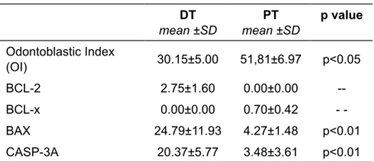
Quantitative data are illustrated in Table 1. Dental pulps from DT were characterized by reduction of odontoblastic layer and occurrence of apoptotic odontoblasts (Figure 1). The observa-tion of odontoblastic layer in the secobserva-tions of dental pulp showed of forty-two (50%) deciduous teeth (DT) if compared to four out of twenty-seven (14.8%) permanent teeth (PT) (p=0.04). To evaluate whether apoptosis may be involved, a sensitive analysis, using ssDna immunostaining in deciduous and permanent dental pulps was applied. SsDna+ cells were observed in the majority of
deciduous dental pulps (66.6%) in contrast to permanent dental pulps (45%) and appeared as single scattered cells located to the odontoblastic layer. Furthermore, the apoptotic index (AI) was
to the pulps of PT (7.80+4.13 vs 4.70+1.68).
compared to PT. Histomorphometric results showed that odonto-blastic layer length was lower in DT compared to PT (10.12+0.81
vs 12.74+
lower in DT compared to PT (30.15+5.00 vs 51.81+6.97, p=0.03). Pro-apoptotic BAX phenotype expression on odontoblasts correlated with the occurrence of numerous activated caspase-3+
odontoblasts in DT (p=0.01).
Immunostaining for the anti-apoptotic BCL-2 and BCL-x proteins (Figure 2 and Figure 3) and the pro-apoptotic BAX protein (Figure 4) was performed in sections obtained from DT and PT. Table 1 exhibits the results of immunohistochemistry. No or rare odontoblasts showed positivity for BCL-x protein in DT and PT. On the other hand, BCL-2 protein was detected in odonto-present. In particular, BCL-2 protein was expressed in 33.3% of DT and counting analysis showed that number of BCL-2+
odon-toblasts was slightly higher in DT compared to PT (2.75+1.60 vs 2.51+
(p
and in rare lymphocytes, if these were present. BAX protein was expressed on odontoblasts in 83.3% of DT and in 40.7% of PT . Moreover, high number of BAX+
-cant (24.79+11.93 vs 4.27+1.48, p
proteins have an inverse role in the regulation of the apoptosis,
+
-cantly higher compared to BCL-2+ cells in the odontoblastic layer
of the DT (24.79+11.93 vs 2.75+1.60, p=0.03), suggesting that odontoblasts have a predominant pro-apoptotic phenotype in DT.
These results were corroborated by detection of nuclear and cyto-plasmic activated caspase 3+, a marker of commitment to apoptosis,
on numerous odontoblasts in DT (20.37+5.77) (Figure 5). Finally, statistically analysis revealed strong linear correlation between numbers of BAX+ and activated caspase-3+ odontoblasts in DT (r=0.908, p=0.01).
Table 1. Extent of odontoblastic layer as determined by odontoblastic index (OI) and odontoblastic cells,
mean ±SD mean ±SD Odontoblastic Index (OI) 30.15±5.00 51,81±6.97 p<0.05 BCL-2 2.75±1.60 0.00±0.00 --BCL-x 0.00±0.00 0.70±0.42 -BAX 24.79±11.93 4.27±1.48 p<0.01 CASP-3A 20.37±5.77 3.48±3.61 p<0.01
The results of the present study support the two research hypoth-eses, showing that: 1) there is a morphological apparent reduction of the odontoblastic layer in dental pulps of DT compared to PT, and 2) there is greater occurrence of apoptotic odontoblasts in DT compared to PT. It can be speculated that the mode of odontoblasts previous studies demonstrating that odontoblasts decrease during their life-cycle by apoptotic cell death. 8,9
-mately is mediated by activated caspase-3 and consequently acti-vation of downstream DNAses.10,11 Indeed, previous
immunohisto-chemical reports demonstrated that apoptosis plays a crucial role not only during embryonic development, but also in the maintenance of dental tissue homeostasis, by eliminating cells that have already achieved their genetic program, thus controlling the pattern, shape and size of the teeth. 12-14
A close correlation exists between the volume of the dental pulp chamber and the age of the teeth, because there is a contin-uous dentin deposition that decreases the volume occupied by the odontoblast/dentin interface. Only one report described this physi-ological event, showing that massive odontoblast apoptosis occurs during a 4-year period.8 Some studies evaluated apoptosis in the
odontoblastic layer of intact and injured permanent human teeth.15,16
However, to the authors knowledge, there are no studies evaluating the odontoblast presence (by means of odontoblastic index) and apoptosis (by means of apoptotic markers such as BAX, BCL-2 and Caspase-3), both in DT and in PT.
Indeed, the morphological observations of the present study between DT and PT. Histomorphometry and immunohistochemistry
allows evaluating the occurrence of a pro-apoptotic phenotype expression on odontoblasts, using in situ immunohistochemistry for apoptotic regulatory BAX, BCL-2 and BCL-x proteins. Both BAX and BCL-2 have been expressed on odontoblasts, but a signif-for BAX expression. Indeed, while PT displayed scattered BAX+
odontoblasts, DT showed higher numbers of BAX+ odontoblastic
prevalence of pro-apoptotic BAX protein over anti-apoptotic BCL-2 protein in odontoblastic cell population from DT, revealing the occurrence of pro-apoptotic phenotype on these cells. Since it has been demonstrated that high BAX/BCL-2 ratio determines the cell susceptibility to apoptosis following trigger signals, we have tried to detect activated caspase-3, one of the key cystein protease of apoptosis.17 Caspase-3 is involved in the proteolytic cleavage of
key downstream proteins, such as poly(ADP-ribose) polymerase (PARP), which ultimately results in DNA fragmentation and apop-totic cell death.
Findings showed that the occurrence of pro-apoptotic pheno-type on odontoblasts of DT might be correlated with the degree of caspase-3 activation, a marker of commitment to apoptosis. 18
+ odontoblasts were
found in DT, as opposed to PT, and they were associated with acti-vation of caspase-3, consequently being committed to apoptosis.
evidenced by histomorphometric changes in DT. Odontoblasts circumpulpal dentin and lay down physiological secondary dentin.
19
development and may change the response to tissue damage, since tertiary dentin is deposited beneath the site of injury by existing odontoblasts. 15,20 According to the present study, the odontoblasts of
DT can be assumed to have a lower reparative activity if compared to odontoblasts of PT. Many reports have documented that the vital pulp treatments used in primary dentition have lower success rates in comparison to permanent dentition but this fact has been no correlated with the extent of odontoblastic layer and the apoptosis of odontoblastic cell population. 17,21-24 Further studies are needed to
According to the present study, the odontoblasts of DT can be assumed to have a lower reparative activity if compared to odonto-blasts of PT. Many reports have documented that the vital pulp treat-ments used in primary dentition have lower success rates in compar-ison to permanent dentition but this fact has been no correlated with the extent of odontoblastic layer and the apoptosis of odontoblastic cell population. 17,21-24 Further studies are needed to evaluate whether
The Journal of Clinical Pediatric Dentistry Volume 40, Number 6/2016 455
1. Goldberg M, Smith AJ. Cells and extracellular matrices of dentin and pulp: a biological basis for repair and tissue engineering. Crit Rev Oral Biol Med 15(1): 13-27, 2004.
2. Lesot H, Bègue-Kirn C, Kübler MD, Meyer JM, Smith AJ, Cassidy N, -ulation during reparative processes. Cell Mater 3: 201-217, 1993. 3. Smith AJ, Lesot H. Induction and regulation of crown dentinogenesis:
embryonic events as a template for dental tissue repair?. Crit Rev Oral Biol Med 12:425-437, 2001.
4. Linde A, Goldberg M. Dentinogenesis. Crit Rev Oral Biol Med 4:679-725, 1993.
5.
Develop Biol 39: 51-68, 1995.
6. Smith AJ, Tobias RS, Cassidy N, Plant CG, Browne RM, Bègue-Kirn C et al. Odontoblast stimulation in ferrets by dentine matrix components. Archives of Oral Biology 39;13-22, 1994.
7. Smith AJ, Cassidy N, Perry H, Bègue-Kirn C, Ruch Jv, Lesot H. Reac-tionary dentinogenesis. Int J Develop Biol 39:273-280, 1995.
8. Franquin JC, Remusat M, Abou Hashieh I, Dejou J. Immunocytochemical detection of apoptosis in human odontoblasts. Eur J Oral Sci 106 Suppl 1: 384-387, 1998.
9. Mitsiadis TA, De Bari C, About I. Apoptosis in developmental and repair-related human tooth remodeling: a view from the inside. Exp Cell Res 314(4): 869-877, 2008.
10. Kerr JF, Wyllie AH, Currie AR. Apoptosis: a basic biological phenom-enon with wide-ranging implications in tissue kinetics. Brit J Cancer 26: 239-257, 1972.
11. Nunez G, Benedict Ma, Hu Y, Inohara N. Caspases: the proteases of the apoptotic pathway. Oncogene 17: 3237-3245, 1998.
12.
association with an embryonic signaling center and suppression by EGF and FGF-4. Develop 122: 121-129, 1996.
13. Kim JY, Cha YG, Cho SW, Kim EJ, Lee MJ, Lee JM et al. Inhibition of apoptosis in early tooth development alters tooth shape and size. J Dent Res 85(6):530-535, 2006.
14.
of an embryonic signaling center: BMP-4 induces p21 and is associated with apoptosis in the mouse tooth enamel knot. Develop 125(2):161-169, 1998.
15. Larmas M. Odontoblast function seen as the response of dentinal tissue to dental caries. Adv Dent Res15: 68-71, 2001.
16. Larmas M, Sándor GK. Enzymes, dentinogenesis and dental caries: a literature review. J Oral Maxillofac Res 29; 5(4):e3, 2014. doi: 10.5037/ jomr.2014.5403
17. Oltvai ZN, Milliman CL, Korsmeyer SJ. Bcl-2 heterodimerizes in vivo with a conserved homolog, Bax, that accelerates programmed cell death. Cell 74: 609-619, 1993.
18. Mazumder S, Plesca D, Almasan A. Caspase-3 activation is a critical determinant of genotoxic stress-induced apoptosis. Meth Molec Biol 414: 13-21, 2008.
19. Lesot H, Lisi S, Peterkova R, Peterka M, Mitolo V, Ruch JV. Epigenetic
. Adv Dent Res 15: 8-13, 2001.
20.
. Adv Dent
Res 15: 14-18, 2001.
21. Fuks AB. Current concepts in vital primary pulp therapy. Eur J Paed Dent 3: 115-120, 2002.
22. Fuks AB. Vital pulp therapy with new materials for primary teeth: new directions and Treatment perspectives. Ped Dent 30: 211-219, 2008. 23. Kopel HM. Considerations for the direct pulp capping procedure in
primary teeth: a review of the literature. ASDC J Dent Child 59: 141-149, 1992.
24. Ranly DM, Garcia-Godoy F. Current and potential pulp therapies for primary and young permanent teeth. J Dent 28: 153-161, 2000.