This is an open access article under the terms of the Creative Commons Attribution License, which permits use, distribution and reproduction in any medium, provided the original work is properly cited.
Fetal cerebral Doppler changes and outcome in late preterm
fetal growth restriction: prospective cohort study
T. STAMPALIJA
1,2, J. THORNTON
3, N. MARLOW
4, R. NAPOLITANO
4,5, A. BHIDE
6,
T. PICKLES
7, C. M. BILARDO
8,9, S. J. GORDIJN
9, W. GYSELAERS
10, H. VALENSISE
11,
K. HECHER
12, R. K. SANDE
13, P. LINDGREN
14, E. BERGMAN
15, B. ARABIN
16,
A. C. BREEZE
17, L. WEE
18, W. GANZEVOORT
8, J. RICHTER
19, A. BERGER
20, J. BRODSZKI
21,
J. DERKS
22, F. MECACCI
23, G. M. MARUOTTI
24, K. MYKLESTAD
25, S. M. LOBMAIER
26,
F. PREFUMO
27, P. KLARITSCH
28, P. CALDA
29, C. EBBING
30, T. FRUSCA
31, L. RAIO
32,
G. H. A. VISSER
33, L. KROFTA
34, I. CETIN
35, E. FERRAZZI
36, E. CESARI
35, H. WOLF
8and
C. C. LEES
37, on behalf of the TRUFFLE-2 Group
#1Unit of Fetal Medicine and Prenatal Diagnosis, Institute for Maternal and Child Health, IRCCS Burlo Garofolo, Trieste, Italy;2Department of Medicine, Surgery and Health Sciences, University of Trieste, Trieste, Italy;3School of Clinical Sciences, University of Nottingham, Division of Obstetrics and Gynaecology, Maternity Department, City Hospital, Nottingham, UK;4UCL Elizabeth Garrett Anderson Institute for Women’s Health, University College London, London, UK;5Fetal Medicine Unit, University College London Hospitals NHS Foundation Trust, London, UK;6Fetal Medicine Unit, St George’s University Hospitals NHS Foundation Trust and Molecular & Clinical Sciences Research Institute, St George’s, University of London, London, UK;7Centre for Trials Research, College of Biomedical and Life Sciences, Cardiff University, Cardiff, UK;8Department of Obstetrics and Gynecology, Amsterdam University Medical Centers, University of Amsterdam, Amsterdam Reproduction and Development Research Institute, Amsterdam, The Netherlands; 9Department of Obstetrics and Gynaecology, University Medical Center Groningen, University of Groningen, Groningen, The Netherlands;10Faculty of Medicine and Life Sciences, Hasselt University, Agoralaan, Diepenbeek, Belgium, Department of Obstetrics & Gynaecology, Ziekenhuis Oost-Limburg, Genk and Department Physiology, Hasselt University, Diepenbeek, Belgium;11Department of Surgery, Division of Obstetrics and Gynecology, Tor Vergata University, Policlinico Casilino Hospital, Rome, Italy;12Department of Obstetrics and Fetal Medicine, University Medical Centre Hamburg-Eppendorf, Hamburg, Germany;13Department of Obstetrics and Gynecology, Stavanger University Hospital, Stavanger and Department of Clinical Science, University of Bergen, Bergen, Norway;14Center for Fetal Medicine, Karolinska University Hospital, Stockholm, Sweden;15Department of Women’s and Children’s Health, Uppsala University, Uppsala, Sweden;16Department of Obstetrics Charite, Humboldt University Berlin and Clara Angela Foundation, Berlin, Germany; 17Fetal Medicine Unit, Leeds General Infirmary, Leeds Teaching Hospitals NHS Trust, Leeds, UK;18The Princess Alexandra Hospital NHS Trust, Harlow, UK;19Department of Gynecology and Obstetrics, UZ Leuven and Department of Regeneration and Development, KU Leuven, Leuven, Belgium;
20Department of Obstetrics and Gynecology, Medical University of Innsbruck, Innsbruck, Austria;21Department of Pediatric Surgery and Neonatology, Lund University, Skane University Hospital, Lund, Sweden;22Department of Perinatal Medicine, University of Utrecht, Utrecht, The Netherlands;23Department of Health Sciences, University of Florence, Obstetrics and Gynecology, Careggi University Hospital, Florence, Italy;24Department of Neurosciences,
Reproductive and Dentistry Sciences, University of Naples ‘Federico II’, Naples, Italy;25St Olav’s Hospital, Trondheim, Norway;26Department of Obstetrics and Gynecology, Klinikum Rechts Der Isar, Technical University of Munich, Munich, Germany;27Department of Obstetrics and Gynecology, ASST Spedali Civili di Brescia and University of Brescia, Brescia, Italy;28Department of Obstetrics and Gynecology, Medical University of Graz, Graz, Austria; 29Department of Obstetrics and Gynaecology, General University Hospital and First Faculty of Medicine, Charles University, Prague, Czech Republic; 30Department of Obstetrics and Gynecology, Haukeland University Hospital, Bergen, Norway;31Department of Obstetrics and Gynecology, University of Parma, Parma, Italy;32Department of Obstetrics & Gynecology, University Hospital of Bern, Bern, Switzerland;33Department of Obstetrics, Division of Woman and Baby, University Medical Center Utrecht, Utrecht, The Netherlands;34Institute for the Care of Mother and Child, Prague, Czech Republic and Third Medical Faculty, Charles University, Prague, Czech Republic;35Department of Obstetrics and Gynecology, Vittore Buzzi Children’s Hospital, University of Milan, Milan, Italy;36Department of Obstetrics and Gynecology, Fondazione IRCCS Ca’ Granda Ospedale Maggiore Policlinico and Department of Clinical Sciences and Community Health, Universit`a degli Studi di Milano, Milan, Italy;37Imperial College School of Medicine, Imperial College London and Department of Fetal Medicine, Queen Charlotte’s and Chelsea Hospital, Imperial College NHS trust, London, UK
KEYWORDS: adverse outcome; Doppler; middle cerebral artery; neonatal; umbilical artery; umbilicocerebral ratio
CONTRIBUTION
What are the novel findings of this work?
In this prospective multicenter observational study of late preterm singleton pregnancies at risk of fetal growth restriction (FGR), fetal cerebral Doppler changes were found to be associated with adverse perinatal outcome.
Correspondence to: Prof. H. Wolf (e-mail: [email protected]) and Prof. C. C. Lees (e-mail: [email protected]) #TRUFFLE-2 Group and collaborating authors are listed at the end of the manuscript.
Accepted: 29 May 2020
What are the clinical implications of this work?
We confirm an association between abnormal fetal cere-bral Doppler and adverse perinatal outcome in late preterm singleton pregnancies at risk of FGR. Whether cerebral flow changes are a marker of fetal compromise and whether intervention changes the risk for poor outcome can be answered only in a randomized intervention study.
ABSTRACT
Objectives To explore the association between fetal
umbilical and middle cerebral artery (MCA) Doppler abnormalities and outcome in late preterm pregnancies at risk of fetal growth restriction.
Methods This was a prospective cohort study of singleton
pregnancies at risk of fetal growth restriction at 32 + 0 to 36 + 6 weeks of gestation, enrolled in 33 European centers between 2017 and 2018, in which umbilical and fetal MCA Doppler velocimetry was performed. Pregnancies were considered at risk of fetal growth restriction if they had estimated fetal weight and/or abdominal circumference (AC) < 10th percentile, abnormal arterial Doppler and/or a fall in AC growth velocity of more than 40 percentile points from the 20-week scan. Composite adverse outcome comprised both immediate adverse birth outcome and major neonatal morbidity. Using a range of cut-off values, the association of MCA pulsatility index and umbilicocerebral ratio (UCR) with composite adverse outcome was explored.
Results The study population comprised 856 women.
There were two (0.2%) intrauterine deaths. Median gestational age at delivery was 38 (interquartile range (IQR), 37–39) weeks and birth weight was 2478 (IQR, 2140–2790) g. Compared with infants with normal outcome, those with composite adverse outcome (n = 93; 11%) were delivered at an earlier gestational age (36 vs 38 weeks) and had a lower birth weight (1900 vs 2540 g). The first Doppler observation of MCA pulsatility index < 5thpercentile and UCR Z-score above gestational-age-specific thresholds (1.5 at 32–33 weeks and 1.0 at 34–36 weeks) had the highest relative risks (RR) for composite adverse outcome (RR 2.2 (95% CI, 1.5–3.2) and RR 2.0 (95% CI, 1.4–3.0), respectively). After adjustment for confounders, the association between UCR Z-score and composite adverse outcome remained significant, although gestational age at delivery and birth-weight Z-score had a stronger association.
Conclusion In this prospective multicenter study, signs
of cerebral blood flow redistribution were found to be associated with adverse outcome in late preterm singleton pregnancies at risk of fetal growth restriction. Whether cerebral redistribution is a marker describing the severity of fetal growth restriction or an independent risk factor for adverse outcome remains unclear, and whether it is useful for clinical management can be answered only in a randomized trial. 2020 The Authors. Ultrasound in Obstetrics & Gynecology published by John Wiley & Sons Ltd on behalf of the International Society of Ultrasound in Obstetrics and Gynecology.
INTRODUCTION
Poor third-trimester fetal growth is associated with adverse perinatal outcome1,2. The only therapeutic option
is timely delivery. This poses a dilemma as delivery too early risks the baby suffering the effects of moderate–late
prematurity, whereas delivery too late risks further fetal compromise increasing the risk of suboptimal outcome or stillbirth. There remains little evidence on which to base the timing of delivery of such babies in the late preterm period. The problem is twofold: first, there is no consensus on how to identify the fetus at risk for compromise and, second, an evidence-based monitoring strategy remains to be defined3.
The association between fetal middle cerebral (MCA) and umbilical artery Doppler impedance ratios and adverse outcome has been well described, deriving mainly from retrospective studies4–6. The key question is whether
abnormal cerebral artery Doppler is a non-injurious response to fetal compromise or is itself a marker of compromise and ongoing damage necessitating early delivery. Without this information, it cannot be known if using these Doppler parameters to decide on intervention by delivery is beneficial for infant outcome. Guidelines of the Royal College of Obstetricians and Gynaecologists on small-for-gestational-age (SGA) fetuses7 state that
‘MCA Doppler may be a more useful test in SGA fetuses detected after 32 weeks of gestation’, but perhaps wisely do not define the parameters that should trigger a decision to deliver. The usefulness of MCA Doppler and cerebral blood flow redistribution in improving perinatal and/or long-term outcome can be assessed only by randomization in a prospective study, but current data are insufficient to decide how such a trial should be designed.
As part of the design process for the TRUFFLE-2 ran-domized trial for determining optimal timing for delivery in the context of late preterm fetal growth restriction, we undertook a prospective observational feasibility study. The objectives of this study were to quantify perinatal morbidity and mortality in contemporary clinical prac-tice and to determine which thresholds for umbilical and MCA Doppler are most strongly associated with adverse perinatal outcome in late preterm singleton pregnancies at risk of fetal growth restriction, and which thus might be most appropriate for evaluation in a future random-ized interventional study in late preterm fetal growth restriction.
METHODS Setting
This was a prospective multicenter observational study conducted between 1 April 2017 and 1 July 2018 in 33 European perinatal centers with fetal medicine and specialized neonatal intensive care services.
Participants
Women were eligible if they had a singleton pregnancy from 32 + 0 to 36 + 6 weeks of gestation with a fetus con-sidered at risk for growth restriction, defined as estimated fetal weight (EFW) and/or abdominal circumference (AC) <10th percentile, abnormal fetal arterial Doppler and/or
a fall in AC growth velocity of more than 40 percentile points from the 20-week scan. The references for EFW, AC and Doppler parameters were based on local charts. In order to be eligible, the fetus had to have positive umbilical artery end-diastolic flow and a normal car-diotocogram (CTG) with short-term variation of the fetal heart rate > 3.0 ms, using Dawes–Redman CTG analysis. Gestational age was calculated from certain menstrual age and/or ultrasound assessment before 22 weeks of gestation. Women were ineligible for inclusion if there was known, planned or impending delivery based on maternal obstetric complications, uterine contractions or rupture of membranes, or if the fetus had a known or suspected structural or chromosomal abnormality. Further monitoring
Women were monitored using fetal biometry and Doppler assessment of umbilical and fetal MCA pulsatility index (PI), with a recommended minimum interval of 1 week between assessments. Z-scores were calculated for MCA-PI and UCR using reference data from normal pregnancies in the study of Arduini and Rizzo8, and
for fetal weight using the Hadlock fetal growth charts9.
UCR Z-scores were calculated as reported previously10.
The study was not blinded and clinicians who were collecting the data were also involved directly in the management and delivery of those women. However, there was no guidance as to what findings should trigger delivery, which was left to local custom. Management, including maternal steroid administration and delivery, was undertaken according to local clinical protocols. Definitions
Maternal hypertension was defined as blood pressure ≥ 140/90 mmHg and proteinuria was defined as > 0.3 g/L on a 24 h collection of urine or urine dipstick result of ‘1+’ or more. Hypertensive disorders were defined as chronic if hypertension existed prior to 20 weeks’ gestation or required treatment prior to pregnancy, or as gestational if the onset of hypertension occurred after 20 weeks in the absence of proteinuria. Pre-eclampsia was defined as hypertension and proteinuria, or hypertension and clinical signs of pre-eclampsia11. HELLP syndrome
was defined as alanine aminotransferase > 70 IU/L with platelets < 100 × 109/L and with evidence of hemolysis
from blood film or lactate dehydrogenase > 600 IU/L. Outcomes
The primary outcome was a composite of abnormal con-dition at birth or major neonatal morbidity. Abnormal condition at birth was defined as at least one of the following: Apgar score < 7 at 5 min, umbilical artery pH < 7.0 or umbilical vein pH < 7.1, resuscitation with intubation, chest compressions or medication, or still-birth. Major neonatal morbidity until first discharge home was defined as at least one of the following:
neurological abnormality (intracerebral hemorrhage Grade 3 or 4, periventricular leukomalacia Grade 2 or 3, encephalopathy, or seizures necessitating anti-epileptic drug treatment); cardiovascular abnormal-ity (hypotensive treatment, ductus arteriosus treatment or disseminated coagulopathy); respiratory morbidity (respiratory support for more than 1 week, mechanical ventilation, meconium aspiration or persistent pulmonary hypertension); or sepsis (clinical sepsis with positive blood culture, necrotizing enterocolitis (Bell’s stage 2 or greater) or meningitis).
Data collection
Data were collected on a secure cloud-based electronic data capture platform (Castor EDC, Amsterdam, The Netherlands) curated by H.W. The database carried no personal identifiers. Participants and their infants could be identified only using unique study identifiers that were stored in their recruiting center.
Analysis
Two analyses were undertaken. First, based on the last fetal Doppler assessment within 1 week before delivery, fetal Doppler parameters were compared between women whose infant had composite adverse outcome and those with normal outcome. Second, based on the first abnormal fetal Doppler assessment after inclusion, several fetal Doppler parameters and values were selected as potential dichotomous predictors of the composite primary outcome.
The selected dichotomous predictors were: absolute MCA-PI < 5th percentile (equivalent to a Z-score of
−1.645) at any gestational age; gestational-age-specific MCA-PI Z-score of < −2 at 32–33 weeks, < −1.5 at 34–35 weeks or < −1 at 36 weeks; absolute UCR value ≥ 95th percentile (Z-score, 1.645) at any gestational age;
UCR Z-score of ≥ 1.0 or ≥ 1.5 at any gestational age; and three gestational-age-specific UCR Z-score ranges: (1) ≥ 1.5 at 32–33 weeks or ≥ 1.0 at 34–36 weeks, (2) ≥ 1.5 at 32–33 weeks, ≥ 1.0 at 34–35 weeks or ≥ 0.5 at 36 weeks, and (3) ≥ 2.0 at 32–33 weeks, ≥ 1.5 at 34–35 weeks or ≥ 1.0 at 36 weeks.
The gestational-age-specific ranges were proposed allowing a graduated range on the basis that clinical decisions for delivery would require a more severely abnormal Doppler at an early gestational age, and the criteria for delivery would be more permissive closer to term. For each cut-off, we compared the incidence of composite adverse outcome between those women who had an abnormal Doppler at least once and those who never had an abnormal Doppler.
Statistical methods
Groups were compared with two-sided tests for statistical significance using the Mann–Whitney U-test or Pearson’s chi-square test, as appropriate. Relative risks for
composite adverse outcome were calculated unadjusted. In women who had the last Doppler measurement obtained within 1 week before delivery, the association of UCR with composite adverse outcome was adjusted using logistic regression analysis for those parameters that differed significantly between women with and those without composite adverse outcome. Heterogeneity between centers was tested using multilevel logistic regression analysis. Data are presented as number (%) or median (interquartile range (IQR)), as required. Statistical calculations were performed using SPSS Statistics software (version 25; IBM Corp., Armonk, NY, USA).
Ethical approval
This study was observational and practice (monitoring, delivery, steroid administration) was based on existing local guidance. Data were recorded anonymously after delivery. In the UK, the Health Research Authority (HRA) did not require the study to undergo ethical review, which was the case in four further countries (14 centers). In six countries (19 centers), ethical approval was required and obtained, and participating women gave informed signed consent.
RESULTS Population
Complete delivery and outcome data were recorded for 873 patients with late preterm singleton pregnancy at risk of fetal growth restriction in the study database. Seventeen cases were excluded because of the presence of major congenital abnormality, leaving 856 women and their fetuses for the final cohort analysis. Demographic, obstetric and fetal Doppler velocimetry characteristics of the women included in the cohort are shown in Table 1. Median gestational age and EFW at inclusion were 34 (IQR, 33–35) weeks and 1894 (IQR, 1624–2145) g, respectively. Indication for inclusion was EFW and/or AC < 10th percentile or a drop in AC growth velocity of
more than 40 percentiles in 842 (98%) fetuses, while in 98 (11%) an abnormal fetal arterial Doppler (umbilical artery or MCA), according to local reference charts, was observed.
Outcome
Median gestational age at delivery was 38 (IQR, 37–39; range, 32–42) weeks and birth weight was 2478 (IQR, 2140–2790; range, 1080–4255) g. The primary composite adverse outcome occurred in 93 (11%) infants, and its components are detailed in Table 2.
Abnormal condition at birth was present in 27 (3%) fetuses. Among these, there were two (0.2%) antenatal deaths, of which one was diagnosed at 33 + 3 weeks (1 week after inclusion) and the second at 34 + 6 weeks (2 weeks after inclusion). In both cases, umbilical artery PI and UCR were < 95th percentile, while MCA-PI was
Table 1 Demographic, obstetric and Doppler variables in study population of 856 late preterm singleton pregnancies at risk of fetal growth restriction
Variable Value
Maternal age (years) 31 (28 to 35)
Nulliparous 524 (61)
BMI (kg/m2) 22.5 (20.3 to 26.0)
Smoker 68 (8)
Diabetes Type 1, 2 or gestational 70 (8)
Chronic hypertension 19 (2)
Other disease 246 (29)
At inclusion
Gestational age (weeks) 34 (33 to 35)
Inclusion indication*
EFW and/or AC < 10thpercentile 792 (93)
AC growth velocity drop > 40 percentiles
50 (6)
Doppler abnormality 98 (11)
EFW (g) 1894 (1624 to 2145)
EFW Z-score −1.52 (−2.0 to −1.1)
Umbilical artery PI Z-score 0.1 (−0.3 to 0.6) Umbilical artery PI ≥ 95thpercentile 34 (4)
Middle cerebral artery PI Z-score −0.4 (−1.2 to 0.5) Middle cerebral artery PI < 5thpercentile 116 (14)
UCR Z-score 0.0 (−0.4 to 0.5)
UCR ≥ 95thpercentile 35 (4)
Pre-eclampsia or HELLP 79 (9)
Any hypertensive disorder of pregnancy 119 (14)
Antihypertensive medication 103 (12)
Corticosteroids for fetal lung maturation (> 24 h before delivery)
98 (11) Total number of arterial Doppler
measurements
2751 Number of arterial Doppler measurements
per woman
4 (3 to 5) Inclusion-to-delivery interval (days) 27 (14 to 37) Delivery
Planned Cesarean section 219 (26)
Indication
Fetal condition (CTG or Doppler) 155/219 (71)
Fetal growth/EFW 25/219 (11)
Maternal condition 39/219 (18)
Induction of labor 369 (43)
Indication
Fetal condition (CTG or Doppler) 112/369 (30)
Fetal growth/EFW 213/369 (58)
Maternal condition 44/369 (12)
Spontaneous onset of labor 268 (31)
Cesarean section after onset of labor 117 (14) Data are given as median (interquartile range), n (%), n or n/N (%). *Multiple indications possible. AC, abdominal circumference; BMI, body mass index; CTG, cardiotocogram; EFW, estimated fetal weight; HELLP, hemolysis, elevated liver enzymes and low platelet count; PI, pulsatility index; UCR, umbilicocerebral ratio.
<5th percentile and birth weight Z-score was < −2. There were no neonatal deaths before discharge. Major neonatal morbidity occurred in 77 (9%) cases; the major contributors were respiratory morbidity (53/77; 69%) and infection (17/77; 22%).
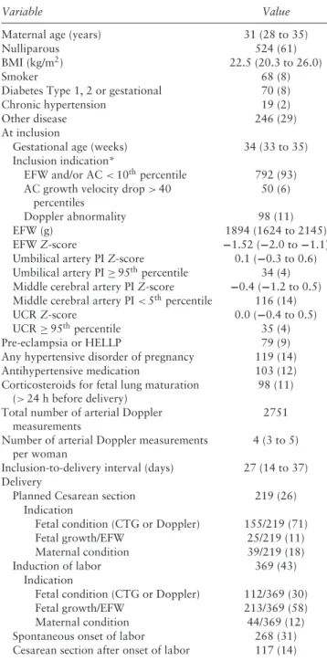
Figure 1 shows the proportion of infants with abnormal condition at birth and/or major neonatal morbidity before discharge, according to gestational age at delivery and birth weight Z-score. In comparison with infants with normal outcome, composite adverse perinatal outcome
Table 2 Perinatal outcome in study population of 856 late preterm singleton pregnancies at risk of fetal growth restriction
Variable Value
Gestational age at delivery (weeks)
38 (37–39) [32–42] Birth weight (g) 2478 (2140–2790) [1080–4255] Birth-weight Z-score −1.7 (−2.3 to −1.1) [−5.6 to 3.2] Birth weight < 10thpercentile 596 (70)
Male fetal sex 372 (43)
Composite adverse outcome* 93 (11) Abnormal condition at birth† 27 (3)
Fetal death 2 (0)
Cord arterial pH < 7.0 or venous pH < 7.1
7/712 (1)
5-min Apgar score < 7 15 (2)
Resuscitation with intubation or medication
10 (1)
Major neonatal morbidity† 77 (9)
Cerebral 7 (1)
Cardiovascular 7 (1)
Respiratory 53 (6)‡
Infection 17 (2)
Abnormal condition at birth and major neonatal morbidity
11 (1)
Neonatal death 0 (0)
Data are given as median (interquartile range) [range], n (%) or n/N (%). *Composite adverse outcome defined as abnormal condition at birth and/or major neonatal morbidity. †Multiple conditions possible. ‡39/53 (74%) had only some respiratory support for short duration in first week.
was associated strongly with lower gestational age at delivery (36 (IQR, 34–38) vs 38 (IQR, 37–39) weeks) and lower birth weight (1900 (IQR, 1557–2355) vs 2540 (IQR, 2220–2810) g). Infants with birth weight Z-score <−2 had a significantly higher risk of composite adverse outcome than did those with a higher birth weight Z-score (RR, 2.7 (95% CI, 1.8–4.0); P < 0.01).
A Doppler evaluation was recorded within 1 week before delivery in 584 (68%) women, of whom, 75 (13%) had composite adverse outcome and 509 (87%) did not. Those with composite adverse outcome had higher umbilical artery PI and lower MCA-PI at the last assessment before delivery, were born at an earlier gestational age and had lower birth weight (Table 3).
The Doppler velocimetry criteria were assessed by comparing women who had an abnormal fetal Doppler at least once at or after inclusion with women who never had an abnormal fetal Doppler, according to the different criteria (Table 4). Pregnancies with abnormal Doppler were delivered earlier in gestation compared with those in which the Doppler velocimetry results remained in the normal range, regardless of the criterion used (P < 0.05 for each comparison). Middle cerebral artery PI < 5th percentile and four of the UCR Z-score
criteria were associated with an increased prevalence of composite adverse outcome. The highest RR of abnormal Doppler for composite adverse outcome was for MCA-PI < 5thpercentile (RR, 2.2 (95% CI, 1.5–3.2))
and high gestational-age-specific UCR Z-score (≥ 1.5
32–33 0 20 40 Percentage 60 80 100 (a) n (%) 34–35
Gestational age (weeks)
36–37 38–39 ≥40 32 (4%) 96 (11%) 274 (32%) 338 (39%) 116 (14%) <–2 0 20 40 Percentage 60 80 100 (b) n (%) –2 Birth-weight Z-score –1 ≥0 318 (37%) 367 (43%) 141 (16%) 30 (4%)
Figure 1 Incidence of composite adverse outcome in 856 late preterm singleton pregnancies at risk of fetal growth restriction, according to gestational age at delivery (a) and birth-weight Z-score (b). Composite adverse outcome defined as abnormal condition at birth and/or major neonatal morbidity. Eleven infants had both abnormal condition at birth and major neonatal morbidity. , normal; , major neonatal morbidity; , abnormal condition at birth + major neonatal morbidity; , abnormal condition at birth.
at 32–33 weeks and ≥ 1.0 at 34–36 weeks) (RR, 2.0 (95% CI, 1.4–3.0)).
Compared with women with normal Doppler, those who had an abnormal Doppler at any point after inclusion, based on either the 5th percentile cut-off of
MCA-PI or the gestational-age-specific UCR Z-score range, underwent prelabor Cesarean section more frequently, had a lower gestational age at delivery and a lower birth weight, and their infants had more frequently composite adverse outcome (Table 5). Table 6 shows that an abnormal Doppler occurred more frequently at an earlier gestational age and that abnormal Doppler before 36 weeks was associated significantly with a higher rate of composite adverse outcome.
Multilevel logistic regression analysis with an uncon-ditional mean model using the participating centers and composite adverse outcome showed a random inter-cept variance of 0.21 (95% CI, 0.03–1.59; P = 0.06). Calculated from this value, the intraclass correlation coefficient was 0.06. This indicated that only 6% of the
chance of having an abnormal composite endpoint was explained by differences between centers, and that further assessment of confounders using logistic regression ana-lysis without taking center into account was appropriate. This analysis was performed in the subgroup of 584
women with the last Doppler measurement obtained within 1 week before delivery, using UCR Z-score and those variables that differed significantly between women with and those without composite adverse outcome (Table 3). The adjusted odds ratio for composite adverse
Table 3 Univariate analysis of demographic, obstetric and Doppler variables in 584 late preterm singleton pregnancies at risk of fetal growth restriction that had last fetal Doppler measurement obtained within 1 week before delivery, according to presence or absence of composite adverse outcome
Composite adverse outcome Parameter
Present (n = 75)
Not present (n = 509)
Maternal age (years) 33 (29 to 37) 32 (28 to 35)*
Maternal age ≥ 35 years 30 (40) 138 (27)*
Nulliparous 49 (65) 311 (61)
Smoker 11 (15) 38 (7)
BMI (kg/m2) 21.8 (20.1 to 25.9) 22.3 (20.2 to 26.1)
Gestational hypertension or pre-eclampsia 24 (32) 78 (15)*
Umbilical artery PI Z-score 0.5 (−0.2 to 1.1) 0.1 (−0.3 to 0.6)*
Umbilical artery PI ≥ 95thpercentile 9 (12) 31 (6)
Umbilical artery PI ≥ 90thpercentile 13 (17) 53 (10)
Umbilical artery A/REDF 3 (4) 5 (1)
Middle cerebral artery PI Z-score −1.4 (−2.0 to −0.4) −1.0 (−1.6 to −0.3)*
Middle cerebral artery PI < 5thpercentile 32 (43) 125 (25)*
UCR Z-score 0.6 (0.2 to 1.3) 0.4 (0.0 to 1.0)
UCR ≥ 95thpercentile 9 (12) 65 (13)
UCR ≥ 90thpercentile 18 (24) 95 (19)
Gestational age at delivery (weeks) 35 (34 to 37) 38 (37 to 39)*
Birth weight (g) 1820 (1520 to 2240) 2420 (2125 to 2725)*
Birth-weight Z-score −2.5 (−3.1 to −1.5) −1.8 (−2.3 to −1.2)*
Birth weight < 10thpercentile 68 (91) 373 (73)*
Male sex 35 (47) 220 (43)
Data are given as median (interquartile range) or n (%). Composite adverse outcome defined as abnormal condition at birth and/or major neonatal morbidity. *P < 0.05 by Mann–Whitney U-test or Fisher’s exact test. A/REDF, absent or reversed end-diastolic flow; BMI, body mass index; PI, pulsatility index; UCR, umbilicocerebral ratio.
Table 4 Effect of different Doppler velocimetry criteria to define abnormal result in 856 late preterm singleton pregnancies: comparison of participants who had abnormal Doppler observation at least once in study with those who never had abnormal Doppler
RR for composite adverse outcome Doppler always normal
Doppler abnormal at least once
Abnormal Doppler criterion Pregnancies First abnormal Doppler to delivery interval (days) Composite adverse outcome GA at delivery (weeks) Pregnancies Composite adverse outcome GA at delivery
(weeks) Value (95% CI) P Middle cerebral artery PI
<5thpercentile 287 (34) 11 (4–20) 49/287 (17)* 38 (36–39)* 569 (66) 44/569 (8) 38 (37–39) 2.2 (1.5–3.2) <0.01
Low GA-specific Z-score 329 (38) 12 (6–22) 43/329 (13) 37 (37–38)* 527 (62) 50/527 (9) 39 (37–40) 1.4 (0.9–2.0) 0.06 Umbilicocerebral ratio
≥ 90thpercentile 168 (20) 7 (3–13) 25/168 (15) 37 (35–38)* 688 (80) 68/688 (10) 38 (37–39) 1.5 (1.0–2.3) 0.07 Z-score ≥ 1.0 223 (26) 8 (3–14) 34/223 (15)* 37 (36–38)* 633 (74) 59/633 (9) 38 (37–39) 1.6 (1.1–2.4) 0.02
Z-score ≥ 1.5 134 (16) 6 (2–11) 24/134 (18)* 37 (35–38)* 722 (84) 69/722 (10) 38 (37–39) 1.9 (1.2–2.9) <0.01 High GA-specific Z-score 1 162 (19) 9 (3–14) 30/162 (19)* 37 (35–37)* 694 (81) 63/694 (9) 38 (37–39) 2.0 (1.4–3.0) <0.01 High GA-specific Z-score 2 233 (27) 10 (5–16) 35/233 (15)* 37 (36–38)* 623 (73) 58/623 (9) 38 (37–40) 1.6 (1.1–2.4) 0.02 High GA-specific Z-score 3 126 (15) 7 (3–12) 19/126 (15) 37 (36–37)* 730 (85) 74/730 (10) 38 (37–39) 1.5 (0.9–2.4) 0.12
Data are given as n (%), median (interquartile range) or n/N (%), unless stated otherwise. Composite adverse outcome defined as abnormal condition at birth and/or major neonatal morbidity. Low GA-specific Z-score: < −2 at 32–33 weeks, < −1.5 at 34–35 weeks, < −1 at 36 weeks; high GA-specific Z-score 1: ≥ 1.5 at 32–33 weeks, ≥ 1.0 at 34–36 weeks; high GA-specific Z-score 2: ≥ 1.5 at 32–33 weeks, ≥ 1.0 at 34–35 weeks, ≥ 0.5 at 36 weeks; high GA-specific Z-score 3: ≥ 2.0 at 32–33 weeks, ≥ 1.5 at 34–35 weeks, ≥ 1.0 at 36 weeks. *P < 0.05 by Mann-Whitney U-test or Fisher’s exact test, compared with pregnancies with Doppler assessment always normal. GA, gestational age; PI, pulsatility index; RR, relative risk.
Table 5 Gestational age (GA) at delivery, birth weight and perinatal outcome in 856 singleton pregnancies at risk of late preterm fetal growth restriction, according to whether Doppler assessment was abnormal
Middle cerebral artery pulsatility index GA-specific UCR Z-score*
Variable <5thpercentile at least once (n = 287) Always normal (n = 569) High at least once (n = 162) Always normal (n = 694)
Inclusion-to-delivery interval (days) 25 (11–35)‡ 28 (16–39) 17 (7–28)‡ 29 (17–39)
First abnormal Doppler to delivery interval (days)
11 (4–21) — 9 (3–14) —
CS before labor 87 (30)‡ 132 (23) 75 (46)‡ 144 (21)
Induction of labor 146 (51)‡ 223 (39) 68 (42) 301 (43)
Spontaneous onset of labor 54 (19)‡ 214 (38) 19 (12)‡ 249 (36)
CS during labor 44 (15) 73 (13) 27 (17) 90 (13)
Vaginal delivery 156 (54) 364 (64) 60 (37)‡ 460 (66)
GA at delivery (weeks) 38 (36–39)‡ 38 (37–39) 37 (35–37)‡ 38 (37–39)
Birth weight (g) 2380 (2000–2655)‡ 2555 (2213–2850) 2045 (1800–2391)‡ 2577 (2260–2830)
Birth weight < 10thpercentile 219 (76)‡ 377 (66) 132 (81)‡ 464 (67)
Composite adverse outcome† 49 (17)‡ 44 (8) 30 (19)‡ 63 (9)
Abnormal condition at birth only 10 (3)§ 6 (1) 3 (2)¶ 13 (2)¶
Major neonatal morbidity only 38 (13)‡ 28 (5) 26 (16)‡ 40 (6)
Abnormal condition at birth and major neonatal morbidity
1 (0) 10 (2) 1 (1) 10 (1)
*GA-specific UCR Z-score cut-offs: ≥ 1.5 at 32–33 weeks and ≥ 1.0 at 34–36 weeks. †Composite adverse outcome defined as abnormal condition at birth and/or major neonatal morbidity. ‡P < 0.05 by Mann–Whitney U-test or Fisher’s exact test, compared with pregnancies with Doppler assessment always normal. §Two cases of fetal death. ¶One case of fetal death. CS, Cesarean section; UCR, umbilicocerebral ratio.
Table 6 Composite adverse outcome and gestational age (GA) at delivery in 856 singleton pregnancies at risk of late preterm fetal growth restriction, according to whether Doppler assessment was abnormal and GA at assessment
Doppler abnormal at least once Doppler always normal RR for composite adverse outcome Abnormal Doppler criterion/GA at assessment Composite adverse outcome GA at delivery (weeks) Composite adverse outcome GA at delivery
(weeks) Value (95% CI) P MCA-PI < 5thpercentile
32–33 weeks 24/63 (38)† 36 (34–38)† 26/282 (9) 38 (37–39) 4.1 (2.6–6.7) <0.01
34–35 weeks 14/94 (15)† 37 (36–38)† 37/508 (7) 38 (37–40) 2.0 (1.2–3.6) 0.02
36 weeks 6/63 (10) 38 (37–38)† 27/562 (5) 39 (38–40) 2.0 (0.9–4.6) 0.1
High GA-specific UCR Z-score*
32–33 weeks 12/31 (39)† 34 (33–36)† 34/316 (11) 38 (37–39) 4.1 (2.2–7.9) <0.01
34–35 weeks 15/86 (17)† 36 (35–37)† 46/558 (8) 39 (37–40) 2.1 (1.2–3.7) 0.02
36 weeks 3/45 (7) 37 (37–38)† 34/620 (5) 39 (38–40) 1.2 (0.4–3.8) 0.7
Data are given as n/N (%) or median (interquartile range), unless stated otherwise. Women who had abnormal Doppler ultrasound were counted only once at gestational age epoch in which first abnormal Doppler was registered. Women with normal Doppler could have ultrasound at more than one gestational age epoch after inclusion; totals are therefore higher than total number of included women. Composite adverse outcome defined as abnormal condition at birth and/or major neonatal morbidity. *GA-specific UCR Z-score cut-offs: ≥ 1.5 at 32–33 weeks and ≥ 1.0 at 34–36 weeks. †P < 0.05 by chi-square test or Mann–Whitney U-test, compared with pregnancies with Doppler assessment always normal. MCA, fetal middle cerebral artery; PI, pulsatility index; RR, relative risk; UCR, umbilicocerebral ratio.
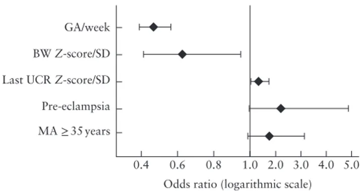
outcome of UCR Z-score was 1.3 per SD of UCR (95% CI, 1.0–1.8; P = 0.04) (Figure 2). This model had sensitivity of 79% at specificity of 75%. Gestational age and birth weight Z-score had the greatest proportional contribution to the model (0.45 and 0.29, respectively), while that of UCR Z-score was 0.12, calculated using multilayer perceptron analysis. In a similar analysis with MCA-PI Z-score, it was observed that, after adjustment, this parameter was not associated significantly with the composite adverse endpoint.
DISCUSSION
In this study, we have shown that, in late preterm singleton pregnancies at risk of fetal growth restriction, fetal cerebral blood flow redistribution detected on Doppler ultrasound within 1 week prior to delivery and the first abnormal Doppler result at any time after inclusion were both associated with composite adverse outcome. The strength of our study is that a wide range of centers were involved, which resulted in rapid prospective recruitment of a large cohort of babies at risk of growth
0.4 MA ≥ 35 years
Pre-eclampsia Last UCR Z-score/SD BW Z-score/SD GA/week
0.6 0.8 1.0
Odds ratio (logarithmic scale) 2.0 3.0 4.0 5.0
Figure 2 Adjusted odds ratios with 95% CI for composite adverse outcome in 584 late preterm singleton pregnancies at risk of fetal growth restriction and with Doppler measurement obtained within 1 week before delivery, calculated by logistic regression analysis, using parameters that were statistically significant on univariate analysis (Table 3). Missing variables from Table 3 were ejected from analysis when P > 0.1. Model had sensitivity of 79% at specificity of 75%, and area under receiver-operating-characteristics curve of 0.84 (95% CI, 0.79–0.89). Composite adverse outcome defined as abnormal condition at birth and/or major neonatal morbidity. BW, birth weight; GA, gestational age at delivery; MA, maternal age; UCR, umbilicocerebral ratio.
restriction. The data provide a snapshot of the natural history of late preterm growth restriction in relation to fetal cerebral redistribution in contemporary practice. We have also defined the prevalence of composite adverse perinatal outcome in these fetuses and infants, according to prespecified outcomes. The main weakness, as with any observational study, is that the Doppler results were revealed to the clinicians, thus making the results susceptible to the treatment paradox.
With the above proviso, which also affects all previous similar series, our results largely confirm the association between fetal cerebral blood flow redistri-bution and short-term adverse outcome in fetal growth restriction4–6,12,13. Our findings are also consistent
with those of recent meta-analyses4,5, in which the
accuracy of fetal Doppler for the prediction of composite adverse outcome was low, with sensitivity of 45–70% and specificity of 75–95%, depending on the Doppler parameter and thresholds used.
There was a significant association between these predefined markers of abnormal Doppler velocimetry and gestational age at delivery and the frequency of our composite primary outcome (Tables 3, 5 and 6). Gesta-tional age at delivery and birth weight Z-score should be interpreted in this cohort as a proxy measure of the severity of fetal growth restriction (Figures 1 and 2). We might hypothesize that the most severe cases will reach the limits of the uteroplacental supply earlier, which may ultimately result in fetal asphyxia or death and increase the risk of neonatal morbidity. The higher incidence of the first finding of an abnormal Doppler parameter at an earlier gestational age epoch, which was associated with more preterm deliveries and a higher rate of composite adverse outcomes, supports this (Table 6). Adjustment for gestational age at delivery, birth weight, maternal age and pre-eclampsia reduced the importance of the association
of UCR Z-score with the composite adverse endpoint, but it remained statistically significant (Figure 2).
These data cannot be used to ascertain when delivery should occur in the context of abnormal Doppler and CTG findings. This can be evaluated only in a well-conducted randomized trial before fetal MCA Doppler can be recommended for this purpose. Hence, these results have important implications for research, in particular for the design of such a trial. The parameters that dichotomized most effectively between normal outcome and composite adverse perinatal outcome were MCA-PI < 5th percentile or a graduated
gestational-age-specific range of UCR Z-score (≥ 1.5 at 32–33 weeks, ≥ 1.0 at 34–36 weeks). The rationale behind choosing gestational-age-specific ranges reflects the higher level of concern about fetal condition that is required to trigger delivery at an earlier gestational age compared with near term at a stage at which neonatal morbidity and mortality are normally very low, extrapolating from population studies14. However,
long-term morbidity from late preterm birth is not negligible15,16. The extent to which this excess morbidity
is caused by late fetal growth restriction is unknown. Furthermore, a ratio between the PI of the umbilical and middle cerebral arteries may have a closer association with perinatal outcome than does each parameter alone4,5.
Most studies on cerebral blood flow redistribution have reported on the cerebroplacental ratio (CPR), which is the inverse of UCR. We have used UCR as we believe this ratio allows for better differentiation in the abnormal range than does CPR10. A brief justification for this is that,
as fetal Doppler changes become more abnormal with lower cerebral and higher umbilical artery impedance, CPR tends towards an asymptote approximating to zero, while UCR tends towards infinity, thus accentuating the differences between abnormal values. Furthermore, most commonly used ratios in medicine become larger with increasing abnormality, similar to UCR.
In conclusion, a randomized trial is required to answer the uncertainties in relation to triggering delivery based on different parameters in late-preterm fetuses with, or those at risk of, growth restriction. These data will help in the selection of the potentially most effective diagnostic method and its cut-off value to guide the design of such a trial.
ACKNOWLEDGMENTS
C.C.L. is supported by the UK National Institute for Health Research Biomedical Research Centre (BRC) based at Imperial College Healthcare National Health Service Trust and Imperial College. The study was supported by a grant from the Imperial College London European Partners Award. The Munich cohort was funded by Else Kr ¨oner-Fresenius-Stiftung (S.M.L.). P.C. is supported by Ministry of Health, Czech Republic – conceptual development of research organization 64165, General University Hospital in Prague, Czech Republic.
TRUFFLE-2 GROUP AND COLLABORATING AUTHORS
C. Brezinka, Department of Obstetrics and Gynecology, Medical University of Innsbruck, Innsbruck, Austria D. Casagrandi, University College London Hospitals NHS Foundation Trust, London, UK
A. Cerny, Department of Obstetrics and Gynaecology, General University Hospital and First Faculty of Medicine, Charles University, Prague, Czech Republic
A. Dall’Asta, Department of Obstetrics and Gynecology, University of Parma, Parma, Italy
R. Devlieger, Department of Gynecology and Obstetrics, UZ Leuven, Leuven and Department of Regeneration and Development, KU Leuven, Leuven, Belgium
J. Duvekot, Erasmus Centre Rotterdam, Rotterdam, The Netherlands
T. M. Eggebo, St Olav’s Hospital, Trondheim, Norway I. Fantasia, Unit of Fetal Medicine and Prenatal Diagnosis, Institute for Maternal and Child Health, IRCCS Burlo Garofolo, Trieste, Italy
F. Ferrari, Obstetrics & Gynecology, Policlinico Univer-sity Hospital of Modena, Modena, Italy
N. Fratelli, Department of Obstetrics and Gynecology, ASST Spedali Civili di Brescia and University of Brescia, Brescia, Italy
T. Ghi, Department of Obstetrics and Gynecology, University of Parma, Parma, Italy
O. Graupner, Department of Obstetrics and Gynecology, Klinikum Rechts Der Isar, Technical University of Munich, Munich, Germany
P. Greimel, Department of Obstetrics and Gynecology, Medical University of Graz, Graz, Austria
C. Hofstaetter, Department of Obstetrics & Gynecology, University Hospital of Bern, Bern, Switzerland
D. Lo Presti, Department of Surgery, Division of Obstetrics and Gynecology, Tor Vergata University, Policlinico Casilino Hospital, Rome, Italy
M. Georg, Helsinki University Central Hospital, Helsinki, Finland
F. Macsali, Department of Obstetrics and Gynecology, Haukeland University Hospital, Bergen, Norway
K. Marsal, Department of Obstetrics and Gynecology, Lund University, Sk˚ane University Hospital, Lund, Sweden
P. Martinelli, Department of Neurosciences, Reproductive and Dentistry Sciences, University of Naples ‘Federico II’, Naples, Italy
B. Mylrea-Foley, Imperial College London, London, UK E. Mullins, Imperial College London, London, UK E. Ostermayer, Department of Obstetrics and Gynecol-ogy, Klinikum Rechts Der Isar, Technical University of Munich, Munich, Germany
A. Papageorghiou, Fetal Medicine Unit, St George’s Uni-versity Hospitals NHS Foundation Trust and Molecular & Clinical Sciences Research Institute, St George’s, Uni-versity of London, London, UK
R. Peasley, Fetal Medicine Unit, University College London Hospitals NHS Foundation Trust, London, UK
A. Ramoni, Department of Obstetrics and Gynecology, Medical University of Innsbruck, Innsbruck, Austria L. Sarno, Department of Neurosciences, Reproductive and Dentistry Sciences, University of Naples ‘Federico II’, Naples, Italy
L. Seikku, Helsinki University Central Hospital, Helsinki, Finland
S. Simeone, Department of Health Sciences, University of Florence, Obstetrics and Gynecology, Careggi University Hospital, Florence, Italy
B. Thilaganathan, Fetal Medicine Unit, St George’s Uni-versity Hospitals NHS Foundation Trust and Molecular & Clinical Sciences Research Institute, St George’s, Uni-versity of London, London, UK
G. Tiralongo, Department of Surgery, Division of Obstetrics and Gynecology, Tor Vergata University, Policlinico Casilino Hospital, Rome, Italy
A. Valcamonico, Department of Obstetrics and Gyne-cology, ASST Spedali Civili di Brescia and University of Brescia, Brescia, Italy
C. Van Holsbeke, Department of Obstetrics & Gynaecol-ogy, Ziekenhuis Oost-Limburg, Genk, Belgium
A. Vietheer, Department of Obstetrics and Gynecology, Haukeland University Hospital, Bergen, Norway
REFERENCES
1. Miller SL, Huppi PS, Mallard C. The consequences of fetal growth restriction on brain structure and neurodevelopmental outcome. J Physiol 2016; 594: 807–823. 2. Baschat AA. Planning management and delivery of the growth-restricted fetus. Best
Pract Res Clin Obstet Gynaecol 2018; 49: 53–65.
3. Ghi T, Frusca T, Lees C. Cerebroplacental ratio in fetal surveillance: an alert bell or a crash sound? AJOG 2016; 214: 297–298.
4. Vollgraff Heidweiller-Schreurs CA, De Boer MA, Heymans MW, Schoonmade LJ, Bossuyt PMM, Mol BWJ, De Groot CJM, Bax CJ. Prognostic accuracy of cerebroplacental ratio and middle cerebral artery Doppler for adverse perinatal outcome: systematic review and meta-analysis. Ultrasound Obstet Gynecol 2018; 51: 313–322.
5. Conde-Agudelo A, Villar J, Kennedy SH, Papageorghiou AT. Predictive accuracy of cerebroplacental ratio for adverse perinatal and neurodevelopmental outcomes in suspected fetal growth restriction: systematic review and meta-analysis. Ultrasound
Obstet Gynecol 2018; 52: 430–441.
6. Meher S, Hernandez-Andrade E, Basheer SN, Lees C. Impact of cerebral redistribution on neurodevelopmental outcome in small-for-gestational-age or growth restricted babies: a systematic review. Ultrasound Obstet Gynecol 2015; 46: 398–404.
7. Royal College of Obstetricians and Gynaecologists (RCOG). Small-for-gestational-age fetus, investigation and manSmall-for-gestational-agement. Green-top Guidelines No. 31. London: RCOG, 2013. https://www.rcog.org.uk/en/guidelines-research-services/guidelines/ gtg31/.
8. Arduini D, Rizzo G. Normal values of Pulastility Index from fetal vessels: A cross-sectional study on 1556 healthy fetuses. J Perinat Med 1990; 18: 165–172. 9. Hadlock FP, Harrist RB, Martinez-Poyer J. In utero analysis of fetal growth: a
sonographic weight standard. Radiology 1991; 181: 129–133.
10. Stampalija T, Arabin B, Wolf H, Bilardo CM, Lees C; TRUFFLE investigators. Is middle cerebral artery Doppler related to neonatal and 2-year infant outcome in early fetal growth restriction? Am J Obstet Gynecol 2017; 216: 521.e1–13. 11. Tranquilli AL, Dekker G, Magee L, Roberts J, Sibai BM, Steyn W, Zeeman GG,
Brown MA. The classification, diagnosis and management of the hypertensive disorders of pregnancy: A revised statement from the ISSHP. Pregnancy Hypertens 2014; 4: 97–104.
12. Hernandez-Andrade E, Stampalija T, Figueras F. Cerebral blood flow studies in the diagnosis and management of intrauterine growth restriction. Curr Opin Obstet
Gynecol 2013; 25: 138–144.
13. DeVore GR. The importance of the cerebroplacental ratio in the evaluation of fetal well-being in SGA and AGA fetuses. Am J Obstet Gynecol 2015; 213: 5–15. 14. Mylrea-Foley B, Bhide A, Mullins E, Thornton J, Marlow N, Stampalija T,
Napolitano R, Lees CC. Building consensus: thresholds for delivery in TRUFFLE-2 randomized intervention study. Ultrasound Obstet Gynecol 2020; 56: 285–287. 15. Natarajan G, Shankaran S. Short- and long-term outcomes of moderate and late
preterm infants. Am J Perinatol 2016; 33: 305–317.
16. Mackay DF, Smith GCS, Dobbie R, Pell JP. Gestational age at delivery and special educational need: Retrospective cohort study of 407,503 schoolchildren. PLoS Med 2010; 7: e1000289.