UNIVERSITÀ DEGLI STUDI DI SALERNO
Dipartimento di Farmacia
Dottorato di Ricerca
in
Scienze del Farmaco
Ciclo XXIX — Anno accademico 2016/2017
Tesi di Dottorato in
Design and synthesis of peptides
involved in the inhibition of influenza
virus infection
Dottorando Tutore
Dott. Maria Carmina Scala Chiar.mo Prof. Pietro Campiglia
Index
Index
Chapter I
Hemagglutinin: a promising target against influenza virus ... - 1 -
Abstract...- 3 -
Keywords ...- 3 -
Abbreviations ... - 3 -
1.1 Introduction ... - 4 -
1.2 Influenza viruses ... - 5 -
1.2.1 Virion structure and organization ... - 7 -
1.2.2 The influenza virus replication cycle ... - 8 -
1.2.2.1 Virus attachment ... - 8 -
1.2.2.2 Virus Entry ... - 9 -
1.2.2.3 Synthesis of Viral RNA ... - 9 -
1.2.2.4 Virus Budding and Release ... - 10 -
1.3 Anti-influenza therapy... - 10 -
1.3.1 Vaccine ... - 10 -
1.3.2 M2 ion channel blockers... - 11 -
1.3.3 Inhibitors of neuraminidase ... - 12 -
1.4 Influenza drug resistance ... - 14 -
1.5 Hemagglutinin: a new promising target ... - 15 -
References ... - 18 -
Chapter II Lactoferrin: a novel drug target for influenza virus ... - 25 -
Abstract... - 27 -
Keywords ... - 27 -
Abbreviations ... - 27 -
2.1. Introduction ... - 28 -
2.2 Structure and properties ... - 29 -
Index
2.4 Scientific background ... - 33 -
References ... - 36 -
Chapter III Lactoferrin-derived Peptides Active towards Influenza: Identification of Three Potent Tetrapeptide Inhibitors ... - 41 -
Abstract ... - 43 -
Keywords ... - 43 -
Abbreviations ... - 43 -
3.1 Introduction ... - 44 -
3.2 Aim of work ... - 44 -
3.3 Design, Results and Discussion ... - 45 -
3.3.1 Synthesis of C-lobe fragments (Peptides 4-9) ... - 45 -
3.3.1.1 Interaction with viral hemagglutinin ... - 45 -
3.3.2 Design of peptides 10-17 ... - 47 -
3.3.2.1 Interaction with viral hemagglutinin ... - 47 -
3.3.2.2 Neutralization of influenza virus ... - 48 -
3.3.3 Peptide 14 modifications (Peptides 18-23) ... - 50 -
3.3.3.1 Interaction with viral hemagglutinin ... - 50 -
3.3.4 Alanine scanning approach (Peptides 24-31) ... - 51 -
3.3.4.1 Interaction with viral hemagglutinin ... - 52 -
3.3.4.2 Neutralization of influenza virus ... - 53 -
3.3.5 Peptidomimetics ... - 54 -
3.3.6 Design of N-methyl peptides (Peptides 32-41) ... - 54 -
3.3.6.1 Interaction with viral hemagglutinin ... - 55 -
3.3.6.2 Neutralization of influenza virus ... - 56 -
3.3.7Design of peptoids (Compounds 42-51)... - 57 -
3.3.7.1 Interaction with viral hemagglutinin ... - 59 -
3.3.7.2 Neutralization of influenza virus ... - 59 -
3.4 NMR analysis of peptide 1 and 17 ... - 60 -
3.5 Chemistry ... - 63 -
Index
3.5.2 Microwave peptide synthesis ... - 65 -
3.5.3 Synthesis of N-methyl peptides ... - 66 -
3.5.4 Synthesis of peptoids ... - 67 -
3.6 Conclusions ... - 70 -
3.7 Experimental section ... - 70 -
3.7.1 Synthesis of linear derivatives (peptides 1, 8, 13-31) ... - 71 -
3.7.2 Microwave peptide synthesis ... - 72 -
3.7.3 Synthesis of N-methyl peptides (compound 32-41) ... - 73 –
3.7.4 Synthesis of peptoids (compound 42-51) ... - 74 -
3.7.5 Purification and characterization ... - 74 -
3.7.6 Biological assay ... - 78 -
3.7.6.1 Virus strains ... - 78 -
3.7.6.2 Cells ... - 78 -
3.7.6.3 Cytotoxicity assay ... - 78 -
3.7.6.4 Hemagglutination inhibition assay (HI) ... - 78 -
3.7.6.5 Neutralization assay ... - 79 -
3.7.7 NMR experiments and structure calculation ... - 79 -
3.8 Supporting information ... - 81 -
References ... - 87 -
Chapter IV Investigation on side-products formation during the preparation of a cyclic peptide contained a lactam bridge ... - 95 -
Abstract... - 97 - Keywords ... - 97 - Abbreviations ... - 97 - 4.1 Introduction ... - 98 - 4.2 Chemistry ...- 103 - 4.2.1. Synthesis of peptides 52-71 ...- 103 -
4.3 Results and discussion ...- 106 -
Index
4.3.1.1 Solid support (peptides 52, 55 and 56) ... - 106 -
4.3.1.2 Base (peptides 52, 57-59) ... - 107 -
4.3.1.3 β-carboxyl protecting group (peptides 52, 60, 61) ... - 109 -
4.3.1.4 Sequence ... - 111 -
4.3.2 β-(2-phenylisopropyl) ester to minimization of aspartimide formation - 112 - 4.4 Conclusions ... - 113 -
4.5 Experimental section ... - 114 -
4.5.1 Synthesis of peptides with Rink-Amide and Fmoc-PAL-PEG-PS resin- 114 - 4.5.2 Synthesis of peptides 56 with 2-Chlorotrityl-cloride resin ... - 115 -
4.5.3 Synthesis of lactam peptide (peptide 71) ... - 116 -
4.5.4 Purification and characterization ... - 116 -
References ... - 119 –
Figure captions
Figure captions
Chapter I:
Figure 1.1 Diagrammatic representation of an Influenza A Virus, the two major surface glycoproteins, hemagglutinin (HA) and neuraminidase (NA), along with small numbers of the matrix 2 (M2) ion channel protein, are embedded in a lipid bilayer (according to Ludwig et al. 2003)... -6- Figure 1.2 Cellular targets for anti-influenza drugs in the context of the replication cycle of influenza virus. Stages of influenza A virus replication are in green. Cellular pathways shown by siRNA screens to be essential for completion of the viral replication cycle are shown in red………... -8- Figure 1.3 Structure of M2 ion channel inhibitors: amantadine (1) and rimantadine (2)... -12- Figure 1.4 Structure of neuraminidase inhibitors: zanamivir (1) and oseltamivir (2)……….. -13- Figure 1.5 Structure of peramivir (1) and laninamivir (2)……….. -14- Figure 1.6 Ribbon diagram of an uncleaved hemagglutinin monomer from an influenza A virus (H1N1). The head contains the sialic acid receptor-binding site, which is surrounded by the five predicted antigenic sites (Sa, Sb, Ca1, Ca2, and Cb). The stem comprises helices A and B and the fusion peptide, as shown……….……… -16-
Chapter II:
Figure 2.1 Schematic diagram of the bovine lactoferrin molecule. The N1 and N2 domains are colored in yellow and pink, respectively, while the C1 and C2 domains are colored in green and blue, respectively. The interconnecting helix between the lobes is colored in orange. The two iron atoms are shown as red spheres………... -30-
Figure captions
Figure 2.2 Structure of the iron-bound (holo) form (A) and iron-free (apo)
form (B) of Lf ………..……... -31-
Figure 2.3 Schematic figure of the iron-binding site of lactoferrin. The iron atom is shown as a red sphere, while the interacting amino acid residues of lactoferrin are in yellow. The residue numbers correspond to N-lobe, while the corresponding residues of C-lobe are in brackets……….. -32-
Figure 2.4 Putative binding mode of bLf C-lobe (white ribbon) with the HA stm (light grey solid surface): (A) close to the fusion peptide (dark grey surface); (B) in the cleft between two monomers. Selected bLf sequences correspond to the numbered black loops of bLf C-lobe: (1) SKHSSLDCVLRP; (2) AGDDQGLDKCVPNSKEK; (3) NGESSADWAKN………. -33-
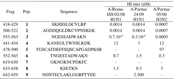
Chapter III: Figure 3.1 Cartoon representation of the bLf C-lobe. The patented sequences are depicted in purple and the newly synthesized sequences in yellow……….……….... -45-
Figure 3.2 Structure of compound 41... -55-
Figure 3.3 The comparision of (a) peptide and (b) peptoid... -58-
Figure 3.4 Structure of compound 51.………... -58-
Figure 3.5 On the left, superposition of backbone atoms of twenty NMR structures of 1 (orange ribbons) generated by using CYANA 2.1. On the right, the average NMR derived structures of 1. The atoms are depicted in tube and colored by atom types (O, red; N, blue; S, yellow; polar hydrogen, white). The backbone C atoms of 1 are colored as for the ribbons and the side chain C atoms are in grey. The dashed lines indicate intramolecular H-bonds responsible of the global fold... -61-
Figure 3.6 a) Superimposition of loop Ser418-Pro429 (yellow and blue ribbon) of lactoferrin (PDB ID: 3IB0) and 1 (orange and black ribbon). The
Figure captions
blue and black portions indicate the helix 310 of Ser418-Pro429 and the
γ-turn of 1, respectively. b) Superimposition of first four amino acids (SKHS) of 1 (cyan) and loop Ser418-Pro429 (tan). The atoms are depicted in tube and colored by atom types (O, red; N, blue). The C atoms of 1 and loop
Ser418-Pro429 are colored as for tube……….……….. -62-
Figure 3.7 The Biotage Initiator + Alstra™……….. -65-
Figure 3.8 Vapourtec Flow Chemistry……… -69-
Figure 3.9 HPLC profile of pure peptide 1………. -77-
Figure S1. 2D-NOESY spectrum of peptide 1 HFA/H2Osolution (600 MHz, 300 K, tmix = 400 ms)……… -83-
Figure S2. NOE connectivities from 2D-NOESY spectra of peptide 1 in HFA/H2O (600 MHz, 300 K, tmix = 400 ms)……… -84-
Figure S3. Ramachandran plot of NMR derived bundle of peptide 1, calculated by PROCHECK1 software………... -84-
Figure S4. HPLC profile of crude peptide 32 synthesized at room temperature………...…… -85-
Figure S5. HPLC profile of crude peptide 32 synthesized using microwave…. -85- Figure S6. HPLC profile of pure peptide 32……….. -86-
Chapter IV: Figure 4.1 Mechanism for aspartimide-related by-product formation... -99-
Figure 4.2 Stucture of peptide 71.…... -100-
Figure 4.3 HPLC elution profile of mixture of compounds and ESI-MS spectrum………... -101-
Figure 4.4 Partial NOESY spectrum of compound 54.…... -102-
Figure 4.5 Competition between N-hydroxylamine and amide backbone proton abstraction... -109-
Figure captions
Figure 4.6 Structures of Asp β-carboxyl protecting groups used... -110- Figure 4.7 Influence of cysteine β-protecting group on aspartimide formation..………. -113- Figure 4.8 HPLC profile of pure piperidinyl derivative……….. -118-
Table captions
Table captions
Chapter II:
Table 2.1 Interaction of SKHSSLDCVLRP, AGDDQGLDKCVPNSKEK, and NGESSADWAKN peptides with viral HA.………... -34- Table 2.2 In vitro antiviral activity of SKHSSLDCVLRP, AGDDQGLDKCVPNSKEK, and NGESSADWAKN peptides towards influenza virus infection...………... -35-
Chapter III:
Table 3.1 Sequence and HI titer of peptides 1-9.………….…………..………… -46- Table 3.2 Sequence and HI titers of peptides 1, 10-17... -48- Table 3.3 In vitro antiviral activity of most potent peptides against influenza virus infection………... -49- Table 3.4 Sequence and HI titers of peptides 14, 18-23…….…………... -51- Table 3.5 Sequence and HI titers of peptides 15, 17, 24-31... -53- Table 3.6 In vitro antiviral activity of most potent peptides against influenza virus infection.……….. -54- Table 3.7 Sequence and HI titers of peptides 15, 17, 32-41.……….….. -56- Table 3.8 In vitro antiviral activity of most potent peptides against influenza virus infection.………..…… -57- Table 3.9 Sequence and HI titers of peptides 15, 17, 42-51.………..……. -59- Table 3.10 In vitro antiviral activity of most potent peptides against influenza
virus infection.………..………… -60-
Table 3.11 Analytical data of peptides 1-31.……… -76- Table 3.12 Analytical data of peptides 32-51.………..…………... -77- Table S1. 1H chemical shifts (ppm) of peptide 1 in HFA/H2O (600 MHz, 300
K)……….. -81-
Table S2. 1H chemical shifts (ppm) of peptide 17 in [D6] DMSO (600 MHz,
Table captions
Table S3. Mean values of φ, ψ and χ1 angles and αC distances relative to the
most representative conformers of peptide 1………...………. -82-
Chapter IV: Table 4.1 Peptides obtained during the synthesis... -101-
Table 4.2 Influence of the solid support on aspartimide and piperidinyl derivatives formation..…... -107-
Table 4.3 Influence of the nature of the base on byproducts formation... -109-
Table 4.4 Influence of β-carboxyl protecting group on aspartimide and piperidinyl derivatives formation..…... -111-
Table 4.5 Peptides synthesized by alanine scanning approach... -111-
Table 4.6 Scramble peptides synthesized………..……... -112-
Table 4.7 Peptides synthesized……….. -113-
Scheme captions
Scheme captions
Chapter III:
Scheme 3.1 Synthesis of peptide 17..………..…….……… -64- Scheme 3.2 Synthesis of peptide 41... -67- Scheme 3.3 Synthesis of compound 51.………... -69-
Chapter IV:
- 1 -
CHAPTER I
Hemagglutinin: a promising target against influenza
virus
Chapter I: Hemagglutinin: a promising target against influenza virus
- 3 - Abstract
Influenza is a highly contagious, acute respiratory illness, which represents one of the main plagues worldwide. Even though some antiviral drugs are available, the alarming increase of virus strains resistant to them, highlights the need to find new antiviral compounds. The high mutation rate of the RNA genome of the influenza virus, combined with assortment of its multiple genomic segments, promotes antigenic diversity and new subtypes, allowing the virus to evade vaccines and become resistant to antiviral drugs. Thus, there is a continuing need for new anti-influenza therapy using novel targets and creative strategies.
On the basis of the above considerations, an ideal target for therapy should be a viral component, whose function is essential for virus infection.
In this contest, the influenza A virus hemagglutinin (HA) represents a very promising target.
Keywords: Influenza, virus, drug, resistance, therapy, hemagglutinin.
Abbreviations
IAVs, Influenza A virus; ORFs, open reading frames, HA, hemagglutinin; NA, neuraminidase; M2, matrix 2; M1, matrix 1; NEP, nuclear export protein; NS2, nonstructural protein 2; RNP, ribonucleoprotein; NP, nucleoprotein; HEF, hemagglutinin-esterase-fusion protein; NLSs, nuclear localization signals; cRNA, complementary RNA; NAIs, neuraminidase inhibitors; DANA, 2-deoxy-2,3-didehydro- N-acetyl neuraminic acid; FANA, 2-deoxy-2,3-dehydro-N-trifluoroacetylneuraminic acid.
Chapter I: Hemagglutinin: a promising target against influenza virus
- 4 - 1.1. Introduction
Influenza is a contagious respiratory infection and it is considered to be one of the life-threatening infectious diseases.[1] It is a viral infection, associated with seasonal outbreaks of respiratory illness during the winter months in regions with temperate climates and during rainy seasons in tropical regions.
In some countries seasonal influenza affects annually up to 40% of the population and 500 million people die from it worldwide every year.[2-4]
The reasons for seasonal epidemics of influenza are not definitely known. They probably involve a combination of environmental factors such as low humidity and low temperature and social behaviors that facilitate person-to-person transmission of influenza virus. At unpredictable intervals, influenza pandemics occur with very high attack rates and severe disease. In the population, influenza follows the general pattern that now appears to characterize essentially all respiratory infections. It can be particularly hazardous to individuals with poor immunity such as children and the elderly, and those with pulmonary, cardiovascular or other complications. Because of a lack of prior immunity, humans can be highly susceptible to infection and disease from these subtypes.
Influenza in otherwise healthy persons is characterized predominantly by fever, myalgias, cough and other respiratory symptoms, and malaise. In most persons, recovery from these symptoms occurs in 5 to 7 days, but even in healthy persons symptoms of fatigue and malaise may not completely resolve for several weeks.
Influenza may cause more severe pulmonary symptoms through direct invasion of the lung (leading to primary viral pneumonia) or by altering lung defense mechanisms in a variety of ways that lead to bacterial superinfection. This superinfection, which may occur simultaneously with influenza or follow it by days to weeks, may be responsible for much of the disease burden
Chapter I: Hemagglutinin: a promising target against influenza virus
- 5 -
associated with influenza. Although the primary target and clinically relevant tissue in influenza virus infection is the respiratory epithelium,[2] facultative infection of other organs, such as the cardiac or skeletal muscle, is possible and has occasionally been documented in cell culture and experimental animal infections.[5-9]
1.2 Influenza viruses
Influenza viruses, belonging to the Orthomyxoviridae family, are enveloped negative-strand RNA viruses with segmented genomes containing seven to eight gene segments.[4]
One genus includes influenza A and B viruses, and the other comprises influenza C viruses. The three virus types differ in host range and pathogenicity.
[10] Type B and C influenza viruses are isolated almost exclusively from humans,
although influenza B viruses have been isolated from seals and influenza C viruses have been isolated from pigs and dogs.[11-12]
Influenza A viruses, however, infect a wide variety of warm-blooded animals, including birds, swine, horses, humans, and other mammals. Influenza A and B viruses have a similar structure, whereas influenza C is more divergent. Influenza A- and B-type viruses contain eight discrete single-stranded RNA gene segments, each encoding at least one protein. Only Influenza A virus (IAVs) pose a significant risk of zoonotic infection, host switch, and the generation of pandemic IAVs. IAVs are enveloped with a host cell-derived lipid membrane. The eight gene segments encode at least 11 open reading frames (ORFs) (Figure 1.1).
Chapter I: Hemagglutinin: a promising target against influenza virus
- 6 -
Figure 1.1 Diagrammatic representation of an Influenza A Virus, the two major surface glycoproteins, hemagglutinin (HA) and neuraminidase (NA), along with small numbers of the matrix 2 (M2) ion channel protein, are embedded in a lipid bilayer (according to Ludwig et al. 2003). Adapted from “Antiviral agents targeting the influenza virus: a review and publication analysis,” by L. Eyer and K. Hruska, 2013, Veterinarni Medicina, 58, 113-185.
The envelope bilayer harbors the two spike glycoproteins, hemagglutinin (HA) and neuraminidase (NA), and the M2 proton channel. HA is a glycosylated type I integral membrane protein with functions both as the viral receptor-binding protein and fusion protein. NA cleaves glycosidic bonds with terminal SA facilitating the release of budding virions from the cell. There are 16 known HA (H1 to H16) and 9 NA (N1 to N9) subtypes in influenza A,[4, 13] leading to
the current HxNy nomenclature. Routine human infections of seasonal influenza are mainly due to H1N1, H1N2 and influenza B; however, H3N2 is gradually becoming more abundant.[14] The small protein M2 is a proton channel necessary for viral replication.
Chapter I: Hemagglutinin: a promising target against influenza virus
- 7 -
1.2.1 Virion structure and organization
Influenza A and B viruses are virtually indistinguishable by electron microscopy. They are spherical or filamentous in shape, with the spherical forms on the order of 100 nm in diameter and the filamentous forms often in excess of 300 nm in length. The influenza A virion is studded with glycoprotein spikes of HA and NA, in a ratio of approximately four to one, projecting from a host cell–derived lipid membrane.[4]
A smaller number of matrix ion channels (M2) traverse the lipid envelope, with an M2:HA ratio on the order of one M2 channel per 101-102 HA molecules.[15] The envelope and its three integral membrane proteins HA, NA, and M2 overlay a matrix of M1 protein, which encloses the virion core. Internal to the M1 matrix are found the nuclear export protein (NEP; also called nonstructural protein 2, NS2) and the ribonucleoprotein (RNP) complex, which consists of the viral RNA segments coated with nucleoprotein (NP) and the heterotrimeric RNAdependent RNA polymerase, composed of two “polymerase basic” and one “polymerase acidic” subunits (PB1, PB2, and PA). The organization of the influenza B virion is similar, with four envelope proteins: HA, NA, and, instead of M2, NB and BM2. Influenza C virions are structurally distinct from those of the A and B viruses; on infected cell surfaces, they can form long cordlike structures on the order of 500 μm. However, influenza C virions are compositionally similar, with a glycoprotein-studded lipid envelope overlying a protein matrix and the RNP complex. The influenza C viruses have only one major surface glycoprotein, the hemagglutinin-esterase-fusion (HEF) protein, which corresponds functionally to the HA and NA of influenza A and B viruses, and one minor envelope protein, CM2.[4]
Chapter I: Hemagglutinin: a promising target against influenza virus
- 8 -
1.2.2 The influenza virus replication cycle
1.2.2.1 Virus attachment
Influenza viruses recognize N-acetylneuraminic (sialic) acid on the host cell surface. Sialic acids are nine-carbon acidic monosaccharides commonly found at the termini of many glycoconjugates. The sialic acid moiety is recognized and bound by the HA spikes on the surface of influenza viruses.[16]
Figure 1.2 Cellular targets for anti-influenza drugs in the context of the replication cycle of influenza virus. Stages of influenza A virus replication are in green. Cellular pathways shown by siRNA screens to be essential for completion of the viral replication cycle are shown in red.Adapted from “Cellular targets for influenza drugs,” by J. Min and K. Subbarao, 2010, Nature Biotechnology, 28, 239-240.
Chapter I: Hemagglutinin: a promising target against influenza virus
- 9 - 1.2.2.2 Virus Entry
Following attachment of the influenza virus HA to sialic acid, the virus is endocytosed. The acidity of the endosomal compartment is crucial to influenza virus uncoating in two ways. First, low pH triggers a conformational change in the HA, exposing a fusion peptide that mediates the merging of the viral envelope with the endosomal membrane, thus opening a pore through which the viral RNP’s are released into the host cell cytoplasm.[17, 18] Second, hydrogen
ions from the endosome are pumped into the virus particle via the M2 ion channel. The M2 protein, a transmembrane ion channel found only in influenza A virus, has portions external to the viral envelope, along with the HA and NA.[19, 20] Internal acidification of the influenza virion via the M2 channel disrupts internal protein-protein interactions, allowing viral RNPs to be released from the viral matrix into the cellular cytoplasm.[21]
1.2.2.3 Synthesis of Viral RNA
Once liberated from the virion, RNPs are trafficked to the host cell nucleus by means of viral proteins’ nuclear localization signals (NLSs), which direct cellular proteins to import the RNPs and other viral proteins into the host cell nucleus.[22] The nucleus is the location of all influenza virus RNA synthesis. The viral RNAdependent RNA polymerase – a component of the RNPs imported into the nucleus – uses the negative-sense vRNA as a template to synthesize two positive-sense RNA species: mRNA templates for viral protein synthesis, and complementary RNA (cRNA) intermediates from which the RNA polymerase subsequently transcribes more copies of negative-sense, genomic vRNA. Nuclear export of vRNA segments, however, is mediated by the viral proteins M1 and NEP/NS2.[22] M1 interacts with both vRNA and NP and it also
associates with the nuclear export protein NEP, which mediates the M1-RNP export via nucleoporins into the cytoplasm.
Chapter I: Hemagglutinin: a promising target against influenza virus
- 10 - 1.2.2.4 Virus Budding and Release
Influenza virus budding occurs at the cell membrane, probably initiated by an accumulation of M1 matrix protein at the cytoplasmic side of the lipid bilayer. When budding is complete, HA spikes continue to bind the virions to the sialic acid on the cell surface until virus particles are actively released by the sialidase activity of the NA protein. The NA is a mushroom-shaped tetramer, anchored to the viral envelope by a transmembrane domain.[23, 24] It possesses receptor destroying activity, cleaving terminal sialic acid residues from cell-surface glycoproteins and gangliosides to release progeny virus from the host cell. In viruses with inactive or absent NA, or in the presence of neuraminidase inhibitors, virus particles clump at the cell surface and infectivity is consequently reduced. The NA also removes sialic acid residues from the virus envelope itself, which prevents viral particle aggregation to enhance infectivity.[25, 26] The NA is also thought to aid virus infectivity by breaking down the mucins in respiratory tract secretions and allowing the virus to penetrate through to the respiratory epithelium, and it may play a role in virus entry into respiratory epithelial cells.[27] Host antibodies to the NA, as well as neuraminidase inhibitors, prevent virus release from infected cells and thus inhibit viral replication.
1.3 Anti-influenza therapy
1.3.1 Vaccine
The vaccination represent the main strategy for preventing infections. However, the vaccines are not able to follow the rapid virus antigenic drift, so that vaccine antigen composition needs to be updated annually based on global influenza surveillance. Efforts to influenza prevention by vaccination are made difficult by the virus ability to rapidly mutate and recombine into antigenically
Chapter I: Hemagglutinin: a promising target against influenza virus
- 11 -
new viral particles, sometimes leading to the emergence of a totally new virus. For this reason, at present, the development of antiviral drugs represents a crucial strategy in the control and prevention of seasonal and pandemic influenza infections.[28] Antiviral drugs can overcome the limitations of vaccination strategies, such as the time-consuming vaccine design, insufficient protection for immunocompromised patients and the unpredictable antigenic changes in influenza strains which render vaccination ineffective.
The anti-influenza drugs are usually classified according to their target in the viral life-cycle, which is schematically depicted in Figure 1.2. Antiviral molecules are particularly used as inhibitors of the following processes: attachment of the virus to host cell receptors, endocytosis and fusion of viral and cell membranes, replication and transcription of the viral genome, synthesis of viral proteins, assembly of the viral progeny and release of the new virions into the outside environment. Two classes of antiviral drugs, the adamantane derivatives (amantadine and rimantadine) and neuraminidase inhibitors (NAIs; zanamivir and oseltamivir), have been approved for treatment and prophylaxis of influenza.[4, 29, 30]
1.3.2 M2 ion channel blockers
M2 ion channel is a transmembrane viral protein (Figure 1.1) that mediates the selective transport of protons into the interior of the influenza virion. Conductance of protons acidifies the internal space of the viral particle and facilitates the haemagglutinin-mediated membrane fusion, which in turn results in the uncoating of the influenza nucleocapsid and import of the viral genome into the nucleus.[31] Adamantanes are potent M2 channel blockers, which are known as the first synthetic anti-influenza drugs described in the mid-1960s.[32]
Two adamantane derivatives, amantadine and rimantadine (Figure 1.3), have been licensed for influenza control and are commercially available under the
Chapter I: Hemagglutinin: a promising target against influenza virus
- 12 -
trademarks Symmetrel® and Flumadine®, respectively.[33] The adamantanes are relatively cheap, highly stable in storage and show strong anti-influenza activity at micromolar concentrations. At present, the application of adamantanes for prevention and treatment of influenza infections is, however, not recommended because of the rapid emergence of drug-resistant virus variants that retain full virulence and transmissibility.[34, 35]
Figure 1.3 Structure of M2 ion channel inhibitors:
amantadine (1) and rimantadine (2).
1.3.3 Inhibitors of neuraminidase
Neuraminidase, also referred to as sialidase, is an antigenic glycoprotein anchored in the surface envelope of the influenza virions, which hydrolytically cleaves the terminal sialic acid from the host cell receptors (Figure 1.2). Thus, it plays a crucial role in the release of viral progeny from the membranes of infected cells, prevents self-aggregation of virions and facilitates the movement of the infectious viral particles in the mucus of the respiratory epithelia.[27, 36] Influenza neuraminidase has been established as a key drug target for the prophylaxis and treatment of influenza infections, predominantly for the following reasons: firstly, the structure of the influenza neuraminidase active site is highly conserved between influenza A and B strains, making neuraminidase an attractive target for the development of broad-spectrum inhibitors.[37] Secondly, resistance to neuraminidase inhibitors develops less
Chapter I: Hemagglutinin: a promising target against influenza virus
- 13 -
commonly than to other anti-influenza drugs. Nevertheless, the intensive application of neuraminidase inhibitors for influenza treatment results in a permanently increasing number of drug-resistant strains.[38] Thirdly, in contrast
to adamantanes, neuraminidase inhibitors are mostly well tolerated in patients under therapy.[39] Finally, neuraminidase protein is a freely accessible target for
antiviral molecules with an extracellular mode of action. The development of neuraminidase inhibitors started in the middle 1970s, when the first structural analogues of sialic acid were described and denoted as DANA (2-deoxy-2,3-didehydro- N-acetyl neuraminic acid) and its trifluoroacetyl derivative FANA (2-deoxy-2,3-dehydro-N-trifluoroacetylneuraminic acid).[40] At present, several licensed anti-influenza medications are available on the market, most notably the inhalant zanamivir with the trademark Releza®, and the orally administered oseltamivir (Tamiflu®) having excellent bioavailability and relatively long half-life in vivo (Figure 1.4).[41, 42]
Figure 1.4 Structure of neuraminidase inhibitors: zanamivir (1) and oseltamivir (2).
In response to the emergence of some oseltamivir-resistant influenza strains, peramivir and laninamivir have been recently developed (Figure 1.5).[43, 44]
Chapter I: Hemagglutinin: a promising target against influenza virus
- 14 -
Figure 1.5 Structure of peramivir (1) and laninamivir (2).
New-generation neuraminidase inhibitors are currently under investigation, e.g., multimeric forms of zanamivir,[45] dual-targeted bifunctional antivirals[46] and several herbal remedies, such as flavonols, alkaloids and saponins.[47]
1.4 Influenza drug resistance
The propagation of viruses in the presence of antiviral drugs increases the selection pressure for mutations in the viral target proteins, which results in the induction of virus drug resistance. As an example, adamantane resistant strains are typically characterised by a single substitution in the transmembrane region of the M2 ion channel.[48, 49] On the other hand, resistance to neuraminidase inhibitors can result from mutations in the neuraminidase active cavity, but also from amino acid substitutions on the molecular surface of the neuraminidase protein.[37, 50] It is noteworthy that resistance to adamantanes is acquired rapidly and by a high number of virus strains,[34] while neuraminidase inhibitor resistance has developed over a longer time period and occurs with a relatively lower frequency.[38] This may be due to the fact that some mutations significantly affect viral infectivity and ability to replicate in the host cell.
Therefore, the capability of viruses to mutate the target proteins represents an obstacle for efficient treatment with these drugs. On the basis of the above it
Chapter I: Hemagglutinin: a promising target against influenza virus
- 15 -
is apparent the need to provide for new compounds against influenza virus able to overcome the disadvantages of the known therapies.[51, 52]
Therefore, the identification of new target for therapy of influenza virus infection and development of new therapeutic agents are the global public health priority.
1.5 Hemagglutinin: a new promising target
An attractive antiviral strategy is the blocking of influenza virus entry into the host cell. This process is mediated by the viral hemagglutinin (HA), a glycosylated type I integral membrane protein. HA is responsible for the binding of the virus to the target cell and, after virus uptake into endosomes, fusion of the virus with the cell membranes.[53]
The crystal structure of the HA molecule is a trimer with two structurally distinct regions: a stem, comprising a triple-stranded coiled-coil of alpha-helices, and a globular head of antiparallel beta-sheet, positioned atop the stem.[54]
The head contains the sialic acid receptor binding site, which is surrounded by the predicted variable antigenic determinants, designated A, B, C, and D in the H3 subtype[55] and Sa, Sb, Ca1, Ca2, and Cb in the H1 subtype (Figure 1.6).[4]
Chapter I: Hemagglutinin: a promising target against influenza virus
- 16 -
Figure 1.6 Ribbon diagram of an uncleaved hemagglutinin monomer from an influenza A virus (H1N1). The head contains the sialic acid receptor-binding site, which is surrounded by the five predicted antigenic sites (Sa, Sb, Ca1, Ca2, and Cb). The stem comprises helices A and B and the fusion peptide, as shown. Adapted from “The biology of influenza viruses,” by N. M. Bouvier and P. Palese, 2008, Vaccine, 26, 1-10.
During virus replication, the HA protein is cleaved by serine proteases into HA1 and HA2; this post-translational modification is necessary for virus infectivity. The HA2 portion is thought to mediate the fusion of virus envelope with cell membranes, while the HA1 portion contains the receptor binding and antigenic sites.[56] Antibodies to HA neutralize virus infectivity, so virus strains evolve frequent amino acid changes at the antigenic sites; however, the stem-head configuration of the HA molecule remains conserved among strains and subtypes. These relatively minor changes accumulate in a process called antigenic drift. Eventually, mutations in multiple antigenic sites result in a virus strain that is no longer effectively neutralized by host antibodies to the parental virus, and the host becomes susceptible again to productive infection by the drifted strain.
Hemagglutinin has been chosen since it is the major surface protein of the Influenza A virus and is essential to the entry process so representing an
Chapter I: Hemagglutinin: a promising target against influenza virus
- 17 -
attractive target for antiviral therapy. An initial attachment of HA to specific receptors on the host cell surface and a membrane fusion of HA matured by protease digestion are required for virus infection. As a matter of fact, neutralizing compounds targeting HA represent a useful tool in neutralizing viral infection, clearing virus, and suppressing viral spread.
Chapter I: Hemagglutinin: a promising target against influenza virus
- 18 - References
[1] Mulder, J.; Hers, J. F. P. Influenza. Groningen: Wolters-Noordhoff.
1972, 287.
[2] Metersky, M. L.; Masterton, R.G.; Lode, H.; File, T. M., Jr.; Babinchak,
T. Epidemiology, microbiology, and treatment considerations for bacterial pneumonia complicating influenza. Int J Infect Dis. 2012, 16, 321-331.
[3] Hale, B. G.; Albrecht, R. A.; García-Sastre, A. Innate immune evasion
strategies of influenza viruses. Future Microbiol. 2010, 5, 23-41.
[4] Palese, P.; Shaw, M. L. Orthomyxoviridae: the viruses and their
replication. In Fields Virology. 5 edition. Edited by: Knipe, D. M.; Howley, P. M. Philadelphia, PA: Lippincott Williams 2007, 1647-1689.
[5] Haessler, S.; Paez, A.; Rothberg, M.; Higgins, T. 2009 pandemic
H1N1-associated myocarditis in a previously healthy adult. Clin Microbiol Infect. 2011, 17, 572-574.
[6] Kumar, K.; Guirgis, M.; Zieroth, S.; Lo, E.; Menkis, A. H.; Arora, R. C.;
Freed, D. H. Influenza myocarditis and myositis: case presentation and review of the literature. Can J Cardiol. 2011, 27, 514-522.
[7] Pan, H. Y.; Yamada, H.; Chida, J.; Wang, S.; Yano, M.; Yao, M.; Zhu,
J.; Kido, H. Upregulation of ectopic trypsins in the myocardium by influenza A virus infection triggers acute myocarditis. Cardiovasc Res. 2011, 89, 595-603.
[8] Ru, Y. X.; Li ,Y.C.; Zhao, Y.; Zhao, S. X.; Yang, J. P.; Zhang, H. M.;
Pang, T.X. Multiple organ invasion by viruses: pathological characteristics in three fatal cases of the 2009 pandemic influenza A/H1N1. Ultrastruct Pathol. 2011, 35, 155-161.
[9] Toffan, A.; Serena Beato, M.; De Nardi, R.; Bertoli, E.; Salviato, A.;
Cattoli, G.; Terregino, C.; Capua, I. Conventional inactivated bivalent H5/H7 vaccine prevents viral localization in muscles of turkeys infected experimentally with low pathogenic avian influenza and highly pathogenic avian influenza H7N1 isolates. Avian Pathol. 2008, 37, 407-412.
[10] Wright, P.F.; Neumann, G.; Kawaoka, Y. Orthomyxoviruses. In: Knipe
DM, Howley PM, editors. Fields Virology. Williams & Wilkins; Philadelphia, Lippincott: 2007, 1691–1740.
Chapter I: Hemagglutinin: a promising target against influenza virus
- 19 -
[11] Osterhaus, A. D.; Rimmelzwaan, G. F.; Martina, B. E.; Bestebroer, T.
M.; Fouchier, R. A. Influenza B virus in seals. Science. 2000, 288, 1051–1053.
[12] Youzbashi, E.; Marschall, M.; Chaloupka, I.; Meier-Ewert, H.
Distribution of influenza C virus infection in dogs and pigs in Bavaria. Tierarztl. Prax. 1996, 24, 337–342.
[13] Webster, R. G.; Bean, W. J.; Gorman, O. T.; Chambers, T. M.; Kawaoka,
Y. Evolution and ecology of influenza A viruses. Microbiol Rev. 1992, 56, 152-179.
[14] Centers for Disease Control and Prevention: Seasonal influenza (flu) -
information on H3N2 variant influenza A viruses.[http://www.cdc.gov/flu/ swineflu/influenza-variant-viruses-h3n2v.htm.
[15] Zebedee, S. L.; Lamb, R. A. Influenza A virus M2 protein: monoclonal
antibody restriction of virus growth and detection of M2 in virions. J Virol. 1988, 62, 2762–2772.
[16] Couceiro, J. N.; Paulson, J. C.; Baum, L. G. Influenza virus strains
selectively recognize sialyloligosaccharides on human respiratory epithelium; the role of the host cell in selection of hemagglutinin receptor specificity. Virus Res. 1993, 29,155–165.
[17] Sieczkarski, S. B.; Whittaker, G. R. Viral entry. Curr Top Microbiol
Immunol. 2005, 285, 1–23.
[18] Stegmann, T. Membrane fusion mechanisms: the influenza
hemagglutinin paradigm and its implications for intracellular fusion. Traffic. 2000, 1, 598–604.
[19] Pinto, L. H.; Holsinger, L. J.; Lamb, R. A. Influenza virus M2 protein
has ion channel activity. Cell. 1992, 69, 517-528.
[20] Wharton, S. A.; Belshe, R. B.; Skehel, J. J.; Hay, A. J. Role of virion M2
protein in influenza virus uncoating: specific reduction in the rate of membrane fusion between virus and liposomes by amantadine. J Gen Virol. 1994, 75, 945-948.
[21] Martin, K.; Helenius, A. Transport of incoming influenza virus
Chapter I: Hemagglutinin: a promising target against influenza virus
- 20 -
[22] Cros, J. F.; Palese, P. Trafficking of viral genomic RNA into and out of
the nucleus: influenza, Thogoto and Borna disease viruses. Virus Res. 2003, 95, 3–12.
[23] Colman, P. M.; Varghese, J. N.; Laver, W. G. Structure of the catalytic
and antigenic sites in influenza virus neuraminidase. Nature. 1983, 303, 41-44.
[24] Varghese, J. N.; Laver, W. G.; Colman, P. M. Structure of the influenza
virus glycoprotein antigen neuraminidase at 2.9 A resolution. Nature. 1983, 303, 35–40.
[25] Palese, P.; Compans, R. W. Inhibition of influenza virus replication in
tissue culture by 2-deoxy-2,3- dehydro-N-trifluoroacetylneuraminic acid (FANA): mechanism of action. J Gen Virol. 1976. 33,159–163.
[26] Palese, P.; Tobita, K.; Ueda, M.; Compans, R. W. Characterization of
temperature sensitive influenza virus mutants defective in neuraminidase. Virology. 1974, 61, 397–410.
[27] Matrosovich, M. N.; Matrosovich, T. Y.; Gray, T.; Roberts, N. A.;
Klenk, H. D. Neuraminidase is important for the initiation of influenza virus infection in human airway epithelium. J Virol. 2004, 78, 12665–12667.
[28] Hayden, F. G. Respiratory viral threats. Curr. Opin. Infect. Dis. 2006,
19, 169–178.
[29] Dolin, R.; Reichman, R. C.; Madore, H. P.; Maynard, R.; Linton, P. N.;
Webber-Jones, J. A controlled trial of amantadine and rimantadine in the prophylaxis of influenza A infection. N Engl J Med. 1982, 307, 580–584.
[30] Nicholson, K. G.; Wood, J. M.; Zambon, M. Influenza. Lancet. 2003,
362, 1733–1745.
[31] Schnell, J. R; Chou, J. J. Structure and mechanism of the M2 proton
channel of influenza A virus. Nature. 2008, 451, 591-595.
[32] Davies, W. L.; Grunert, R. R.; Haff, R. F.;, Mcgahen, J. W.; Neumayer,
E. M.; Paulshock, M.; Watts, J. C.; Wood, T. R.; Hermann, E. C.; Hoffmann, C. E. Antiviral activity of 1-adamantanamine (amantadine). Science. 1964, 144, 862-863.
Chapter I: Hemagglutinin: a promising target against influenza virus
- 21 -
[33] Alves Galvão, M. G.; Rocha Crispino Santos, M. A.; Alves da Cunha,
A. J. Amantadine and rimantadine for influenza A in children and the elderly. Cochrane Database of Systematic Rev. 2012, 23, 1-116.
[34] Bright, R. A.; Medina, M. J.; Xu, X.Y.; Perez-Oronoz, G.; Wallis, T. R.;
Davis, X. H. M.; Povinelli, L.; Cox, N. J.; Klimov, A. I. Incidence of adamantane resistance among influenza A (H3N2) viruses isolated worldwide from 1994 to 2005: a cause for concern. Lancet. 2005, 366, 1175–1181.
[35] Barr, I. G.; Deng, Y. M.; Iannello, P.; Hurt, A. C.; Komadina, N.
Adamantane resistance in influenza A(H1) viruses increased in 2007 in South East Asia but decreased in Australia and some other countries. Antiviral Research. 2008, 80, 200–205.
[36] Suzuki, T.; Takahashi, T.; Guo, C. T.; Hidari, K. I. P. J.; Miyamoto, D.;
Goto, H.; Kawaoka, Y.; Suzuki, Y. Sialidase activity of influenza A virus in an endocytic pathway enhances viral replication. Journal of Virology. 2005, 79, 11705–11715.
[37] Yen, H. L.; Hoffmann, E.; Taylor, G.; Scholtissek, C.; Monto, A. S.;
Webster, R. G.; Govorkova, E. A. Importance of neuraminidase active-site residues to the neuraminidase inhibitor resistance of influenza viruses. Journal of Virology. 2006, 80, 8787–8795.
[38] Garcia, J.; Sovero, M.; Torres, A. L.; Gomez, J.; Douce, R.; Barrantes;
M.; Sanchez, F.; Jimenez, M.; Comach, G.; de Rivera I.; Agudo, R.; Kochel, T. Antiviral resistance in influenza viruses circulating in Central and South America based on the detection of established genetic markers. Influenza Other Respiratory Viruses. 2009, 3, 69–74.
[39] Cao, B.; Wang, D. Y.; Yu, X. M.; We, L. Q.; Pu, Z. H.; Gao, Y.; Wang,
J.; Dong, J. P.; Li, X. L.; Xu, Q.; Hu, K.; Chen, B. Y.; Yu, Y. S.; Song, S. F.; Shu, Y. L.; Wang, C. An uncontrolled openlabel, multicenter study to monitor the antiviral activity and safety of inhaled zanamivir (as Rotadisk via Diskhaler device) among Chinese adolescents and adults with influenza-like illness. Chinese Medical Journal. 2012, 125, 3002–3007.
[40] Schulman, J. L. and Palese, P. Susceptibility of different strains of
influenza A virus to inhibitory effects of 2-deoxy-2,3-dehydro-N-trifluoroacetylneuraminic acid (FANA). Virology. 1975, 63, 98–104.
Chapter I: Hemagglutinin: a promising target against influenza virus
- 22 -
[41] He, G.; Massarella, J.; Ward, P. Clinical pharmacokinetics of the
prodrug oseltamivir and its active metabolite Ro 64-0802. Clinical Pharmacokinetics. 1999, 37, 471–484.
[42] Greengard, O.; Poltoratskaia, N.; Leikina, E.; Zimmerberg, J.; Moscona,
A. The anti-influenza virus ahent 4-GU-DANA (Zanamivir) inhibits cell fusion mediated by human parainfluenza virus and influenza virus HA. Journal of Virology. 2000, 74, 11108–11114.
[43] Bantia, S.; Arnold, C. S.; Parker, C. D.; Upshaw, R.; Chand, P.
Anti-influenza virus activity of peramivir in mice with single intramuscular injection. Antiviral Research. 2006, 69, 39–45.
[44] Kubo, S.; Tomozawa, T.; Kakuta, M.; Tokumitsu, A.; Yamashita, M.
Laninamivir prodrug CS-8958, a long-acting neuraminidase inhibitor, shows superior anti-influenza virus activity after a single administration. Antimicrobial Agents and Chemotherapy. 2010, 54, 1256–1264.
[45] Watson, K. G. et al. Highly Potent and Long-Acting Trimeric and
Tetrameric Inhibitors of Influenza Virus Neuraminidase. Bioorg Med Chem Lett. 2004, 14, 1589-1592.
[46] Liu, K. C.; Fang, J. M.; Jan, J. T.; Cheng, T. J. R.; Wang, S. Y.;
Yang, S. T.; Cheng, Y. S. E.; Wong, C. H. Enhanced Anti-influenza Agents Conjugated with Anti-inflammatory Activity. J. Med. Chem. 2012, 55, 8493– 8501.
[47] Jeong, H. J.; Ryu, Y. B.; Park, S. J.; Kim, J. H.; Kwon, H. J.; Kim, J.
H.; Park, K. H.; Rho, M. C.; Lee, W. S. Neuraminidase inhibitory activities of flavonols isolated from Rhodiola rosea roots and their in vitro anti-influenza viral activities. Bioorg Med Chem. 2009, 17, 6816-23.
[48] Saito, R.; Sakai, T.; Sato, I.; Sano,Y.; Oshitani, H.; Sato,M.; Suzuki, H.
Frequency of Amantadine-Resistant Influenza A Viruses during Two Seasons Featuring Cocirculation of H1N1 and H3N2. J Clin Microbiol. 2003, 41, 2164– 2165.
[49] Shiraishi, K.; Mitamura, K.; Sakai-Tagawa, Y.; Goto, H.; Sugaya, N.;
Kawaoka, Y. High frequency of resistant viruses harboring different mutations in amantadine-treated children with influenza. J Infect Dis. 2003, 188, 57-61.
Chapter I: Hemagglutinin: a promising target against influenza virus
- 23 -
[50] Du, Q. S.; Wang, S. Q.; Huang, R. B.; Chou K. C. Computational 3D
structures of drug-targeting proteins in the 2009-H1N1 influenza A virus. Chemical Physics Letters. 2010, 485, 191–195.
[51] Belshe, R. B.; Burk, B.; Newman, F.; Cerruti, R. L.; Sim, I. S. Resistance
of influenza A virus to amantadine and rimantadine: results of one decade of surveillance. J Infect Dis. 1989, 159, 430–435.
[52] Bertram, S.; Glowacka, I.; Steffen, I.; Kuhl, A.; Pohlmann, S. Novel
insights into proteolytic cleavage of influenza virus hemagglutinin. Rev Med Virol. 2010, 20, 298–310.
[53] Skehel, J. J. and Wiley. D. C. Receptor binding and membrane fusion in
virus entry: The influenza hemagglutinin. Annual Review of Biochemistry. 2000, 69, 531–569.
[54] Wilson, I. A.; Skehel, J. J.; Wiley, D. C. Structure of the haemagglutinin
membrane glycoprotein of influenza virus at 3 A resolution. Nature. 1981, 289, 366–73.
[55] Webster, R. G.; Laver, W. G.; Air, G. M. Antigenic Variation Among
Type A Influenza Viruses. In: Palese, P.; Kingsbury, D. W., editors. Genetics of Influenza Viruses. Vienna: Springer-Verlag. 1983, 127-68.
[56] Steinhauer, D. A. Role of hemagglutinin cleavage for the pathogenicity
- 25 -
CHAPTER II
Bovine lactoferrin: a novel drug target for the
inhibition of influenza virus infection
Chapter II: Bovine lactoferrin: a novel drug target for the inhibition of influenza virus infection
- 27 - Abstract
Bovine lactoferrin (bLf) is a multifunctional glycoprotein that plays an important role in innate immunity against infections, including influenza.
Therefore, bLf was considered a novel drug target for the inhibition of influenza virus infection. Previously, it was shown that inhibition of influenza virus hemagglutination and cell infection is entirely attributable to the C-lobe and that all major virus subtypes, including H1N1 and H3N2, are inhibited. By far-western blotting and sequencing studies, bLf was shown to bind to the HA2
subunit, an HA region which is known to contain the universally conserved HA epitope. Moreover, molecular docking studies have identified some C-lobe fragments which inhibited virus hemagglutination and infection at picomolar concentration range.
Besides contributing to explain the broad anti-influenza activity of bLf, these findings lay the foundations for exploiting bLf fragments as source of potential anti-influenza therapeutics.
Keywords: Influenza, Bovine lactoferrin, C-lobe, Peptides, Antiviral.
Abbreviations
LF, lactoferrin; BLf, bovine lactoferrin; HI, hemagglutination inhibition assay; SI, selectivity index; HA, hemagglutinin.
Chapter II: Bovine lactoferrin: a novel drug target for the inhibition of influenza virus infection
- 28 - 2.1 Introduction
Lactoferrin (LF) is a non-haem iron-binding protein that is part of the transferrin protein family, whose function is to transport iron in blood serum.[1, 2]
Lactoferrin was first isolated by Sorensen and Sorensen from bovine milk in 1939. In 1960 it was concurrently determined to be the main iron binding protein in human milk by three independent laboratories.[3-5]
LF is commonly found in various secretory fluids, such as saliva, tears, nasal secretions, seminal and vaginal fluids, and in granules of polymorphonuclear leukocytes[6] of different mammalian species, including humans, cows, goats, horses, dogs, and several rodents.[7, 8]
It is also found in considerable amounts in secondary neutrophil granules (15µg/106 neutrophils),[9] where it plays a significant physiological role. LF is an essential player of the natural immunity. LF possesses a greater iron-binding affinity and is the only transferrin with the ability to retain this metal over a wide pH range,[10] including extremely acidic pH. It also exhibits a
greater resistance to proteolysis. In addition to these differences, LF’s net positive charge and its distribution in various tissues make it a multifunctional protein. It is involved in several physiological functions, including: regulation of iron absorption in the bowel; immune response; antioxidant, anticarcinogenic and anti-inflammatory properties; protection against microbial infection, which is the most widely studied function to date; and inhibiting activity towards different pathogens.[7, 11-13]
In particular, bLf has been recognized as potent inhibitor of different enveloped viruses, such as human cytomegalovirus,[14] herpes simplex viruses types 1 and 2,[15-18] human immunodeficiency virus,[19] human
Chapter II: Bovine lactoferrin: a novel drug target for the inhibition of influenza virus infection
- 29 -
hepatitis C virus,[20] hantavirus,[21] hepatitis B virus,[22] respiratory syncytial virus,[23] flavivirus,[24] alphavirus,[25] and phlebovirus.[26]
2.2 Structure and properties
Bovine lactoferrin (bLf) is a glycoprotein consisting of a single polypeptide chain of 689 amino acidic residues, with a molecular mass of 76 kDa, which binds two iron atoms with very high affinity.[27]
BLf, like lactoferrin of other mammalian species, is folded in two symmetric and globular lobes: N-lobe (residues 1-333) and C-lobe (residues 345-676) which are highly homologous with one another (33–41% homology). It is made up of α-helix and β-pleated sheet structures, which create two domains for each lobe (domains I and II). In bovine lactoferrin, the N1 stands for the sequences 1-90 and 251-333, N2 for 91-250, C1 for 345-431 and 593-676, and C2 for 432-592.[28, 29] These two lobes are linked
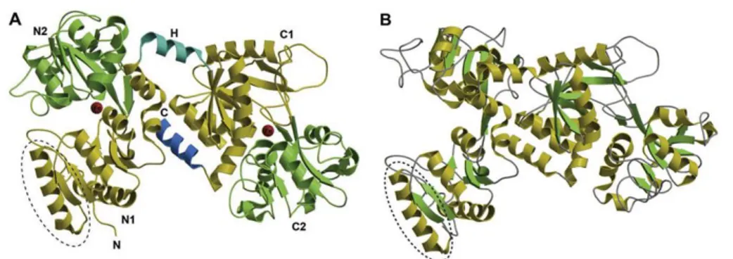
by a three-turn connecting helix, residues 334 and 344, which provide additional flexibility to the molecule (Figure 2.1).[28] Each lobe can bind a
metal atom in synergy with the carbonate ion (CO3 2−). The metals that it
binds are the Fe2+ or Fe3+ ions, but it has also been observed bound to Cu2+, Zn2+ and Mn2+ ions.[7]
Chapter II: Bovine lactoferrin: a novel drug target for the inhibition of influenza virus infection
- 30 -
Figure 2.1 Schematic diagram of the bovine lactoferrin molecule. The N1 and N2 domains are colored in yellow and pink, respectively, while the C1 and C2 domains are colored in green and blue, respectively. The interconnecting helix between the lobes is colored in orange. The two iron atoms are shown as red spheres. Adapted from “C-Lobe of Lactoferrin: The whole story of the half-molecule,” by S. Sharma, M. Sinha, S. Kaushik, P. Kaur and T. P. Singh, 2013, Biochemistry Research International, 2013, 1-8.
Because of its ability to reversibly bind Fe3+, LF can exist free of Fe3+ (apo-LF) or associated with it (holo-LF),[30] and it has a different three-dimensional conformation depending on whether it is binding Fe3+.[31] Apo-LF has an open conformation, whilst holo-Apo-LF is a closed molecule with greater resistance to proteolysis (Figura 2.2).[8]
Chapter II: Bovine lactoferrin: a novel drug target for the inhibition of influenza virus infection
- 31 -
Figure 2.2 Structure of the iron-bound (holo) form (A) and iron-free (apo) form (B) of Lf. Adapted from “A structural framework for understanding the multifunctional character of lactoferrin,” by E. N. Baker and H. M. Baker, 2009, Biochimie, 91, 3–10.
The iron-binding site is situated inside the interdomain cleft in each lobe. The iron-binding site consists of four residues: 2 tyrosines, 1 aspartate, and 1 histidine. The iron-binding residues in N-lobe are Asp 60, Tyr 92, Tyr 192, and His 253 while the corresponding iron-binding residues in C-lobe are Asp 395, Tyr 433, Tyr 526, and His 595. The iron-binding residues are coordinated to the ferric ion and a synergistic bidentate carbonate anion (Figura 2.3).[31] LF is a basic, positively charged protein with an isoelectric
Chapter II: Bovine lactoferrin: a novel drug target for the inhibition of influenza virus infection
- 32 -
Figure 2.3 Schematic figure of the iron-binding site of lactoferrin. The iron atom is shown as a red sphere, while the interacting amino acid residues of lactoferrin are in yellow. The residue numbers correspond to N-lobe, while the corresponding residues of C-lobe are in brackets. Adapted from “C-Lobe of Lactoferrin: The whole story of the half-molecule,” by S. Sharma, M. Sinha, S. Kaushik, P. Kaur and T. P. Singh, 2013, Biochemistry Research International, 2013, 1-8.
2.3 Biological functions of lactoferrin
Several functions have been attributed to LF. It is considered a key component in the host’s first line of defence, as it has the ability to respond to a variety of physiological and environmental changes.[32] The structural
characteristics of LF provide functionality in addition to the Fe3+
homeostasis function common to all transferrins: strong antimicrobial activity against a broad spectrum of bacteria, fungi, yeasts, viruses[33] and parasites;[34] anti-inflammatory and anticarcinogenic activities;[32] several enzymatic functions[35] and anti-influenza activity.[36]
Chapter II: Bovine lactoferrin: a novel drug target for the inhibition of influenza virus infection
- 33 - 2.4 Scientific background
Previously, Superti et al. have demonstrated that bLf binds to viral HA and inhibits hemagglutination and infection of all major virus subtypes, including H1N1 and H3N2.[37]
In particular, by far-western blotting and sequencing studies, it demonstrated that lactoferrin binds to the HA2 subunit of viral HA,
particularly to the fusion peptide, the only universally conserved epitope in all influenza virus hemagglutinin.[38] This behaviour explains the broad specificity of bLf and its C-lobe anti-influenza activity.[37]
Moreover, molecular docking studies have shown that the bLf C-lobe binding to HA was mediated, in most cases, by three surface-exposed loops characterized by the following amino acid sequences: SKHSSLDCVLRP (aa 418–429, 1), AGDDQGLDKCVPNSKEK (aa 506–522, 2) and NGESSADWAKN, (3) (Figure 2.4).
Figure 2.4 Putative binding mode of bLf C-lobe (white ribbon) with the HA stm (light grey solid surface): (A) close to the fusion peptide (dark grey surface); (B) in the cleft between two monomers. Selected bLf sequences correspond to the numbered black loops of bLf C-lobe: (1) SKHSSLDCVLRP; (2)
Chapter II: Bovine lactoferrin: a novel drug target for the inhibition of influenza virus infection
- 34 -
AGDDQGLDKCVPNSKEK; (3) NGESSADWAKN. Adapted from “Bovine lactoferrin-derived peptides as novel broad-spectrum inhibitors of influenza virus,” by M. G. Ammendolia, M. Agamennone, A. Pietrantoni, F. Lannutti, R. A. Siciliano, B. De Giulio, C. Amici, F. Superti, 2012, Pathog. Glob. Health., 106, 12-19.
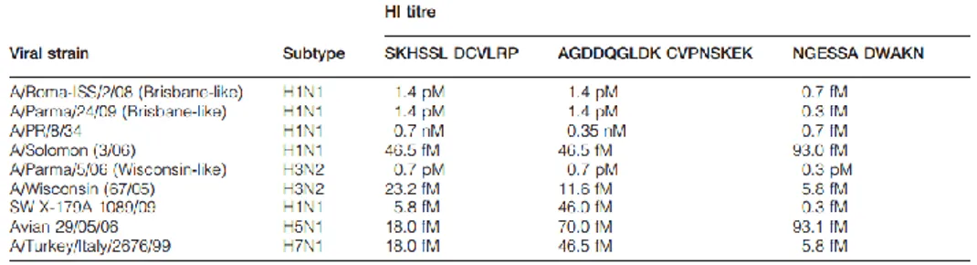
Therefore, each peptide, tested by HI, has shown to be able to inhibit HA activity of all tested virus strains at concentrations much lower than those shown by the C-lobe (Table 2.1).
Table 2.1 Interaction of SKHSSLDCVLRP, AGDDQGLDKCVPNSKEK, and NGESSADWAKN peptides with viral HA. Adapted from “Bovine lactoferrin-derived peptides as novel broad-spectrum inhibitors of influenza virus,” by M. G. Ammendolia, M. Agamennone, A. Pietrantoni, F. Lannutti, R. A. Siciliano, B. De Giulio, C. Amici, F. Superti, 2012, Pathog. Glob. Health., 106, 12-19.
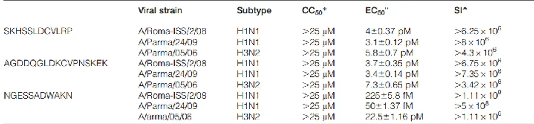
Similarly to HI data, bLf-derived peptides were better inhibitors than the entire protein, their selectivity index being about one or two order of magnitude higher, depending on virus strain (Table 2.2).
Chapter II: Bovine lactoferrin: a novel drug target for the inhibition of influenza virus infection
- 35 -
Table 2.2 In vitro antiviral activity of SKHSSLDCVLRP, AGDDQGLDKCVPNSKEK, and NGESSADWAKN peptides towards influenza virus infection. Adapted from “Bovine lactoferrin-derived peptides as novel broad-spectrum inhibitors of influenza virus,” by M. G. Ammendolia, M. Agamennone, A. Pietrantoni, F. Lannutti, R. A. Siciliano, B. De Giulio, C. Amici, F. Superti, 2012, Pathog. Glob. Health., 106, 12-19.
*CC50 the reciprocal substance dilution at which 50%of cells were protected from substance toxicity; °EC50 the reciprocal substance dilution at which 50% of cells were protected from the virus induced killing;ˆSI (selectivity index) the ratio between CC50 and EC50.
The mean values of three independent experiments with standard errors are shown.
This is the first demonstration that viral hemagglutination can be inhibited by a specific interaction with the HA2 subunit. As a matter of fact,
neutralizing antibodies against influenza virus have been found to act by two different mechanisms, mirroring the dual functions of hemagglutinin: (i) prevention of attachment to target cells, (ii) inhibition of entry (membrane fusion).
Chapter II: Bovine lactoferrin: a novel drug target for the inhibition of influenza virus infection
- 36 - References
[1] Anderson, B. F.; Baker, H. M.; Dodson, E. J.; Norris, G. E.; Rumball, S.
V.; Waters, J. M.; Baker, E. N. Structure of human lactoferrin at 3.2 A resolution. Proc. Natl. Acad. Sci. USA. 1987, 84, 1769–1773.
[2] Moore, S. A.; Anderson, B. F.; Groom, C. R.; Haridas, M.; Baker, E. N.
Threedimensional structure of diferric bovine lactoferrin at 2.8 A resolution. J. Mol. Biol. 1997, 274, 222–236.
[3] Groves, M. L. The isolation of a red protein from milk. Journal of the
American Chemical Society. 1960, 82, 3345–3350.
[4] Johanson, B. Isolation of an iron containing red protein from human
milk. Acta Chemica Scandinavica. 1960, 14, 510–512.
[5] Montreuil, J.; Tonnelat, J.; Mullet, S. Preparation and properties of
lactosiderophilin (lactotransferrin) of human milk. Biochimica et Biophysica Acta. 1960, 45, 413–421.
[6] Gennaro, R.; Dewald, B.; Horisberger, U.; Gubler, H. U.; Baggiolini, M.
A novelty of cytoplasmic granule in bovine neutrophils. J Cell Biol. 1983, 96, 1651–1661.
[7] van der Strate, B. W. A.; Belijaars, L.; Molema, G.; Harmsen, M. C.;
Meijer, D. K. Antiviral activities of lactoferrin. Antiviral Res. 2001, 52, 225– 239.
[8] Öztas-Yesim E. R.; Özgünes¸ N. Lactoferrin: a multifunctional protein.
Adv. Mol. Med. 2005, 1, 149–154.
[9] Bennett, R. M. and Kokocinski, T. Lactoferrin content of peripheral
blood cells. Br. J. Haematol. 1987, 39, 509–521.
[10] Aisen, P. and Leibman, A. Lactoferrin and transferrin: a comparative
study. Biochim. Biophys. Acta. 1972, 257, 314–323.
[11] Levay, P. F. and Viljoen, M. Lactoferrin: A general review.