DIPARTIMENTO DI SCIENZE DELLA VITA
DOTTORATO DI RICERCA IN SCIENZE DELLA VITA
Immunohistochemical studies of pulmonary remodeling in
mice exposed to chronic cigarette smoke
Settore Scientifico Disciplinare: MED
Relatore: Prof.ssa Monica Lucattelli
Dipartimento di Medicina molecolare e dello sviluppo
Coordinatore: Prof. Massimo Valoti
DIPARTIMENTO DI SCIENZE DELLA VITA
DOTTORATO DI RICERCA IN SCIENZE DELLA VITA
XXXIII CICLO
Immunohistochemical studies of pulmonary remodeling in
mice exposed to chronic cigarette smoke
Settore Scientifico Disciplinare: MED-04
Monica Lucattelli
Dipartimento di Medicina molecolare e dello sviluppo
Coordinatore: Prof. Massimo Valoti
Dott.ssa Emilia
Anno Accademico 2020/2021
DIPARTIMENTO DI SCIENZE DELLA VITA
DOTTORATO DI RICERCA IN SCIENZE DELLA VITA
Immunohistochemical studies of pulmonary remodeling in
mice exposed to chronic cigarette smoke
Tesi di:
Dott.ssa Emilia Balzano
2
INDEX
Abstract pag. 3
Introduction pag. 5
Aim of the study pag. 21
Materials and Methods pag. 22
Results pag. 27
Discussion pag. 59
Conclusions pag. 64
3
ABSTRACT:
Chronic Obstructive Pulmonary Disease (COPD) is a progressive and debilitating disease, associated primarily with cigarette smoke exposure, and it is characterized by chronic inflammation of the airways and lung parenchyma and changes in the pulmonary vasculature.
Four anatomic lesions are recognizable in COPD: emphysema, small airway remodeling (SAR), vascular remodeling, which may be associated with pulmonary hypertension, and chronic bronchitis, characterized by excessive mucus secretion. Several mechanisms are presumed to cause these changes and support related symptoms.
In particular, chronic inflammation and its related consequences, which include epithelial necrosis and apoptosis, changes in cell phenotype and function, proliferation and compartmentalization of specific cells in some pulmonary areas, and deposition of excessive extracellular matrix, have been implicated as the cause of the different clinical presentations of COPD.
Although a lot of studies have been carried out in the last twenty years, many facets of the pathogenesis of COPD are not fully understood.
Some murine strains mirror some human phenotypes after smoke exposure. Therefore, it was of interest to investigate in these strains whether changes in different endogenous factors, whose expression can influence alveolar destruction, repair and anatomical remodeling, are associated with changes characterizing different phenotypes of the disease. This study has been approached by using immunohistochemistry in order to have information on the expression and distribution of these factors in pulmonary structures at selected time points after the start of smoke exposure.
4
By using this methodological approach, the expression of important fibrogenic cytokines (i.e. TGF-β, PDGF-B and CTGF) at various time points after cigarette smoke exposure have been investigated in C57 Bl/6J mice, which develop significant emphysema, and DBA/2 mice that develop changes similar to those of the "pulmonary fibrosis /emphysema syndrome”. Some other factors that are indicative of apoptosis (cleaved caspase-3), senescence (p16ink4A and p21), regeneration (PCNA and Ki-67) or are implicated in fibrosis resolution and in apoptosis resistance (MyoD) were studied. In order to evaluate the involvement of these factors in parenchymal (i.e., vascular and peri-bronchiolar fibrosis) and airways remodeling (such as goblet cell metaplasia, fibrous and muscular remodeling), we used specific staining techniques (i.e., Masson’s trichrome and PAS staining) or an immunohistochemical analysis for -SMA, at the various experimental time points.
A further investigation has been carried out to investigate the expression of three purinergic receptors (i.e. P2X4, P2X7 and P2Y2), which have been recently involved in
COPD pathogenesis by acting as danger signals and important mediators of inflammation. The data obtained suggest that apoptosis, senescence and proliferation, that are induced at different rate and time points by inflammatory and fibrotic cytokines, play a role in early or late appearance of the remodeling processes that we observe in these strains of mice. Additionally, the necrosis of alveolar epithelial cells caused by enzyme release and oxidative damage (as revealed by MMP-9 and 8-OHdG positivity) characterize, at different extent, the lung responses of C57 Bl/6J and DBA/2 mice. The activation of purinergic receptors may be due to the release of alarmins, such as ATP and UTP, (which are the main ligands for P2X4, P2X7 and P2Y2) following the necrosis of alveolar epithelial cells.
5
INTRODUCTION:
Chronic Obstructive Pulmonary Disease (COPD) is a major cause of chronic morbidity and mortality throughout the world. It has been defined by GOLD as a disease state characterized by a progressive airflow limitation, measured during forced expiration, that is not fully reversible [Curtis JL et al, 2007]. The airflow limitation is usually associated with an abnormal inflammatory response of the lungs to noxious particles or gases [Pauwels RA et al, 2001].
The irreversible and progressive airflow limitation is caused by an increased resistance of small conducting airways, due mainly to the inflammatory and structural changes described in small airways, and by an increase in lung compliance due to emphysematous lung destruction [Hogg JC, 2004].
Risk factors for COPD include both host factors and environmental exposure. Although cigarette smoking has been firmly established as the most important risk factor for the development of COPD, other factors may be involved, including a genetic predisposition, which explains why only a small proportion of chronic heavy cigarette smokers develop COPD. Other risk factors include particulates or gases in environmental pollution and exposure to biomass combustion which could explain why some patients who develop COPD are never-smokers [Chung KF et al, 2008].
It is still difficult to give an exact definition of COPD because COPD is not per se a definite disease entity but rather a complex of conditions that contribute to airflow obstruction [Jeffery PK, 1999]. Four anatomic lesions are recognizable in COPD: emphysema, small airway remodeling (SAR), vascular remodeling, that may be associated with pulmonary hypertension, and excessive large airways mucus secretion (chronic
6
bronchitis) [Churg A et al, 2009]. Not all these lesions are always recognizable simultaneously in all COPD patients and they may be found differently associated in the individual patient.
Emphysema is probably the most studied anatomic lesion of COPD and is defined as an abnormal and permanent enlargement of airspace distal to terminal bronchioles accompanied by destruction of alveolar walls and without obvious fibrosis [Snider GL, 1989]. Over time several theories have been proposed to explain the pathobiology of emphysema: the protease-antiprotease hypothesis, the oxidant-antioxidant imbalance hypothesis or other theories that lay at the basis of the disease an inflammatory mechanism.
Oxidant-antioxidant imbalance is an important mechanism for the onset of COPD; CS and other harmful particles, upon inhalation, can produce excessive oxides, like superoxide anion (O2-) and nitric oxide (NO), which can directly damage lung tissue [Liang GB et al, 2019].Oxides can not only directly destroy a lot of biochemical macromolecules such as proteins, lipids, and nucleic acids, thereby causing cell dysfunction and cell death, but can also destroy the extracellular matrix causing protease-antiprotease imbalance [Deslee G et al, 2009]. Destruction of elastic fibers is probably the central event in the pathogenesis of smoking-induced emphysema and several elastolytic enzymes in the lung have been suggested to be responsible for the onset of emphysema. In COPD patients, increased release of proteases such as matrix metalloproteinase and neutrophil elastase, which are capable of digesting structural proteins of the lung, may be found. Normally lung tissue is protected from proteolytic damage by a shield of protease inhibitors, principally derived from blood but also synthesized in loco, and emphysema occurs when protease-antiprotease imbalance favors proteolytic activity [Senior RM et al, 1998]. COPD is
7
characterized by chronic inflammation throughout the airways, lung parenchyma and pulmonary vasculature.
The number of inflammatory cells is increased approximately two to three times in lung of smokers. These cells may contribute to the local release of proteases and also to the production of reactive oxygen species (ROS) which are involved in alveolar destruction. Evidence supporting a role for these factors comes from human and animal studies and it is now accepted that they may all be differently involved.
Airway hypersecretion and ciliary dysfunction contribute to the morbidity of COPD leading to chronic bronchitis [Pauwels RA et al, 2001]. Epithelial goblet cells and mucus secreting glands are the major source of luminal mucus. Chronic irritation by cigarette smoke causes alteration in the biochemistry and in mucus rheology, as well as in the number and activity of secretory cells. Enlarged mucus-secreting glands (submucosal gland hypertrophy) and an increase in number of goblet cells (goblet cell hyperplasia) also in small bronchi or bronchioles are associated with mucus hypersecretion that contributes to the development of airflow obstruction in COPD. The end stage of COPD can also be characterized by epithelial atrophy, a decrease in the number of ciliated cells and ciliary abnormalities, such as giant cilia derived from the fusion of ciliary shafts, which may interfere with mucociliary clearance causing pooling of bronchial secretion [Jeffery PK, 1999 - Lungarella G et al, 1983 - Cosio MG et al, 1980].
Pulmonary arterial hypertension (PAH), which usually develops in the late course of COPD, is the main cardiovascular complication of the disease and it is associated with the development of cor pulmonale and a poor prognosis [McNee W, 1994 (1)]. PAH is characterized by a progressive increase in pulmonary vascular resistance leading to right ventricular failure and ultimately death. Remodeling of small pulmonary arteries represents
8
the main pathologic finding related to PAH, with endothelial dysfunction and marked proliferation of pulmonary artery smooth muscle cells, ending in the obstruction of resistance pulmonary arteries [Humbert M et al, 2004]. Medial hypertrophy in the muscular pulmonary arteries, and less commonly fibrinoid necrosis in these vessels, have been reported in patients with COPD who develop sustained pulmonary arterial hypertension. Small pulmonary arteries also develop accumulations of vascular smooth muscle cells in their intima and intimal thickening is an early event that occurs in association with progressive airflow limitation [McNee W, 1994 (2)]. Churg et al suggest that smoke exerts direct and rapid effects on vascular expression of vasoconstrictive, vasoproliferative and vasodilatatory mediators with resulting endothelial dysfunction and vascular remodeling [Churg A et al, 2009].
A crucial pathologic feature of COPD is airway inflammation and remodeling at the level of small airways, defined as bronchioles that are less than 2 mm in diameter [Kim V et al, 2008]. Structural alteration and inflammation occurring at this level are considered to be the most important contributors to the airflow limitation in COPD patient accelerating the decline in FEV1(Forced expiratory volume in the 1st second). Small airway remodeling
(SAR) is a variable combination of structural changes including epithelial denudation, myofibroblast proliferation, bronchiolar smooth muscle hypertrophy, mural edema, peribronchiolar and sub-epithelial fibrosis as well as the presence of inflammatory cells in the walls of membranous and respiratory bronchioles. Such changes may cause narrowing and distortion of airways lumina [Jeffery PK, 1999]. The pathogenesis of SAR is still being debated; two hypotheses have been formulated. One is that SAR represents a response to repeated inflammatory insults evoked by cigarette smoke so that the resulting changes in airway structure are a manifestation of aberrant healing processes induced by inflammatory cells. An alternative theory suggests that SAR is induced by an excessive production of
9
growth factors as a response to chronic injury and repair of the airway epithelium mediated by the inciting agent (es. cigarette smoke) which leads to increased muscle and fibrous tissue [Churg A et al, 2006].
Airway remodeling, though, does not affect only small airways but also central airways in COPD patients. An abnormal deposition of ECM proteins in sub-epithelial layers and an increase in smooth muscle mass has been demonstrated in the large airways of COPD patients when compared to control smokers. Sub-epithelial collagen deposition inversely correlates with the FEV1/ FVC ratio and FEV1 suggesting that an abnormal protein
deposition at this level could also play a role in the development of airflow obstruction [Pini L et al, 2014].
One of the critical steps in the remodeling process is the proliferation of fibroblasts and myofibroblasts and the thickening of bronchial walls, which is mainly related to an increase in connective tissue deposition induced by fibroblasts activation. Some growth factors are able to modulate the proliferation of fibroblasts and the synthesis of extracellular matrix components [Kovacs EJ et al, 1994] and their involvement in COPD pathogenesis has been studied. Growth factors like transforming growth factor β (TGF-β) has been demonstrated to be up-regulated in chronic bronchitis and the increased expression of TGF-β is usually significantly correlated with the thickness of the basal membrane and the number of fibroblasts [Vignola AM et al, 1997]. TGF-β enhances airway smooth muscle proliferation and ECM deposition by activated fibroblasts and modulates airway smooth muscle shortening and hyperresponsiveness [Saito A et al, 2018].
TGF-β signaling, through a Smad dependent pathway, drives the production of other genes like Connective Tissue Growth Factor (CTGF) which is believed to be the proximate
10
mediator of collagen induction in fibroblasts. CTGF gene expression has been demonstrated to be up-regulated after chronic exposure to cigarette smoke [Churg A et al, 2006]. Moreover, since the combined action of TGF-β and CTGF induces TIMPs, thereby preventing matrix degradation [Bonniaud P et al, 2004], it has been suggested that CTGF is necessary for the progression and persistence of a fibrogenic [Lasky JA et al, 1998] response.
Myofibroblasts are multifunctional cells and they play critical roles in wound healing and in fibrosis [Wynn T, 2008]; they regulate connective tissue remodeling by combining the extracellular matrix (ECM)–synthesizing features of fibroblasts with cytoskeletal characteristics of contractile smooth muscle cells. Myofibroblasts are present in few normal tissues with high remodeling capacity like lungs [Phan SH, 2002]. In injured tissue, quiescent fibroblasts can differentiate into myofibroblasts [Li B et al, 2011]. Smooth muscle cells (SMC) and fibrocytes from blood are also potential source of myofibroblasts. The differentiation of myofibroblasts from fibroblasts and SMC is a common response in wound healing. This process is triggered by many cytokines and growth factors including TGF-β and CTGF. Additionally, TGF-β induces myogenic differentiation factor D (MyoD) which is a master regulator of the terminal differentiation of skeletal muscle and is capable of inducing fibroblasts to differentiate into myofibroblasts and muscle cells to de-differentiate into myofibroblasts [Hecker L et al, 2011 - Serrano AL et al, 2010]. Once the repairing process is over, myofibroblasts are thought to undergo apoptosis, but their inability to terminate the reparative process associated with persistent activation and accumulation of these cells in tissue can lead to resemble fibrosis. It has also been suggested that epithelial and endothelial cells may change their phenotype to mesenchymal cells, in response to TGF-β signaling [Saitoh M, 2015], through processes described as epithelial-mesenchymal transition (EMT) and endothelial to mesenchymal transition
11
(EndMT) respectively. Such mechanisms may also be source of myofibroblasts in pulmonary fibrosis [Willis BC et al, 2006].
Figure 1. Multiple origins of myofibroblasts.
Activated myofibroblasts express α-smooth muscle actin (α-SMA) and show an increased proliferation, migratory ability and production of cytokines and interstitial matrix [Hinz B et al, 2012].
Cellular senescence is defined as an irreversible cell cycle arrest so that the proliferation is blocked even in presence of mitogen factors. Several markers are associated with cellular senescence among which p16ink4A and p21CIP1/WAF1, cyclin-dependent kinase inhibitors that act as sensors of cellular stress and are involved in cell-cycle growth arrest and eventually apoptosis. Chronic smoke exposure has been reported to increase the expression of p21 in alveolar epithelial cells type II and bronchial cells [Chiappara G et al, 2014]. Senescence and apoptotic resistance are other mechanisms that have been postulated to be involved in the fibrogenic process; recent studies have demonstrated that senescent myofibroblasts accumulate in the lung of IPF patients. They persistently express MyoD
12
and are resistant to apoptosis and dedifferentiation thus contributing to established fibrosis [Kato K et al, 2020].
Platelet derived growth factor (PDGF)is considered a potent mitogen and chemo-attractant for myofibroblasts that drives the recruitment and replication of these cells at sites of tissue injury. In human disease and in animal models of pulmonary fibrosis, PDGF expression correlates with the expansion of the myofibroblasts population that ultimately contributes to the production of extracellular matrix proteins such as collagen, fibronectin, and glycosaminoglycans [Bonner JC, 2004]. PDGF can be produced by airway and alveolar epithelial cells in fibrotic condition [Antoniades HN et al, 1990] and may play a role in airway remodeling in asthma [Ingram JL et al, 2004]. Interestingly, the inhibition of PDGF signaling can prevent fibrosis in some murine model [Abdollahi A et al, 2005]. Expression of PDGF and PDGF receptors is also increased in the lung arteries of patients with pulmonary arterial hypertension and induces proliferation and migration of smooth muscle cells in human pulmonary artery [Perros F et al, 2008].
It is increasingly clear that subtle fibrotic reactions are associated with airway and vascular remodeling in diseases such as asthma, chronic bronchitis and chronic obstructive pulmonary disease and that all the mechanisms mentioned above may contribute to the remodeling typical of COPD patients.
As previously said, COPD is characterized by chronic inflammation throughout the airways, parenchyma and pulmonary vasculature. Chronic inflammation is characterized by accumulation of neutrophils, macrophages, B-cells and CD8+ T-cells. Each of these inflammatory cell types is involved in the pathogenesis of COPD through the stimulation of the synthesis of inflammatory mediators such as cytokines, chemokines, growth factors, nitric oxide and reactive oxygen species (ROS). ROS can directly damage lung cells or can
13
promote the release of damage associated molecular patterns (DAMPs), which can further trigger the inflammatory response. Recently numerous studies have generated data supporting the hypothesis that extracellular adenosine 5-triphosphate (ATP) is involved in the pathogenesis of COPD acting as an important danger signal and an important mediator of inflammation [Mortaz E et al, 2009]. The extracellular concentration of ATP, that is usually low and tightly regulated by ectonucleotidases, can markedly rise during hypoxia, infection or inflammation, either by active or passive release from various cell types and/or by down-regulation of ectonucleotidases [Aliagas E et al, 2018].
Increased extracellular ATP level has been found in BALF from COPD patients and its signaling has been suggested to be involved in smoke induced lung injury and emphysema [Lommatzsch M et al, 2010].
Purines and pyrimidines actions are mediated by purinergic receptors which are distinguished in two types and are identified as P1, which binds adenosine, and P2, which binds mostly ATP.
14
P1 receptors are G protein-coupled membrane receptors which bind adenosine and comprises A1, A2A, A2B and A3 receptors.
P2 receptors instead bind mainly ATP and can be divided in 2 sub-families:
P2Y receptors, which are 7 domain trans-membrane metabotropic receptors and P2X receptors, which are ionotropic receptors [Abbracchio MP et al, 1998]
P2X receptors are a family of ligand-gated, non-selective cation channels that open in response to the binding of extracellular ATP. P2X receptor subunits are encoded by seven genes and are 36–48% identical to one another at the amino acid level. Each subunit has two hydrophobic transmembrane domains separated by an extracellular domain, a short NH2 terminus (20-30 aa) and longer COOH tail which are cytoplasmatic [Newbolt A et al,
1998]. The extracellular domains contain 10 cysteine residues that bind to form disulfide bonds, which allow the preservation of the structure needed to link the binding site for ATP and the ion channel [Ralevic V et al, 1998]. All P2X receptor subunits have three to six consensus sequences for N-linked Glycosilation in the extracellular loop and the deletion of two of them greatly impairs the functional expression of the receptor [Torres GE et al, 1998]. COOH terminus modifications, instead, alter the kinetics, the desensitization and the permeability of the channel in different ways [Smith FM et al, 1999]. P2X subunits are 379 (P2X6) to 595 (P2X7) amino acids long and include a binding
site for protein kinase C in the short NH2 terminus [Boue-Grabot E et al, 2000] and an
ATP binding site in the extracellular loop, probably near the channel vestibule [Ennion S et al, 2000].
Figure 3. Schematic representation of P2X receptor.
The functional P2X receptor
heteromers and at least two different subunits can contribute to the ion channel.
P2X subtype P2X1 P2X2 P2X3 P2X4 P2X5 P2X6 P2X7 Tab 1. P2X subunits
Seven homomeric receptors, named from one to seven, P2X1/5, P2X2/6 and P2X4/6
receptors are permeable to smal or anion permeability as well
15 Figure 3. Schematic representation of P2X receptor.
receptor is a multimeric unit, which can form as homomers or and at least two different subunits can contribute to the ion channel.
Length aa Main agonist
399 471 397 388 422 441 595
Seven homomeric receptors, named from one to seven, and four heterotrimeric (P2X
6) receptors have been identified and characterized.
o small monovalent cations but some have a significant calcium as well. The opening of P2X channels induces an
form as homomers or and at least two different subunits can contribute to the ion channel.
Main agonist ATP ATP ATP ATP ATP ATP ATP heterotrimeric (P2X2/3,
identified and characterized. All P2X a significant calcium n influx of ions like
16
Na+ and Ca2+ and a K+ efflux through the membrane, which lead to plasma membrane depolarization and the activation of Ca2+ signaling cascade such as p38 MAPK or phospholipase A2 activation following the increase of intracellular Ca2+ levels [Idzko M et
al, 2014]. P2X2, P2X4 and P2X7 seem to have at least two pore dilatations (I1 and I2) and
different permeability following ATP concentration. Low concentration of ATP or a brief stimulation lead to a small ion channel (0,9 nm in P2X7), which is not permeable to organic
or large cations (I1). The opening happens within few milliseconds. Sustained activation of
these channel, instead, consents the development of a larger pore (3-5 nm), which allows the flow of large cations for about a hundred milliseconds (I2) [Virginio C et al, 1999
(1)-Virginio C et al, 1999 (2)]. P2X receptors play key roles in various physiological process such as nerve transmission, pain sensation and immune response [North RA, 2002].
P2X4 preferentially assembles as homotrimers and is exclusively activated by ATP. High
level of expression has been found in bronchial epithelial cells, alveolar epithelial type I cells, lamellar bodies of alveolar type II cells, alveolar macrophages and smooth muscle cells [Burnstock G et al, 2012]. P2X4 is also the most abundantly expressed P2X receptor
in vascular endothelial cells. Several studies demonstrated the involvement of this receptor in allergen induced airway inflammation [Zech A et al, 2016]. A strict relation has also been found between P2X4 and inflammation-induced mucous metaplasia and hyperplasia
[Winkelmann VE et al, 2019], alterations of endothelial cell response to flow stimulation, and with hypoxia, leading to structural remodeling of vessels [Yamamoto K et al, 2006]. Chen et al. also demonstrated that the signaling originated from the binding of ATP to P2X4R may contribute to airway remodeling in allergic asthma in mice and it could be
required for collagen deposition [Chen H et al, 2016]. Furthermore, Wang L. and colleagues suggested that P2X4 contributes to airway remodeling in a mouse model of
17
allergic asthma by favoring the proliferation of bronchial smooth muscle cells induced by PDGF-B through the expression of p38MAPK [Wang L et al, 2018].
P2X7 is the last cloned P2X receptor and it is activated by ATP, especially in its
tetra-anionic form (ATP4-), while ADP and AMP are very weak agonists. It assembles only as a homotrimer and it shows no desensitization; prolonged ATP exposure or higher concentrations of ATP lead to the transition from an ion channel to an unselective pore that allows the transit of bigger molecules [North RA, 2002]. P2X7 differs from other P2X
receptors because of a long COOH terminus which seems to be necessary for the formation of the pore. The formation of the pore is not reversible unless the ligand is removed so this phenomenon is usually associated with cito-toxicity. [Surprenant A et al, 1996]. After 30 seconds of application of 2′(3′)-O-(Benzoyl-4-benzoyl)-ATP or ATP the plasma membrane begins to develop large blebs, which become multiple and sometimes coalesce in a few minutes. Membrane blebs then develop as large and hemispherical protrusions of plasma membrane [North RA, 2002].
P2X7 is expressed in airway epithelium and in fibroblasts and there is increasing evidence
to support that the P2X7 receptor is involved in signaling between macrophages or other
cells involved in the immune response and target cells. Chronic smoke induces inflammation, which is associated with increased levels of this receptor on blood and airway neutrophils, alveolar macrophages and in whole lung tissue. P2X7 is implicated in
the release of reactive oxygen species (ROS), cytokines like IL-6 and IL-1β and matrix metalloprotease-9 (MMP-9) by immune cells and recent studies suggest the involvement of this receptor in the pathogenesis of the emphysema associated with COPD [Lucattelli M et al, 2011 - Eltom S et al, 2011]. IL-6 and IL-1β have been shown upregulate the expression
of fibrotic factors like TGF process [Robert S et al, 2016
Riteau and colleagues demonstrated that ATP and P2X remodeling process in a mouse model of b
et al, 2010].
A compensatory mechanism for the expression of mouse lung epithelial cells
stimulate the up-regulation of the other [Weinhold K
P2Y receptors show the characteristic seven h are 328-377 aa long with a
terminal for a total weight [Abbracchio MP et al, 1998] heterogeneous. The structural diver
among the different P2Y subtypes determine the coupling with [Fischer W et al, 2007].
Figure 4. Schematic representation of P2Y receptor
18
of fibrotic factors like TGF-β via an NF-κB/AP1 pathway, thus contributing to the fibrotic , 2016- Saito F et al, 2008].
s demonstrated that ATP and P2X7R are involved in inflammation and
ss in a mouse model of bleomycin induced pulmonary fibrosis [Riteau N
compensatory mechanism for the expression of P2X7 and P2X4 has been documented in
lung epithelial cells and in rodent immune cells: the down regulation of the other [Weinhold K et al, 2010].
show the characteristic seven hydrophobic transmembrane domain
aa long with a short extracellular NH2 terminal and an intracellular COOH
total weight, after glycosilation (N11,27,223,197
, 1998]. The amino acid sequence of these receptor is highly The structural diversity of the intracellular loops and the COOH
P2Y subtypes determine the coupling with different
Figure 4. Schematic representation of P2Y receptor.
contributing to the fibrotic
R are involved in inflammation and induced pulmonary fibrosis [Riteau N
has been documented in down-regulation of one
ydrophobic transmembrane domains. They intracellular COOH (N11,27,223,197), of 41-53 kDa acid sequence of these receptor is highly sity of the intracellular loops and the COOH-terminal different G proteins
19
Moreover, the C-terminus has consensus binding motifs for protein kinases A and C. These receptors are activated by low concentration of ATP and the duration of the signal may last from a hundred of milliseconds to a few seconds even if they are sensitive to different nucleotide. ADP is the main agonist of P2Y1, P2Y12 and P2Y13; ATP and UTP are
equivalent in activating P2Y2, while UTP and UDP are the main agonists of P2Y4 and
P2Y6; P2Y11 is activated only by ATP but it is not expressed in mice. P2Y14 is different
from other receptors: it is activated by UDP-glucose [Chambers JK et al, 2000].
Some positively charged amino acid residues in transmembrane regions 3, 6, and 7 may be involved in the binding to phosphates of ATP. Neutralization of positive amino acids in transmembrane regions 6 and 7 causes a 100- to 850-fold decrease in the potency of ATP and UTP [Erb L et al, 1995].
P2Y subtype Length in aa Main agonist
P2Y1 373 ADP
P2Y2 377 UTP, ATP
P2Y4 365 UTP P2Y6 328 UDP P2Y11 374 ATP P2Y12 342 ADP P2Y13 354 ADP P2Y14 338 UDP-glucose
Tab.2 P2Y receptors
The P2Y receptor family is further divided in 2 subfamilies: the P2Y1 subfamily, which
includes receptors coupled with a Gq protein (P2Y1, P2Y2, P2Y4, P2Y6 and P2Y11)
20
coupled with Gi protein (P2Y12, P2Y13 and P2Y14) [Harden TK et al, 2010]. The binding
between ATP and a P2Y receptor coupled with Gq protein leads to calcium release from
intracellular storage via phospholipase C/IP3 activation, while the binding with a Gi
-coupled receptor inhibits adenylate cyclase [Idzko M et al, 2014]. Interaction between P2Y receptors has been demonstrated and formation of oligomers greatly increases the diversity of purinergic signaling. Interaction between a P2Y receptor and other G protein-coupled receptors, for example adenosine A1 receptor, has been demonstrated [Burnstock G, 2007]. P2Y receptors are widely expressed in brain, airways epithelia, gastrointestinal tract, eye, vascular system and immune and inflammatory cells [Harden TK et al, 2010]. The activation of several mitogen-activated protein kinases (MAPKs),in particular extracellular signal-regulated protein kinase 1/2 (ERK 1/2), is commonly associated with the stimulation of several P2Y receptors [Burnstock G, 2007].
P2Y2 is a Gq protein coupled receptor, and both ATP and UTP have the same potency as
agonists. P2Y2 receptor is widely and highly expressed in lung, heart, skeletal muscle,
myofibroblasts and immune cells such as lymphocytes and macrophages; it is also expressed in smooth muscle cells and in endothelial cells of vessels, where it induces vasodilatation [Ralevic V et al, 1998]. Activation of the P2Y2 receptor promotes
proliferation of many cell types, including airway epithelial cells, and it is crucial for some leukocyte function such as neutrophils chemotaxis and the production of mediators by neutrophils, eosinophils and macrophages. Interestingly, several studies suggest an ambivalent function for P2Y2; it can act as a "friend" in case of a bacterial infection but, in
case of chronic lung disease, inappropriate activation can lead to uncontrolled inflammation and fibrotic remodeling [Idzko M et al, 2014]. ATP, through the activation of P2Y2, acts as an autocrine and paracrine mediator of vascular remodeling and adventitial
21
extracellular zinc endopeptidase with the ability to cleave extracellular matrix components. It can also be involved together with his inhibitor TIMP-1, in the pathogenesis of lung fibrosis [Riteau N et al, 2010]. It has been demonstrated that MMP-9 may be induced by P2Y2 signaling [van der Vliet A et al, 2011]. P2Y2R is also upregulated in mice model of
pulmonary fibrosis and studies carried out on fibroblasts derived from wild type and P2Y2-/- mice showed an increased migration and proliferation. This suggests a role of this
receptor in tissue remodeling and fibrosis.
Other studies suggest that P2Y2 signaling may induce IL-6 secretion [Müller T et al, 2017],
as well as tissue destruction and lung inflammation following cigarette smoke exposure [Cicko S et al, 2010].
AIM OF THE STUDY:
The aim of this study was to investigate the mechanism underlying the pulmonary remodeling associated with COPD in mice chronically exposed to cigarette smoke. In particular, we sought to understand the role of proliferation, senescence and apoptosis of the various cell types involved in the disease, and the role of profibrotic cytokines in the induction of the process.
Moreover, since the involvement of purinergic receptor(s) in the pathogenesis of some anatomic changes in COPD, like inflammation and emphysema, has been previously proved, we tried to determine if these receptors may play a role also in the remodeling processes.
22
MATERIALS AND METHODS:
Animals: C57 BL/6J and DBA/2 male two months old mice (purchased from Charles River Italia, Calco, Italy) were used in this study. The mice were housed at room temperature (22 to 24°C) with relative humidity of 40-50%. Food and water were supplied ad libitum.
Chronic exposure to cigarette smoke: C57 BL/6J and DBA/2 mice were exposed to the smoke from three cigarettes (CS) (Marlboro Red, 10 mg of tar and 0.8 mg of nicotine) per day, 5 days/week for 1, 4, 7 and 11 months in specifically designed cages according to Cavarra and colleagues. The smoke is produced by the burning of a cigarette and introduced into the chamber with the airflow generated by a mechanical ventilator at the rate of 33 ml/min; a second ventilator is used to provide room air for dilution 1:8 of the smoke stream [Cavarra E et al, 2001]. Control mice were exposed to room air under the same conditions. At the different time points animals were anesthetized and sacrificed by severing the abdominal aorta.
Morphology and morphometry: Mice lungs were fixed intra-tracheally with buffered formalin (5%) at a constant pressure of 20 cm H2O for 5 hours then dehydrated, cleared in
toluene and embedded in paraffin. Multiple 6 µm latero-sagittal lung section were made.
Two lung sections were stained with Masson’s trichrome staining in order to evaluate the entity of collagen deposition. Goblet cell metaplasia was evaluated by Periodic acid–Schiff (PAS) reaction. Because usually there aren't any goblet cells in murine bronchi, a mouse is considered to have developed goblet cell metaplasia when at least one or more middle size bronchi show positive PAS staining. The percentage of animals developing goblet cell metaplasia is calculated evaluating pulmonary sections from 8 mice.
23
The morphometrical evaluation of emphysema was performed on hematoxylin-eosin stained sections using the determination of the "mean linear intercepts" (Lm) and of the internal surface area (ISA) calculated according to the formula ISA=4xVL/Lm where VL is the post fixation lung volume [Thurlbeck WM, 1967 (1)]. For the determination of Lm two blinded pathologists evaluated 40 histologic fields for each pair of lungs, both vertically and horizontally [Thurlbeck WM, 1967 (2)].
Immunohistochemical analysis: 5 µm tissue sections from mice exposed to room air or CS for 1, 4, 7 and 11 months were stained for α-smooth muscle actin (α-SMA), transforming growth factor-β (TGF- β), connective tissue growth factor (CTGF), platelet derived growth factor-B (PDGF-B), 8-oxo-7,8-dihydro-2′-deoxyguanosine (8-OHdG), cleaved caspase-3, p16ink4A, p21, proliferating cell nuclear agent (PCNA), Ki-67, MyoD, P2Y2, P2X4, P2X7,
interleukin-6 (IL-6) and Matrix Metalloproteinase 9 (MMP-9).
Antigen retrieval was performed by heating the sections in a microwave (750W) in 0.01 M pH 6.0 citrate buffer for:
20 min for the immunodetection of 8-OHdG, p16ink4A, PCNA, Ki-67, caspase-3
and MyoD,
16 min for the immunodetection of PDGF-B, 15 minutes for the immunodetection of p21, 12 minutes for TGF-β,
10 minutes for the immunodetection of P2X7 and P2Y2,
8 minutes for the immunodetection of MMP-9 and 5 minutes for P2X4
For the immunodetection of IL-6 antigen retrieval was performed by heating the sections for 10 minutes in a microwave at 600W in 0.01 M pH 6.0 citrate. All the sections were
24
allowed to cool slowly to room temperature (RT). No antigen retrieval was used for the immunodetection of α-SMA and CTGF. All the sections, except the ones used for the immunodetection of P2Y2 and Caspase-3, were pre-treated with 3% hydrogen peroxide for
blocking the endogenous peroxidase and then incubated with 3% bovine serum albumin for 30 minutes at room temperature to block non-specific antibody binding.
Sections were incubated overnight at 4°C with the primary antibodies: rabbit polyclonal Ab to TGF-β (Novus Biological, Centennial, CO, USA) diluted 1:50, rabbit polyclonal Ab to CTGF (Abcam, Cambridge, UK) diluted 1:200, rabbit polyclonal Ab to PDGF-B (Abcam, Cambridge, UK) diluted 1:100, rabbit polyclonal Ab to Caspase-3 (Cell signaling technology, Danvers, MA, USA) diluted 1:300, rabbit polyclonal Ab to Ki-67 (Abcam, Cambridge, UK) diluted 1:100, rabbit polyclonal Ab to P2Y2 (Novus Biological,
Centennial, CO, USA) diluted 1:150, rabbit polyclonal Ab to P2X4 (Biorbyt, Cambridge,
UK) diluted 1:100, rabbit polyclonal Ab to P2X7 (Novus Biological, Centennial, CO,
USA) diluted 1:350 and rabbit polyclonal Ab to MMP-9 (Novus Biological, Centennial, CO, USA) diluted 1:400.
After rinsing with PBS, TGF-β, Ki-67, P2X4 and P2X7 slides were incubated with sheep
anti-rabbit IgG diluted 1:200 (Sigma, St. Louis, MO, USA) and PDGF-B slides with the same antibody diluted 1:100 for 30 minutes at RT followed by incubation with peroxidase-antiperoxidase complex (Sigma, St. Louis, MO, USA) prepared from rabbit serum.
CTGF and MMP-9 sections were incubated with the appropriate biotin-conjugated secondary antibody (Vector Labs, Burlingame, CA) diluted 1:200 and subsequently with streptavidin-HRP complex (BD bioscience, San Diego, CA, USA) diluted 1:1000 with PBS+BSA.
25
Color development was performed using 3,3′-diaminobenzidine tetra hydrochloride as a chromogen (DAB; Vector Laboratories, Burlingame, CA).
The sections for P2Y2 and cleaved caspase-3 were rinsed in PBS and incubated with goat
polyclonal anti-rabbit biotinylated IgG (1:200) (Vector Labs, Burlingame, CA) for 30 minutes at room temperature. The staining was revealed by adding Streptavidin-alkaline phosphatase (BD Pharmingen, Buccinasco, Italy).
Color development was performed using nitro-blue tetrazolium chloride (NBT) and 5-bromo-4-chloro-3'-indolyphosphate p-toluidine salt (BCIP).
α-SMA protein, 8-OHdG, p16ink4A, p21, PCNA, IL-6, and MyoD were immunohistochemically evaluated using the Vector® M.O.M™ immunodetection kit (Vector Laboratories, Burlingame, CA), designed specifically to localize mouse primary monoclonal and polyclonal antibodies on mouse tissues by using a novel blocking agent and reducing undesired background staining, and DAB (Vector Laboratories, Burlingame, CA) as substrate. Paraffin sections were incubated using mouse monoclonal Ab to α-SMA (Sigma, St. Louis, MO, USA) diluted 1:300, mouse monoclonal Ab to 8-OHdG (Abcam, Cambridge, UK) diluted 1:500, mouse monoclonal Ab to p16ink4A (Abcam, Cambridge, UK) diluted 1:200, mouse monoclonal Ab to p21 (Novus Biological, Centennial, CO, USA) diluted 1:300, mouse monoclonal Ab to PCNA (Dakocytomation, Glostrup, Denmark) diluted 1:100, Mouse monoclonal Ab to MyoD (Novus Biological, Centennial, CO, USA) diluted 1:50 and mouse monoclonal Ab to IL-6 (Proteintech group, Rosemont, IL, USA) diluted 1:300. Slides immunostained for MyoD were then counterstained with hematoxylin in order to highlight nuclei.
As negative controls, all primary antibodies were replaced by non-immunized specific serum.
26 Antigen Antigen unmasking Primary antibody Secondary andibody Revelation
α-SMA No 1:300 30 minutes Anti-mouse biotinylated Vectastain®
TGF-β 12 minutes 1:50 Overnight Anti-rabbit 1:200 PAP 1:200 CTGF No 1:200 Overnight Anti-rabbit biotinylated 1:200 Streptavidin-HRP 1:1000 PDGF-B 16 minutes 1:100 Overnight Anti-rabbit 1:100 PAP 1:200
8-OHdG 20 minutes 1:500 30minutes Anti-mouse biotinylated Vectastain ® Cleaved Caspase-3 20 minutes 1:300 Overnight Anti-rabbit biotinylated 1:200 Streptavidin-APK
p16ink4A 20 minutes 1:200 30 minutes Anti-mouse biotinylated Vectastain® p21 15 minutes 1:300 Overnight Anti-mouse biotinylated Vectastain® PCNA 20 minutes 1:100 30 minutes Anti-mouse biotinylated Vectastain® Ki-67 20 minutes 1:100 Overnight Anti-rabbit 1:200 PAP 1:200
MyoD 20 minutes 1:50
Overnight
Anti-mouse
biotinylated Vectastain ®
P2Y2 10 minutes 1:150 Overnight
Anti-rabbit biotinylated 1:200
Streptavidin-APK
P2X4 5 minutes 1:100 Overnight Anti-rabbit 1:200 PAP 1:200 P2X7 10 minutes 1:350 Overnight Anti-rabbit 1:200 PAP 1:200 IL-6 10 minutes 600W 1:300 30 minutes Anti-mouse biotinylated Vectastain®
MMP-9 8 minutes 1:400 Overnight Anti-rabbit biotinylated 1:200 Streptavidin-HRP 1:1000
Tab.3 Immunohistochemical Analysis
Statistical analysis: Data are presented as mean ± standard deviation. The significance of the differences was calculated using one way analysis of variance. A p value of less than 0,05 was considered significant.
27
RESULTS:
Emphysema
The entity of emphysema, quantified by using morphometric analysis, is reported in Figure 5. A light microscopy examination of the lungs harvested from C57 Bl/6J and DBA/2 mice exposed to cigarette smoke for 7 and 11 months showed significant higher values of mean linear intercepts (Lm) and significant lower values of internal surface area of lungs (ISA) when compared to mice exposed to room air of the same strain at the same time points. Data obtained confirmed the data previously observed in our laboratory.
Figure 5. Quantification of emphysema. * p< 0.05 vs the air control of the same strain at the same time of exposure
Bronchial and bronchiolar remodeling
In order to evaluate the entity of peri-bronchiolar fibrosis, Massons’s trichrome staining on tissue slides was performed. As we can see in Figure 6 (B, C, E, F) C57 Bl/6J mice show, after cigarette smoke exposure, an increased deposition of collagen (stained in light green) around bronchioles, distal and main bronchi in lung parenchyma, when slides are compared with those from control animals at the same time points (A and D). After 7 months of smoke exposure, we found only a small amount of collagen (Fig. 6B and C),
28
accompanied by a massive infiltration of inflammatory cells (arrowheads), that increases progressively during the following 4 months of treatment (Fig. 6E and F). No changes in morphology areseen in control mice at all age (Fig. 6A and D).
Same features are observed in air exposed and smoking DBA/2 mice. Following chronic smoke exposure, they show a marked peri-bronchial fibrosis with a progressive deposition of collagen, which is extended also near small bronchi and vessels. These lesions are not evenly distributed in the parenchyma and are more consistently present in the mice exposed to cigarette smoke for 11 months (Fig. 7E and F). DBA/2 control animals show only a faint green stain around cartilaginous bronchi (Fig. 7A and D).
Of interest, DBA/2 mice show a more consistent deposition of collagen around bronchi and bronchioles when compared to C57 Bl/6J mice at all time points reported in figures 6 and 7.
29
Figure 6. Airway remodeling in representative C57 Bl/6J mice pulmonary sections. (A) and (D): Large bronchi of control animals exposed to air for 7 and 11 months, respectively, are reported. (Inserts) Parenchymal section showing small bronchi and avessel. (B) and (C) Medium and small bronchi and cartilaginous bronchus from a mouse exposed to cigarette smoke for 7 months. In(E) and (F) medium and small bronchi and a large bronchus from mice exposed to cigarette smoke for 11 months are shown. Masson's trichrome stain, Original Magnification x100.
30
Figure 7. Airway remodeling in representative DBA/2 mouse pulmonary sections. (A) and (D) Large bronchi of control animal exposed to air for 7 and 11 months. (Inserts) Parenchymal sections showing small bronchi and vessel. (B) and (C) Medium and small bronchi and a large bronchus from a mouse exposed to cigarette smoke for 7 months. In(E) and (F) medium and small airways and large bronchus from a mouse exposed to cigarette smoke for 11 months are seen. Masson's trichrome stain. Original magnification x100.
31
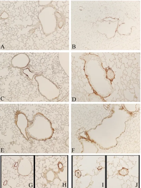
The entity of the peri-bronchiolar and bronchial remodeling was also approached by using α-SMA immunohistochemical staining. The α-smooth muscle actin (α-SMA) is the actin isoform that predominates within smooth muscle cells and it is expressed also by myofibroblasts. These cells play an important role in fibrogenesis. Control mice show only a few positive stained spots (Fig. 8A and B) while mice chronically exposed to cigarette smoke for 7 (Fig. 8C and D) and 11 months (Fig. 8E and F) show thicker layers of α-SMA positive cells, which progressively increase with time and circled main (not shown), distal bronchi and small vessels (Fig. 8G, H, I, and J). It is interesting to note that DBA/2 mice show a more evident intensity of α-SMA reaction from 7 months onward. Additionally, the presence of α-SMA positive cells is widely distributed in airways and intraparenchymal vessels in respect to C57 Bl/6J mice. In the latter strain α-SMA positive cells are unevenly distributed throughout lung parenchyma.
32
Figure 8. Representative pulmonary sections stained for α-SMA. (A) and (B) Tissue sections from C57 Bl/6J and DBA/2 control mice exposed to room air. (C) and (D) Small airways and vessels from C57 Bl/6J and DBA/2 mice, respectively, exposed to cigarette smoke for 7 months. (E) and (F) Small bronchi and vessels from C57 Bl/6J and DBA/2 mice, respectively, exposed to cigarette smoke for 11 months. (G) and (H) Intraparenchymal vessels of C57 Bl/6J mouse exposed to cigarette smoke for 11 months. (I) and (J) Intraparenchymal vessels of DBA/2 mouse exposed to
33
cigarette smoke for 11 months. (A-F) original magnification x100; (G-J) original magnification x200.
Goblet cell Metaplasia
Goblet cell metaplasia (GCM) was evaluated after PAS staining of tissue slides. In Figure 9, lung representative sections of a C57 Bl/6J air-control (A) and of a mouse exposed to cigarette smoke (B) are reported. As can be seen there is a clear difference between control and smoker mice. Additionally, we found no appreciable difference between air-controls, as well as between smoking mice within this mouse strain at 7, when 75% of mice develop GCM), and 11 months of exposure (70%). After chronic exposure to cigarette smoke, C57 Bl/6J mice develop a goblet cell metaplasia both in large and small airways (Fig. 9B).
Of interest, DBA/2 mice show only a few goblet cells at 7 months of cigarette smoke exposure that are localized mainly in the large bronchi (Fig. 10C). A more consistent goblet cell metaplasia is detected after 11 months of exposure. At this time point, PAS positive cells can be seen in large bronchi (Fig. 10F) and also in some small bronchi (Fig. 10E) even if only 35% of animals of this strain develop GCM. Control animals show no positively stained PAS cells at any time (Fig. 10A and D).
34
Figure 9. PAS staining of C57 Bl/6J pulmonary sections (A): Representative section from air-control mouse. (B): Small airways of smoke exposed mouse. (Insert) large bronchus of a mice exposed to chronic cigarette smoke for 11 months. Original magnification x40.
Figure 10. PAS staining of DBA/2 (A) and (D) are representative section
small intra-parenchymal airways and small vessels
smoke for 7 months are reported. (E) and (F) bronchioles, vessels
from mice exposed to cigarette smoke for 11 months are shown. Original magnification x100.
35
. PAS staining of DBA/2 mice pulmonary sections.
epresentative sections of DBA/2 control mice exposed to room air. parenchymal airways and small vessels, and large airways of mice
smoke for 7 months are reported. (E) and (F) bronchioles, vessels, and a cartilaginous bronchus from mice exposed to cigarette smoke for 11 months are shown. Original magnification x100.
of DBA/2 control mice exposed to room air. In(B) and (C) of mice exposed to cigarette , and a cartilaginous bronchus from mice exposed to cigarette smoke for 11 months are shown. Original magnification x100.
36
Growth factors expression
Several growth factors were analyzed in order to investigate mechanism(s) underlying the remodeling associated with smoke induced COPD. TGF-β expression was evaluated and an increased level of the growth factor is appreciated in main and small bronchi of both C57 Bl/6J (Fig. 11B and C) and DBA/2 (Fig. 11E and F) smoking mice. A faint reaction is seen in control mice (Fig. 11A and D) at the examined time points. In inserts of Figure 11, a positive stain of the muscular layer is appreciable beneath the bronchial epithelium.
CTGF expression showed a different pattern between the two mouse strains, and at the various examined times. A progressive increase in stain positivity is seen in C57 Bl/6J mice at 7 months of exposure (Fig. 12C), (when the expression is localized mostly in bronchiolar epithelium) and at 11 months of exposure (Fig. 12E). At 11 months, CTGF expression is localized in epithelial cells as well as in the muscolar layers that circle bronchioles and vessels (Fig. 12G and H). No positive stain is appreciated in control mice (Fig. 12A).
On other hand, DBA/2 mice showed no appreciable difference in CTGF expression around parenchymal bronchioles at 7 and 11 months of smoke exposure (Fig. 12D and F). We observe higher expression of this fibrogenic cytokine in smoking mice when compared with that present in control ones (Fig. 12B). An increased expression of CTGF is seen in the muscolar layer of large airways (Fig. 12I) and in small vessels (Fig. 12H) of DBA/2 mice after 11 months of smoke exposure.
Finally, PDGF-B expression is highly increased in intra-parenchymal airways of C57 Bl/6J mice after 7 months exposure to cigarette smoke (Fig. 13B). After 11 months of exposure, a significant decrease of PDGF-B expression isseen in small bronchi (Fig. 13C). However, it remains expressed mostly in airways and vessel smooth muscle cells (Fig.
37
13C, arrowheads) and inflammatory cells (Fig. 13B and C, arrows). On the other hand, DBA/2 mice showed a comparable PDGF-B expression in large and small airways at 7 (Fig. 13E) and 11 months (Fig. 13F) of exposure.
Only a faint positivity can be seen in lung sections of control mice (Fig. 13A and D).
Figure 11. Immunohistochemical reaction for TGF-β on representative tissue section of C57 Bl/6J mice (A) and DBA/2 mice (D) exposed to room air. In (B) and (C) a rapresentative section of large airway and parenchymal bronchioles of C57 Bl/6J mice exposed to chronic cigarette smoke is shown. In (E and F) Cartilagineous bronchus, small airways and intraparenchymal vessel of DBA/2 mice exposed to cigarette smoke are reported. (A-F) original magnification x100. (Inserts)
38
show a higher magnification (x200) which better highlight the positive staining on the muscle layer under the bronchiolar epithelium.
Figure 12. Immunohistochemical reaction for CTGF. (A) and (B) report control sections of C57 Bl/6J mice and DBA/2 mice, respectively. Intraparenchymal bronchioles of C57 Bl/6J (C) and
39
DBA/2 mice (D) exposed to cigarette smoke for 7 months are shown. Intraparenchymal bronchioles of C57 Bl/6J (E) and DBA/2 mice (F) exposed to cigarette smoke for 11 months. Small vessels from C57 Bl/6J (G and H) and DBA/2 mice (J), as well as a large airway from a DBA mouse (I) exposed cigarette smoke for 11 months are shown. (A-G) original magnification x100. (H-J) original magnification x200.
Figure 13. Immunohistochemical reaction for PDGF-B. Representative sections of mouse lungs from room air exposed (A) and cigarette smoke exposed C57Bl/6J mice for 7 (B) and 11 months (C). Arrowheadsindicate smooth muscle cells; arrows point to inflammatory cells. Representative lungsections from a control DBA/2 (D) and cigarette smoke exposed mice at 7 (E) and 11 months (F). (A-F) original magnification x100. (Inserts) magnification x200.
40
Immunohistochemical stain for 8-oxo-7,8-dihydro-2′-deoxyguanosine (8-OHdG)
Previous work carried out by our laboratory suggested a different sensitivity to oxidative stress for C57 Bl/6J and DBA/2 mice that may condition parenchymal and airway changes induced by cigarette smoke. In order to evaluate the oxidative damage of DNA induced by chronic smoke we perform an immunohistochemical stain for 8-oxo-7,8-dihydro-2′-deoxyguanosine (8-OHdG). After 4 months of cigarette smoke exposure, an increased positivity for 8-OHdG is appreciated on nuclei of parenchymal (Fig. 14B and D) and bronchiolar cells (Fig. 15B and D). In Fig.14A and D, and Fig.15A and D, tissue slides from control mice after 8-OHdG staining are reported. Of interest, also fibroblasts, myofibroblasts and SMC are damaged by oxidants in cigarette smoke groups (Fig. 15B and D, arrow heads). Moreover, the proportion of staining nuclei is more consistent in DBA/2 (Fig. 14D and Fig. 15D) if compared to C57 Bl/6J smokingmice (Fig. 14B and Fig. 15B).
Figure 14. Immunohistochemical reaction for 8-OHdG. Representative sections of lungsfrom room air (A) and cigarette smoke exposed (B) C57Bl/6 mice at 4 months. Representative sections of
41
lungs from room air (C) and cigarette smoke exposed (D) DBA/2 at 4 months. Original magnification x200.
Figure 15. Immunohistochemical reaction for 8-OHdG. Representative sections of bronchi from room air (A) and cigarette smoke exposed (B) C57Bl/6J mice at 4 months. Representative sections of bronchi from room air (C) and cigarette smoke exposed(D) DBA/2 mice, at 4 months are shown. Arrowheads in (B) and (D) point to cells of the fibro-muscular layer. Original magnification x200.
Oxidative damage to DNA may lead cells to different consequences: repair, apoptosis and cellular senescence associated with apoptosis resistance. Both apoptosis and cellular senescence are suggested to participate in the pathogenesis of the morphological changes associated with COPD.
42
Immunohistochemical stain for Cleaved Caspase-3
Apoptosis has been evaluated by examining cleaved caspase-3 immunostaining in pulmonary tissue slides. In C57 Bl/6J control mice only a few positive stained cells in patchy areas are seen in bronchial and bronchiolar epithelial cells and in the subepithelial layer, and no positive stained cells are appreciated in lung parenchyma (Fig. 16A and B). After 7 months of cigarette smoke exposure, massive increase in cells positivity in lung parenchyma (Fig. 16D) is seen, that seems to be related with the increased extent of emphysema observed. At this time, an almost absent positivity is seen in sub-bronchiolar fibro-muscular layer (Fig. 16C). At 11 months of exposure, cleaved caspase-3 is absent around bronchi and its positivity is decreased in lung parenchyma. This suggests that oxidative damage in C57 Bl/6J mice occurs at the maximal extent between 7 and 11 months of smoke exposure.
DBA/2 mice show a similar immunohistochemical pattern in lung parenchyma, where no positive cells in control mice are found (Fig. 17B) but a widespread positivity for caspase-3 is detected after 7 months exposure to cigarette smoke (Fig. 17D). Of interest, DBA/2 mice show an increased expression of cleaved caspase-3 also in epithelial bronchial cells after 7 months exposure (Fig. 17C) when compared with control animal (Fig. 17A).
43
Figure 16. Immunohistochemical staining for Cleaved caspase-3. Representative sections of pulmonary tissue of C57 Bl/6J mice. (A) Intraparenchymal bronchus and (B) lung parenchyma of control mouse. Intraparenchymal bronchi(C) and lung parenchyma (D) of 7 months smoking mouse. Original magnification x200.
44
Figure 17. Immunohistochemical staining for Cleaved caspase-3. Representative sections of the pulmonary tissue of DBA/2 mice. (A) Intra-parenchymal bronchus and (B) lung parenchyma of control mouse. Intra-parenchymal bronchi (C) and parenchyma (D) of 7 months smoking mouse. Original magnification x200.
Immunohistochemical stain for p16ink4A
p16ink4A immunohistochemistry was performed in order to evaluate cellular senescence induced by cigarette smoke. The absence of p16ink4A positivity in C57 Bl/6J mice exposed to room air for 1 month (Fig. 18A) indicate that no cellular senescence is induced in these mice at this time point. Additionally, only a few positive cells are seen in mature mice of the same strain exposed to room air for 11 months (Fig. 18B).
It is interest to note that after 4 months of exposure to cigarette smoke an increase in cell positivity for p16ink4A is found in bronchial epithelial cells, fibroblasts, and myofibroblasts
45
localized in the sub-epithelial layer (Fig. 18C) and lung alveolar type II cells (Fig. 18D, arrows). DBA/2 mice show a similar p16ink4A staining pattern in lung parenchyma after 4 month of smoke exposure (Fig. 19D), while a more marked staining is found in bronchiolar epithelial cells after 4 months of exposure (Fig. 19C) when it is compared to that observed on bronchi of C57 Bl/6J mice (Fig. 18C). Only a faint staining is appreciable in pulmonary sections of control mice (Fig. 19A and B). p21 immunohistochemistry has been also performed in order to evaluate cellular senescence. The pattern of expression of p21 observed in pulmonary cells confirmed that seen for p16ink4A (data not shown).
Figure 18. Immunohistochemical staining for P16ink4A on representative sections of C57 Bl/6J mice.
(A) and (B) depict bronchi and lung parenchyma of a mouse exposed to room air for 1 month and 11 months, respectively. (C) and (D) depict bronchi and lung parenchyma from a mouse exposed to cigarette smoke for 4 months. Arrows in (D) point to alveolar type II cells. Original magnification x200; insert in (C) magnification x400.
46
Figure 19. Immunohistochemical staining for P16ink4A on representative sections of DBA/2 mice. In
(A) and (B) bronchi and lung parenchyma of a mouse exposed to room air for 1 month and 11 months, respectively, are shown. (C) and (D) show bronchi and lung parenchyma from a mouse exposed to cigarette smoke for 4 months. Arrows in (D) point to alveolar type II cells. Original magnification x200.
Immunohistochemical stain for Proliferating Cell Nuclear Agent (PCNA)
Finally, we evaluated cell proliferation through an immuno-localization in pulmonary tissue of PCNA. Only a few number of cells are positive for PCNA stain in lung sections of air-control mice from C57 Bl/6J (Fig. 20A) and DBA/2 (Fig. 20C) strains. However, after cigarette smoke exposure for 4 months both strains show in lung cells an increased number of nuclei positive for PCNA. A more pronounced increase is observed in C57 Bl/6J bronchial cells (Fig. 20B) than in DBA/2 ones (Fig. 20D). In DBA/2 mice the greatest increase in positive cells is localized in the sub-bronchiolar fibro-muscular layer (Fig. 20D)and in the subpleural areas (Fig. 20F), whereas in C57 Bl/6J mice a low number
47
of proliferating cells is present in the subpleural areas (Fig. 20E). PCNA staining was sided by an immunostaining for Ki-67 (not shown), which confirmed the same pattern of positivity we observe with PCNA.
Figure 20. Immunohistochemical stain for PCNA. (A) and (C) report control sections of C57 Bl/6J and DBA/2 mice, respectively. Lung section of C57 Bl/6J (B) and DBA/2 mice (D) exposed to cigarette smoke for 4 months are shown. Sub-pleural areas of C57 Bl/6J (E) and DBA/2 mice (F) exposed to cigarette smoke for 4 months. (A-E) original magnification x200.
48
Immunohistochemical staining for MyoD
MyoD is a transcription factor which plays a critical role in myogenic differentiation and its expression has been associated with myofibroblasts presence in tissue repair/fibrosis. In control animals no MyoD expression can be seen in bronchioles and subpleural areas of both C57 Bl/6J (Fig. 21A and C) and DBA/2 strains (Fig. 22A and C). An appreciable increase in MyoD expression is detectable in DBA/2 mice exposed to cigarette smoke for 4 months in sub-bronchiolar fibro-muscular layer (Fig. 22B) as well as in subpleural areas (Fig. 22D). No positive cells to MyoD stain can be appreciated in C57 Bl/6J bronchi (Fig. 21B) and only a few in subpleural areas (Fig. 21D).
Figure 21. Immunohistochemical staining for MyoD counterstained with hematoxylin on representative sections of C57 Bl/6J mice. (A) and (C) depict bronchi and lung parenchyma of a mouse exposed to room air for 4 months. (B) and (D) depict bronchi and lung parenchyma from a mouse exposed to cigarette smoke for 4 months. Original magnification x200.
49
Figure 22. Immunohistochemical staining for MyoD counterstained with hematoxylin on representative sections of DBA/2 mice. (A) and (C) depict bronchi and subpleurical area of a mouse exposed to room air for 4 months. (B) and (D) depict bronchi and subpleurical area from a mouse exposed to cigarette smoke for 4 months. Original magnification x200.
50
Purinergic Receptors
ATP signaling has been implicated in the pathogenesis of anatomic lesions associated with COPD. Therefore, the expression of P2X4 and P2Y2 has been evaluated in both strains of
mice after chronic exposure to cigarette smoke.
P2Y2 expression seems to be slightly increased in C57 Bl/6J mice exposed to cigarette
smoke for 7 (Fig. 23B) and 11 months (Fig. 23C) if compared to control mice (Fig. 23A). The increased expression is localized mostly on large and small airways epithelial cells and in lung parenchyma with no appreciable difference between the time points we examined. Of interest, DBA/2 mice show a different pattern of expression. Only few bronchiolar cells show a positive staining for P2Y2 in control mouse (Fig. 23D) and a strong increase in
positivity is seen in lung tissues from mice exposed to cigarette smoke for 7 months (Fig. 23E). After 11 months of CS exposure the positive cells for P2Y2 immunostaining are
decreased (Fig. 23F) when compared to those that tested positive at 7 months. However, their numbers remain increased in respect to those observed control mice.
The expression of P2X4 is constitutively present at low levels in C57 Bl/6J (Fig. 24A) and
DBA/2 mice (Fig. 24B) in large and small airways. After chronic exposure to cigarette smoke, C57 Bl/6J mice show an up-regulation of P2X4 that is comparable at 7 (Fig. 24C)
and 11 months (Fig. 24E) of exposure. At 7 months, DBA/2 mice show a positive reaction especially in large airways (Fig. 24D). Up-regulation of P2X4 in bronchioles of DBA/2
mice is seen after 11 months from the beginning of the exposure (Fig. 24F).
An increase in P2X4 and P2Y2 is seen also in immune cells after cigarette smoke exposure
51
The immunohistochemical staining for P2X7 confirmed data previously obtained in our
laboratory where P2X7 expression is mainly increased, after chronic exposure to cigarette
smoke, on macrophages, neutrophils and lung tissue both at 7 and 11 months (data not shown).
Figure 23. Immunohistochemical staining for P2Y2 on representative mouse sections of pulmonary
tissue. (A) and (D) C57 Bl/6J and DBA/2 mice exposed to room air. In (B) and (E) C57 Bl/6J and DBA/2 mice exposed to cigarette smoke for 7 months are shown, respectively. (C) and (F) are
52
sections from C57 Bl/6J and DBA/2 mice exposed to cigarette smoke for 11 months. Original magnification x100. (Inserts) original magnification x200.
Figure 24. Immunohistochemical staining for P2X4 on representative mouse sections of pulmonary
tissue. (A) and (B) C57 Bl/6J and DBA/2 sections from mice exposed to room air. In (C) and (D) C57 Bl/6J and DBA/2 mice exposed to cigarette smoke for 7 months, respectively, are shown. (E) and (F) C57 Bl/6J and DBA/2 are from mice exposed to cigarette smoke for 11 months. Original magnification x100.
53
Purinergic signaling has been also suggested to be implicated in the pathogenesis of remodeling through the induction of different pro-fibrotic stimuli. Among them, we investigated the expression of IL-6 in our mice. In Figure 25, we show the expression of this cytokine in C57 Bl/6J (C) and DBA/2 (D) mice exposed to cigarette smoke for 7 and 11 months (E and F). IL-6 is absent in air-control C57 Bl/6J (A) and DBA/2 (B) mice, increase progressively on bronchial epithelial cells between 7 (C) and 11 (E) months exposure in C57 Bl/6J. Differently, DBA/2 mice show only a faint stain at 7 months (D) and an increased expression at 11 months (F) of exposure.
Purinergic receptors, expressed in inflammatory cells like macrophages, promote and increase the expression of Matrix Metalloproteinase-9 (MMP-9). The immunohistochemical reaction of MMP-9 on pulmonary tissue from smoking C57 Bl/6J (Fig. 26D) and DBA/2 mice (Fig. 26B) confirmed the increased expression of the enzyme when compared to air-control animals of the same strain (Fig. 26A and C).
54
Figure 25. Immunohistochemical reaction for IL-6. Representative sections of mouse lungs from room air exposed (A) and cigarette smoke exposed C57Bl/6J mouse for 7(C) and 11 months (E). Representative lung sections from a control DBA/2 (B) and cigarette smoke exposed mouse at 7 (D) and 11 months (F). (A-F) original magnification x200.
55
Figure 26. Immunohistochemical reaction for MMP-9. Representative sections of lung tissue from room air-exposed (A) and a cigarette smoke exposed (B) DBA/2 mouse. Representative lung sections from a control (C) and a cigarette smoke exposed (D) C57Bl/J mouse. (A-D) Original magnification x200.