UNIVERSITÀ DEGLI STUDI DELLA TUSCIA DI VITERBO
DIPARTIMENTO DI PER L’INNOVAZIONE NEI SITEMI BIOLOGICI,AGROALIMENTARI E FORESTALI
Corso di Dottorato di Ricerca in SCIENZE AMBIENTALI - XXVIII Ciclo
STRUCTURAL ANALYSIS OF ENDOGENOUS
BIOACTIVE PEPTIDES
(s.s.d CHIM/02)
Tesi di dottorato di:
Dott. Cristina Olivieri
Coordinatore del corso Tutore
Prof. Maurizio Petruccioli Prof. Fernando Porcelli
Co-tutore
Prof. Gianluigi Veglia
Hope lies in dreams,
in imagination and in the courage
of those who dare to make
dreams into reality
Acknowledgments
I would like to acknowledge many people for helping me to achieve this accomplishment, but first and foremost I want to especially thank both my advi-sors, Prof. Fernando Porcelli and Gianluigi Veglia, for their guidance as well as for their scientific enthusiasm and creativity. Their willingness to pursue new and difficult challenges has been a source of inspiration and motivation during my graduate career. I am thankful for their continuous support and for challeng-ing me to grow and improve not only as a scientist, but as a person as well.
I want to extend a thank you to Dr. Francesco Buonocore and Prof. Giuseppe Scapigliati for the collaboration in Chionodracine projects
I also want to thank Dr. Maria Giordani who has always been a source of inspiration and sincere and trusted friend.
I would like to thank all the members of Veglia Lab, past and present, for their support and friendship through the period that I have spent there as a visit-ing PhD student. I want to thank, Dr. Manu V. Surahmanian, Geoffrey Li, Aly-sha A. Dicke, Sarah E.D. Nelson, Adak N. Karamafrooz, Erik K. Larsen and Caitlin Walker. I am honored to have had the opportunity to work with such great scientists. I would like to especially thank Dr. Jonggul Kim for the quality discussions that we had, for being an exceptional role model and friend.
Lastly I want to thank my family and friends for their love and support dur-ing this “journey”, especially my father, for supportdur-ing me in every sdur-ingle mo-ment. He taught me to believe in myself and to not be afraid to dream. I love you dad.
To my family
that has always been by my side
in the good and bad times.
Contents
Acknowledgments
iDedication
iiList of Tables
viList of Figures
viiChapter 1
Preface
1.1 Unconventional protein……….1Chapter 2
Scope of the work
4Chapter 3
Introduction
7 3.1 Antimicrobial peptide ……….……….……...73.2 Intrinsically disordered protein………...15
3.3 Protein kinase ……….…...20
3.3.1 cAMP-dependent protein kinase A………...22
3.3.1.1 Regulatory subunits………….……….…...28
3.3.1.2 Protein kinase A inhibitor PKI…………...…………...30
Chapter 4
Structure and membrane interaction of the
antimicrobial peptide Chionodracine
4.1 Chionodracine ………...………..354.2 Materials and methods……….………...…37
4.2.1 Peptide synthesis………..…....…...37
4.2.2 Phospholipid vesicle preparation………...…….37
4.2.3 Steady-state fluorescence experiments………38
4.2.3.1 Outer membrane permeability assay………...39
4.2.3.2 Partition studies ………..…………39
4.2.3.3 Iodide quenching experiments………...…..41
4.2.3.4 Calcein leakage studies………..….41
4.2.4 NMR spectroscopy study………..…..43
4.2.4.1 Sample preparation and spectroscopy………43
4.3 Results……….45
4.3.1 Outer membrane permeability assay………..……….45
4.3.2 Partitioning studies……….…….45
4.3.3 Intrinsic fluorescence quenching experiments…………...…….49
4.3.4 Calcein leakage studies………..….52
4.3.5 Chionodracine structure determination…………...………...….55
4.4 Discussion and conclusion……….……….….59
Chapter 5
Characterization of membrane interaction
abilities of Chi onodracine mutants using
fluo-rescence spectroscopy
625.1 Chionodracine mutants……….62
5.2 Materials and methods……….……….65
5.2.1 Peptide synthesis………..65
5.2.2 Phospholipid vesicle preparation………...….….65
5.2.3 Steady-state fluorescence experiments………...……….65
5.2.3.1 Outer membrane permeability assay……….…..65
5.2.3.2 Partition studies………..66
5.2.3.3 Iodide quenching experiments………...……..67
5.2.3.4 Calcein leakage studies………...68
5.3 Results……….………..…..69
5.3.1 Outer membrane permeability assay………..…….69
5.3.2 Partitioning studies……….……….71
5.3.3 Intrinsic fluorescence quenching experiments………...……….75
5.3.4 Calcein leakage studies………...79
5.4 Discussion and conclusion……….83
Chapter 6
Conformational landscape of protein kinase A
inhibitor PKI studied by NMR spectroscopy
926.1 Materials and methods………..…92
6.1.1 Sample preparation………..92
6.1.1.2 Expression and purification of recombinant PKIα…...93
6.1.2 NMR experiments………...92
6.1.2.1 Sample preparation………...94
6.1.2.2 Assignment of PKIα free………..94
6.1.2.3 Assignment of PKIα bound to PKA-C……….95
6.1.2.4 Chemical shift analysis………95
6.1.2.5 NMR relaxation experiments………...96
6.1.2.6 Paramagnetic relaxation enhancement………...97
6.2 Results……….99
6.2.1 Assignment and secondary structure of PKIα………...99
6.2.2 Assignment and secondary structure of PKIα bound PKA-C...102
6.2.3 Transient tertiary structure of PKIα………..106
6.3 Discussion and conclusion………..….…110
Abstract
115Appendix
117A.1 NMR Spectroscopy……….……....117
A.1.1 NMR spectroscopy on the study of AMPs………....122
A.1.2 NMR spectroscopy on the study of IDPs………...127
A.2. Fluorescence spectroscopy……….…129
A.2.1 Fluorescence quenching...131
Reference
List of tables
Table 4.1. Partition parameters for CND calculated from the titration of the peptide with LUV of different phospholipid composition.
Table 4.2. Stern-Volmer quenching constant (KSV) and percentage of I- quenching for
CND in the presence of vesicles of different lipid composition. The pep-tide/lipid molar ratio was 1:100 in all the case.
Table 4.3. Relative leakage capabilities of CND, ad different peptide/lipid molar ratio, in presence of LUVs of different lipid composition.
Table 4.4. Kinetic parameters for calcein release from LUVs of different lipid compo-sition upon addition of increasing amount of CND. The parameters derived from the data fitting with the equation 4.5.
Table 5.1. Sequences and physical-chemical characteristics of CND and its design mu-tants. Most of the values reported were obtained using ExPASy-Prot Param Tool and Helical wheel project (Raphael Zidovetzki PhD, UCR).In red are highlighted the mutated residues.
Table 5.2. Partition parameters for CND and its mutants calculated from the titration of the peptides (1.0 M) with LUVs of different phospholipid composition. Table 5.3. Stern-Volmer quenching constant (KSV) and percentage of I- quenching for
CND and the derived mutants, in the presence of vesicles of different lipid composition. The peptide/lipid molar ratio was 1:100 in all the case.
Table 5.4. Relative leakage capabilities of CND and its mutant in presence of LUVs of different lipid composition and at different peptide/lipid molar ratio.
Table 5.5Kinetic parameters for calcein release from 25 M LUVs of different lipid composition upon addition of increasing amount of CND mutants. The parame-ters derived from the data fitting showed in figure 5.7.
List of figures
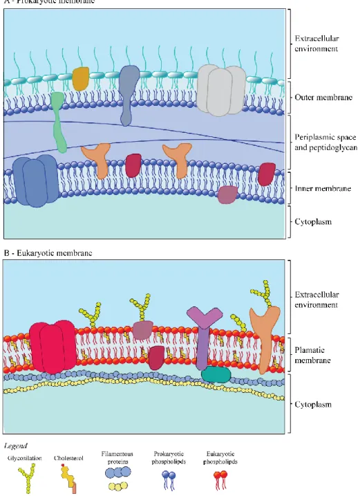
Figure 3.1. Schematic representation of biological membrane. (A) Gram-negative membrane schematization. The Gram-negative bacteria have two different membrane (outer and inner membrane) that differ on phospholipid and protein composition. The outer membrane contain the lipopolysaccharides (LPS) that contribute to the structural integrity of the membrane. (B) Model of eukaryotic membrane, composed by neutral phospholipids.
Figure 3.2.Principal phospholipids present in biological membrane. In the top are reported the phospholipids typically compose the eukaryotic membrane, while in the bottom structure are phospholipid present almost exclusively in prokary-otic organism. The letter R indicate the different fatty acid chains that can be coordinate to the glycerophosphoric acid group.
Figure 3.3. Proposed model for antimicrobial activity of AMP. The antimicrobial peptides pile up on the bilayer surface (A) until they reach a threshold concen-tration. Following an aggregation process, they disrupt the bilayer integrity through; a barrel-stave model (b), toroidal model (c), and carpet-like model (figure adapted from [1, 2]).
Figure 3.4 Schematic representation of the continuum model for protein struc-ture. The protein are ranging from highly dynamic conformation (organ) to a well-defined folded proteins. SUMO-1 (PDB 2N1A) and dystrophin (PDB 3UMM) are chosen as a prototypes for disordered-loop and well-folded pro-teins, respectively. (Figure adapted from [3])
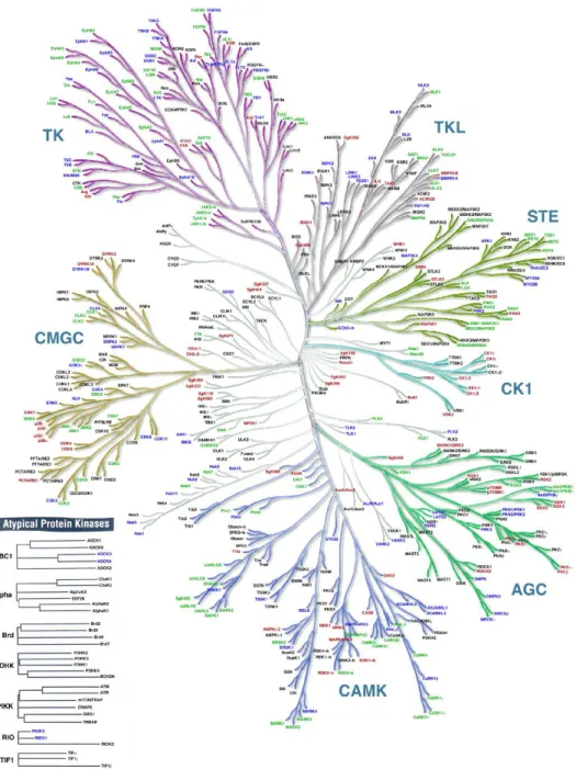
Figure 3.5 Human kinome. Classification of all the protein kinases present in the hu-man genome (adapted from Manning et. al. [4]).
Figure 3.6 3D structure of cAMP-dependent protein kinase A. X-ray structure of protein kinase A holoenzyme formed by two catalytic subunits and the dimer of RIIβ regulatory subunits (PDB-3TNP, [5]).
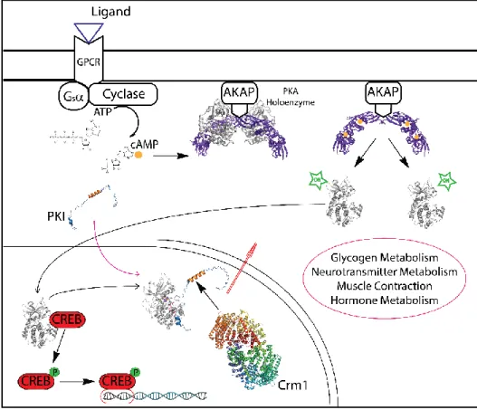
Figure 3.7 Intracellular signaling pathway of transcription factor activation. Sig-naling cascade for the activation of the cAMP-responsive element-binding pro-tein (CREB). The binding of a ligand (in the specific case glucagon) to the G protein-coupled receptor (GPCR), causes the translocation of the α-subunit of the coupled G protein to the membrane-bound adenylyl cyclase, with the con-comitant transformation of GTP from GDP. cAMP is produced and, through the binding to the regulatory subunit of the cAMP-dependent protein kinase A (PKA) (PDB-3TNP) holoenzyme, induces the release of the catalytic subunits (PKA-C). PKA-C is then translocate to the nucleus, where phosphorylates CREB, an IDP transcription factor. Inside the nucleus, the heat-stable protein kinase inhibitor regulates the activity of PKA-C and mediate the cytoplasmic translocation of the enzyme through the nuclear export receptor CRM1
(PDB-3GJX). The PKI:PKA-C, was modeled in the basis of the X-ray crystal struc-ture 1ATP [6]. (adapted from [7].
Figure 3.8 Crystal structure of the catalytic subunit of protein kinase A. 3D struc-ture of PKA-C, in presence of Mg2+ ions and ATP (PDB-1ATP [6]
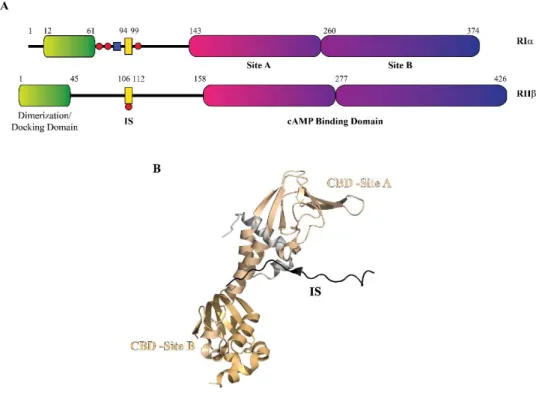
Figure 3.10 Organization of the regulatory subunits. (a) Schematic domain organi-zation of RIα and RIIβ. Red circles indicate heterologous and auto-phosphorylation sites. (B) Domain organization on the structure of RIIβ (PDB-3TNP, [5]).
Figure 4.1. Sequence comparison inside the Piscidin family. The primary sequence of CND is aligned with the sequence of the other member of the family. The green boxes highlight the shared amino acids in the protein sequence. (Adapted [8]).
Figure 4.2. Outer membrane permeability assay. Permeabilization of the external membrane of the two Gram-negative bacteria, E.coli BL21 (DE3) (A) and
Psy-chrobacter TAD1 (B).
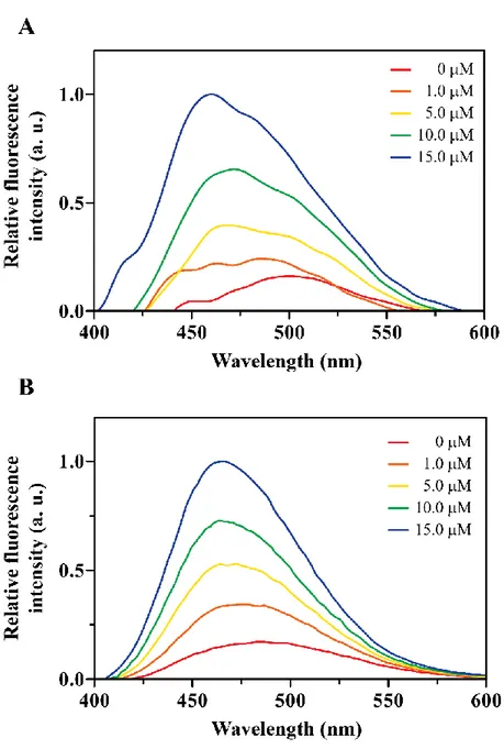
Figure 4.3. Partition experiments. Cnd fluorescence in absence and presence of in-creasing concentration of LUVs composed by (A) 100% of PC and (B) a mix-ture of PC/PG (70:30 molar ratio). In C is reported the binging isotherm for the peptide upon addition of increasing amount of vesicles. The CND concentra-tion was 10.0 M.
Figure 4.4. Iodide quenching experiments. Quenching of the intrinsic fluorescence of CND in absence (A) and in presence of phospholipid vesicles of different com-position: (B) PC-100%, (C) PC/PG (70:30), and (D) E. coli total extract. Figure 4.5. Stern-Volmer plots for the Tyr quenching of CND. The graph reports
the plots for the intrinsic fluorescence quenching of the peptide in aqueous buffer and vesicles made from PC-100%, PC/PG (70:30) and E. coli lipid ex-tract.
Figure 4.6. Kinetic of calcein leakage from LUVs after addition of CND. From 01 M through 10 M. The curve are experimental data normalized by Triton-X. In panel A and B are reported the calcein release kinetic recoded for 25 M and 50 M of PC-100% LUVs, respectively. Panels C and D are the kinetic of cal-cein efflux for 25 M and 50 M of PC/PG (70:30) LUVs.
Figure 4.7. Fingerprint region of CDN extracted from the 2D [1H-1H] NOESY experiment at 300 ms mixing time. The peptide was reconstituted in DPC mi-celles. The spectrum was used to solve the 3D-structure of CND.
Figure 4.8. Summary of structural parameters. Backbone NOEs pattern for CDP in DPC micelles. The thick line correspond to strong NOE, while the thinner line represent medium and weak NOEs (A). In panel B is reported the predicted or-der parameter RCI-S2.
Figure 4.9. NMR structure model of CND. (A) Histogram of backbone RMSD vs residues for the final 40 conformers. (B) Conformational ensemble showing the
convergence of the structures for heavy atoms of the backbone. (C) Representa-tive structure=re of CND in DPC micelle. The peptide assume a conical am-phipathic -helix, with the repartition of the hydrophobic residues on one side on f the helix (blue), and the hydrophilic in the other side (green).
Figure 5.1 CND and its derived mutants. (A) CND in amphipathic -helical confor-mation (structure obtained as describe in chapter 4).In the panels B, C, and D are showed the sequence and the structure of CND mutants modeled by Pymol software. In blue is presented the hydrophobic side of the helix, while in pink is presented the hydrophilic portion. The red dots are indicating the positive charged amino acids present in the sequence.
Figure 5.2.Outer membrane permeability assay. Permeabilization of the external membrane of E.coli BL21 (DE3) by the three CND mutants (A-C).In panel D is reported the ANS Uptake for the wild-type peptide and the mutants.
Figure 5.3. Partition experiments. Emission spectra of CND mutants recorded in presence of increasing concentration of (A-C-E) 100% of PC and (B-D-F) a mixture of PC/PG (70:30 molar ratio) LUVs. The Imax of each spectra was used
to calculate the Kx, according to equation 4.3. The spectra were recorded at 25
°C using 1.0 M of peptide.
Figure 5.4. Binding isotherms for CND and its mutants interacting with lipid bi-layer vesicles. The fluorescence intensity of the Try/Trp residue was measured as LUVs were titrated into the sample. Before registered the spectrum, the sys-tem was allowed to equilibrate at least for 10 minutes.
Figure 5.5. Iodide quenching experiments. Spectra recorded for quenching of the Trp residues of CND mutants presence of 0.5 mM PC-100% (A-C-E) and PC/PG (70:30) (B-D-F) LUVs. For each mutant was also performed the experiment in absence of LUVs (spectra not showed).
Figure 5.6. Stern-Volmer plots for the quenching of Trp residue of CND by Io-dide. For each mutants, the quenching experiments were performed in buffer and in presence of 0.5 mM of PC-100%, PC/PC (70:30) and E. coli total extract LUVs. The final concentration of peptides was 5 M.
Figure 5.7. CND mutants cause efficient calcein release from vesicles. Kinetics of calcein release from 25 M PC-100% (A-C-E) and PC/PG (70:30) (B-D-F) LUVs, upon the addition of increasing amount of mutants. The LUVs were loaded with 30 mM of calcein. All the experiments were performed at 25 °C. Each curve is normalized by Triton-X
Figure 6.1. Simulated structure of the full-length PKI bound to PKA-C. Full-length PKI sequence is docked in the structure of PKA-C (PDB-1ATP). In the two boxes are reported the crystal of truncated PKI peptides bound to CRM1 (top, PDB-3GJX) PDB ant to PKA-C (bottom, PDB-1ATP), respectively.
Figure 6.2. 1H-15N HSQC spectrum of of PKIα. (A) 1H-15N HSQC spectrum showing the assigments of the resonance peaks for PKIα in solution. (B) Selected 1H-13C planes from the 3D HNCACB (red and green) and 3D-CBCA(CO)NH (red) spectra taken at different nitrogen frequencies.
Figure 6.3. Chemical shift and HX-NOE for PKIα. Top: primary sequence of PKIα, where are highlighted in red the PSS residues and in blue the NES residues, re-spectively. (A) Graph of the ΔδCα-ΔδCβ calculated for each residue of PKIα. Positive values of CSI are indicative of α-helical secondary structure. (B) HX-NOE values obtained from each residues of PKIα sequence. In the box is en-large the region between -0.5 and 1. Values of HX-NOE lower than 0.6 are in-dicative of highly dynamic (in the ps-ns time scale) residues.
Figure 6.4. Mapping the interactions between PKIα and PKA-C. (A) 1H-15N HSQC
spectrum with reported the assigment of the resonance peaks. (B) Selected 1 H-13C planes of overlaid 3D HNCA taken at the nitrogen frequencies of indicate backbone amide resonances, for the PSS residues.
Figure 6.5. Chemical shift and HX-NOE for PKIα. On the top of the graphs is re-ported the primary sequence of PKIα, where are highlighted in red the PSS res-idues and in blue the NES resres-idues, respectively. (A) Graph of the ΔδCα-ΔδCβ calculated for each residue of PKIα. Positive values of CSI are indicative of α-helical secondary structure. (B) HX-NOE values obtained from each residues of PKIα sequence. In the box is enlarge the region between -0.5 and 1. Values of HX-NOE lower.
Figure 6.6. Chemical shift perturbation. (a) Chemical shift values obtained for each PKI residues. In pink and orange are highlighted the portion of the graph rela-tive to the PSS and NES sequence, respecrela-tively. The residues that show more chemical shift changes from PKIα free to PKIα bound to PKA-C, are localized primarily in the N-terminal patron of the peptide. (B) Model of the structure of the complex between PKA-C and full-length PKI. In the PKI sequence are re-ported the CSP trends estimated for each amino acids.
Figure 6.7. 1H-PRE-Γ
2rates for three different PKIα mutants. Intermolecular PRE
profile observed for the three PKI mutants: (A) V3C, (B) S28C and (C) S59C, respectively, labeled with MTCL.
Figure 6.8. Intramolecular paramagnetic relaxation enhancement of PKI mutants, free in solution. Plots of intensity ratios for the observed for (A) V3C, (B) S28C, and (C) S59C PKI mutant spin labeled with MTSL.
Figure A.1.1. Schematic representation of a spin in presence of an external magnetic field (Bo) (A). Zeeman levels, in absence and in presence of Bo.
Figure A.1.2. Dihedral angles in proteins. In a polypeptide chain three dihedral an-gles are defined as ψ (psi) (C’-Ca bound), φ (phi) (Ca-N bound), and (N-C’, peptide bound). Due the planarity of the peptide bound, the values that can assume is 180° (in trans) or 0° (in cis).
Figure A.1.3. Size, shape and composition of membrane mimicking system used in NMR spectroscopy.
Chapter 1
Preface
1.1 Unconventional Protein
For over fifty years, Anfinsen’s Principle has remained the driving force for understanding the relationship between protein structure and function [9, 10]. According to this principle, the primary sequence of the protein, derived from the translation information encoded in the mRNA, determines a singular unique structure for the protein. This unique fold is what governs the function of the protein. While there can be no doubt that protein structure and function are linked, numerous studies have demonstrated that almost 30% of the eukaryotic genome encodes for protein sequences that do not fold into a stable globular structure [11-13]. These proteins were initially classified as unconventional pro-teins and later as intrinsically disordered propro-teins (IDPs). These IDPs are bio-logically active in physiological conditions with no well-defined 3D structure[14, 15]. Intrinsic disorder in proteins is structurally very diverse. These can range from small disordered linkers to molten globule structure to complete absence of structure.
However what is the purpose for intrinsic disorder for function? Genomic analyses have found that IDPs are frequently involved in regulation, recogni-tion, and signaling; where high-specificity and low affinity interactions with
multiple partners are fundamental prerequisites for function. Specificity is commonly achieved by recognition of a linear amino acid motif on the IDP or a general environmental change. Upon binding, often a subsequent disorder-to-order transition (coupled binding and folding) occurs, typically that of a -helix, while others remain disordered [16-18]. The increasing interest in these proteins is due to their prominent roles in many pathophysiological cellular mechanisms. As a result, there has been a growing awareness that proteins with intrinsically disordered regions (IDR) or fully-disordered (IDPs) are biologically active and play important roles in both eukaryotic and prokaryotic cells [15, 19].
There are small endogenous peptides that interact with biological mem-branes and share many characteristics with other IDPs, but are not fully recog-nized as IDPs in the literature. These peptides, better known as membrane-active peptides, are unstructured in solution, but undergo a disorder-to-order transition upon interaction with phospholipid membranes [20]. However, these membrane-active peptides are not involved in signaling and transcriptional reg-ulation. Membrane-active peptides can be divided in three groups: i, the amy-loid peptides that are characterized by their ability to form stable--sheet-rich structures known as amyloid fibers present in Alzheimer disease [21]; ii, the cell-penetrating peptides that show the intrinsic capacity to translocate across biological membranes [22]; and iii, the antimicrobial peptides (AMPs), that be-long to the ancient component of the eukaryotic innate immune system directed against the bacterial infections[23]. In the last twenty years, the research on the characterization of the structure and the biological function of these peptides
has shown their importance in biology and human health. In this work, I will argue that these classifications are arbitrary as membrane-associated peptides and IDPs fundamentally share the same mechanism, where disorder imparts function and activity is exhibited by the environment in which the protein is present in.
Chapter 2
Scope of the work
In my PhD work I have focused on elucidating the structure, mechanism and function of several peptides which are characteristic of IDPs. In the follow-ing PhD thesis I elucidated the antimicrobial activity of Chionodracine (CND), an antimicrobial peptide, isolated in the immune system of the gills of the Ant-arctic teleost Chionodraco hamatus [24]. I used steady-state fluorescence spec-troscopy to characterize the membrane disrupting properties of the peptide in in-teraction with synthetic and natural membrane environments. Nuclear magnetic resonance (NMR) spectroscopy was used to solve the 3D structure in presence of dodecyl-phosphocholine (DPC) micelles. I demonstrated that CND can bind to membranes of different lipid compositions with a preference for membranes mimicking those of prokaryotic cells. CND also interacts with Escherichia coli BL21 (DE3) and Psychrobacter sp. TAD1 membranes and cause the lysis of the bacterial cells. Studies of fluorescence quenching demonstrated that, upon the binding, this peptide remains adsorbed at the lipid surface, only partially ex-posed to the aqueous environment. This behavior is also confirmed by the NMR structure where, the peptide assumes a classical amphipathic -helix, with the hydrophobic residues on one side on the helix, and the hydrophilic on the other side [8].
Due to the interest of discovering of new AMPs with higher activity to be used as an antibiotic-substitute, I introduced rational mutations in CND to increase its antimicrobial activity. I reasoned from the NMR structure that CND with a stronger hydrophobic/hydrophilic interface would increase antimicrobial activity. I designed three different mutants, with increased net positive charge and hydrophobicity, and characterized their interaction with synthetic and natu-ral membranes. As with wild-type CND, the characterization of the mutant’s function, in the presence of different phospholipid membranes, was assessed us-ing steady-state fluorescence spectroscopy experiments. These studies demon-strated that the mutations have increased antimicrobial properties of CND. The mutants showed a stronger ability to interact, bind and disrupt membranes of different lipid compositions that can be directly correlated to the number of pos-itively charged amino acids present in the sequence. All of my work on CND, was carried out in collaboration with Professor Francesco Buonocore, Ph.D, from University of Tuscia (Department for Innovation in Biological, Agro-food and Forest systems, University of Tuscia, Viterbo, Italy) and Professor Gianlui-gi Veglia, Ph.D, from the University of Minnesota (Biochemistry, Molecular Biology and Biophysics and Chemistry Department, University of Minnesota – Twin Cities, Minneapolis, MN, United States of America).
In the second part of my PhD work I focused on the elucidation of the mechanism of inhibition of the catalytic subunit of cAMP-dependent protein ki-nase A (PKA-C), by the endogenous Protein Kiki-nase Inhibitor peptide (PKI) [25]. PKI is a fully disordered 75 amino acid protein that regulates the activity
and the intracellular localization of PKA-C [26, 27]. To understand the molecu-lar mechanism underlying recognition of PKA-C by PKI, I characterized the change in the conformational landscape that PKI undergoes upon binding to PKA-C (Apo to PKA-C bound). These studies were carried out using NMR spectroscopy, the only structural technique to provide atomic resolution detail on IDPs. Using chemical shift analysis and nuclear spin relaxation experiments, I demonstrated that PKI alone has minimal secondary structural elements with only two transient helixes. However, paramagnetic relaxation enhancement (PRE) experiments [28] show that PKI is not fully disordered, but has transient tertiary intramolecular interaction between the C- and N-terminus. When PKI is bound to the kinase, in the presence of the non-hydrolysable ATP mimic, β,γ-Imidoadenosine 5′-triphosphate (ATPN), only the sequence that directly inter-acts with the catalytic subunit undergoes significant structural rearrangement while the majority of the peptide is still unfolded. By clarifying the mechanisms of inhibition and regulation of PKA-C, we will be able to provide new insights into enzymatic regulation at the cellular level.
Chapter 3
Introduction
3.1.
Antimicrobial peptide
Due to the indiscriminate use of antibiotics, from agricultural and human health, the last thirty years we have observed an increase in the resistance of conventional antibiotics [23]. Antimicrobial peptides (AMPs) have emerged as a potential class of agents that may replace antibiotics, due to the inability of pathogenic organisms to develop resistance. The AMPs are a fundamental and ancestral component of the innate immune system of eukaryotic organisms against bacteria, protozoa, fungi, and virus infections. Although they have been identified in a wide range of tissues, they are prevalent in epithelial tissue where they represent a “first line of defense” against pathogen infections [23, 29-31].
AMPs have been described in prokaryotic cells since 1939, when substanc-es isolated from Bacillus brevis, named gramicidin, showed antimicrobial ac-tivity both in vivo and in vitro against a wide range of Gram-positive bacteria [32, 33]. Nevertheless systematic studies on this class of membrane interacting peptides started only at the beginning of the 1980’s, when the primary structure of cepropin (AMP from insect) was published by Hans Boman and co-workers [34-36]. Since then, hundreds of AMPs have been characterized and, even though they show a widely diverse amino acid sequences, they share some
pe-culiar features. Typically, natural AMPs are composed of 12-60 amino acid res-idues, predominantly of positively charged and hydrophobic residues. Due to their considerable genetic variability, AMPs cannot be easily classified on the base of size or physico-chemical parameters. However they can be categorized on the base of their secondary structure in: -sheet, -helix, extended, and loop peptides [37]. Among this structural groups, -helix and -sheet are the more common [31, 38] and -helical peptides are the most studied AMPs. The best known examples of -helical AMPs are protegrin, magainin, cyclic indolicin and coiled inolicin [39]. Usually, in -helical structures, the distance between two adjacent amino acid residues is around 0.15 nm and the angle between them with regard to the center id around 100 degree from the top view. -sheet pep-tides are composed of at least two -strands with disulfide-bonds between the strands [31]. However, most AMPs do not assume a defined secondary structure and they cannot be classified in this categories. Also, many -helical peptides assume an active structure only when they interact with the membranes of target organism. For example, indolicin assume a globular and amphipathic confor-mation in aqueous solution, while it is wedge-shaped in presence of phospholip-id bilayers [40]. Instead the majority of -sheet peptphospholip-ides do not undergo to dras-tically conformational transition, but preserve their secondary structure even in absence of phospholipid membranes [37]. AMPs can also be classified accord-ing to their (i) biosynthetic pathways, (ii) mechanism of biological activity, and (iii) structural properties. AMPs can be ribosomally-synthetized, (this is
typical-ly of higher eukaryotes) or non ribosomaltypical-ly-synthetized (when produced by lower eukaryotes and prokaryotic organisms) [41]. On the base of their biologi-cal activity, AMPs are classified in antiviral, antifungal, anticancer, antipara-sital, insecticidal, spermicidal, anti-HIV, and/ or with chemotactic nature [42, 43].
The most important features of all AMPs are their ability to selectively in-teract with microbial biological membranes and kill the pathogen. AMP’s abil-ity to distinguish between target and host cells depends largely on the differen-tial membrane composition between the prokaryotic and eukaryotic membranes (Figure 3.1) [37, 44]. All biological membranes are a fluid mosaic composed of proteins and phospholipids [45]. By definition, the phospholipid bilayer is am-phipathic with a hydrophobic surface and hydrophilic interior. Each organism has an asymmetric composition of the phospholipids in the bilayer. Eukaryotic membranes are rich in zwitterionic neutral phospholipids, like phosphatidylcho-line (PC), phosphatidylethanolamine (PE), and sphingomyelin (SM) (Figure 3.2) [44]. Prokaryotic membranes are composed predominantly of negative charged phospholipids; phosphatidylglycerol (PG), cardiolipin (CL, that is a dimer of PG), and phosphatidylserine (PS).
Figure 3.1. Schematic representation of biological membrane. (A) Gram-negative membrane schematization. The Gram-negative bacteria have two different membrane (outer and inner membrane) that differ on phospholipid and protein composition. The outer membrane contain the lipopolysaccharides (LPS) that contribute to the structural integrity of the membrane. (B) Model of eukaryotic membrane, composed by neutral phospholipids.
Figure 3.2.Principal phospholipids present in biological membrane.
In the top are reported the phospholipids typically compose the eukaryotic membrane, while in the bottom structure are phospholipid present almost exclusively in prokaryot-ic organism. The letter R indprokaryot-icate the different fatty acid chains that can be coordinate to the glycerophosphoric acid group.
From this prospective, it follows that the prokaryotic membranes are generally more electronegative than eukaryotic membranes, and the membrane itself can work as a pseudo-receptor for AMP recruitment [23, 46, 47]. Therefore, the in-teraction of the cationic AMPs with target cells does not require macromolecu-lar recognition, but is macromolecu-largely due to the electrostatics [48, 49].
Many AMPs exhibit their killer-activity against the pathogens through the interaction and disruption of phospholipid membrane integrity [37, 50]. In this case, no matter the mechanism used to perturb the target membrane, all the pro-posed models start with an electrostatic interaction between the AMPs and the charged phospholipids of the membrane (attraction step) [50]. Upon the pep-tide/lipid interaction (attachment step) [51-54], the peptide undergoes a struc-tural rearrangement (folding-upon-binding mechanism) that allows the insertion into the lipid bilayer (insertion step) [55-58]. These peptides induce structural
changes in the prokaryotic membrane through formation of pores (pore for-mation model) or solubilization of the membrane (carpet-like model) [2]. In both cases, the disruption of the membrane integrity causes the dissipation of the electrochemical gradients, necessary for cell survival, and the leakage of cy-toplasmic metabolites. The most popular pore forming models are the
barrel-stave, toroidal, and the carpet-like model (Figure 3.2), in which the AMPs
oli-gomerize and form pores in the membrane [2, 43]. In the barrel-stave model, the pore is formed by peptides that aggregate to form a bundle of monomers aligned parallel to the membrane [59, 60]. In the toroidal model, binding of AMP on the membrane induces a curvature strain on the leaflets on the bilayer until the two sides of the bilayer fuse to form a peptide/lipid lined pore [59, 61, 62]. In the carpet-like model, AMPs accumulate on the surface of the membrane and, once an effective concentration threshold is reached, a micellization of the membrane is induced. This model is often called also a detergent-like model, for the similarity with the detergent mechanism of action [59, 62-64].
Parallel to the structural models, two thermodynamic models have been de-veloped in the last two years to describe the mechanism of action [51, 52]. Both models, graded or all-or-none model, are based on the analysis of AMP capa-bility to induce leakage of a fluorescence dye entrapped inside a phospholipid vesicle [51, 52, 65-67]. If the addition of AMP to the vesicle suspension induces the disruption of all the vesicles, highlighted by a massive release of fluores-cence dye, the peptide acts according to the grade model [51, 52, 65, 68]. If
on-ly a fraction of the vesicles release the content and the other part do not lose any dye, the peptide perform a dye release according to all-or-none [69, 70].
The structural and binding characterization of AMPs in presence in native lipid environments is fundamental to understand underlying mechanism of their antimicrobial activity. Nuclear magnetic resonance spectroscopy (NMR) and X-ray crystallography are the most important techniques for the determination of atomic resolution structure of biomolecules [2, 43]. Unfortunately, it is nearly impossible to obtain high quality crystals of AMPs in a lipid environment, due to their small size and intrinsic flexibility. Fortunately NMR has been fruitful in determining high resolution atomic structures of AMPs in membrane-like envi-ronments [2, 43, 71]. This has primarily been achieved using solution-NMR techniques using micelles as membrane-like environments [72, 73]. However, solid state NMR spectroscopy, in particular using oriented solid state NMR (OSS-NMR) techniques, has emerged as a powerful tool to determine the struc-ture of AMPs [74, 75]. Not only does OSS-NMR allow for atomic resolution structure determination in full native lipid environments, but also allows for the determination of the topology of AMPs along the membrane.
For the characterization of the binding and kinetic mechanism of
AMP/membrane interaction, fluorescence spectroscopy is largely used. This sensitive spectroscopy allows the calculation of kinetic and thermodynamics pa-rameters that describe the interaction of AMPs with natural or synthetic phos-pholipid bilayer [51, 52, 67, 68, 76, 77].
3.2.
Intrinsically disordered protein
The classical Anfinsen's principle has stated the structure-function relation-ship in proteins for decades [9]. This paradigm of states that the primary se-quence is defines the structure of the protein, and the fold determines its func-tion. In the past 20 years, numerous studies have demonstrated that a large por-tion of the proteome is formed by proteins that are partial or fully unstructured in solution, nevertheless are functional [14, 18, 78-90]. Over the 30% of the human proteome is predicted to be composed by proteins that exhibit intrinsic disorder, including folded proteins with intrinsically disordered regions (IDRs) and entirely structure-less entities [83, 91]. From this prospective, it follows that “structured proteins” classify the proteins that do not possess IDRs, while pro-teins that are fully unstructured are called intrinsically disordered propro-teins (IDPs).
The ability of a protein to fold or not under physiological conditions is en-coded in its amino acid sequence [14, 18, 81]. A bioinformatic study conducted over 275 natively folded and 91 natively unfolded proteins has demonstrated that is the combination between a low mean hydrophobicity and a high net charge the essential pre-requisite for the absence of a compact structure in pro-teins [81]. IDRs and IDPs lack in a bulky hydrophobic core that driving the folding events of globular proteins, but they are rich in charged a polar amino acids.
IDRs and IDPs are involved in numerous cell processes and, based on their mode of action, are grouped in six functional classes: (i) assemblers, (ii) chap-erons, (iii) display site, (iv) effectors, (v) entropic chain, and (vi) scavengers [15, 92]. Eukaryotes and viruses use IDRs/IDPs essentially for mediating transient protein-protein interactions in signaling and regulation processes. Conversely, prokaryotes use disordered proteins mainly for larger lasting interaction in-volved in formation of complex between two or more partners [93]. IDPs were almost never involve in (a) catalysis, (b) biosynthesis, (c) metabolism, (d) transport processes [11, 16].
Currently, the important role that IDRs/IDPs play in the cell is globally recognized and, in light of this, new theories have been developed [3]. For ex-ample, proteins have been proposed to function within a conformational
contin-uum, ranging from complete disordered to a fully structured [86] (Figure 3.3).
In this model, that does not contemplate the presence of boundaries between the described states and the native proteins, IDRs/IDPs fluctuate stochastically be-tween several different states (transiently sampling coil-like states, localized secondary structure, and more globular states). What determines the structural characteristic and the population of the individual states in the conformational ensemble is the nature of the amino acid sequence and their distribution. Hence, in the native unbound state, IDRs/IDPs exist as a dynamic ensemble of rapidly interconverting conformations [94-96].
Figure 3.3. Proposed model for antimicrobial activity of AMP. The antimicrobial peptides pile up on the bilayer surface (A) until they reach a threshold concentration. Following an aggregation process, they disrupt the bilayer integrity through; a barrel-stave model (b), toroidal model (c), and carpet-like model (figure adapted from [1, 2]).
It was proposed the mechanism of IDR/IDP-mediating molecular interac-tion works using a combinainterac-tion of conformainterac-tional selecinterac-tion and induced
fold-ing. These two models are not mutually exclusive. The dominant mechanism
depends on the concentrations of the individual proteins (conformational
selec-tion) [97] and on the association rate constant (induced folding) [98]. Briefly, in
the conformational selection model, the addition of binding partners can result in a shift of the population in the conformational ensemble of the IDP that is most favorable for the binding [99-104]. This mechanism was observed in pro-tein-protein and protein-nucleic acids interaction [101]. On the other hand, the
induced folding model asserts that the IDP undergoes a disorder-to-order
transi-tion upon associatransi-tion with the binding partner [100, 105-107]. Usually, disor-dered segments assume a well-defined 3D-structure upon the interaction with
the binding target. The IDPs retain a significant conformational freedom even when they are bound to the target [108].
To study the structure of partial or fully disordered proteins, a variety of experimental techniques are used including NMR, small-angle X-ray scattering (SAXS), and single-molecule fluorescence. SAXS is the elastic scattering of X-rays by biomolecules at small angles, reporting on the size and shape [109, 110]. Although straightforward with simple experimental conditions, SAXS is inherently a low resolution technique. Single-molecule (sm) techniques, like sm-fluorescence resonance energy transfer (smFRET), are able to minimize av-eraging over the heterogeneous ensembles of conformations in which IDPs nat-urally exit. smFRET can measure the dynamics and individual conformations of the unbound ensemble, intermediates during induce folding, and internal fric-tion in the folding process of IDPs [111-113] in a time resolved manner. How-ever, the spatial resolution is limited by the placement of the FRET probes. NMR has emerged as a powerful technique to study the conformational ensem-ble of IDPs since this is the only technique that can provide atomic resolution structural insights of IDPs [95, 114-116]. Application of NMR to the study of IDPs has shown the existence of transient secondary and tertiary structural ele-ments that are invisible to other techniques and quantifies protein dynamics in IDPs along a wide range of timescales (ps to seconds). Correlating structural dynamics of IDPs to functional consequences remains an on-going challenge in understanding IDP function and mechanism (Appendix).
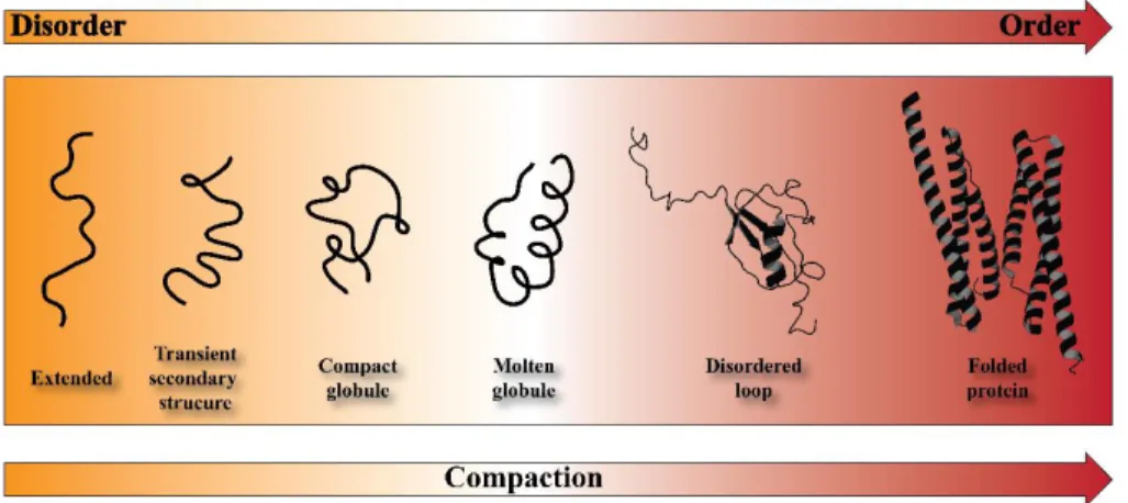
Figure 3.4 Schematic representation of the continuum model for protein structure. The protein are ranging from highly dynamic conformation (organ) to a well-defined folded proteins. SUMO-1 (PDB 2N1A) and dystrophin (PDB 3UMM) are chosen as a prototypes for disordered-loop and well-folded proteins, respectively. (Figure adapted from [3]).
3.3
Protein kinases
The phosphorylation of target proteins is one of the most common signaling pathway used by the cells to regulate fundamental intra-cellular processes like cellular cycle, metabolism and apoptosis [1]. Since 1955, when the importance of protein phosphorylation was first elucidate as a regulatory cell mechanism [2], numerous studies have demonstrated the crucial role that this protein modi-fication plays in regulation processes of mammalian cells [3, 4]. The enzymes that perform and regulate phosphorylation inside the cells are the protein kinas-es. This enzymatic family is composed by over 500 members and accounts for approximately 2% of the entire mammalian genome (kinome – Figure 3.5) [5, 6], and 4% of the plant genome [7]. The canonical function of these enzymes is to transfer the γ-phosphate of adenosine triphosphate (ATP) to Ser/Thr/Tyr resi-dues of the target substrates [2], causing the activation or deactivation of differ-ent signaling pathways [8-11]. Inside the cell, protein kinases play a crucial role as a molecular switches, and thus they are highly dynamics protein that can ex-ist between several different conformational states [12]. Most of these enzymes are also phosphoproteins, and those phosphates are essential for both structure and protein function [3]. For instance, phosphorylation sites can be used as docking sites for other proteins or as an organizing points for a specific struc-ture conformation [3]. The majority of protein kinases take part to signaling events through a dual-function: they not only catalyze the phosphoryl transfer but they can act as scaffolds to modulate, integrate, or compete in the signaling
cascade [3, 13]. Over a decade ago, a new classes of kinases with a non-canonical function was described by Manning and coworkers [14].
Figure 3.5 Human kinome. Classification of all the protein kinases present in the hu-man genome (adapted from Manning et. al. [4]).
They discover that approximately 10% of kinome contained mutations in ca-nonical amino acid motifs thought to be fundamental for the catalytic function.
These proteins, called pseudo-kinases, are partial or completely devoid of cata-lytic activity [15, 16], but they may have a more general essential non-catacata-lytic role in signaling pathways, [16-18]. Conversely, a dead kinase is a protein that cannot neither perform the catalytic activity nor act as a scaffold for binding substrates and inhibitors [19]. Due to the crucial processes in which kinases take part, their activity is also finely tuned by cofactors and/or accessory proteins that coordinate the protein activation or deactivation over time and space [20]. Typically, protein kinase are thought to be maintained in a basal, inactive state and recruited to action transiently by extracellular stimuli [21].
3.3.1 cAMP-dependent protein kinase A
Within the large and diversified kinase families that compose the kinome [14, 22], cAMP-dependent protein kinase A (cAPK), known also as protein ki-nase A (PKA), is considered the prototype of the entire super-family [23-25]. Inside the cell, PKA exist as an inactive heterotetramer formed by two catalytic subunits (PKA-C) bound to a dimer of regularity (R) subunits (Figure 3.6). The cooperative binding of the cellular second messenger cyclic adenosine mono-phosphate (cAMP) to R subunits causes the disassembling of holoenzyme and the releasing of the active PKA-C [26]. PKA-C targets a diverse array of sub-strates, including protein localized in the cytoplasm, mitochondria, plasma membrane, sarcoplasmic reticulum membrane and nucleus [27], and it is in-volve in numerous cellular signaling cascades induce by β-adrenergic stimula-tions (Figure 3.7).
Figure 3.6 3D structure of cAMP-dependent protein kinase A. X-ray structure of protein kinase A holoenzyme formed by two catalytic subunits and the dimer of RIIβ regulatory subunits (PDB-3TNP, [5]).
Figure 3.7 Intracellular signaling pathway of transcription factor activation. Sig-naling cascade for the activation of the cAMP-responsive element-binding protein (CREB). The binding of a ligand (in the specific case glucagon) to the G protein-coupled receptor (GPCR), causes the translocation of the α-subunit of the protein-coupled G protein to the membrane-bound adenylyl cyclase, with the concomitant transformation of GTP from GDP. cAMP is produced and, through the binding to the regulatory subu-nit of the cAMP-dependent protein kinase A (PKA) (PDB-3TNP) holoenzyme, induces the release of the catalytic subunits (PKA-C). PKA-C is then translocate to the nucleus, where phosphorylates CREB, an IDP transcription factor. Inside the nucleus, the heat-stable protein kinase inhibitor regulates the activity of PKA-C and mediate the cyto-plasmic translocation of the enzyme through the nuclear export receptor CRM1
(PDB-3GJX). The PKI:PKA-C, was modeled in the basis of the X-ray crystal structure 1ATP [6]. (adapted from [7]).
The first 3D structure of a protein kinase was the structure of PKA-C, solved in 1991 by Sunan Taylor and coworkers [28]. This pioneering work gave the possibility to map on a 3D structure the eleven conserved regions (I-XI) shared by all kinases, individuated through sequence aligning by Hanks and Hunter [5, 29]. Thank to that, it was possible understand that these residues not only are conserved, but are organized in secondary structure that are superim-posable in all the member of the kinome (when the enzymes are in their catalyt-ic active conformation) [30-33]. The catalytcatalyt-ic subunit of PKA is a 40 kDa pro-tein that folds in a bean-shape structure with two lobes: a small lobe formed by β-strands with only a short helical segment at the N-terminus, and a large lobe at C-terminus that is mostly helical [28] (Figure 3.8).
Figure 3.8 Crystal structure of the catalytic subunit of protein kinase A. 3D structure of PKA-C, in presence of Mg2+ ions and ATP (PDB-1ATP [6].
The small lobe is primary responsible of binding and positioning of ATP, while the large lobe provide a docking surface for substrates or inhibitor proteins. The active site cleft is positioned at the interface of the two lobes and harbor the nu-cleotide binging site. Through the coordination of two Mg+2 ions, the nucleotide is positioned several amino acids from various catalytic motif such as the DFG loop, glycine-rich loop, and catalytic loop [34]. The nucleotide’s adenine ring completes the architecture of the catalytic spine (C spine), an array of hydro-phobic residues that are involve in intramolecular allosteric signaling and acti-vation [35]. The regulatory spine (R spine) instead is assembled upon phos-phorylation of the activation loop [35, 36]. The PKA-C spine architecture was also observed in other members of the kinome, suggesting the relevant role that both the spines have in allosteric signaling [36] and regulation [37, 38].
The phosphoryl transfer catalyzed by PKA-C has been extremely studied [39, 40] and comprises three major steps: ligand (ATP and substrate) binding, chemical step (γ-phosphate transfer) and product release [20]. The fastest step is the chemical while the product release is the rate-determining step of the cata-lytic cycle [39]. During the catalysis the kinase undergoes to significant struc-tural rearrangement and three major conformational states have been identifies by X-ray crystallography: open (apo form); intermediate (binary or nucleotide-bound form); and a close (ternary complex) [3]. It was hypothesized that ATP acts as an allosteric effector arranging the kinase for substrate binding [35, 41-43]. It was also observed that ATP binding shifts the conformational ensemble of the enzyme from the open to the intermediate state, and increases substrates
affinity through a K-type binding cooperativity [13, 19, 44-46]. The binding of substrate further shifts the ensemble toward the close form state [3].
Unlike the majority of the members of kinase family, whose activity is reg-ulated by the transient addition of phosphate in the activation loop, PKA-C is assembled as a fully active enzyme and kept inactive by inhibitory proteins [3]. There are two principal classes of endogenous inhibitors that bind with high af-finity and specificity PKA-C: the R subunit itself and the heat-stable protein ki-nase inhibitor (PKI). Both proteins are structurally very dynamic with major re-gions of disorder, globular domains and/or well-defined secondary structures. In addition, both containing a substrate-mimicking sequence that dock inside the active site cleft preventing the bind of substrates (Figure 3.9) [3].
Figure 3.9 Consensus sequence for the PKA-C
Alignment of consensus sequences of natural PKA substrates, such as phospholamban (PLN) and kemptide, and inhibitor proteins.
3.3.1.1 Regulatory subunit
The regulatory subunits are modular, multifunctional and very stable pro-teins, and although there are different isoforms [47-52], all share a common domain organization (Figure 3.10).
There are identified two major classes of R subunit, RI and RII, which are func-tionally non-redundant and differ for the molecular weight, isoelectric points, amino acid sequence, auto-phosphorylation capacities, and antigenicity [53-56]. Each R-type presents two isoforms, α and β: (RIα, RIβ, RIIα and RIIβ), with RIα and RIIα expressed ubiquitously [49]. The R-β isoform is prominent pressed in brain [57], adipose tissues and in the liver [58]. Gene knockout ex-periments demonstrated that R subunit isoforms are not functionally inter-changeable and the isoform diversity is critical biologically [58-62]. R subunits have a conserved domain structure composed by a dimerization /docking do-main (D/D) at N-terminal, followed by two tandem cyclic-nucleotide binding domains (CNB) at the C-terminus, and a variable linker region between them. The linker region contains a substrate-like inhibitor sequence (IS), that docks to the active site cleft of C subunit, and multiple phosphorylation and ubiquitina-tion sites [3, 53, 63, 64]. The D/D domain maintains the protomers as a dimer, and provides a docking surface for the A kinase anchoring proteins (AKAPs), that, in turn, anchor the holoenzyme to specific subcellular structure [65-70]. The CNB domains contains the high affinity cAMP binding site. In the presence of cAMP, each regulatory subunit binds two molecules of cAMP at separate al-losteric binding sites and the holoenzyme dissociates into a regulatory subunit
dimer and two monomeric catalytic subunits [71-75]. The PKA-C activation, mediated by the second messenger cAMP, is a high cooperatively processes, that involves drastic structural rearranges in R, ending in the distinct localiza-tion release of the active C [3].
Figure 3.10 Organization of the regulatory subunits. (a) Schematic domain organi-zation of RIα and RIIβ. Red circles indicate heterologous and auto-phosphorylation sites. (B) Domain organization on the structure of RIIβ (PDB-3TNP, [5]).
3.3.1.2 Protein kinase A inhibitor PKI
The heat-stable protein kinase A inhibitor, PKI, is a small peptide identified for the first time in 1962, as a contaminant substance that prevented the activa-tion of phosphorylase kinase in skeletal muscle extracts [76-78]. Only few years later, Walsh and coworkers successfully purified the “substance” [79, 80] and demonstrated that selectively inhibits the activity of the free catalytic subunit of PKA, following cAMP mediated dissociation of the holoenzyme [81, 82]. PKI is a polypeptide (70-75 amino acids) that participated in the cellular regulation and localization of PKA-C. Like R subunit, PKI contains two functional do-mains: the inhibitory sequence and a PKA-C localization sequence (nuclear ex-port signal) [3, 83].
The peptide is a potent competitive inhibitor of PKA-C [42, 84] and its binding activity are matched only by R subunits [85]. These two PKA-C endog-enous inhibitors share the same recognition inhibitory sequence (pseudo-substrate sites) (Figure 9) however, PKI lacks a specific binding site for cAMP, so the PKI-mediated inhibition mechanism occurs specifically in the presence of cAMP [83]. The PKI pseudo-substrate sequence (PSS) is positioned near the N-terminus, within residues 15-22 (R15TGRRNAI22) [86, 87]. This sequence differ
from a typical cAMP-dependent protein kinase substrate which contains Ser/Thr residues that can be phosphorylate by PKA-C (Arg–X–X–Arg–Arg–X–(Ser or Thr)–X) (Figure 3.9) [88]. In addition, the formation of high affinity complex with PKA-C, require the presence of both Mg2+ and ATP [42, 89]. ATP has a Km of 10 µM for catalysis but binds to C:PKI complex with a Kd of 60 nM,
while PKI alone has a Kd of 230 nM but, in presence of ATP, the Ki is about 0.2
nM [90, 91]. Studies of mutational and structure analysis have point out the im-portance of arginine residues (Arg-15, Arg-18, Arg-19) and isoleucine (Ile-22), that are required for the potent inhibition activity [28, 86, 87, 92, 93].
In addition to PSS, PKI contain a nuclear export sequence (NES), com-posed by several hydrophobic Leucine residues (37-46, LALKLAGLDI), that make the sequence one of the strongest nuclear export signals identified [94, 95]. Thanks to this sequence, PKI can enter inside the nucleus, bind PKA-C and translocate the enzyme in the cytoplasm [94, 96-98], where it can re-associate with R subunit to form the holoenzyme. Briefly, the catalytic subunit of PKA shuttles between the cytoplasm and the nucleus and phosphorylates proteins in both cellular compartments (Figure 3.7). The enzyme enters inside the nucleus by passive diffusion through pores in the nuclear membrane. Once inside PKA-C phosphorylates nuclear proteins, such as the transcription factor called cAMP-response element binding protein (CREB) [99, 100], that in turn acts as cAMP responsive element (CRE) promoters to activate gene transcription [99, 101-103]. The translocation of PKI:C complex is mediate by Chromosomal Maintenance 1 (CRM1), also known as Exportin1 [104, 105]. This 115 kDA protein is a nuclear protein export receptor that mediates the export of leucine-rich NES-bearing proteins through the nuclear pore complexes (NPCs) [106-110]. The cytoplasmic relocation of PKI happens only when the peptide is com-plexed with the kinase. As mentioned before, PKI is a fully disordered protein, and it is believed that only when PKI is bond to PKA-C, the NES become more
structured, and can binds CRM1 and mediate the nuclear-exportation of the en-zyme [97, 111].
At present, three endogenous PKI isoforms (PKIα, PKIβ, PKIγ) have been identified in humans and mice that arise from three different genes. Each iso-form is expressed in a variety of tissues throughout the body including skeletal muscle, testis, and heart [83]. Between the three isoforms, PKIα is the most widespread in the tissues and is also the most powerful inhibitor (Ki 0.22 nM) against PKA-C [112, 113]. This peptide is a fully disordered protein, composed by 75 amino acids. Crystallographic studies performed using a truncated version of peptide (PKIα5-24) containing the PSS [114], and NMR studies on full-length PKI [115] and on a peptide containing only NES sequence [116] have demonstrated the two functional domains assume a well-defined secondary structure upon binding with the respective binding partners. However, the other portions of the peptide remain highly dynamic, even upon the interaction with targets protein [3, 115].
PKIβ is a 70 amino acid peptide and share only 41% on amino acid identity with PKIα. Although the conserved residue between the two isoforms are locat-ed in both PSS and NES portions, PKIβ is significantly less potent inhibitor (Ki 7.1 nM) [112]. This significant discrepancy between the two peptide is attribut-ed to the absence of a tyrosine residue (Try-7) and also to the presence of more unstructured portions [117].
PKIγ is the “youngest” within the three isoforms. It is composed by 75 res-idues and shares 35% homology sequence with PKIα [113]. As for PKIβ, the
conserved amino acids belong to PSS and NES portions and, as inhibitor (Ki 0.44 nM), is less effective than PKIα, but more than PKIβ. The unique feature of this peptide is the presence of a cysteine residue in position 13 [113].
PKI is a potent and selective endogenous inhibitor of the catalytic subunit of cAMP-dependent protein kinase A, that also is involved in the enzyme cellu-lar localization. Initially, PKI was believed to be responsible for the regulation of basal activity of PKA-C [118], due to the fact the in certain tissues, such as rat heart and rabbit skeletal muscle, the peptide concentration would allow for the inhibition of approximately 20 % of total cellular PKA-C [119]. However, later studies revealed that tissue levels of PKI are probably much higher than those originally determinate [83]. Studies on PKIα knockout mice have high-lighted that PKI play a role more crucial than a simply regulator of the basal level of the PKA-C activity [120]. The second physiological function of PKI is to regulate the nuclear activity of PKA-C and, therefore, regulate gene expres-sion [121]. For example, studies conducted by Kawakami and Nakanishi, have demonstrated that PKIα is involved in brain morphogenesis and symmetrical left-right axis formation [122]. Several characteristics possessed by all the PKI members suggest that each isoform have a unique physiological functions that have not yet elucidate. For instance, the different isoforms exhibit a cell-type specific expression patterns in certain tissues, while the expression and intracel-lular distribution are developmentally, hormonally a cell-cycle regulated [83, 111, 123, 124].
Chapter 4
Structure and membrane interaction of
the antimicrobial peptide Chionodracine
4.1.
Chionodracine
Chionodracine (Cnd) is an anti-microbial peptide isolated from
Chionodra-co hamatus, an Antarctic teleost icefish [24]. Cnd is expressed predominantly in
the gills and head kidney as an 80 amino acid precursor and cleaved into a ma-ture 22 amino acid peptide (FFGHLYRGITSVVKHVHGLLSG). Sequence alignment suggests that Cnd belongs to the antimicrobial family of Piscidin (Figure 4.1) [117, 118]. This family is composed by antimicrobial peptide de-rived from fish with a high conserved histidine- and phenylalanine-rich N-terminus and a variable C-N-terminus [119]. The piscidin members (piscidin 1, 2, 3, and 4) displays a broad spectrum of antimicrobial activity against fish patho-gens and, in vitro, display also anti-tumor activity against several cancer cell lines, such as HL60 [120], Hela, and 4T1 [121]. Previous studies on other pis-cidins, suggest that these peptides interact and disrupt the target membranes through the formation of pores [122].
The activity of Cnd was tested against various pathogens [24]. The peptide shows antimicrobial properties against Psychrobacter sp. TAD1 and TA144 (a natural icefish pathogens), and it is active toward Gram-positive (Bacillus
cere-us) ad Gram-negative (Escherichia coli) bacteria. No significant hemolytic
ac-tivity toward human erythrocytes was showed by Cnd. Consequently, Cnd is a promising template to further develop small molecules with anti-microbial ac-tivity.
In this PhD thesis, we elucidate the membranolytic properties and structural characterization of Cnd using steady-state fluorescence spectroscopy and solu-tion-state NMR. We found that Cnd is able to permeabilize membranes of bac-terial organisms (E. coli and Psychrobacter sp.) and it has a high preference to interact with model negatively charged membranes, made by synthetic or natu-ral phospholipids. The structure determined by solution-state NMR shows Cnd adopting a classic amphipathic helix, where the hydrophobic amino acids are embedded in the membrane while the positively charged residues lie along the membrane [8].
Figure 4.1. Sequence comparison inside the Piscidin family. The primary sequence of CND is aligned with the sequence of the other member of the family. The green boxes highlight the shared amino acids in the protein sequence. (Adapted [8]).
4.2
Material and Methods
4.2.1 Peptide synthesis
The Cnd peptide (FFGHLYRGITSVVKHVHGLLSG) was purchased from United Biosystems Inc., USA. The peptide concentration was estimated by light absorption at 280 nm (ε280 = 1490 M-1 cm-1) before each sample preparation.
4.2.2 Phospholipid vesicle preparation
Large unilamellar vesicles (LUVs) with a diameter of 100 nm were chosen as membrane mimicking system (MMS) model. These vesicles were prepared by extrusion with an Avanti Polar mini-extruder through a polycarbonate mem-brane with 100 nm pore size (Avanti Polar Lipids Inc., USA). Briefly, appropri-ate amount of phospholipid powder of oleoyl-sn-glycero-3-phosphocoline (POPC or PC-100%); mixtures of POPC and 1-palmitoyl-2-oleoyl-sn-glycero-3-phosphoglycerol (POPG) in 70:30 molar ratio, and E. coli B (ATCC 11303) total phospholipid extract (Avanti Polar Lipids Inc.) [having a composition (wt/wt %) of PE (phosphatidyl-ethanolamine) 57.5%, PG 15.1%, CA (cardiolipin) 9.8% and unknown 17.6%.] was dissolved in chloroform in a heart-glass flask and dried using a rotary evaporator. The lipid film was, then, rehydrated with an appropriate buffer, incubated for about 2 hour, at 50 °C, in a water-bath, vortexed occasionally and subjected to 5 freezing-thawing cycles. The obtained multi-lamellar vesicle (MLVs) suspension was then extruded, more of 20 times, through a polycarbonate filter using Avanti Polar mini-extruder. Depending on the type of experiment performed, the rehydration
buff-er used was diffbuff-erent. Briefly, in calcein leakage studies, the buffbuff-er was com-posed by 20 mM MOPS at pH 7.4 containing 0.8 mM EDTA, 70 mM NaCl, and 30 mM calcein. The excess of free calcein was eliminated from the calcein-loaded LUVs performing a size exclusion chromatography using a Sephadex G-50 package (Sigma-Aldrich). The calcein-encapsulated LUVs were eluted in the void volume of the mini-column, recovered and then resuspended in buffer with an appropriate osmolarity. For the other experiments, the rehydration buffer used is the analysis buffer itself. In both the preparations, the LUVs concentra-tion was calculated by measuring the light scattering at 550 nm [123] . The PC-LUV (100%) is taken as a model of eukaryotic membranes while PC/PG (70:30)-LUV, is taken as a model of prokaryotic membrane. It was also used the total E. coli lipid extract whose phospholipid composition is not full character-ized. They are all natural lipids, so we used them to monitoring the peptide be-havior with natural phospholipids against synthetic.
4.2.3 Steady-state fluorescence experiments
A Perkin Elmer LS55 steady-state fluorescence spectrometer equipped with a thermostatic cell holder with magnetic stirrer was used for all the steady-state fluorescence studies. All the experiments were recorded at 25 °C and, to correct the polarization effects and to reduce the direct contributions from vesicle light scattering, some measurements were carried out with a cross-oriented configu-ration of polarizers (Polem=0° and Polexc=90°) [77].


![Figure 3.8 Crystal structure of the catalytic subunit of protein kinase A. 3D structure of PKA-C, in presence of Mg2+ ions and ATP (PDB-1ATP [6]](https://thumb-eu.123doks.com/thumbv2/123dokorg/2776100.1372/37.893.348.602.625.976/figure-crystal-structure-catalytic-subunit-protein-structure-presence.webp)