A
A
l
l
m
m
a
a
M
M
a
a
t
t
e
e
r
r
S
S
t
t
u
u
d
d
i
i
o
o
r
r
u
u
m
m
–
–
U
U
n
n
i
i
v
v
e
e
r
r
s
s
i
i
t
t
à
à
d
d
i
i
B
B
o
o
l
l
o
o
g
g
n
n
a
a
DOTTORATO DI RICERCA IN BIOINGEGNERIA
Ciclo XXIII - ING-INF/06
ANALYSIS OF HUMAN BODY KINEMATICS USING A HYBRID
MARKERLESS VIDEO ACQUISITION AND PROCESSING
ELIF SURER
Coordinatore Dottorato
Relatore
Prof. ANGELO CAPPELLO
Prof. UGO DELLA CROCE
To the beloved memory of Dr. Ali Vahit Şahiner To my grandparents
SUMMARY
A main objective of the human movement analysis is the quantitative description of joint kinematics and kinetics. This information may have great possibility to address clinical problems both in orthopaedics and motor rehabilitation. Previous studies have shown that the assessment of kinematics and kinetics from stereophotogrammetric data necessitates a setup phase, special equipment and expertise to operate. Besides, this procedure may cause feeling of uneasiness on the subjects and may hinder with their walking. The general aim of this thesis is the implementation and evaluation of new 2D markerless techniques, in order to contribute to the development of an alternative technique to the traditional stereophotogrammetric techniques.
At first, the focus of the study has been the estimation of the ankle-foot complex kinematics during stance phase of the gait. Two particular cases were considered: subjects barefoot and subjects wearing ankle socks. The use of socks was investigated in view of the development of the hybrid method proposed in this work. Different algorithms were analyzed, evaluated and implemented in order to have a 2D markerless solution to estimate the kinematics for both cases. The validation of the proposed technique was done with a traditional stereophotogrammetric system. The implementation of the technique leads towards an easy to configure (and more comfortable for the subject) alternative to the traditional stereophotogrammetric system.
Then, the abovementioned technique has been improved so that the measurement of knee flexion/extension could be done with a 2D markerless technique. The main changes on the implementation were on occlusion handling and background segmentation. With the additional constraints, the proposed technique was applied to the estimation of knee flexion/extension and compared with a traditional stereophotogrammetric system. Results showed that the knee flexion/extension estimation from traditional stereophotogrammetric system and the proposed markerless system were highly comparable, making the latter a potential alternative for clinical use.
A contribution has also been given in the estimation of lower limb kinematics of the children with cerebral palsy (CP). For this purpose, a hybrid technique, which uses high-cut underwear and ankle socks as “segmental markers” in combination with a markerless methodology, was proposed. The proposed hybrid technique is different than the abovementioned markerless technique in terms of the algorithm chosen. Results showed that the proposed hybrid technique can become a simple and low-cost alternative to the traditional stereophotogrammetric systems.
SOMMARIO
Uno dei principali obiettivi dell’analisi del movimento umano è la descrizione quantitativa della cinematica e della dinamica delle articolazioni. Questa informazione può avere grandi potenzialità nell’individuazione di approcci clinici sia in ortopedia che in riabilitazione motoria. Alcuni studi hanno mostrato che la stima della cinematica e dinamica da dati stereofotogrammetrici richiede una fase di preparazione, della strumentazione molto specifica e delle competenze per utilizzarla. Inoltre tali procedure possono creare nei pazienti un senso di impaccio e possono modificarne il cammino naturale. Lo scopo generale di questa tesi è l’implementazione e la valutazione di alcune nuove tecniche markerless in due dimensioni, come contributo allo sviluppo di tecniche alternative alle tradizionali tecniche stereo fotogrammetriche.
Inizialmente, lo studio è stato focalizzato sulla stima della cinematica del complesso caviglia-piede durante la fase di appoggio del cammino. Sono stati considerati due casi particolari: soggetti scalzi e soggetti che indossano calzini sportivi alla caviglia. L’uso dei calzini è stato analizzato in previsione degli studi successivi che richiedono l’uso di marker segmentali. Sono stati analizzati diversi algoritmi, valutati e implementati per avere una soluzione markerless in due dimensioni per la stima della cinematica in entrambi i casi. La validazione della tecnica proposta è stata svolta con un sistema stereofotogrammetrico tradizionale. L’implementazione della tecnica si muove verso un’alternativa ai tradizionali sistemi stereo fotogrammetrici, che sia di facile configurazione e meglio accettata dal paziente.
La tecnica sviluppata è stata poi migliorata in modo che la stima della flesso/estensione del ginocchio potesse essere svolta con la tecnica markerless a due dimensioni. Le modifiche principali nell’implementazione hanno riguardato la gestione delle occlusioni e la segmentazione dello sfondo. Con l’aggiunta di altri vincoli, la tecnica proposta è stata applicata alla stima della flesso/estensione del ginocchio e confrontata con un sistema stereo fotogrammetrico tradizionale. I risultati hanno mostrato che la stima della flesso/estensione ottenuta con un sistema stereo fotogrammetrico.
E’ stato anche sviluppato un contributo per la stima della cinematica dell’arto inferiore durante il cammino di bambini con paralisi cerebrale infantile. In questo caso, è stata sviluppata una tecnica markerless ibrida che utilizza la diversa colorazione della maglieria intima e dei calzini per identificare dei marker segmentali. Gli algoritmi utilizzati in quest’ultima applicazione sono diversi dai precedenti. I risultati hanno mostrato che la tecnica ibrida proposta può diventare una alternativa ai tradizionali sistemi stereo fotogrammetrici di semplice uso e a basso costo.
GLOSSARY OF TERMS
The following nomenclature is used throughout the thesis:
AF: anatomical frame AL: anatomical landmark CA: calcaneous
CAST: calibrated anatomical system technique CCD: charged-coupled device
CP: cerebral palsy
CSG: constructive solid geometry CSP: colored surface points DLT: direct linear transformation DOF: degree of freedom
GF: global frame
GLT: Gauss–Laguerre transform HF: head of fibula
Hyb: hybrid
LE: lateral epicondyle LED: light emitting diode LM: lateral malleolus
MAT: medial axis transform
Mb: marker-based ME: medial epicondyle Ml: markerless
MoG: mixture of gaussians NSS: nonlinear spherical shells OBE: oriented bounding ellipsoid RI: reference image
RMSD: root mean square deviation RMSDv: intra-subject variability ROI: region of interest
SMAC: simultaneous multi-frame analytical calibration SPM: scaled prismatic models
STS: sit-to-stand TF: technical frame TOE: big toe VH: visual hull VI: visual intersection VM: fifth metatarsal head
INTRODUCTION
Clinical gait analysis performed with video systems usually requires the use of markers to be positioned to the patient’s body surface. In some occasions, the presence of markers may represent a source of uneasiness and discomfort, may interfere with natural walking. Moreover, operators are required to spend some time to set-up the patient, increasing the cost of the evaluation. To overcome the abovementioned limitations, markerless techniques are proposed. The main goal of this thesis is to develop new 2D markerless approaches for the analysis of gait.
The thesis is organized as follows.
Chapter 1 is a summary of the history of human movement analysis. A brief chronology of the devices and methods used throughout the history are presented.
Chapter 2 is about the state of the art of the methods to analyze human movement. Human movement is analyzed under the headings of marker-based and markerless human movement analysis. Theoretical background, applications and limitations of both methodologies are presented thoroughly.
Chapter 3 defines the aims of the thesis.
Chapter 4 presents a review of the algorithms used in the image processing implementation of this thesis. Besides, a brief comparison of the algorithms in the literature and their limitations are taken into consideration.
Chapter 5 presents a study focusing on the analysis of the 2D kinematics of the ankle-foot complex during the stance phase of gait from markerless images. The proposed technique is explained in detail with the sections of material and methods, results and discussion.
Chapter 6 presents an extension to the study proposed in Chapter 5. The proposed technique is applied to the knee flexion/extension estimation of children with CP.
Chapter 7 describes a hybrid technique, which is a combination of a markerless methodology and “segmental markers”. The proposed technique is used in analyzing the lower limb kinematics of the children with CP. The purpose of the study, material and methods and results are explained in detail.
INDEX
SUMMARY ... v
SOMMARIO ...vi
GLOSSARY OF TERMS...vii
INTRODUCTION...viii
CHAPTER 1 HUMAN MOVEMENT ANALYSIS: HISTORICAL NOTES ... 1
CHAPTER 2 HUMAN MOVEMENT ANALYSIS: STATE OF THE ART... 6
2.1 Marker-Based Human Movement Analysis... 7
2.1.1 Theoretical Background ... 7
2.1.2 Calibration of Anatomical Landmarks... 8
2.1.3 Protocols... 9
2.1.4 Sources of errors ... 10
2.2 Markerless Human Movement Analysis... 11
2.2.1 2D Markerless Techniques... 12
2.2.2 3D Markerless Techniques... 17
2.2.3 Goal-Oriented Classification of the Markerless Studies... 27
2.2.4 Limitations ... 28
CHAPTER 3 AIMS OF THE THESIS... 29
CHAPTER 4 IMAGE PROCESSING: REVIEW OF THE ALGORITHMS EMPLOYED ... 31
4.1 Image Segmentation... 32
4.2 Cross-Correlation ... 36
4.3 Skeletonization... 37
4.4 Convex Hull ... 38
CHAPTER 5 A MARKERLESS ESTIMATION OF THE ANKLE-FOOT COMPLEX 2D KINEMATICS DURING STANCE ... 40
5.1 Introduction ... 42
5.2 Materials and methods ... 43
5.2.1 Acquisition Setup... 43
5.2.2 Video Acquisitions... 44
5.2.3 Segmentation... 44
5.2.4 Multi-Segment Model ... 45
5.2.5 Anatomical Axes Definition ... 45
5.2.7 Data Analysis ... 47
5.3 Results... 48
5.4 Discussion and Conclusion ... 48
CHAPTER 6 MEASUREMENT OF KNEE FLEXION/EXTENSION USING A 2D MARKERLESS TECHNIQUE ... 53
6.1 Introduction ... 55
6.2 Materials and methods ... 56
6.2.1 Acquisition Setup... 56
6.2.2 Segmentation... 57
6.2.3 Model and Axes Definition... 57
6.2.4 Cross Correlation ... 58
6.3 Results and Discussion... 59
CHAPTER 7 2D GAIT ANALYSIS OF CHILDREN WITH CEREBRAL PALSY USING SEGMENTAL MARKERS AND A MARKERLESS APPROACH... 61
7.1 Introduction ... 63
7.2 Materials and methods ... 64
7.2.1 Acquisition Setup... 64
7.2.2 Anthropometric Measurements and Calibration ... 64
7.2.3 Video Acquisitions... 65
7.2.4 Segmentation... 65
7.2.5 Skeletonization... 66
7.2.6 Thresholding and Labeling of the Garments... 66
7.2.7 Edge Detection and Extracting Body Segments ... 67
7.2.8 Occlusion Handling... 68
7.2.9 Data Analysis ... 68
7.3 Results and discussion ... 69
CHAPTER 8 CONCLUSIONS ... 75
ACKNOWLEDGEMENTS... 78
CHAPTER 1
The historical notes was written on the basis of the book “Biolocomotion: A century of research using moving pictures” (Cappozzo A., Marchetti M, Tosi V, 1992, Rome: Promograph).
Cinematography has been an essential instrument for the study and interpretation of animal motion. Experimental physiology has served as a catalyst for this technique, which has become a key tool for the progress in biological research. Two early scientists, German physiologist Karl Ludwig and French physiologist Etienne-Jules Marey - influenced this development.
Development of cinematography had an important effect on the development of the analysis of animal locomotion. During the 17th century, the new physics became embedded in Alfonso Borelli’s work on animal motion. This work (Figure 1-1), which is a complete textbook of Physiology, claims that “every function in the living body, animal or vegetable, manifests itself through movement: macroscopic and apparent, as in locomotion, or microscopic, on an atomic dimension, as the movement in which atoms come in contact to form living matter” (Borelli, 1681). The fundamental aim of Borelli was to integrate physiology and physical science.
Fig. 1-1. Sample page from the book “De Motu Animalium” that shows the illustrations of biomechanical studies of
Adoption of the graphic method by Marey in 1857 was a breakthrough for the studies of animal locomotion. He used this approach during the following 20 years and applied it to humans, animals such as horse and dove together with the mechanical detectors he had designed to complex movement of locomotor acts. Marey published what he obtained using this approach in his books La Machine Animale”, which was published in 1873, and “La Methode Graphique”, published five years later.
Precisely at that time photography was begun to be used in the physiology in order to advance further the studies of biolocomotion. Leland Stanford has been claimed to be the first person to propose using photography to prove the real positions of a horse’s leg during galloping.
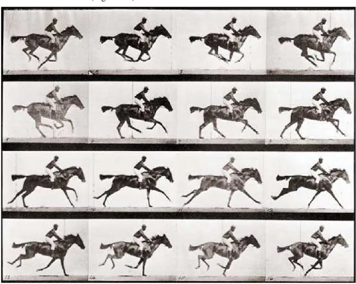
English photographer Edward Muybridge made use of the idea of Stanford’s with a series of cameras whose shutters were triggered by running horses, which was the beginning of his studies on biolocomotion. The most important contribution of Muybridge was the 781 plates he created, each having 1, 2 or 3 dozens of serial shots for a total of 20,000 images. These were published with the title “Animal Locomotion” (Figure 1-2).
Using photography for the biolocomotion studies also inspired Marey and he envisioned a new device in order to obtain multiple images of moving objects at equal time intervals on a single photographic plate. This device - “fusil photographique” – was the first device of photographic apparatuses he invented for the study of locomotion. The invention of the process was named as “chronophotographie” by him (Figure 1-3).
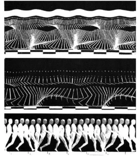
Fig. 1-3. Serial images of a man walking acquired on a still plate by Marey with his “chronophotograph”. The model
wears a black suit with white stripes and radiant points to indicate the position of one arm, one leg and the joints (from Musée Marey, Beaune).
Another French scientist, Jules Jannsen, had also contributed to his field with the photographic device he invented. This device – “revolver astronomique” - was able to record, on a single, circular-shaped, photosensitive plate having a rotating motion with regular intermittence, up to 48 consecutive images, spaced by constant time intervals, of an object in motion. Jannsen applied this device to the telescope and obtained the permanent recording of the transit of the planet Venus across the sun on December 8, 1874.
On October 29, 1888 Marey presented the device “chambre chronophotographique” which is claimed to contain all the principal components of the modern cine-camera. In 1893, Marey
constructed his projector, which was an important inspiration for the improvement of the cinematographic technique.
When the history of motion picture is analyzed, it can be realized that this invention cannot be attributed to a single person; it is collective work of optical, chemical and mechanical studies. The integration of these interdisciplinary contributions by the science of physiology was a great catalyst for the improvements in motion picture. Applying the graphic method to the study of biolocomotion was the key point in this contribution.
The growth of cinematography also contributed to physiology and introduction of cinematography can be considered as the “Renaissance” of biomechanics. Marey’s publications have contributed to the knowledge of motor phenomena, but the results were largely qualitative. In 1895, two physiologists, Wilhelm Braune and Otto Fischer, started publishing their works on human locomotion which were very important for the science of biomechanics. Besides being able to estimate the locomotor act, they were the first to present a three-dimensional analysis of human movement by using stereophotogrammetry. They were also the first measure the forces acting on the human body.
In the years between 1927 and 1936, Nikolai Bernstein improved the work of Braune and Fischer by increasing the shutter frequency from 26 to 70-100 and in some cases 120-156 frames per second. This improvement enabled the details of the human movement to be observed. The analysis of coordinated movements became the study of biomechanics which involve kinematics and dynamics.
Research handled at the University of California at Berkeley between 1945 and 1947 on both normal and pathological human locomotion was also a great contribution to biomechanics. Electromyographic apparatus and the force platform were begun to be used in biomechanics laboratories.
In the mid-1960s many specialized biomechanics laboratories were founded both in Europe and in the United States of America. Bioengineering Unit of Strathclyde University in Glasgow, Scotland and Institute of Human Physiology of the University of Milan, Italy were among the most important of these institutions.
Starting in the 1970s, the optoelectronic technique began to be used which resulted in easier movement recording and faster data reduction. The data can easily elaborated by digital computer which enables the analyses to be performed conveniently and fast.
CHAPTER 2
2.1
Marker-Based Human Movement Analysis
2.1.1 Theoretical Background
“Acquisition of quantitative information about the mechanics of the musculo-skeletal system during the execution of a motor task is the main goal of the human motion analysis” (Cappozzo et al., 2005). In order to pursue this goal, motion capture is frequently used in biomechanics. Human motion capture is widely used in order to study musculoskeletal biomechanics and clinical problems. In this context, estimating joint kinematics is of extreme relevance. For this purpose, video-based optoelectronic systems are commonly preferred among the human motion capture systems.
Gait analysis is generally carried out by mounting retro-reflective markers on the body of the subjects and reconstructing their 3D position using video-based optoelectronic systems (Figure 2-1). Retro-reflective markers and infrared illumination - produced by light-emitting diodes (LEDs) around the lens of the cameras - are used for the 3D reconstruction. By adjusting the camera thresholds, reflective markers are sampled and the recognition of the markers in the video frames is performed.
The 3D position of a marker in a reference frame fixed to the laboratory (global frame - GF) can be reconstructed if the marker is visible from at least two cameras at the same time. Visibility from additional cameras is usually beneficial (Chiari et al., 2005). Additional reference frames associated to body segments (technical frame - TF) can be defined from the position in the GF of cluster of markers attached to the same body segments.
The pose of the TFs in the GF can then be determined. However, although considered fixed to the underlying bone, the TFs are not necessarily representative of the anatomy of the body segment they are attached to. For this reason for each body segment being analyzed an additional frame is defined: the anatomical frame (AF). The AF has a time invariant relationship with the respective TF. To define AFs, it is necessary to determine the location of selected anatomical landmarks (ALs) with respect to the relevant TF (Cappozzo, 1995). Standards for the definitions of AFs have been proposed (Wu et al., 2002; Wu et al., 2005).
The pose of an AF is the orientation and position in space of a body segment. Given the pose of the AFs of two adjacent body segments, the kinematics of the joint between the two body segments can be determined.
Fig. 2-1. The human movement analysis laboratory with basic measurement instruments, with their systems of axes (p:
photogrammetry; d: dynamometry). When level walking is analysed, the motor task frame may overlap with the frame of one of the two force plates (from Cappozzo et al., 2005).
2.1.2 Calibration of Anatomical Landmarks
ALs are either bony prominences or bone points of geometrical relevance. In the first case they are normally identified by palpation, in the second case, they can be identified using imaging, regression equations or functional movements (Cappozzo et al., 2005). In any case, once identified, their location with respect to the relevant TF has to be determined. Once the location of ALs in their relevant TF is determined, it is possible to reconstruct their position in the GF by simple coordinate transformations (Figure 2-2). The Calibrated Anatomical System Technique (CAST) is an experimental methodology that formalizes the concept of AL calibration and allows the implementation of various calibration methods.
The AL calibration can be implemented using a) a marker positioned on the AL during a static acquisition, b) a pointer where a minimum of two markers are mounted with a known distance from its tip, pointing at the AL during a static acquisition, c) determining the centre of rotation of recorded functional movements (for joint centres, such as the hip centre), d) by imaging of the bone and the relevant TF (Cappozzo et al., 1995; Benedetti et al., 1998).
Recently, the CAST methodology was updated, by adding information on the subject-specific bone geometry. By determining the position of unlabelled points (UPs) situated over all prominent
parts of the bone surface, initial estimation is employed. After the estimation step, a digital model of a template-bone is matched to them. The estimated subject-specific bone contains all relevant anatomical landmark locations. The technique, UP-CAST, is evaluated in terms of repeatability and accuracy on average weight subjects (Donati et al., 2007).
Fig. 2-2. Anatomical calibration using stereophotogrammetry. The following external, palpable, anatomical landmarks
are indicated: prominence of the greater trochanter external surface (GT), medial epicondyle (ME), lateral epicondyle (LE). The location of the external anatomical landmarks relative to the marker cluster technical frame (xc, yc, zc) may be reconstructed using markers denoting the anatomical landmarks, or using a wand which carries a cluster of at least two markers. Prior to recording, the end point of the wand, the position of which relative to the latter cluster of markers is accurately known, is made to coincide with the target anatomical landmark (from Cappozzo et al., 2005).
2.1.3 Protocols
Human movement analysis and gait analysis in particular, typically makes use of the theory of multi- rigid body systems. The portion of human body is modelled with a number of rigid segments. Adjacent segments are connected by joints. The number of degrees of freedom of the each modelled joint characterizes the multi-body system model employed. Protocols – data collection and reduction practices – have been proposed in gait analysis offering various ways of modelling the system of rigid bodies of interest. Often, in clinical gait analysis, all model joints are rotational (either cylindrical or spherical) and AFs are defined based also on this assumption. The rationale behind this choice is related to the errors affecting the human movement recordings (see following section).
Proposed protocols also differ in the marker-sets used to identify AFs and joint centre locations. Typically data acquired with different protocols cannot be compared.
From these protocols, “Newington model” is the pioneer and the most commonly used practice for gait data acquisition and reduction which has also been used by the commercial applications like Plug-in Gait (PiG—Vicon Motion Systems, Oxford, UK). “Servizio di Analisi della Funzioni Locomotoria” developed their protocol named “SAFLo” - which differs from the Newington model in terms of segmental anatomical references and anatomical marker configurations. Then, “Calibration Anatomical System Technique” (CAST) was introduced which standardizes and defines references, internal anatomical landmarks and external technical markers. Then, protocols of “Laboratorio per l’Analisi del Movimento nel Bambino” (LAMB) and “Istituiti Ortopedici Rizzoli Gait” were proposed, of which the latter was the basis of the software “Total 3D Gait” (T3Dg-Aurion s.r.l, Milan, Italy) (Ferrari et al., 2008; Baker, 2006).
Ferrari et al. compared these commonly used protocols and find out that same gait cycles revealed good intra-protocol repeatability. Regardless of the known significant differences among the techniques, reasonable correlations are observed for most of the gait variables. It was pointed out that model conventions and definitions seem to be more important than the design of the relevant marker-sets. Sharing the model conventions and definitions can be sufficient for worldwide clinical gait analysis data comparison (Ferrari et al., 2008).
2.1.4 Sources of errors
Human movement analysis performed with stereo-photogrammetry is affected by three major sources of errors.
- Instrumental errors: these errors are the results of both instrumental noise and volume calibration inaccuracies. These errors have been studied intensively in the 80s and 90s (Fioretti and Jetto, 1989; Chen et al., 1994), tests for estimating them have been proposed (Della Croce and Cappozzo, 2000). The instrumental noise can be substantially reduced by low pass filtering. The volume calibration inaccuracies stem from the inadequate number of cameras and the volume calibration algorithm chosen for the application. Direct linear transformation (DLT) algorithm (Abdel-Aziz and Karara, 1971) is broadly used, but when the volume of interest is large, the construction of a suitable calibration object to be used with DLT becomes restrictive. Simultaneous multi-frame analytical calibration (SMAC) (Woltring, 1980) - a technique based on a planar calibration object with a grid of known control points - suffices the recording of the calibration object by at least two convergent cameras. SMAC allows covering larger volumes when compared with DLT, but for very large volumes, analytical self-calibration is more appropriate (Chiari et al.,
2005). Thus, the volume calibration inaccuracies can be remarkably lowered by increasing the number of cameras and improving the volume calibration algorithms.
The contribution of the instrumental errors to the total error is currently considered negligible. - Soft tissues artefacts: the markers captured by the cameras can be directly attached to the skin or arranged in clusters and positioned with fixtures over a body segment. Due to its origin, this error has the same frequency content as the bone movement. Thus, there is no way of separating the artefact from the actual bone movement by simply using a filter, as opposed to most instrumental errors. However, its effect on the end results may be reduced in the following ways. First of all marker locations (marker points) must be chosen so that the above-mentioned relative displacement is minimal, and secondly through a proper choice of the mathematical operator which estimates position and orientation of the bone from skin marker positions (Lucchetti et al., 1998; Alexander and Andriacchi, 2001). Operators that cope with this problem in an optimization context have been proposed” and their use in movement analysis is being developed.
Knowledge regarding the characteristics of the artefact movement in different body segments is required in order to utilize the mentioned countermeasures against the experimental artefacts.
- Anatomical landmark misplacement: The incorrect location of subcutaneous bony ALs through palpation can be caused by three main factors: (1) the palpable ALs are not points but surfaces, sometimes large and irregular; (2) a soft tissue layer of variable thickness and composition covers the ALs; (3) the identification of the location of the ALs depends on which palpation procedure was used. Studies showed that AL position uncertainty and consequently the erroneous determination of AF axes may result in erroneous clinical interpretations of the estimation (Della Croce et al., 2005).
In addition to the abovementioned sources of errors, marker based movement analysis is affected by the influence of markers attached to the body on the subject’s movement and the need of an extended setup time for marker placement (Corazza et al., 2006).
2.2
Markerless Human Movement Analysis
Some of the limitations of marker based systems can be overcome using a completely different approach. Markerless systems of human motion capture have been proposed where cameras can be utilized without the necessity of using special clothing or hardware (Deutscher et al., 2000). Markerless motion capture ensures an important reduction of the amount of time for setup preparation in comparison to marker-based techniques. Besides, the problem of inter-operator
variability is removed because no specialized operator is needed to place markers on the skin (Corazza et al., 2006).
Markerless techniques can be classified into model-based and model-free techniques both for the cases of 2D and 3D applications. Model-based approaches utilize an a priori human body model and are composed of two stages: modelling and estimation.
Modelling is the building step of a likelihood function by taking camera model, image descriptors, body model, and matching function into consideration. Estimation step is fitting the optimum pose in the likelihood domain planned in the modelling step. Model-free approaches do not use an a priori model, but “implicit model variations in pose configuration, body shape, camera viewpoint and appearance” (Poppe, 2007).
In the next section, current markerless techniques, brief algorithm explanations and their limitations are analyzed and exemplified with current applications.
2.2.1 2D Markerless Techniques
Much of the work on motion analysis uses detailed 3D kinematic models and 3D motion estimation. These techniques require multiple camera viewpoints, but motion analysis can also be operated using a single camera input (Cham and Rehg, 1999).
Motion capture with a single camera is a significant task since data acquisition is very simple, besides being an interesting computer vision challenge that focuses on inference as much as movement (Howe et al., 2000).
Hence, 2D markerless techniques, which can be classified as model-based and model-free, have been proposed. In the following section, the algorithms and the applications of these techniques are introduced.
2.2.1.1 Model-Based Techniques
Ju et al. (Ju et al., 1996) uses a cardboard model to define the human body as a set of connected planar patches and to approximate the limbs as planar regions (Figure 2-3). The main assumption behind this model is that, the motions of the limb planes are assumed to be the same at the points of articulation. The motion of each patch is estimated using the energy minimization (annealing) concept and estimated motions are called “absolute motions”. After the estimation of the absolute motions, it is necessary to estimate the articulated motions. To estimate the articulated motions, the motions of limbs which are relative to their preceding (parent) patches should be recovered. The relative motion of the patch is calculated using the displacements of the connected patches.
The estimated articulated motion between two frames is used in the tracking step in order to predict the location of each patch in the next frame. In the first frame, each patch is manually defined by its four corners. For every patch, the first two corners are defined as the articulated points, whose corresponding points are the last two corners of its previous patch. This shows that two connected patches share an “edge”. After the “chain” structure definition step is over, automatic object tracking starts. The articulated motion between two frames is used to predict the location of each patch in the next frame. Then, the location of each of the four corners of each patch is updated by applying its estimated planar motion to it.
The experiments demonstrate that the image motion models are able to track motion correctly during long sequences. In this study, optical flow is estimated with the parameterized model, 3D model is not necessary and edges are not used.
In the study of Deutscher et al. (Deutscher et al., 2000), the idea of annealing is adapted to perform a particle based stochastic search. The adapted algorithm is called annealed particle filtering and is capable of recovering full articulated body motion. The authors focused on the problem of constraining the search space along which the real posture of the subject is investigated. Other studies (Hogg, 1983; Goncalves et al., 1995; Bregler and Malik, 1998) in the literature do this making the following assumptions: 1) assuming that the subject is walking, 2) assuming a constant angle of view, 3) performing a hierarchical search using color cues. The study of Deutscher et al. does not depend on these assumptions and reduces the dimensionality of the search space through annealed particle filtering (Condensation algorithm).
This algorithm includes an edge detection procedure followed by a particle filtering, developed to match a 3D human body model of 29 DOF to the 2D image edge (Figure 2-4). However, this matching process has a very high computational cost. Also, a number of particles are required to the posterior density representations, which increase with the size of the model’s configuration space. In order to solve these problems, a multi-layer method using simulated annealing approach was implemented. Even if knee and ankle joints are modelled as simple hinge joints, the algorithm performance is in general satisfactory. The tracking performance of the algorithm was compared to the standard Condensation algorithm (i.e. Particle filtering algorithm which is used for tracking objects in clutter (Isard and Blake, 1996)) and resulted to perform better even if it uses fewer particles.
2.2.1.2 Model-Free Techniques
When there is not a priori human body model, there has to be a mapping between the image output and pose. Model-free algorithms, which do not suffer from (re)initialization problems, can be used for initialization of model-based pose estimation approaches (Poppe, 2007).
Fig. 2-4: The model is based on a kinematic chain consisting of 17 segments (a). Six degrees of freedom are given to
base translation and rotation. The shoulder and hip joints are treated as sockets with 3 degrees of freedom, the clavicle joints are given 2 degrees of freedom and the remaining joints are modelled as hinges requiring only one. This results in a model with 29 degrees of freedom. The model is fleshed out by conical sections (b) (from Deutscher et al., 2000).
Mori and Malik (Mori and Malik, 2006) estimate body pose in 3D by placing the joint points in a single 2D image with a human figure. First, a number of example views of the human body in different viewpoints with respect to the camera, are acquired. Each of the views are manually marked from the body joints and labelled. Then, the input figure is matched to each stored view using the shape context matching with a kinematic chain-based deformation model. By extracting
external and internal contours of an object, shape contexts are employed to encode the edges (Figure 2-5).
Fig. 2-5. The deformation model. (a) Underlying kinematic chain. (b) Automatic assignment of sample points to
kinematic chain segments on an exemplar. Each different symbol denotes a different chain segment. (c) Sample points deformed using the kinematic chain (from Mori and Malik, 2006).
Following the correspondence step, the locations of the body joints are then moved from the example views to the test figure. The 3D body configuration and pose are then estimated using the existing algorithm of Taylor with the 2D joint locations (Taylor, 2000) which uses point correspondence in a single image. In an estimation step, the stored example images are deformed to match the image observation. The 2D joint estimate is found by enforcing 2D image distance consistency between body parts. This technique can be applied to each frame of a video sequence so that tracking recognition becomes repeated for every frame. The experiments of this study are performed with CMU MoBo Database (Gross and Shi, 2001) and the main contribution of this study was demonstrating the use of deformable template matching to example views in order to localize human body joint positions.
In another 2D model-free markerless application, Elgammal and Lee (Elgammal and Lee, 2004) use human silhouettes extracted from a single camera to derive 3D poses. The purpose of this study is to recover the intrinsic body configuration and reconstruct the silhouette excluding the outliers from the visual input. To recover intrinsic body configurations from the silhouette, manifolds are learned from the visual input and afterwards mappings are learned from manifolds to visual input and 3D poses (Figure 2-6).
The experiments demonstrate that the model can be learned from the data of one person and successfully adapt to recovering poses for other people from noisy data. When compared to previous approaches for inferring 3D body pose from visual input, this approach has certain advantages and limitations. This framework makes interpolation of intermediate 3D poses easy even if they are not part of the training data. This approach constrains the mapping to the learned manifold which facilitates robust pose recovery from noisy inputs as well as for reconstruction of
the input. It is based on learning activity manifold and so its application is limited to recovery of poses for the learned activities only. Although in this study the focus is gait, the framework is general and can be applied to other activities by learning their manifolds. In the experiments, validation was done with a sequence obtained from the Georgia Institute of Technology (Atlanta, GA, USA) data compared to relevant motion capture data. CMU Mobo Gait database was used to demonstrate that the proposed approach, which is based on “learning” from the data of a single person, is also applicable to different people. CMU Mobo Gait database contains six views of each walking person. Five views from one person were used for the learning process of the study. In order to evaluate the 3D reconstruction, five sequences (five people with five views, each) were used. Overall correct classification rate from a single frame was 93.05%, while it increased to 99.63% after five frames were used for the classification.
Fig. 2-6. Embedded gait manifold for a side view of the walker. Sample frames from a walking cycle along the
manifold with the frame numbers shown to indicate the order. Ten walking cycles are shown (from Elgammal and Lee, 2004).
In another markerless 2D study, Goffredo et al. (Goffredo et al., 2009) use a region of interest (ROI) based tracking approach for the kinematic analysis of sit-to-stand (STS) tasks. Their approach uses Gauss–Laguerre transform (GLT) since image features such as edges, lines and orthogonal crosses are enhanced easily regardless of their orientations. A 4-segment human body
model was applied (only for the purpose of pose reconstruction of the body segments); each segment was assumed to be rigidly connected the next segment with an ideal rotational joint. Four body markers on ankle, knee, hip and shoulder joints were selected in the initial frame of every sequence and the GLT algorithm was applied to these ROIs. By computing the corresponding candidate points via calculating the log-likelihood function between the textures of consecutive frames, points relevant to motion estimation were tracked (Figure 2-7). The authors used GLT-based motion estimation method in a 2D setting, but this method could be used in stereo vision applications where GLT coefficient matching can be applied for motion and disparity field estimations. Estimating both the translations and the rotations of related anatomical segments in the transform domain with a pattern algorithm appeared to be a good solution for the movement reconstruction. For validation purposes, marker-based results from the study of Gross et al. (Gross et al., 1998) were used. The results of the validation are reported in Figure 2-8.
Fig. 2-7. Estimated trajectories obtained with the proposed method (upper panel: elderly subject I; lower panel: young
subject D). The circles are the points of interest on which the GLT algorithm has been applied. The lines are the estimated trajectories at the end of the phases characterizing the STS task (from Goffredo et al., 2009).
2.2.2 3D Markerless Techniques
When the observation is limited to a single camera, the 3D motion of humans is not determined thoroughly, due to the inherent 3D ambiguity of 2D video (Howe et al., 2000). In order to overcome this ambiguity, 3D markerless techniques are proposed.
Fig. 2-8. Hip, knee, ankle, and γ (shin orientation) angles at liftoff during chair rise. The results obtained with the
proposed markerless system (gray bars) are compared with the one obtained by Gross et al. (black bars) with a marker-based system (from Goffredo et al., 2009).
2.2.2.1 Model-Based Techniques
An important study exemplifying 3D model-based markerless technique is the study of Bottino and Laurentini (Bottino and Laurentini, 2001). Bottino and Laurentini presented a technique to reconstruct unconstrained motion from multiple-view images, which were computed using volume intersection data. First, views of the human body were acquired using different cameras and their 2D silhouette was extracted from each of the images. Then, a volumetric description was formed by intersecting the cones derived by back-projecting from each viewpoint of the corresponding silhouette. This step, called the volume intersection (VI), provided the final voxel representation. Then, a human body model was fitted to the extracted volume (Figure 2-9). Model fitting was done with the minimization of a distance function between the volume and the model via a search through the space of pose parameters. Pose recovery was based on a search through the 32 dimensional space of pose parameters and entailed finding the pose of the model closest to the actual appearance of the moving subject. The approximation accuracy was measured by a similarity function between the current model pose and the volume obtained by VI. This function was obtained by summing the squared distance between each voxel center to the closest segment of the model. Each segment was approximated with an oriented bounding ellipsoid (OBE) at the first step of the reconstruction algorithm in order to reduce the number of computations. The size of the axes of each OBE was the same as the dimensions of the boundary box of the related segment. The posture recovery was a two-step process: first, the OBEs were fitted to the volume reconstructed and then, fitting was employed to the model. In order to recover the motion of the model, the procedure mentioned above was applied to every frame, followed by the implicit filtering to avoid the phantom volumes.
Fig. 2-9. Human body model used in the study of Bottino and Laurentini (from Bottino and Laurentini, 2001).
The experimental setup is composed of two phases. First, the system was tested in a virtual environment in order to investigate the precision of 3D direct reconstruction. Second, the proposed approach was applied to real image sequences (Figure 2-10). Results showed that the proposed approach could reconstruct unconstrained human motion without using markers or external devices attached to the subject’s body.
Fig. 2-10. Original images, reconstructed voxel models, and parameterized shape models for a bow sequence (from
Corazza et al. (Corazza et al., 2006) also used the annealing approach in order to implement a markerless technique making use of visual hull reconstruction. The full body model contained morphological information (surface with 1600 points) and kinematics information on the possible movements of the model. The morphological information was provided by a laser scan of a reference pose of the subject. The model was segmented into parts corresponding to twelve anatomical segments (pelvis, thighs, shanks, feet, arms, forearms, and combined torso and head). The full body kinematic model had 33 degrees of freedom (DOF) where joints were modelled as ball-and-socket joints or as hinge joints. The geometrical formulation of the model was flexible such that each joint model could be modified separately without readjusting the others. The completed model was formed by rigidly combining the morphological representations to the kinematic model. (Figure 2-11). Visual hull was reconstructed, and matched to the model with an adapted fast simulated annealing approach. The validation of the technique was done in a virtual environment where an animated virtual character with known kinematics, provided the gold standard. The validation results are reported in Table 2-1.
Fig. 2-11. Results of the matching algorithm (colored points) applied to the virtual environment sequence superimposed
over the virtual character (gray surface) (from Corazza et al., 2006).
Mean Error (º) Standard Deviation (º) RMS Error (º)
Hip flexion/extension 2.0 3.0 3.6 Hip adduction/abduction 1.1 1.7 2.0 Knee flexion/extension 1.5 3.9 4.2 Knee adduction/abduction 2.0 2.3 3.1 Ankle plantar/dorsiflexion 3.5 8.2 9.0 Ankle inversion/eversion 4.7 2.8 5.9 Shoulder flexion/extension 1.2 4.2 4.4 Shoulder adduction/abduction 3.8 1.2 4.0
Another application to 3D model-based application is proposed by Bregler and Malik (Bregler and Malik, 1998). Bregler and Malik presented a motion estimation technique capable to extract high degree-of-freedom articulated human body configurations from complex video sequences using exponential maps and twist motions. The product of exponential maps and twist motions and their integration into differential motion estimation is a significant parameterization. The pose of each body segment was defined with respect to its “parent” segment to which is attached through a revolute joint. Moreover, the formulation is a very simple linear representation of the motion model. The visual tracking was based on an initial frame, in which the angular configuration was known beforehand. The 2D joint locations in all views were manually marked by a user. The 3D poses and the image projection of the matching configuration was found by minimizing the sum of squared differences between the joint locations of the projected model and of the marked model. The study provided a new technique for articulated visual motion tracking. The tracking results were qualitatively tested on video recordings of moving subjects, and on the Muybridge photographic sequences (Figure 2-12), but no quantitative information on the tracking parameters was given.
In the studies of Cheung et al. (Cheung et al., 2005 (Part I and Part II)) voxel based surface from silhouette algorithms combined with a new colour based approach, were used. Colored Surface Points (CSP) – multi-view stereo points – were extracted from the surface of the object and used in a 3D alignment algorithm, rigid motion between visual hulls determined and recursively refined and the silhouette images were used to refine the object’s shape. Figure 2-13 shows the CSPs on the visual hull of the subject.
Fig. 2-12. Muybridge’s Woman Walking: Motion Capture outputs. This shows the tracked angular configurations and
A sample of shape reconstruction and digital model rendering are reported in Figure 2-14. Tracking algorithm is evaluated with ground-truth data, obtained from synthetic sequences generated with OpenGL. Figure 2-15 shows the results of this validation. The use of the method requires a controlled environment since the algorithm is based on color information.
Fig. 2-13. The Shape-From-Silhouette problem scenario: a head shaped object O is surrounded by four cameras at time
t1. The silhouette images and camera centres are represented by Skj and Ck respectively (from Cheung et al., 2005).
Fig. 2-14. Articulated model of (a) synthetic virtual person, (b) Subject E, (c) Subject G and (d) Subject S. In (a) and
(b), the CSPs are shown with their original colours. In (c) and (d), the CSPs of different body parts are shown with different colours. For display clarity, the CSPs drawn are down-sampled in the ratio of one in two in total number of points (from Cheung et al., 2005).
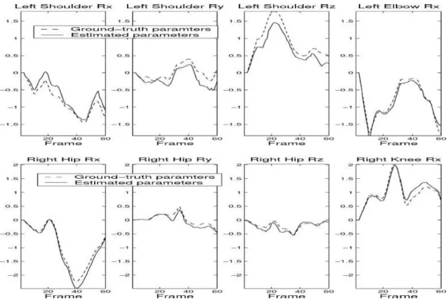
Fig. 2-15. Graphs comparing ground-truth and estimated joint angles of the left arm and right leg of the synthetic
sequence. The estimated joint angles closely follow the ground-truth values throughout the whole sequence (from Cheung et al., 2005).
Sundaresan and Chellappa (Sundaresan and Chellappa, 2006) modelled the human body with a set of articulated super-quadrics and proposed algorithms in order to estimate the model parameters from video sequences. The model was a combination of different body segments and some labelled joints. Each body segment was modelled with a tapered super-quadric (i.e. 3D parametric objects which give the flexibility of defining a large variety of shapes in geometric modelling (Barr, 1981)). The trunk segment was the base, and together with neck, head and four limbs formed the kinematic chain of the human body. Figure 2-16 shows the 3D body model used in the study. A 3D scanned model was used to obtain the dimensions of the super-quadrics. The trunk segment had 6 DOF while the other segments had at most 3 rotational DOF with respect to the trunk. The body model involved the shape and the joint locations of the body segments. Given the pose at time t , the pose at time t+1 was computed by using the images at time t and t+1. The pose estimation required the prediction step and the correction step. Pixel displacement estimation, pose prediction and silhouette-motion combination were necessary to estimate the pose at t+1. The authors claimed the method to be accurate and robust using a visual feedback. Accuracy of the method is strongly dependent on the quality of the estimation of joint location during the model acquisition. The flexibility of the model on some joints (e.g. shoulder joint) affects the performance of the method.
Fig. 2-16. Tracking results using both motion and spatial cues (from Sundaresan and Chellappa, 2006).
Gagalowicz and Quah (Gagalowicz and Quah, 2009), proposed a novel 3D model-based framework and algorithm that can manage clutters and occlusions, is proposed. This method uses a 3D geometrical human model similar to the subject, in order to synthesize the candidate posture producing the image minimizing the matching error with the real image. In this approach, segmentation is performed through the direct projection, texturing and shading via the 3D geometrical human model onto the images (Figure 2-17). The use of analysis-by-synthesis and error feedback allows avoiding the ill-posed problem of standard segmentation. Results of tracking the arms in the presence of occlusions and clutters were presented.
2.2.2.2 Model-Free Techniques
An example study in the 3D model-free markerless application is the study presented by Chu et al. (Chu et al., 2003). They proposed an approach that generates “underlying nonlinear axes” (or skeleton curve) from a volume of a human subject. Multiple cameras were used for human volume capture and skeleton curves estimated the kinematic model and posture for each volume. Skeleton curves were used to automatically produce kinematic motion. Isomap transformation was used in order to map a set of 3D points describing a human body volume into a “pose-invariant intrinsic space posture”. This transformation allowed finding a correspondence between volume points in both Euclidean and intrinsic spaces. By building principal curves in intrinsic space and mapping back to the volume feature produces a skeleton curve. An a priori body model is not used (Figure 2-18). This is a fast technique to be applied to image sequences and manages to define the posture without the help of an a priori model. However, it is not known if the technique gives position and orientations of body segments accurately enough for clinical purposes since no validation is presented. The technique can be used as the initialization step of the marker-based techniques.
Fig. 2-18. The outline of the approach. (1) A human viewed in multiple cameras is used to build (2) a Euclidean space
point volume of the human. This volume is transformed into (3) an intrinsic space pose invariant volume and its (4) principle curves are found. The principal curves are projected back into Euclidean space to provide (5) a skeleton curve. The skeleton curve is used to determine (6) the posture of the human. Using the postures of a volume sequence, (7) the kinematic motion of the human is found and (8) actuated on the Adonis humanoid simulation (from Chu et al., 2003).
In another application, Grauman et al. (Grauman et al., 2003) presented an image-based approach to infer 3D structure parameters. By using a probability density of multi-view silhouette images with known 3D structure parameters, a probabilistic shape and structure model was created (Figure 2-19). This probabilistic model was merged with a model of the observation uncertainty of the silhouettes seen in each camera to compute Bayesian estimate of structure parameters. This was the first study where an image-based statistical shape model was used for the inference of 3D structure. Besides, by using a computer graphics model of articulated bodies, a database of views augmented with the known 3D feature locations were formed in order to learn the image-based models from known 3D shape models. This synthetic training set removed the necessity of labelled real data. The study’s novelty was the use of a probabilistic multi-view shape model to narrow the possible object shape and configurations to those that are more “probable” given the class of the object and the current observation.
Fig. 2-19. Top row shows noisy input silhouettes, middle row shows contour reconstructions, and bottom row shows
2.2.3 Goal-Oriented Classification of the Markerless Studies
In this review, the mentioned markerless studies were analyzed in terms of their shape representations, existence of models and application space. Yet it is also possible to make a classification based on the goal of these studies.
Markerless studies can be classified in terms of the goal of the applications: 1) studies that merely provide graphical representations of the human body, 2) studies that aim to estimate joint kinematics. The table below (Table 2-2) depicts this classification and provides an overview of the abovementioned markerless studies together with their validation information.
Graphical Representation Joint Kinematics Estimation Notes Validation Ju et al., 1996 - - -
Deutscher et al., 2000 Comparison with Standard
Condensation Algorithm
Mori and Malik, 2006 CMU MoBo database
Elgammal and Lee, 2004
Georgia Tech gait data with ground-truth
2D
Goffredo et al., 2009 Hip, knee and ankle angles are
estimated.
Comparison with the results of the marker-based study by Gross et al. (Gross et al., 1998)
Bottino and Laurentini, 2001
Corazza et al., 2006 Shoulder, hip, knee, ankle angles are estimated
Comparison with the ground-truth provided by virtual environment
Bregler and Malik, 2002
Qualitatively validated by the Muybridge sequence, but
quantitative information regarding the parameters are not presented. Cheung et al., 2005 Shoulder, hip, and
elbow angles are estimated
Synthetic sequences with ground-truth
Sundaresan and Chellappa, 2006 Gagalowicz and Quah, 2009
Chu et al., 2003
3D
Grauman et al., 2003
2.2.4 Limitations
Markerless techniques are quite promising in terms of proposing an alternative to marker-based techniques, easy setup and low-cost solution. However, the use of markerless techniques to capture human movement for biomechanical or clinical applications has been restricted by the complexity of acquiring accurate 3D kinematics. The general problem of estimating the free motion of the human body or more generally of an object without markers, from multiple camera views, is under-constrained when compared with marker-based systems.
Existing computer vision approaches focusing on markerless movement analysis may have great potential to be used in biomechanical applications, but most of them have not been validated for these applications. Evaluation of these approaches in terms of applicability to clinical applications is essential.
For the purpose of enhancing computational performance, simple or generic models of human body with fewer joints or reduced number of degrees of freedom are frequently used. Nevertheless, detailed and accurate representation of 3D joint mechanics is required in biomechanical and clinical applications.
Another challenge for the whole-body movement capture is the non-rigid nature of human body segments and the variability of human motion, the presence of self-occlusion or occluding objects (Mündermann et al., 2006). This diversity causes the some predefined parameters to be created or assumptions to be made, which restrict the analysis (Poppe, 2007; Bray, 2001).
To sum up, the field of markerless movement analysis is a promising and active research which will continue to evolve by considering the abovementioned challenges as a roadmap.
CHAPTER 3
Marker-based human movement analysis suffers from the instrumental errors, soft tissues artefacts and anatomical landmark misplacement. Besides, markers hinder with the subject’s movement and an extended setup time is required. To solve these limitations, markerless human movement analysis has been introduced. Even though the markerless techniques mentioned in the previous chapter are promising, they often lack validation and accurate representations, which are crucial for the clinical applications. Therefore, this thesis aims to provide new 2D markerless techniques to overcome the abovementioned difficulties of both systems.
In particular, the following issues are dealt with listed in order of relevance:
1) Development of two different markerless techniques to determine joint kinematics: Two different markerless techniques (cross-correlation-based and skeletonization-based) are implemented.
2) Validation of the proposed markerless techniques with the marker-based techniques: The proposed markerless techniques are validated with traditional stereophotogrammetric marker-based systems by recording the same trials at the same time.
3) Extraction of additional information from anthropometric measurements and garments used during the acquisition (high-cut underwear and ankle socks used as “segmental markers”) to be combined to the information extracted with a markerless methodology, implementing a hybrid technique applied to children with CP.
4) Analysis of the influence of the presence of socks on the performance of the markerless technique.
The present work was conducted at the Biomedical Sciences Department of University of Sassari, Sassari, Italy. The experiments of the study in Chapter 5 were done in University of Rome, “Foro Italico”, Rome, Italy while the experiments of the studies in Chapter 6 and 7 took place in the Motion Analysis Lab at Spaulding Rehabilitation Hospital, Boston, MA, USA.
CHAPTER 4
IMAGE PROCESSING: REVIEW OF THE ALGORITHMS
EMPLOYED
4.1
Image Segmentation
Vision is a deduction problem where the aim is to find out the source of the outputs from a given model and measurements. The main difficulties in the deduction problem of vision are the abundance of data and the ambiguity on whether a specific data item is a part of the deduction problem or not. To overcome these difficulties, image data is generally represented by grouping the features that highlight its main properties, as in segmentation. There are several algorithms for image segmentation depending on the application. In an application where there is a static background, removing an estimate of the background from the image would be functional as image segmentation. However, when the backgrounds change over time, this approach would not work fruitfully (Figure 4-1 and Figure 4-2) (Forsyth and Ponce, 2003).
Fig. 4-1. Subject with barefoot, short socks and long socks.
Fig. 4-2. Output of Fig. 4-1 after background subtraction.
Estimating the background using a moving average is a better solution compared to simple background subtraction. Instead of removing the static background, the value of the background is computed by calculating the weighted average of the previous values of the background pixels. Thus, the pixels from the initial frames have a weight of zero and the moving average adapts to the changes in the background. Even though this method can be useful for coarse scale
images, in the dynamic scenes the performance of the adaptation is low (Forsyth and Ponce, 2003).
The motion of the objects in 3D space causes motion on the image plane which can be computed by the displacements of the image points or displacement vectors of the entire image place, which are defined as “optical flow”. Even though optical flow is highly used on the images where there is motion, ambiguity stemming from 3D to 2D projection is still a major problem (De Micheli et al. 1993; Fermuller et al., 2001).
Many segmentation methods suffer from the changes in the lighting. Ridder et al. (Ridder et al., 1995) used Kalman Filter to model the pixels, which enabled the lighting changes to be handled. Even though this methods has a “pixel-wise automatic threshold”, recovering from the light changes is slow and not effective with bimodal backgrounds. Yet, this method has been successfully used in an automatic traffic monitoring application by Koller et al. (Koller et al., 1994). In another application, Pfinder (Wren et al., 1997), background is modelled by a single Gaussian per pixel and a multi-class statistical model is preferred for the object tracking. In the Pfinder application, the system works well after an initialization phase where the room is empty. However, the performance of the tracker in outdoor scenes is not reported.
Expectation-Maximization (EM) is highly used for image segmentation; Friedman and Russell (Friedman and Russell, 1997) used this method to develop a pixel-wise EM framework for vehicle detection. With this method, pixel values are classified into three separate distributions based on the road color, the shadow color and vehicle color. This system manages the effects of the shadows, but the behaviour of the system when these three distributions are not available, is not known. The performance of the method would be affected if there is a single background or a multiple colored background stemming from motion, shadow or reflectance.
Mixture of Gaussians (MoG) (Hu et al., 2004) is a commonly used method which computes the dynamic features from the image sequence and in this thesis, it has been used as the segmentation method. In this structure, the underlying principle is to describe each single pixel in the image statistically, through a set of Gaussian probability distributions. With this model, the variability of each pixel over time is characterized.
In the study of Stauffer and Grimson (Stauffer and Grimson, 2000), the value of a particular pixel is modelled as a mixture of Gaussians, instead of using a single Gaussian distribution. Gaussians that form the background are determined by calculating the variance of each of the Gaussians of the mixture. Pixels that do not fit to this estimated background are
grouped as foreground pixels. This approach manages “lighting changes, slow-moving objects and introducing or removing objects” successfully (Figure 4-3).
Fig. 4-3. The execution of the program. (a) the current image, (b) an image composed of the means of the most
probable Gaussians in the background model, (c) the foreground pixels, (d) the current image with tracking information (from Stauffer and Grimson, 1999).
In this approach, there are two important parameters –
α
, the learning constant and T, the percentage of the data that has be used for by the background. Every image pixel in the image sequence can be statistically described as a series of values changing over time:{
X1,K,Xt} {
= I(x0,y0,t):1≤t≤T}
where I(x,y,t) represents the intensity value of the pixel at position (x,y) and time t, in the image sequence. The latest changes of the intensities can be modelled as the mixture of K Gaussian probability density distributions:
)
(
∑
= ⋅∑
= K i it t it it t X X P 1 , , , , , ) (ω
η
µ
where K is the number of Gaussian distributions,
ωi,t
is the individual weight of each Gaussian at time t (the sum being equal to 1),µi,t
andΣ
i,t are the mean the covariance matrixassociated with the ith Gaussian at time t.
The general formula of the Gaussian distribution is:
(a) (b)