University of Messina
PhD School in Surgical and Medical Biotechnologies
XXIX
Cycle
Coordinator: Prof. G. Raimondo
Functional and molecular characterization of PbsP
(Plasminogen binding surface Protein)
of Streptococcus agalactiae
PhD student
Romeo Letizia
Supervisor
Prof. Concetta Beninati
Introduction
The plasminogen system in the pathogenesis of group B
streptococcal infections
Bacterial pathogens have evolved a wide range of strategies to adhere to cells and subsequently invade host tissues (Kline et al., 2009; Nobbs et al., 2009). In particular, many microorganisms, including both pathogens and commensals, are able to bind host plasminogen (Plg) on their surface, where its activation can be controlled by host or bacterial factors (Lahteenmaki et al.,
2001; Bergmann and Hammerschmidt, 2007; Fulde et al., 2013).
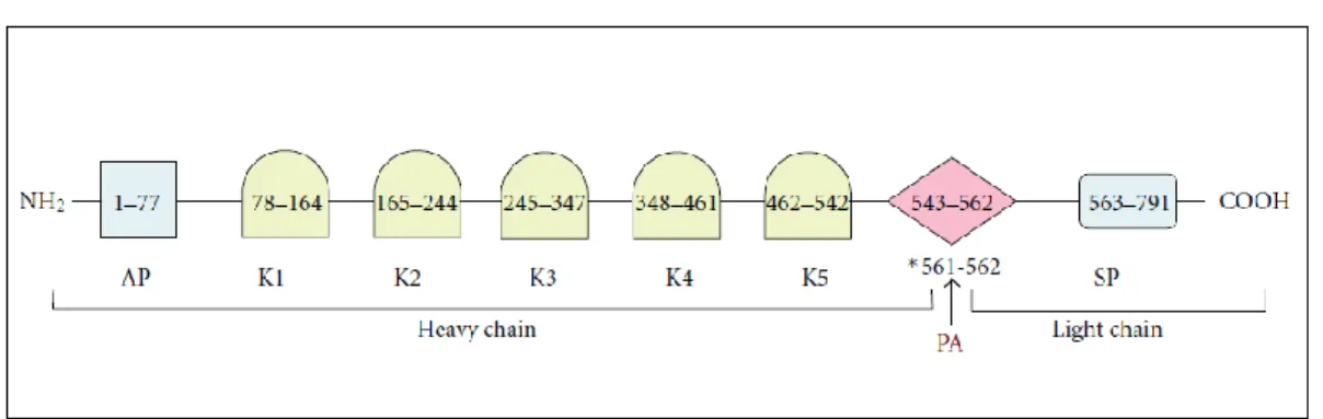
Human plasminogen (Plg) is a 92 kDa glycoprotein which is present in high concentrations within human plasma as precursor (a zymogen) of the serine protease plasmin (Pln) (F. J. Castellino 2005; Bergman et al., 2007;
Bhattacharya et al., 2012 ). The Plg molecule contains a total of seven structural
domains, each with different properties. The N-terminal portion of the molecule consists of an activation peptide (AP) followed by a series of 5 repeating homologous triple-disulfide-linked peptides of approximately 80
amino acids termed kringles (K1–K5) while the C-terminal portion contains the cleavage (activation) loop (CL) for plasminogen activator (PA) followed by a serine protease (SP) domain (F. J. Castellino 2005; Bhattacharya et al., 2012) (Figure 1). The function of the five kringles is primarily to mediate protein-protein interactions, such as those involving binding of Plg or Pln to fibrin, ECM targets, lysine-containing receptors/analogues and bacterial receptors (Suenson E et al., 1981; Miles LA et al., 1988; Urano T et al.,1987 M. L.
Sanderson-Smith et al .2008; S. Bhattacharya et al. 2012).
The activation of inactive Plg into its active form (Pln) is mediated by a proteolytic mechanism usually catalyzed by the host plasminogen activators tissue-type (tPA) and urokinase-type (uPA) Plg activators (Fan ZQ, et al., 1998;
Violand BNet al., 1976). In order to maintain tissue homeostasis and avoid
random tissue damage, Pln activity must be tightly controlled (O'Mullane MJet
al.,1998; O'Mullane MJet al.,1999). This regulation is achieved through the
expression of Plg receptors (PlgRs), Plg activators (PAs) and Pln inhibitors (E.
Figure 1. Schematic representation of the human Plg structure. Starting from the N- terminus of the mature protein, the 77-residue activation peptide (AP) is followed by 5 kringle domains (K1– K5) containing lysine binding sites and the catalytic SP domain. The R561-V562 amide bond, which is proteolytically cleaved by activators to generate Pln, is shown with an asterisk and an arrow. The heavy chain and light chains generated after proteolytic cleavage are also shown.
S. Bhattacharya et al., 2012
Bacteria have independently evolved systems to exploit the host Plg/Pln system to their advantage. They can interact with this system via three distinct mechanisms (Bergman et al., 2007): i) bacteria produce PAs, which activate Plg by complex formation or by proteolysis; ii) Plg is recruited to the bacterial cell surface by bacterial PlgRs and then converted into plasmin by host-derived activators; iii) bacteria bind to Plg on the surface of host cells to promote colonization or penetration of body barriers through adhesion to and/or invasion of mucosal or endothelial layers. Bacteria are able to bind to host Plg/Pln directly, through specific surface adhesins or conserved ubiquitous moonlighting proteins such as enolase or GAPDH, or indirectly, through fibrinogen receptors enabling the formation of fibrinogen-plasminogen
complexes (Figure 2). Multiple Plg/Pln/Fng receptors can be expressed at different steps of the colonization or invasion processes and can thus be simultaneously or sequentially involved in pathogenesis at specific stages of disease development. For example, Plg expressed on the surface of epithelial cells could be initially be used for mucosal colonization. Then, Pln bound to the bacterial surface could be exploited by bacteria for proteolytic degradation of components of tight junctions or basal membranes, facilitating bacterial translocation across cell barriers and dissemination within tissues. The relevance of Plg binding in streptococcal pathogenesis (particularly in the pathogenesis of infections by group A streptococci and pneumococci) is well documented (Bergmann et al., 2001, 2005; Sun et al., 2004; Sanderson-Smith et al.,
2008;Fulde et al., 2011; Siemens et al., 2011; Teles et al., 2012; Agarwal et al., 2013).
Figure 2. Mechanisms of bacterial cell surface plasmin(ogen) acquisition and its role in bacterial-host interactions. Plasmin(ogen) can be bound directly to the bacterial cell surface via cell-membrane-anchored receptors, nonanchored-cell-surface-associated receptors or indirectly through interactions with fibrinogen and cell surface fibrinogen receptors. Plasmin(ogen) localised on the bacterial cell surface is involved in four main processes; (1) ECM degradation via activated metalloproteases and plasmin; (2) fibrinolysis via plasmin; (3) immune evasion through plasmin-mediated degradation of immune effectors, including complement components and immunoglobulins; (4) adherence to host cells via plasminogen linker interactions with host cell surface receptors. ECM: extracellularmatrix; IgG: immunoglobulin G; RBC: red blood cell; SAK: staphylokinase; SEN: streptococcal α-enolase; Ska: streptokinase; tPA: tissue plasminogen activator; uPA; urokinase plasminogen activator. M.L. Sanderson-Smith 2012
Streptococcus agalactiae (also referred to as group B Streptococcus, GBS) is
a Gram-positive encapsulated commensal bacterium of the human gastrointestinal tract, which is also present in the genital tract of 15–30% of healthy women (Campbell et al., 2000; Edwards et al., 2006 Le Doare and Heath,
2013). In the last two decades it has emerged as one of the leading causes of
septicemia or meningitis (Trivalle et al., 1998; Schuchat et al., 1999; Edwards and
Baker, 2005). GBS is also a significant cause of morbidity and mortality in
nonpregnant adults, particularly those with underlying diseases such as diabetes, cirrhosis or malignancy (Farley et al., 1993). Neonatal diseases result from transmission of the organism from the pregnant mother to the neonate. Two distinct GBS-associated clinical syndromes, referred to as early-onset disease (EOD) and late-onset disease (LOD), have been recognized in neonates and characterized by onset of disease during their first 6 days of life or after (from day 7 up to 3 months of age), respectively (Edwards and Baker, 2005). For EOD, the mode of transmission in newborns is thought to be vertical, by inhalation of GBS-contaminated amniotic or vaginal fluid during parturition, followed by bacterial translocation across the respiratory epithelium and subsequent systemic infection (Edwards and Baker, 2005). In contrast, for LOD, the mode of transmission and the infection route remain elusive, although mother-to-child transmission might also be involved. A plausible scenario would involve early intestinal colonization by GBS with intraluminal multiplication, followed by translocation across the intestinal epithelium and
access to the bloodstream. Indeed, an intestinal portal of entry for LOD is supported by several lines of evidence: (a) 60 and 40% of the neonates asymptomatically colonized with GBS at birth remain positive for bacteria at the rectal level at 4 and 12 wk of life, respectively (Weindling et al., 1981); and (b) a longitudinal study of GBS vaginal and rectal colonization in women during and after pregnancy has revealed that carriers are usually colonized for up to 2 yr by a single clone, which is also frequently found in newborn feces for up to 1 yr (Hansen et al., 2004). Although intrapartum antibiotic prophylaxis for women at risk for GBS infection has markedly decreased the incidence of EOD, it did not change that of LOD (Poyart et al., 2008).
Epidemiological data have reported that serotype III is the most frequently identified GBS serotype causing invasive neonatal disease in different countries (Poyart et al., 2008; Edmond et al., 2012). Surprisingly, multi-locus sequence typing (MLST) of GBS isolated from different countries showed that most human carriage and clinical isolates cluster into only five major clonal complexes (CC) (CC1, CC10, CC17, CC19 and CC23) (Jones N, et
complexes. Among these, a substantial proportion of EOD and the majority of LODs are associated with ‘hypervirulent’ serotype III strains included in the clonal complex (CC) 17 (CC-17) (Musser et al., 1989; Lamy et al., 2006; Lin et al.,
2006; Phares et al., 2008; Poyart et al., 2008). These CC17 GBS strains are derived
from the same genetic pool (Teatero et al., 2016) and are more likely to cause neonatal meningitis than other genotypes (Jones et al., 2003; Bisharat et al., 2004;
Luan et al., 2005; Jones et al., 2006; Lin et al., 2006; Bohnsack et al., 2008; Manning et al., 2009).
Figure3: Multi-locus sequence typing (MLST) of GBS isolated from different countries; most human carriage and clinical isolates cluster into only five major clonal complexes (CC) (CC1, CC10, CC17, CC19 and CC23). Da Cunha et al., 2007
As mentioned before, GBS normally resides with its human host as an asymptomatic colonizer of the genital and gastrointestinal tracts. However ,in certain circumstances, it can turn into an invasive pathogen causing severe infections. The transition from a commensal to pathogenic lifestyle is related to adaptive regulated changes in expression of specific bacterial genes in response to diverse host environments. This dynamic adaptation of GBS to the human host involves mechanisms for sensing environmental changes. Bacteria respond to changes in environmental stimuli using signal transduction systems (STS), which regulate the expression and transcription of multiple genes whose products are potentially related to virulence or play a role in metabolic pathways. The most common STS in bacteria are the two-component signal transduction systems (TCS). The genome sequence of GBS has revealed the presence of 17–20 TCS that can respond to changes in the external environment (Poyart et al., 2001; Glaser et al., 2002; Spellerberg et al.,
2002; Tettelin et al., 2002; Tettelin et al., 2005; Quach et al., 2008; Faralla et al., 2014). The most studied TCS used by pathogenic streptococci to adapt to host
Jiang et al., 2005; Jiang et al., 2008). It consists of a membrane-associated
histidine kinase sensor (CovS) and a cytoplasmic transcriptional regulator (CovR). The CovRS system act as a global regulator possessing numerous target genes that belong to different functional categories, such as cell envelope, stress response, metabolism and transport proteins, which often function as virulence factors (Lamy et al., 2004).
Therefore, the ability to sense the surrounding host microenvironment can result in the expression of specific virulence factors that allow bacterial persistance, dissemination through the blood and colonization of distant host districts. Indeed, GBS relies on specific adhesins for harmless colonization or host invasion. Many of the GBS adhesins characterized thus far are cell-wall proteins, which are anchored to bacterial surface by a sortase A-dependent mechanism (Mazmanian et al., 1999; Nobbs et al., 2009) and are therefore accessible to specific receptors present on host cells or in the blood (Tazi et al.,
2010; Papaseri et al., 2011; Buscetta et al., 2014; Jiang and Wessels, 2014; Wang et al., 2014; Mu et al., 2014; Six et al., 2015). For example, GBS is capable to bind
tPA (or uPA), Plg is converted to Pln conferring to this pathogen surface proteolytic activity (Magalhaes et al., 2007). A recent study indicated that Pg activation on the surface of GBS occurs through cell surface moonlighting proteins, such as α-enolase, glyceraldehyde dehydrogenase and phosphoglycerate kinase that can bind Plg and Pln (Magalhaes et al., 2007;
Boone and Tyrrell, 2012; Oliveira et al., 2012). Moreover, GBS release
extracellularly a protein named Skizzle that can bind Plg and accelerate its conversion to Pln by host activators, although the actual role of this protein in the pathogenesis of GBS disease has not been studied (Wiles et al., 2010). In addition, different GBS strains also express unrelated fibrinogen receptors (FbsA, FbsB or FbsC) that might indirectly bind to Plg (Gutekunst et al., 2004;
Pietrocola et al., 2005; Buscetta et al., 2014). In the context of the hypervirulent
CC17 clonal complex, a multifunctional adhesin, the serine rich repeat (Srr2) glycoprotein, that is able to bind both Plg and Fbg can promote GBS invasiveness and the development of meningitis (Brochet et al., 2006; Da Cunha
The aim of the studies presented in this thesis was to identify novel virulence factors of GBS that might be involved in binding to host factors, particularly Plg. First, a novel protein (Gbs0428 or PbsP) was identified by proteomics analysis of the GBS exoproteome and found to display two SSURE domains (Bumbaca et al., 2004), homologous with those of the previously characterized Plasminogen and fibronectin binding protein B (PfbB, also called PavB) of Streptococcus pneumoniae (Papasergi et al., 2010). Next, the role of Gbs0428 in the contest of GBS pathogenesis was investigated. After showing that Gbs0428 is a cell-wall-anchored surface protein that binds Plg, it was found that this adhesin is expressed by isolates belonging to all of the main GBS lineages, including strains of the hypervirulent clonal complex 17 (CC17), which is responsible for a large number of neonatal GBS infections, particularly meningitis cases. In the context of the CC17 prototype BM110 strain, PbsP was found to be markedly upregulated in vivo. PbsP was also found to bind Plg largely through the interaction between lysine-binding sites in the kringle 4 domain of Plg with a novel protein domain that was named MK-rich region. Moreover, PbsP-mediated Plg binding conferred proteolytic
activity to GBS surface, which ensured GBS transmigration across endothelial cells and colonization of the brain. All together these data indicate that PbsP has an important role in the pathogenesis of GBS infections and might represent a target in alternative strategies to control GBS infection.
Materials and Methods
Bacterial strains and reagents
In this study we used the following reference GBS strains: NEM316 (serotype III, CC23), 6313 (serotype III, CC 23), BM110 (serotype III, CC17), COH1 (serotype III, CC17), A909 (serotype Ia, CC1) and 2603V/R (serotype V, CC19) (Glaser et al., 2002; Tettelin et al., 2005; Da Cunha et al., 2014). The relevant characteristics of the other bacterial strains and plasmids used in this study are summarized in Table 1. GBS were grown at 37°C in Todd-Hewitt (TH) broth (Difco Laboratories). Antibiotics (Sigma-Aldrich) were used at the following concentrations for Escherichia coli: ticarcillin,100 mg/ml; erythromycin, 150 mg/ml, kanamycin, 25 mg/ml; and for GBS: erythromycin, 10 mg/ml; kanamycin, 500 mg/ml. Human Fnt, Plg and C Reactive Protein (Calbiochem), Factor H, Factor I, Factor B, C1q and C3 (Complement Technologies) and Collagen (Sigma-Aldrich) were purchased and Fng was prepared as previously described (Pietrocola et al., 2005). Kringle 1-3 and Kringle 1-4 were purchased from Sigma and MyBioSource, respectively. The mini-PLG (residues Val442-Asn790) was obtained by digestion of PLG with porcine
pancreatic elastase (Sigma) as described previously (Christensen, U. and Mølgaard, L. 1992, Váli, Z. and Patthy, L. 1982). Aminocaproic acid (6-ACA), L-Lysine or L -Alanine, used as inhibitors, were purchased from Sigma-Aldrich. N-terminal Mk-rich (residues 423–443), Fr1-Cterminal MK-rich (residues 444–468), and Fr2-Cterminal MK-rich (residues 469–492) were purchased as biotinylated peptides from GenScript (Table4).
DNA manipulation and mutant construction
Purification of GBS genomic DNA and E. coli plasmid DNA were performed with the DNeasy Blood and Tissue kit and the Quiaprep Spin Minipreps kit (Qiagen) respectively. Oligonucleotides were provided by Eurofins MWG Operon or Sigma-Aldrich and are listed in Table 3. Analytical PCR used standard Taq polymerase (Invitrogen, Life Technologies). Preparative PCR for cloning and PCR for sequencing (GATC Biotech) were performed with a high-fidelity polymerase (MyFi or Phusion DNA polymerase, Bioline and Thermo-Scientific respectively). The pG1_DpbsP vector used for pbsP deletion was constructed as described (Firon et al., 2013) using a splicing by overlap extension method (Heckman and Pease, 2007) with
primers pAF375_EcoRI1pAF376_D0428 and pAF377_D04281pAF378_BamHI. After GBS transformation with pG1_DpbsP and selection of pG1_DpbsP integration and de-recombination events, marker-less deletion of pbsP was confirmed on genomic DNA with primers pAF3791380 (positive PCR product in case of pbsP deletion) and pAF3811382 (positive PCR product in case of a WT pbsP gene). Primers pAF553_BamHI1pAF554_PstI were used to amplify the full-lenght pbsP gene with its predicted Shine-Dalgarno sequence from NEM316 WT genomic DNA. The PCR product was cloned into the pTCV_Ptet vector at the corresponding restriction sites to give the constitutive pbsP expressing vector pTCV_Ptet_pbsP. The full length insert was sequenced to confirm the absence of mutations. The pTCV_Ptet_pbsP plasmid was introduced in GBS by electroporation and transformants were selected on TH agar supplemented with kanamycin.
Production of recombinant rPbsP and rPbsP domains
Recombinant PbsP (rPbsP) was produced as described (Garibaldi et al.,
2010; Papasergi et al., 2010; Buscetta et al., 2014). Briefly, the pbsP gene was
cloned into the pGEX-SN bacterial expression vector (Cardaci et al., 2012). The corresponding pGEX-SN_PbsP allows the expression of the rPbsP fused to a glutathione S-transferase (GST) tag at its amino-terminal end. To produce recombinant PbsP fragments, the corresponding gene regions were amplified using the following primers: MKrich423_up and MK-rich492_low (rMK-rich); SSURE123_up and SSURE422_low (rSSURE1-2); SSURE273_up and SSURE422_
low (rSSURE2) and Cterminal MK-rich444_up and MK-rich492_low
(rCterminal Mk-rich) (Table 3). Amplified fragments containing att recombination sequences were used to transform the pDEST15 vector by Gateway cloning according to the manufacturer’s instructions (Thermo Fisher Scientific). The resulting plasmids pDEST15_MK-rich, pDEST15_SSURE112, pDEST15_SSURE2 and pDEST15_Cterminal Mk-rich (Table2) were used to transform E. coli BL21 (DE3). After induction, the recombinant fusion proteins were purified from the cytoplasm of bacterial cells using affinity chromatography (Papasergi et al., 2010). Recombinant GST was produced and purified using the same method and used as a negative control. With the aim of speeding up the workflow, we purchased biotinylated peptides that
encompassed MK-rich region from GenScript company (Table 4). To have a negative control we used the PinPoint ™ Xa Control Vector (Promega) containing the CAT gene fused to the biotinylated protein tag. After induction, cells containing the PinPoint ™ Xa Control Vector produces a 40kDa protein composed by the CAT peptide (27kDa) and by a 13kDa biotinylated peptide. Biotinylated fusion protein is produced in E. coli and are affinity-purified using the SoftLink™ Soft Release Avidin Resin (Promega).
Production of anti-PbsP antisera
CD1 mice (5 weeks old, Charles River Labs) were injected intraperitoneally with 20 ug of rPbsP or GST (in a total volume of 0.2 ml) in complete (first injection, day 0) or incomplete (second and third injections on days 14 and 28 respectively) Freund’s adjuvant emulsions. The use of complete Freund’s adjuvant in the first immunization was justified by our previous observations that high-titer sera were more consistently obtained with this adjuvant. However, care was taken to minimize discomfort to the animals by injecting a low volume (0.1 ml, containing 0.05 mg of mycobacteria) of the oily component of the emulsion. Under these conditions,
no significant abdominal distension or other complications at the injection site were observed throughout the experimental period. The mice were bled at 2 weeks after the last immunization, and the sera were tested for reactivity to the purified antigen using ELISA and western blot assays.
Bacterial extracts and immunoblots
Cell wall extracts were prepared by digestion with mutanolysin (Sigma-Aldrich) of bacterial cells grown to the exponential phase in an osmo-protective buffer, as described (Lalioui et al., 2005; Garibaldi et al., 2010). A total of 30 ug of cell wall proteins (Bradford assay) were run on gels (SDS-PAGE), transferred to nitrocellulose membranes and hybridizations were performed with mouse anti-PbsP serum followed by horseradish peroxidase conjugated goat antimouse IgG (R&D Systems). Loading controls consisted of parallel Coomassie-stained gels. For Far western analyses, Plg (10 ug) was run on 12% acrylamide gels, transferred on nitrocellulose, and overlaid with 0.15 lM rPbsP or GST in 1% of nonfat dry milk supplemented with 0.05% Tween 20. Complex formation was detected using goat anti-GST IgG (1:4000: GE Healthcare) followed by alkaline phosphatase-conjugated anti-goat IgG
(1:5000) (Sigma-Aldrich). rPbsP or GST (0.15 uM) were run on 12% acrylamide gels, transferred on nitrocellulose and overlaid with Plg (1 ug/ml). Complex formation was detected using anti-Plg rabbit polyclonal antibody (1:5000) and horseradish peroxidase-conjugated goat anti-rabbit IgG (1:10,000; Amersham Biosciences).
Flow cytometry analysis
Surface-expressed PbsP was visualized using flow cytometry immunofluorescence analysis, as previously described (Buscetta et al., 2014). Briefly, GBS strains grown to the log phase in TH were washed in PBS, fixed with 3.7% formaldehyde, and blocked using PBS supplemented with 1% dry milk (mPBS). Then bacteria were incubated with anti-rPbsP or anti-GST serum diluted 1:250 followed by FITC or phycoerythrin-conjugated goat anti-mouse IgG (diluted 1:1000 or 1:200 respectively; Sigma-Aldrich), as described (Boone
et al., 2011; Cardaci et al., 2012). Fluorescent bacteria were analysed with a
MACS Quant VYB, FACS-CantoII flow cytometer using the FlowJo software (BD Biosciences).
Coating of microspheres with recombinant proteins
Fluorescent beads (Fluoresbrite YG 1.00-m microspheres, Polysciences) were conjugated with recombinant PbsP or PbsP fragments (or with GST, as a control) according to the manufacturer’s instructions using a protein concentration of 300 ug/ml. The amount of protein coupled on beads was calculated by subtracting from this amount the quantity of protein present in the supernatant after adsorption. Care was taken to use in each experiment beads with similar amounts of coupled proteins. We coupled Fluorescent beads with rPbsP (GST-PbsP), SSURE1-2 (GST SSURE1-2), SSURE2 (GST
SSURE2), rMK-rich (GST-Mk rich), rC-terminal Mk-rich (GST C-ter Mkrich),
rGST (as a negative control), N-terminal Mk-rich (Biotin N-ter Mk-rich purchased from GenScript), Fr1C terminal MK-rich (Biotin Fr1Cter Mk-rich purchased from GenScript), Fr2C terminal MK-rich (Biotin Fr2Cter Mk-rich purchased from GenScript) and CAT-Biotin (as a negative control).
Adhesion of PbsP conjugated microspheres to immobilized
human proteins
For the microsphere adhesion assay, silane-treated 18-mm2 glass
coverslips were incubated overnight at 4°C with the proteins indicated below (50 ug/ml in carbonate buffer 0.1M,pH9.6), blocked with PBS supplemented with 5% BSA for 2 h at 20 °C, and exposed to 108 conjugated- beads in 1 ml of
PBS supplemented with 2% BSA. After incubation at 20 °C overnight, the slides were washed and observed under a fluorescent microscope. Results were expressed as number of particles per field of vision at the indicated magnification. At least 20 different fields per slide were counted. The following substrates were used: plasminogen (Plg), collagen (Coll), fibronectin (Fnt), fibrinogen (Fng), C-reactive protein (CRP) or the complement components factor H (FH), factor I (FI), factor B (FB), C1q and C3. For competitive assay, silane-treated 18-mm2 glass coverslips were incubated
overnight at 4 °C with the plasminogen (50 ug/ml in carbonate buffer 0.1M,pH9.6), blocked with PBS supplemented with 5% BSA for 2 h at 20 °C and exposed O.N to 108 rPbsP conjugated-beads, in the presence of the
following inhibitors: 0–300 mM aminocaproic acid (6-ACA), Lysine or L-Alanine (Sigma-Aldrich). The same procedure was used to test the binding of the whole protein rPbsP and its fragments SSURE1 / 2 SSURE2 and Mkrich to glass coverslips immobilized plasminogen and to identify the minimal region of the MK-rich domain that still maintains the ability to bind Plg. To this end we used N-terminal and C-terminal Mk-rich and Fr1C and Fr2C terminal MK-rich conjugated-beads.
Bacterial attachment to immobilized plasminogen
Microtiter plates were coated overnight at 4 °C with plasminogen at the indicated concentrations in PBS. The wells were washed three times with PBS before the addition of 105 CFU of GBS to each well, and the plates were then
incubated for 1 h at 37 °C. After extensive washing, the wells were treated with trypsin (2.5 mg/ml, Sigma) for 10 min at 37°C to release the attached bacteria, which were then enumerated by agar plate counts.
ELISA binding assay using whole plasminogen or its fragments
Binding of PbsP to whole plasminogen or to its fragments (kringle 1-3, Kringle 1-4 and Mini-Plg) was determined using ELISA assays. Microplates coating was done with 100 ul of 5ug/ml solutions of whole plasminogen or its fragments in 0.1 M carbonate buffer (pH 9.0) overnight at 4°C. The wells were incubated for 1 h at 22°C with 200 ml of 2% bovine serum albumin (BSA, Sigma-Aldrich) in PBS for blocking, followed by addition of 0.15 uM of rPbsP or rGST for one hour, and incubation with goat anti-GST (diluted 1:10,000 in PBS 0.1% BSA) for 90 min. After adding peroxidase-conjugated secondary anti-goat IgG (diluted 1:1000) for 1 h, the peroxydase substrate o-phenylenediaminedihydrochloride was used to reveal bound rPbsP by measuring absorbance at 490 nm and by substracting the values obtained with the GST control.
Bacterial adhesion and invasion
Human epithelial (Caco-2 and A549) cell lines were obtained from the American Type Culture Collection. The human brain endothelial cell line hCMEC/D3 was provided by P.O. Couraud (INSERM, Paris, France) and the
adherence and invasion assays were performed as described (Weksler et al.,
2005; Buscetta et al., 2014, Papasergi et al.,2010). Briefly, bacteria (Nem316 WT,
Nem ΔPbsP, Nem316 ΔPbsP+PbsP) were grown to the mid-log phase and added to confluent monolayers at a multiplicity of infection (MOI) of 10 bacteria/cell. After one hour incubation, monolayers were washed with PBS to remove non-adherent bacteria, lysed, and plated to enumerate cell-associated bacteria. For the invasion assay, after washing, the monolayers were further incubated for 1 h with medium supplemented with penicillin and streptomycin (200 units/ml and 200 ug/ml respectively) to kill extracellular bacteria. Percentages of bacterial adhesion and invasion were calculated as recovered CFU/initial inoculum CFU x 100.
Adhesion of conjugated microspheres to cultured cells
A549 cells were prepared as described above under “bacterial adhesion and invasion,” and protein-coupled beads were added in serum-free DMEM medium with 2% of BSA at a concentration of 108 beads/ml. After 1h of
incubation at 37 °C, monolayers were washed three times, and attached beads were counted by using a fluorescent microscope. At least 300 cells were
counted. To eliminate GST background the monolayers were pre-incubated with 100ug/ml of rGST in serum-free DMEM and antibiotics O.N. at 37°C ,and extensively washed, and the assay was performed as described above. To discriminate the number of beads associated to cell line, cells were fixed with formaldehyde , and stained with Coumarin- Conjugated Phalloidin (Sigma Aldrich)-to label actin filaments- and DAPI (Sigma Aldrich) for nucleic acid staining according to the manufacturer’s instructions.
GBS migration assay across hCMECs
An endothelial blood-brain barrier in vitro model was established by cultivating hCMECs on collagen-coated polycarbonate transwell membrane inserts with a pore size of 3 um (Corning). This in vitro model allows separate access to the upper chamber (blood side) and lower chamber (brain side) and mimics GBS penetration into the brain. The hCMEC monolayer was grown by seeding 500 ml of growth medium containing 1x 10⁶ cells in the upper channel
and 1.5 ml growth medium in the bottom chamber of 12 wells tissue culture inserts. The hCMEC were grown for 5–7 days at 37°C in a humidified chamber containing 5% CO2 to reach confluence. Only monolayers with a
transendothelial electric resistance (TEER) greater than 200 ohm cm22 were used, as measured with a Millicell ERS-2 meter (Millipore). Prior to the assay, hCMECs were washed and resuspended in serum-free culture medium without antibiotics. Log-phase GBS cells untreated or treated with a final concentration of human Plg/Pln of 50 ug plus tPA (20 nM) were applied to the apical chamber (total volume of 500 ml with a MOI of 10). At 2 h post-infection, the lower chamber medium was entirely removed and plated onto TH agar to enumerate bacteria crossing the hCMEC monolayer. Simultaneously, the integrity of the hCMEC monolayer was assessed by TEER measurement.
In vitro qRT-PCR analysis of PbsP in GBS strains
THB broth was inoculated (1:20) with an overnight culture of GBS strain and incubated at 37°C. Exponentially growing cells (OD560 0.5–0.6) were harvested for 1 min at 13000 rpm at RT, washed twice in PBS and resuspended in 400μl of Tris 10mM pH8 added with 0.4 g of glass beads (425-600 μm diameter, G8772 Sigma). The cells were then mechanically disrupted by vortexing and chilling on ice for several times. After centrifugation at 13 000 g
for 5 min and the supernatants was transferred to a fresh tube.Total RNA was qualitatively analyzed on agarose 1% gel, quantified by nanodrop 1000spectrophotometer (Thermo Scientific) and purified using the “High Pure RNA Isolation Kit”(Roche). cDNA synthesis was performed on purified RNA with random primers using SuperScript II reverse transcription (Invitrogen). For the quantification of PbsP mRNA, qRT-PCR analysis were conducted, in duplicate, with an Applied Biosystems 7500 (Applied Biosystems) using primers and probes listed in Table 3. Primers and TaqMan MGB probes were purchased from Applied Biosystems.PCR conditions were as follows: 95°C, 10 min; (95°C, 15 s; 60°C, 1 min) × 40 cycles. Real-time PCR data were normalized in each individual sample by the level of GyrA expression. Gene expression was measured by the comparative CT method (ΔΔCT) and was reported as the n-fold difference relative to the normalized expression of BM 110 WT
strain.
Animal model of GBS infection
All studies involving mice were performed in strict accordance with the European Union guidelines for the use of laboratory animals. The procedures
were approved by the Ethics Committee of the University of Messina (OPBA permit no. 18052010) and by the Ministero della Salute of Italy (permit no. 665/2015). To investigated about in vivo role of PbsP into the context of the hypervirulent CC17 lineage, we used BM110, an CC17 prototype strain. Virulence of GBS strains (BM110 and its ΔpbsP isogenic mutant) were tested with 8-week-old CD1 mice infected intravenously (i.v.) with 6x 10⁷ bacteria, as
described (Buscetta et al., 2014). Mice were monitored at least once a day for lethality and signs of disease for a total of 11 days after challenge, as described (Garibaldi et al., 2010; Cardaci et al., 2012). Animals with signs of irreversible sepsis were euthanized and GBS invasion of organs confirmed as the cause of disease. In a second set of experiments, GBS-infected mice were sacrificed at 48 h after infection to collect blood, brains and kidneys. The number of CFU was measured in organ homogenates using standard methods (Cardaci et al.,
2012). To study the protective effect of PbsP immunization, CD1 mice (5 weeks
old, Charles River Labs) were injected intraperitoneally (i.p.) with 50 ug of rPbsP or GST in complete (first injection) or incomplete (second and third injections) Freund’s adjuvant emulsions (in a total volume of 0.2 ml) on day 0,
14 and 28. Three weeks after the last immunization, mice were challenged i.v. with 9 x 10⁷ CFUs of the BM110 WT strain. To verify if the PbsP immunization
results in a change of bacterial CFU in brains and kidneys, in further experiments GBS infected mice were sacrificed at 48 h after infection to collect organs and to count the number of CFU as indicated above.
Ex vivo qRT-PCR analysis
Mouse samples were obtained from individual animals immediately after the sacrifice. Whole brains and kidneys were collected from mice i.v. infected with 6 x 10⁷ bacteria (BM110 WT) and homogenated in 2ml of DPBS.
All samples were put on ice and subjected immediately to a low speed centrifugation (800 g, 5 min, 4°C) to remove the cell debris. The supernatant, containing the bacteria, was transferred into a new tube and centrifuged (10 000 g, 5 min, 4°C). The pellet of all samples was resuspended in 400μl of Tris 10mM pH8 added with 0.4 g of glass beads (425-600 μm diameter, G8772 Sigma). The cells were then mechanically disrupted by vortexing and chilling on ice for several times. After centrifugation at 13 000 g for 5 min and the supernatants was transferred to a fresh tube. Total RNA analysis, purification
and cDNA synthesis were performed as described before. Gene expression was measured by the comparative CT method (ΔΔCT) and was reported as the n-fold difference relative to the normalized expression of BM 110 WT strain grown in THB broth.
Results
Identification of PbsP by proteomics
In an effort to identify novel virulence factors of Streptococcus agalactiae, we first performed a proteomic analysis, which was used to gain a comprehensive view of the complex of extracellular proteins (i.e. the exoproteome) secreted by GBS under varying gene expression conditions. To this end, we compared, by mass spectrometry analysis, the exoproteome of the WT strain NEM316 with that of an isogenic mutant (ΔcovRS), bearing a deletion encompassing both the covR and covS genes, which encode for a two-component, master regulator of virulence gene expression in GBS. Whole supernatants of GBS NEM316 WT and its covRS deletion mutant (ΔcovRS) grown in CCDM medium were collected during the late exponential phase of growth using a synthetic, entirely dialyzable medium (CCDM ;Carey's chemically defined medium -Carey et al.,
1980) and directly subjected to LC–MS/MS analysis after precipitation and
trypsin treatment. After data acquisition, individual MS/MS spectra were combined, smoothed, and centroided using ProteinLynx version 3.5 (Waters) to obtain the peak list file. Search and identification of peptides were performed in
batch mode with Mascot
(http://www.matrixscience.com/cgi/search_form.pl?FORMVER=2&SEARCH=PMF),
using the NCBI database (http://www.ncbi.nlm.nih.gov/). We found, in the supernatants of the ΔcovRS, but not the WT strain, one peptide (KTTITAPTANTSTNVESSTDKA) that mapped to the hypothetical protein
gbs0428 in the genome of the NEM 316 stain (Glaser et al., 2002).
Bioinformatics analysis of PbsP
Sequence analysis indicated that gbs0428 is an open reading frame (ORF) encoding for a hypothetical cell wall-bound protein, based on the presence of a signal peptide and an LPXTG motif. The protein displays two SSURE domains homologous with those of the recently characterized Plasminogen- and
fibronectin-binding protein B (PfbB) of Streptococcus pneumoniae (S. Papasergi., et al. 2010). PfbB contains a variable number of repetitive sequences (Jensch et al., 2010; Papasergi et al., 2010), referred to as Streptococcal SUrface REpeats (SSURE),
initially described as a fibronectin (Fnt) binding motif (Bumbaca et al). These findings led us to hypothesize that the protein might play a role as a bacterial adhesin capable of binding to extracellular matrix components. Based on its
homology with PfbB, we tentatively named the protein as plasminongen-binding surface protein (PbsP). The Pfam database (http://pfam.xfam.org/search/sequence) described the SSURE domains of the GBS0428 protein (or PbsP) as being 68 amino acids long, which was in contrast with the original description from Bumbaca et al (Bumbaca et al., 2004) of 150 aa-long domains. However, through a manual scrutiny of the sequences of these domains and the use of the RADAR program (http://www.ebi.ac.uk/Tools/pfa/radar/), we determined that the total length of these repeats is actually 150 amino acids, in accordance with the work of Bumbaca et al. (Bumbaca et al., 2004). Fig. 1 summarizes the structural features of PbsP. The protein is 521-aa long and contains an NH2- signal peptide with a YSIRK sorting motif (Carlsson et al., 2006; Brega et al., 2013), two 150-aa SSURE domains (Bumbaca et al., 2004) displaying 77% identity, a methionine and lysine-rich (MK-rich) region, and a COOH cell wall-anchoring LPXTG motif (Fig. 1A and B). Further analysis indicated that the gbs0428 locus is conserved within the family of Streptococcaceae. All sequenced GBS human clinical isolates, including strains belonging to the hypervirulent clonal complex
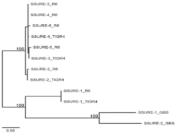
17 (CC17), display a highly homologous (99.3%) identical gbs0428 (or pbsP) locus, containing two SSURE domains (Da Cunha et al., 2014). Moreover, no major difference was observed between the pbsP promoter regions among GBS. GBS SSURE domains are closely related to similar domains present in other streptococcal species, particularly S. pneumoniae, as shown in Fig. 2. Sequences displaying significant homology with GBS SSURE domains are found only in
Streptococcaceae, particularly in S. gordonii, S. mitis, S. sanguinis, S. cristatus, S. oralis, S. anginosus, S. intermedius, in addition to S. agalactiae and S. pneumoniae.
Fig1 A) Schematic representation of PbsP. SP, signal peptide; N-terminal, N-terminal domain; SSURE1 and SSURE2, streptococcal surface repeats 1 and 2; MK rich: methionine and lysine-rich region; LPXTG, cell wall anchoring motif. B) Sequence alignment of the SSURE1 and SSURE2 domains. Asterisk (*), identity; colon (:), residues with strongly similar properties (> 0.5 in the Gonnet PAM 250 matrix); period (.) residues with weakly similar properties (< 0.5 in the Gonnet PAM 250 matrix)
Fig2 Sequence divergence between S. pneumoniae and S. agalactiae SSURE domains. The two SSURE domains (SSURE-1 and -2) of the GBS protein PbsP from strain NEM316,the four and six SSURE domains (SSURE-1 to -6) of the pneumococcal protein PfbB (PavB from, respectively, strains TIGR4 and R6 were aligned and a phylogenetic tree was computedusing the Neighbor Joining method. This analysis defines three clusters supported by significant bootstrap values (100%) calculated from 1,000 replicates. The scale bar (neighbour-joining distance) represents the percentage sequence divergence.
PbsP is expressed in the cell wall and on the surface of GBS
To verify the presence of the PbsP protein on the bacterial surface, we undertook flow cytometry and Western blot analysis of cell-wall-extracts using a polyclonal mouse serum (pAb) raised against a recombinant form of PbsP. Preparation of recombinant PbsP and pAb is reported in the Materials and Methods section. We first compared wild type (WT) NEM 316 with ΔpbsP and
ΔcovRS mutant strains. In the ΔpbsP strain, the gene encoding PbsP was deleted
in-frame in the NEM316 chromosome, while –as indicated above- the ΔcovRS mutant bears a deletion encompassing both the covR and covS genes.In addition we used a strain (srtA*) bearing a defective form of sortase A, an enzyme that is
mutants showed similar viability, morphology and growth in Todd-Hewitt broth, as compared to the WT parental strain (data not shown).
Western Blot Analysis (Fig. 3A) revealed an immunoreactive band in the WT, but not in the ΔpbsP, strain, migrating with an apparent molecular weight of 48 KDa, which is slightly higher than the expected mol weight (40 kDa). This discrepancy is likely linked to a relative lack of precision of SDS-PAGE analysis in determining the molecular weight of elongated proteins such as those found on the surface of gram positive bacteria. The PbsP protein was markedly overexpressed in the ΔcovRS mutant, in agreement with previous transcriptomics and proteomics studies (Lamy et al., 2004; Firon et al., 2013). Notably, the protein was not present in cell wall extracts of the srtA*, strain, indicating a requirement for sortase A for expression in the cell wall of this LPXTG protein. Further evidence for surface expression of PbsP was sought by Facs analysis using pAb. As expected, we detected PbsP on the surface of the WT NEM316 strain, but not on the ΔpbsP mutant, while the protein was highly expressed on ΔcovRS (Fig3B).
To gain a comprehensive view about the expression of this protein in the context of GBS strains belonging to different capsular serotypes (CPS) and clonal complexes (CC), we included in these experiments the representative strains GBS 6313 (CPSIII/CC23, as NEM316), A909 (CPSIa/CC1) BM110 and COH1 (both CPSIII/CC17) and 2603V/R (CPSV/CC19). PbsP expression on the bacterial surface was detected in all of these strains, with the lowest grade of
non-capsulated NEM316 mutant produced a signal of the same intensity as the WT strain, suggesting that the polysaccharide capsule did not significantly mask the display of the protein at the bacterial surface (data not shown).
Fig3 PbsP is a conserved cell surface protein. The following GBS strains were compared: WT, wild-type strain NEM316; pbsP, deletion mutant lacking PbsP; srtA*, mutant with an inactive form of sortase A; covRS deletion mutant lacking the twocomponent system CovRS.
A e B) SDS Page and Western blot analysis of cell wall extracts. A)Left panel, coomassie staining of cell wall extracts separated by polyacrylamide gel electrophoresis; B) right panel, corresponding western blot analysis using polyclonal anti- PbsP mouse serum
C C) Expression of PbsP on the GBS surface. Immunofluorescen ce flow cytometry analysis of PbsP espression on different bacterial strains using mouse polyclonal anti-PbsP serum (blue line) or control anti-GST serum (red line). GBS wild-type strains of different serotypes and clonal complexes are indicated by their common name (6313, A909, COH1, BM110, 2603V/R).
In order to investigate the functional role of PbsP in bacterial adherence, we measured binding of the recombinant protein to different substrates immobilized on glass coverslips. Recombinant PbsP was absorbed to fluorescent beads with a diameter of 1 micron, to mimic surface exposure of this protein on the bacterial surface. Binding of the beads to extracellular matrix or blood components (such as Plg, Fng, Fnt, collagen, C-reactive protein and the complement components factor-H, factor I, factor-B, C1q and C3) was assessed microscopically. These experiments showed that PbsP-coated beads bound Plg, but not other plasma/matrix components (Fig4A). To confirm binding of PbsP to Plg we performed Far western blotting experiments in which the recombinant protein was used as a probe to capture soluble Plg, or vice-versa. As shown in Fig. 4C, the ability of PbsP to bind Plg was confirmed using both approaches.
The kringle domains of plasminogen contain lysine-binding motifs that mediate the binding of plasminogen to fibrin, ECM components and several bacterial surface proteins (Martina L. Sanderson-Smith et al ., 2012).
To understand whether interactions between plasminogen and PbsP occur through the involvement of lysine binding sites on Plg, increasing concentrations of lysine or of the lysine analog ε-aminocaproic acid (6-ACA) were used to analyze the binding of rPbsP-coated beads to Plg immobilized on glass coverslips. Before adding the rPbsP-beads, slides were pre-incubated with increasing concentration of putative inhibitors. L-alanine, a non-charged amino
but not L-alanine, PbsP-Plg interaction were inhibited in a dose dependent manner, suggesting an involvment of the lysine-binding motifs in the plasminogen kringle domains (Fig.4B).
To test the contribution of PbsP to the overall ability of GBS to bind plasminogen, we compared NEM316 WT and ΔpbsP mutant for their capabilities to adhere to plastic plates coated with plasminogen. Under these conditions, adherence of the ΔpbsP mutant to plasminogen was almost completely abrogated in comparison with the WT strain (Fig4D). Genetic complementation of the ΔpbsP mutant restored PbsP expression and its ability to bind immobilized plasminogen, thus confirming that the inability of the mutant strain to bind plasminogen is due to deletion of the pbsP gene. All together these data indicate that PbsP is an important adhesin largely mediating binding of GBS to Plg, at least of in the context of CC23 strains such as NEM316.
Fig4. PbsP binds human plasminogen in vitro.
A) Selective binding of PbsP to immobilized plasminogen. Binding of the rPbsP-coated beads to immobilized human plasminogen (Plg), collagen (Coll), fibronectin (Fnt), fibrinogen (Fng), C-reactive protein (CRP) or to the complement components factor H (FH), factor I (FI), factor B (FB), C1q and C3 was quantified microscopically counting the number of fluorescent beads atthached on the plasminogen cotated coverslips. Columns and bars indicate means ± SD from three independent experiments.
B) Selective inhibition of PbsP-Plg interactions. Competitive binding assays were done with immobilized Plg to which rPbsP cotated beads were added in the presence of the 6-aminocaproic acid (6-ACA), L-Lysine, or L-Alanine, used as a negative control. Points and bars indicate means ± SD from three independent experiments.
C) Far Western blot analysis of PbsP/Plg interactions. Far western blot analyses were done with rPbsP and GST (as negative control) on membranes probed and revealed with Plg and anti-Plg (left panel), and, reciprocally, with Plg on membranes probed and revealed with rPbsP and anti-rPbsP (right panel). Numbers indicate the molecular mass of protein standards in kDa. D) Contribution of PbsP to the overall ability of GBS to bind plasminogen. Wild-type NEM316 (WT) was compared with its PbsP deletion mutant (ΔPbsP) or ΔpbsP strain carrying a complementing vector with constitutive pbsP expression (ΔpbsP+PbsP). Binding of GBS strains to plasminogen (Plg) immobilized on plastic plates was detected counting the number of CFU after shaving with trypsin. The results shown are mean ± S.D. of CFU counts from three independent experiments. * = p ≤ 0.05 by analysis of variance and Student’s Neuman Keuls test.
The Plg molecule contains a total of seven structural domains, each with different properties. The N-terminal portion of the molecule consists of an activation peptide (AP) followed by a series of 5 repeating homologous triple-disulfide-linked peptide regions, approximately 80 amino acids in length, termed kringles (K1–K5) containing lysine binding sites and the catalytic SP domain (Sarbani Bhattacharya et al., 2012). The function of the five kringles in the heavy chain of Plg is primarily to mediate protein-protein interactions, such as those between Plg or Pm and fibrin, ECM targets, and lysine-containing receptors (Sarbani Bhattacharya et al 2012). To identify the plasminogen domain predominantly responsible for interaction with PbsP, we developed an ELISA assay where the whole plasminogen molecule or its fragments (K1-K3, K1-K4 and Mini-Plg a complex of Kringle5 and serine protease domain) are coated on a microplate and binding of PbsP-GST is detected using anti-GST antibodies . This assay showed that Kringle 4, but not the others Kringle domains, was capable to bind PbsP tp the same level as that of the whole plasminogen molecule (Fig. 5).
Fig5. Plasminogen Kingle4 is involved in PbsP/Plg interaction Elisa of PbsP/Plg interactions. Elisa were done coating Plasminogen and its fragment on a microplate and the binding with rPbsP and GST (as negative control) was detected using anti-GST antiboby.
binding
In order to identify the PbsP domain predominantly involved in binding plasminogen, we carried out additional experiments using fluorescent beads coated with different PbsP-fragments. We first obtained recombinant fragments that encompassed different PbsP regions, such as the two SSURE domain (SSURE 1-2) region, the SSURE 2 domain (SSURE2) and the methionine and
lysine-rich region (MK-rich) (Fig6A).
This essay revealed that the MK-rich region bound Plg almost as efficiently as the entire protein. Moderate binding was observed with the fragment consisting of the two SSURE domains (SSURE 1-2) while the single domain fragment
(SSURE2) did not display significant interactions with Plg (Fig6 B). To identify
the minimal region of the MK-rich domain that still maintains the ability to bind Plg, we used two fragments encompassing the Mk-rich region and designated as N-terminalMk-rich and C-terminalMk-rich, respectively (Fig6A). N-terminalMk-rich is a biotinylated peptide purchased from GenScript and C-terminalMk-rich is a peptide in fusion with GST cloned in our laboratory.
To analyze the Plg-adhering activities of these fragments, we coupled these fragment with fluorescent beads as described before and we used GST and CAT-Biotin as a negative control. Under these conditions, the C-terminalMk-rich fragment bound Plg as efficiently as the entire MK-rich region and significantly better than N-terminalMK-rich, which showed only slight, but significant, binding (Fig6 C).
predominantly involved in Plg binding, we obtained an others two biotinylated peptides encompassing C-terminalMk-rich that were designated as Fr1C-terminal MK-rich (Fr1C) and Fr2C-Fr1C-terminalMK-rich (Fr2C) (Fig6A). We used these fragments coupled to fluorescent beads, as indicated before. Under these conditions neither Fr1C nor Fr2C efficiently bound Plg, compared to C-terminalMk-rich, indicating that both C- and N-terminal truncation of Mk-rich impaired its capability to bind Plg.
Fig 6. B) fluorescent latex microspheres were coated with the whole PbsP protein, with the SSURE1-2, SSURE2 or
Mk-rich PbsP fragments expressed as GST fusions (GST-SSURE1-2, GST
SSURE2, GST-Mk-rich) or with GST.
After incubation with plasminogen-sensitized coverslips, adhering particles were counted microscopically.
C and D). In the same manner N-terminal-Mk-rich, C-terminal-Mk-N-terminal-Mk-rich, Fr1C-teminalMk-rich (FR1C) and Fr2C-terminalMk-rich (FR2C) were coupled with fluorescent latex beads. Since N-terminalMk-rich and FR1C and Fr2C were purchased as biotin fragment , in these tests we used CAT-Biotin and GST coupled beads as a control. Results are expressed as particles per field of vision (FOV) at the indicated magnification and represent means ± S.D. of three independent experiments.*, significantly different (p<0.05) from GST-coated beads (A) as assessed by Student’s t test.
Attachment to and invasion of epithelial cells by GBS play a crucial role in the initial stages of infection. Alveolar and intestinal epithelia are considered as likely entry sites in, respectively, early- and late-onset GBS disease (Hansen Et
al., 2004). In early onset neonatal disease, transmission of GBS generally occurs via aspiration of vaginal contents during passage through the birth canal, or of
infected amniotic fluid, after the organism has traveled through intact or ruptured amniotic membranes. By this way, infection can remain localized in the lungs, causing pneumonia, or progress to sepsis (in 72% of the cases) and meningitis (2.5%) (Poyart et al., 2008, Doran and Nizet, 2004). In late onset disease, the organism is transmitted by the mother or other persons coming into contact with the neonate and is believed to colonize the intestinal mucosa before crossing it (Tazi, A., et al., 2010).
Since PbsP is an ortolog of PfbB (plasminogen and fibronectin binding protein B) of Streptococcus pneumoniae, which contributes to the ability of this bacterium to adhere to epithelia, we tested whether PbsP contributes to interactions between GBS and epithelial cells. We first compared the ability of NEM316 WT and ΔpbsP to adhere to Caco-2 (intestinal) and A549 (pulmonary) human epithelial cell lines. Binding of the ΔpbsP strain to either CaCo-2 or A549 cells was significantly reduced compared with NEM316, indicating that PbsP is required for optimal bacterial adherence (Fig. 7A). Complementation of the pbsP mutation with the wild-type allele restored bacterial adherence to A549
epithelial cells are at least partially dependent on surface-expressed PbsP.
Fig7. PbsP plays a non-reduntant role in the association of GBS NEM316 with epithelial cells. The following GBS strains were compared: WT, NEM316 wild-type strain; ΔpbsP, isogenic pbsP deletion mutant; ΔpbsP+pbsP, ΔpbsP strain carrying a complementing vector with constitutive pbsP expression; Each panel shows the mean ± S.D. of CFU counts from three independent experiments; *, p<0.05 by analysis of variance and Student’s Neuman Keuls test. A, adhesion and invasion to respiratory epithelial cell lines (A549). B, adhesion and invasion to intestinal epithelial cell lines (CACO-2)
pulmonary epithelial cells
To determine whether PbsP SSURE domains contribute to bacterial adhesion to epithelial cells, we analyzed the ability of SSURE domains to bind to lung cells in comparison with the C-terminal portion of the protein (MK-rich fragment). To this end, we used fluorescent beads coated with PbsP fragments or with whole PbsP, used as a positive control. The beads were incubated with A549 pulmonary epithelial cells grown on sterile coverslips and, after cell staining, the number of cell-associated beads was counted in 10 different microscopic fields. This experiment revealed that fragments containing SSURE domains bind efficiently to pulmonary epithelial cells. A fragment containing both SSURE domains (SSURE 1-2, Fig. 8) adhered only slightly less efficiently to
these cells than the whole protein and this difference was not statistically significant. On the other hand SSURE 1-2 bound to the cells more efficiently than
the fragment encompassing the MK-rich, C-terminal region. The fragment made up by only one SSURE domain (SSURE 2 in Fig. 8) adhered as efficiently
than the MK-rich fragment. Together, our data indicated that the ranking order of binding to pulmonary epithelial cells of the various fragments (PbsP= SSURE1-2 > SSURE2 =MK-rich, Fig. 8) differed from the ranking order of binding
to plasminogen (PbsP=MK-rich> SSURE1-2 = SSURE2, Fig. 6). Therefore, binding
to Plg might not account, by itself, for the ability of PbsP to bind to epithelials cells, which raises the possibility that PbsP protein is a multifunctional adhesin that can to another receptor on these cells, in addition to Plg. Further studies
implicated in binding of PbsP to pulmunary epithelial cells.
Fig8. Direct binding of PbsP fragments toA549cells. Fluorescent latex microspheres were coated with whole PbsP and PbsP fragments, all expressed as GST fusion proteins (GST SSURE1-2, GST SSURE2 and GST Mk-rich). GST was used as a negative control. After incubation
with A549 cells, adhering beads were counted microscopically (A-B). A549 cells were labeled with 4’,6-diamidino-2-phenylindole for nuclear staining. Columns and bars in panel B represent means ± S.D. of three independent experiments. *, significantly different (p≤0.05) from GST-coated beads, as assessed by Student’s t test.
The experiments reported above indicated that, in the context of the prototype CC23 GBS strain NEM316, PbsP plays a non-redundant role in mediating bacterial Plg binding and adherence to epithelial cells. We were interested to determine whether a similar requirement applies to stains belonging to the hypervirulent CC17 lineage, which is responsible for a large number of neonatal cases of GBS disease, particularly meningitis. Our studies were also prompted by FACS analysis experiments which suggested that PbsP might be less expressed on the surface of GBS CC17 strain COH1 relative to CC23 strains NEM316 and 6313. As a first step, we analyzed the expression of the protein in strain BM110, an additional CC17 prototype strain and whether such expression is under the control of the two component regulatory system CovRS, which, as mentioned above, plays a crucial role as a global regulator of multiple genes involved in host colonization and virulence (Lamy et al., 2004; Jiang et al.,
2008). To this end, we measured PbsP mRNA levels by real time RT-PCR using
strain BM110, and a ∆covR deletion mutant of this strain lacking the response regulator (R) component of the signaling system. For comparison, we also tested wild-type (WT) strain NEM316 and its ∆covR deletion mutant. The housekeeping gen gyrA was used as an internal control. After growth to the exponential phase in Todd-Hewitt broth, the levels of PbsP mRNA were 10-fold lower in wt BM110 relative to wt NEM316. It was found that PbsP expression in BM110 is down regulated by the CovR repressor, since 5- fold higher PbsP mRNA levels were measured in BM110 ∆covR bacteria, respectively, as compared with the parental
Western-blot analysis of GBS cell wall extracts using an anti-PbsP serum, which produced a fainter PbsP band in wt BM110 in comparison with BM110 ∆covR (Fig.9B).
Collectively, data from this first set of experiments indicated that PbsP expression is strongly repressed by the CovR regulator in the CC17 prototype strain BM110, at least during in vitro growth. This feature may explain low-level expression of the protein in CC17 strains, as observed in the present study.
Fig9. qRT-PCR analysis of PbsP in GBS strains(A) and Pbsp expression in cell wall extract of BM110 strain and its mutans (B). A) We used GBS NEM316 and BM110 wild-type strains and ΔpbsP and ΔcovR theirs isogenic deletion mutants. Gene expression was measured by the comparative CT method (ΔΔ CT) and reported as the n fold difference relative to the normalized expression of housekeeping gene (GyrA). Results shown are representative of two independent experiments performed in triplicate. B) protein level of PbsP was detected by Western Blot analysis using polyclonal anti- PbsP mouse serum. The PbsP level in cell-wall extract of wild-type BM110 is compared with its PbsP deletion mutant (ΔPbsP) or ∆covR deletion mutant lacking the response regulator (R) component of the signaling system.
As the CsrRS system is involved in the upregulation of virulence genes during GBS infection (Lamy et al., 2004; Jiang et al., 2005; Jiang et al., 2008), we hypothesized that PbsP expression, which is tightly controlled by CovR, is upregulated in vivo. To investigate this, we performed quantitative RT-PCR (qRT-PCR) analysis of bacterial RNA extracted from kidneys and brains of mice infected i.v. with the BM110 stains. Results were compared with those obtained on in vitro grown bacteria using the housekeeping gyrA gene as an internal control. As shown in Fig. 10, PbsP mRNA levels were approximately 7-fold higher in vivo than in vitro. To investigate the role of PbsP in the pathogenesis of CC17 GBS infection, the gene encoding PbsP was deleted in-frame in the BM110 chromosome. The viability, morphology and growth in Todd-Hewitt broth of the
∆pbsP mutant were similar to those of the WT parental strain (data not shown).
We next compared the virulence of BM110 WT with that of a ∆pbsP mutant using a mouse model of invasive GBS infection. Under these conditions, only 20% (3 out of 16) of mice infected with the mutant strain died, while 70% of mice infected with the WT strains succumbed to infection by day 4(Fig.11). To further study the role of PbsP in this model, we measured the bacterial load in the blood, brains and kidneys of mice infected with BM110 WT or a ∆pbsP mutant as detailed above. At 48h after infection, mice infected with the WT strain had significantly higher bacterial counts than those injected with the ΔPbsP mutant both in brains and in kidneys but not in the blood (Fig. 12 A, B and C). These data indicate that PbsP may play an important role in the pathogenesis of
such as the brain. PbsP is highly conserved among clinical isolates of GBS, which could make it as an interesting vaccine candidate. For this reason we next investigated if the protein has the ability to prevent invasive disease after immunization. Adult mice (n 16 per group) were immunized with rPbsP or GST, used as a negative control, and challenged intravenously three weeks later with BM110 WT. Under these conditions, immunization with rPbsP resulted in increased survival (75% or 12/16 versus 37% or 6/16 in control animals; P<0.05 by log-rank (Mantel Cox) test; Fig. 13) and in decreased bacterial colony counts in the brains and kidney (Fig. 14A and B). These data indicate PbsP immunization can induce significant protection against subsequent systemic infection with GBS.
Figure 10. qRT-PCR analysis of PbsP in GBS BM110 in vitro and in vivo. Values are presented as a ratio of expression in brain, and Kidneys of infected CD1 mice relative to expression in TH broth medium. Results shown are representative of two independent experiments performed in triplicate.
Figure 11. PbsP is a virulence factor of GBS. Comparison of lethality in mice infected with wild-type BM110 (WT) and its pbsP deletion (ΔpbsP) mutant. Mice were monitored at least once a day for a total of 11 days after infection with 6x 10⁷ bacteria. *, p < 0.05 by log-rank (Mantel Cox) test.
Figure12. Effects of PbsP on organs colonization. Bacterial Burden in bood(A) brains (B) and kidneys (C) were quantified 48hours after i.v. challenge with 6x 10⁷ CFUs of WT or ΔpbsP strains. Orizontal
Figure13. Protective effects of GST-PbsP immunization against lethal GBS infection in mice. Animals were immunized with the recombinant GST-PbsP protein or with the GST as a control and challenged i.v. with 9x10⁷ CFU/mouse of wild type strain BM110. *, p < 0.05 by log-rank (Mantel Cox)
test.
Figure 14. Effects of PbsP immunization on organs colonization. Bacterial Burden in brains (A) and Kidneys (B) were quantified 48hours after i.v. challenge with 9x 10⁷ CFUs of BM110 strain. Orizontal
Bars indicate the geometrical means. *= p < 0.05 by the Mann Whitney U test. .