University of Catania
International PhD in
Translational Biomedicine
XXVIII cycle
Coordinator: Prof. Daniele Filippo Condorelli
Giorgia Spampinato
Genomic analysis in human solid tumours:
Copper Homeostasis Genes in Colorectal cancer
PhD Thesis
Tutor:
Prof. Vincenza Barresi
2
INDEX
Introduction
pag. 3
Aim of the work
pag. 32
Materials and Methods
pag. 34
Results
pag. 47
Discussion and Conclusions
pag. 75
Concluding Remarks
pag. 83
3
1.1INTRODUCTION
1.1.1 COPPER: AN ESSENTIAL TRACE NUTRIENT
Metals are integral components of nearly all biological processes. Paradoxically, some metals are essential in trace amounts and toxic at elevated levels, some are simply toxic, while others are tolerated at excessively high levels by living cells. Uranium, arsenic, copper, and lead have all recently been implicated in novel biological roles [Yannone et al., 2012]. It is common knowledge that the elemental building blocks of life as carbon, hydrogen, oxygen and nitrogen are considered, always as the elements essential to life. Really these fundamental components are predominate only in terms of mass in fact it is important to note that all life forms have an absolute requirement for a number of metals that are considered ‘trace’ elements and that play a key role to most biological processes.
Copper (Cu), zinc (Zn), and iron (Fe) are essential micronutrients for maintenance of life. These micronutrients are involved in many complex enzyme systems functioning in various biological processes. The metals are defined as essential when they are needed for the life of the cell and for the proper functioning of the physiological processes; The iron, for example, is an enzyme cofactor in higher eukaryotes that ensures normal functioning of hemoglobin, also the copper acts as a cofactor for many enzymes (eg. Superoxide dismutase, cytochrome oxidase, etc..) allowing their activity within the cell. Copper is required for respiration, connective tissue formation, iron metabolism, and many others processes. In human cells, copper is utilized in several cells compartments, and the intracellular distribution of copper is regulated in response to metabolic demands and changes in cell environment [Lutsenko
2010].
However even if the copper and the iron are involved in various physiological processes important to the cell at high concentrations, can lead to the formation of free radicals and promote lipid peroxidation, leading to a rapid decline in the integrity of biological membranes but can also cause damage DNA and RNA, for this reason the concentration of free copper in the cell is maintained at ≤ 10-18 M [Howell et al., 2010]. The life-supporting properties of Cu
incur a significant challenge to cells that must not only exquisitely balance intracellular Cu concentrations, but also chaperone this redox-active metal from its point of cellular entry to
4
its ultimate destination so as to avert the potential for inappropriate biochemical interactions or generation of damaging reactive oxidative species (ROS) [Nevitt et al., 2012].
For these important characteristics the ability of an organism to accumulate in appropriate concentrations these metal species, considered therefore as essential trace elements, is critical to your health.
1.1.2 COPPER HOMEOSTASIS
Copper (Cu), a transition metal with two redox states, is an essential trace element in living organisms that acts as a catalytic cofactor for a number of enzymes involved in critical biological processes including protection against free radicals (superoxide dismutase), oxidative phosphorylation (cytochrome c oxidase), pigmentation (tyrosinase) collagen maturation (lysyl oxidase) and neuropeptide and peptide hormone production (peptidylglycine alpha amidating mono-oxygenase, PAM). [Mufti et al., 2007]
Copper functions as the active centre of cuproenzymes, such as cytochrome c oxidase (CCO), Cu,Zn-superoxide dismutase (Cu,Zn-SOD, SOD1), ceruloplasmin, lysyl oxidase, tyrosinase, and dopamine β-hydroxylase. However, free copper is very toxic due to its ability to react with hydrogen peroxide and generate highly reactive hydroxyl radicals [Mufti et al.,
2007] (Fig. 1). Other possible molecular events that contribute to the toxicity of copper in
vivo include inappropriate insertion of copper into metalloproteins (so-called protein hijacking), copper-dependent perturbation of membrane potential, and inhibition of membrane ATPases.
Figure 1. Chemical reactions of copper.
Consequently, free copper in the cell is almost undetectable and complex homeostatic mechanisms have evolved to regulate intracellular copper distribution and excretion (Fig. 2). Copper (Cu+) is transported into the cell via the high affinity Copper transporter (Ctr)
proteins, which are highly conserved from yeast to humans. [Lee et al., 2001] All Ctr proteins
O2+ Cu(II) → Cu(I) + O2
Cu (I) + H2O2→ OH-+ OH·+ Cu(II)
Overall Reaction: O2+ H2O2→ OH-+ OH·+O
5
contain three transmembrane domains, and most possess extracellular methionine rich motifs (MxxM, MxM) at the amino terminal and an additional MxxxM motif in the second transmembrane domain. [Guo et al., 2004] These domains are essential for binding extracellular copper and facilitating transport into the cell.
Metalloreductases, Step2, bound to the plasma membrane, and Steap3-4 located on membrane of intracellular vesicles. Both are responsible for reducing of Cu2+ to Cu1+ prior to
transport through the membrane. [Puing et al., 2002]
Figure2. Copper distribution pathways in a generalized mammalian cell.
Two Ctr homologs, Ctr1 (SLC31A1 gene) and Ctr2 (SLC31A2 gene), have also been identified in vertebrates, and of these, Ctr1 is the main protein responsible for intracellular copper transport. The exact function of Ctr2 in vertebrates remains unclear. [Rees et al.,
2004] Due to its toxicity, intracellular copper trafficking is very tightly regulated and
evolutionarily conserved pathways have evolved for the handling of copper upon entry into the cell involving proteins, known as chaperones, whose function is the delivery of copper to specific targets (Fig. 2). [Mufti et al., 2007] Delivery of copper to superoxide dismutase (SOD1), a zinc and copper containing protein, occurs via the Copper Chaperone for SOD (CCS), which is found in both the cytoplasm and the intermembrane mitochondrial space (IMS). SOD1 is an important enzyme, which catalyzes the conversion of superoxide anion to hydrogen peroxide and oxygen (Fig. 3).
6
Figure 3. SOD catalytic mechanism.
Mutations in SOD1 are associated with amyotrophic lateral sclerosis (ALS), a neurodegenerative disorder characterized by the loss of upper and lower motor neurons and muscle atrophy. [Rowland et al., 2002]
Another example of intracellular copper handling involves copper delivery to Cytochrome c Oxidase (CcO), the terminal enzyme in the respiratory chain (Fig. 2). CcO enzyme in mammals is located within the mitochondrial inner membrane needs three copper ions to perform its function. The CcO and is constituted by 13 subunits, two of which contain two copper centres. One, called CuB, is a monocopper site in subunit I, and the other, called CuA, is a binuclear site in subunit II. [Banci et al., 2008] A number of proteins are required for the metallation of these sites. For copper insertion into the CuA site, Cox17, Sco1, and Sco2 proteins have been proposed to have essential non-overlapping roles within the mitochondrial intermembrane space (IMS), the first working as a metallochaperone to Sco1 and Sco2 [Horng et al., 2004.Leary et al., 2004], whereas the other two have a cochaperone
function during the copper insertion in the CuA site.
Antioxidant protein 1, or Atox1, is a cytosolic copper chaperone , like CCS and / or small thiol molecules such as glutathione, binds the ‘‘Labile’’ Cu pool, entered in the cytosolic environment by SLC31A1 transporter, coordinating its further intracellular destination while the excess copper is sequestered by metallothioneins to avoid build-up to toxic levels (‘‘Immobilized’’ copper pool). [Hatori and Lutsenko 2013]. Atox1 is responsible for the shuttling of Cu1+ to the export pumps ATP7A/B (Fig. 4). These are the main transporters
responsible for copper excretion from cells, and are both found in the trans-Golgi network, contain six MxCxxC metal binding domains (MBDs) at their amino termini and each MBD is capable of binding one atom of copper [Lutsenko et al., 2002]. Following binding to copper and under normal or low copper conditions, ATP7A/7B translocate Cu1+ into the lumen of the
secretory vesicles for integration into cuproenzymes such as ceruloplasmin and tyrosinase [Miyayama et al., 2009]. The exact manner in which copper is loaded onto cuproenzymes in the secretory compartment is not known.
SOD-Cu(II) + O
2·↔ SOD-Cu(I) + O
2
SOD-Cu(II) + O
2·↔ SOD-Cu(II) + H
7
Figure 4. Atox1 role in cellular copper trafficking pathways, taken from Hatori et al., 2013.
ATP7A is present in most tissues of the body and is highly expressed in intestinal epithelial cells but is absent in hepatic tissue. In the intestinal epithelium, ATP7A is required to mediate delivery of copper from the cell to the systemic circulation and thus it is essential for the systemic absorption of copper from the gastrointestinal tract. [Voskoboinik et al.,
2002] In the presence of elevated copper levels, ATP7A translocates to the plasma membrane
and pumps excess copper out of the cell.
Like ATP7A, in elevated copper states, ATP7B moves from post-Golgi vesicles to the plasma membrane, specifically the biliary canalicular membrane allowing copper excretion into bile. This constitutes the main pathway for copper efflux from the body. Because of mutations in the gene ATP7B, the copper accumulates in the liver. First inducing the production of metallothioneins (MTs), a family of small proteins (7 kDa) containing a high proportion of cysteins. SH groups of MTs can bind metal ions with high affinity acting as a buffer system or a reservoir for essential copper and zinc ions. [Zalewska et al., 2014] These proteins are able to sequester toxic metal ions and exchange them for zinc [Aicha Ba et al.,
2009] or can apparently maintain copper in a relatively harmless state. Subsequently, this
accumulation of copper causes damage to mitochondria initially, and eventually leads to destruction of the hepatocyte. In Wilson’s disease, the copper is also deposited in renal tubules, and kidney damage occurs to varying degrees. A combination of copper accumulation
8
in the blood as well as impaired expression of ATP7B in the brain, leads to neurological disease, by mechanisms not entirely explained.
X-linked inhibitor of apoptosis (XIAP), known primarily for its caspase inhibitory properties, has recently been shown to interact with and regulate the levels of COMMD1, a protein associated with a form of canine copper toxicosis.
It was recently reported that COMMD1, copper metabolism (Murr1) domain containing 1, interacts with ATP7B (Fig. 5), to mediate the efflux of copper from the cell [Tao et al., 2003], although, the functional role of this interaction remains to be elucidated. COMMD1 is the prototypical member of a family of ten homologous and highly conserved proteins known as COMMD1 to 10 [Burstein et al., 2005]. The defining characteristic is the presence of a unique carboxy-terminal domain termed the COMM domain (copper metabolism gene MURR1).
In addition to its role in copper homeostasis, COMMD1 and other COMMD proteins also function as negative regulators of the transcription factor NF-κB.
Figure 5. Model of copper uptake and metabolism taken from Mufti et al., 2007.
XIAP-associated factor 1 (XAF1) has been identified as an X-linked inhibitor of apoptosis (XIAP) binding partner (Mufti et al., 2007). XAF1 interacts with endogenous XIAP and results in XIAP sequestration in nuclear inclusions. XAF1 mRNA is expressed ubiquitously in all normal adult and fetal tissues, in contrast to very low or undetectable levels in various cancer
↓XAF1 → ↑ XIAP
9
cell lines. Reduced or absent XAF1 expression seems to be a frequent event in human cancer tissues. Transient over expression of XAF1 sensitizes tumour cells to the proapoptotic effects and antagonizes the XIAP-mediated inhibition of caspase 3 activity. Moreover, loss of XAF1 expression correlates strongly with tumour staging, implicating loss of XAF1 function in tumour progression [Shui Ping Tu et al., 2010].
1.1.3 ATOX1: COPPER CHAPERONE ANTIOXIDANT-1
One of the human copper chaperones is HAH1 (also called human Atx1 or Atox1), a small soluble protein [Klomp et al., 1997] which is capable of delivering copper(I) both to the Menkes and the Wilson disease proteins (ATP7A and ATP7B, respectively). [Petris et al.,
1996]
The Atox1 gene is located on chromosome 5 and contains 4 exons and 3 introns. Atox1 encodes for a ubiquitously expressed protein of 68 amino acids and it is characterized by a fold βαββαβ. Antioxidant-1 (Atox1) contains a single N-terminal copy of the conserved MXCXXC copper binding motif (Fig. 6A) suggesting an interaction between Atox1 and ATP7A or ATP7B, in fact has been demonstrated that Atox1 in which the Cys residues, Cys12 and Cys15, of the MBS were mutated was unable to interact with ATP7B, indicating that the MBSs coordinate these interactions (Fig. 6B). [Strausak et al., 2003] Another conserved lysine-rich region (KKTGK) is localized at C-terminus is (Fig. 6A), several data show that this domain represents the potential NLS (nuclear localization signal) and is involved in recruiting Atox1 to the nucleus, data confirmed in vitro [Itoh et al., 2008. Hamza et al., 1999] and in vivo
[Ozumi et al., 2012]. In these studies, it has been tested the hypothesis that, in mouse embryonic fibroblast cells (MEFs), nuclear Atox1 may be involved in regulating copper-induced cell proliferation and that copper stimulates cell proliferation in an Atox1-dependent manner, demonstrating also that the copper induces Atox1 nuclear translocation, binding to DNA at novel cis elements (GAAAGA; Atox1-responsive elements)(Atox1-RE) in the promoter of the cyclin D1 promoting cellular proliferation. [Itoh et al., 2008. Muller and Klomp, 2009]
10
Figure 6 Structure of ATOX1 and homo / heterodimerization.
Tetrathiomolybdate (TTM) is a copper chelator, which blocks Atox1 function [Alvarez et
al., 2010], preventing tumor progression in clinical trials [Brewer et al., 2005]
X-ray crystallography and biochemical analyses also revealed copper-dependent ATOX1 homodimerization. [Wernimont et al., 2000] In these homodimers, two Cys residues of each monomer formed a three-coordinate Cu1+ complex with a loose interaction with the fourth
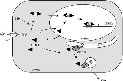
Cys residue. These studies propose that Atox1 can function as a copper chaperone that delivers copper to the export pumps ATP7A or ATP7B and, alternatively, it can function as a copper-dependent transcription factor in which homodimerization copper-induced of Atox1 drives the expression of CCND1 and possibly other genes. In conclusion, Atox1 might shuttle between the nucleus and might form homodimers to activate transcription (Fig. 7). [Muller
and Klomp, 2009]
Figure 7. Cellular model of Atox1 function taken from Muller and Klomp, 2009.
-C N- MTCGGC KKTGK 1 11 16 56 60 68 MBS NLS C X X C X M M X C X X C C X X C X M M X C X X C Cu ATOX1 ATP7A/B ATOX1 ATP7A/B A B
11
CTR1: COPPER TRANSPORTER
The high-affinity copper transporter (Ctr1) (SCLC31A1 gene) is the main copper influx transporter and plays an important role in regulating copper homeostasis because Cu deficiency is detrimental to many important cellular functions while its excess is toxic.
The SLC31A1 gene is localized on long arm of 9 chromosome coding for the human Ctr1 protein. The Ctr1 proteins are integral membrane proteins have a length of 190 amino acids with a ~67 aa extra-cellular amino-terminal (ecto) domain and a ~15 aa carboxyl-terminal cytosolic tail. These transporters harboring three trans-membrane domains (TMD) and multiple copper-binding ligands (predominantly methionine, histidine or cysteine) within the extracellular amino-terminus, at the extracellular boundary of TMD2 and in the cytosolic carboxyl-terminus (Fig. 8). [Puing et al., 2002. Klomp et al., 2003]
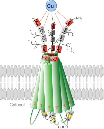
Figure 8. Structural model of three Ctr1 monomers in a lipid bilayer taken from Ohrvik and Thiele, 2014.
Recent insights into the 3D structure of human Ctr1 revealed a homo-trimeric complex arranged in a tail-to-tail orientation in which the carboxy-termini face each other within the cytosol, generating a cone-like pore that is narrow at the extracellular face (approximately 8 Å across) and wide at the intracellular aqueous exit (approximately 22 Å across). [De Feo et al.,
12
2009] Additionally, the position of the conserved M-X3-M Cu-binding motif on TM2 along the
pore interface of the symmetric Ctr1 trimer, suggests that Cu+ moves through the Ctr1 pore by
a series of ligand exchange reactions between distinct binding sites and that these reactions induce conformational changes that mediate Cu+ movement through the pore up to the
HCH-motif near the carboxyl terminus of the protein. The higher stability of Cu+-cysteine
interactions when compared to those between Cu+-methionine, would thus favor cation
enrichment (in the form of a thermodynamic sink) at the wider aqueous intracellular exit, thereby obviating the energetic requirement for ATP hydrolysis to drive Cu transport.
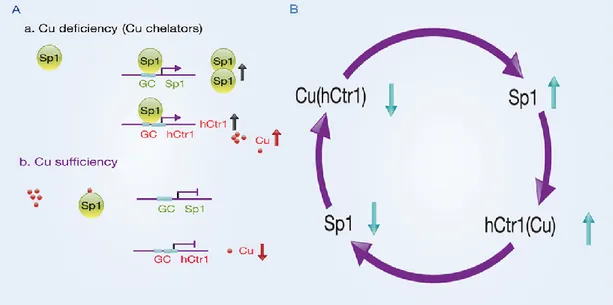
Ctr1 is an important regulator in response to environmental Cu stresses. Recently, it has been demonstrated that Cu deficiency induced by treating human cancer cells with Cu chelators up-regulates hCtr1 expression; whereas Cu sufficiency achieved by treating cells with CuSO4, down-regulates endogenous hCtr1 expression. [Liang et al., 2012. Song et al., 2008] Moreover, it has been revealed that human Cu homeostasis is tightly controlled by
interregulatory circuitry involving Cu, Sp1 and hCtr1. This circuitry uses Sp1 transcription factor as a Cu sensor in modulating hCtr1 expression which in turn controls cellular Cu and Sp1 levels in a three-way mutual regulatory loop. [Kuo et al., 2012] Not only hCtr1, but also Sp1 itself, is regulated by Cu bioavailability (Fig.9).
13
1.1.4 THE RELATIONSHIP BETWEEN TRACE METALS AND CANCER
The roles of metals in the development and inhibition of cancer have a complex character and have risen many questions because of their essential and toxic effects on human health. Question of whether trace metal concentrations in tissues are increased or decreased in cancerous patients in comparison with noncancerous patients has not been answered yet, due to the fact that the data known in this field is rare and have contradictory results. Although Zn and Cu concentrations in serum and tissues of cancerous patients have been extensively studied, the precise role of these metals in carcinogenesis is not clearly understood. The differences in literature on the increases or decreases in trace metal concentrations of cancerous tissues in comparison with non-cancerous tissues may be attributed to a few reasons such as the tissue basis-dry or wet weight, different sensitivities and basis of analysis methods that affect the accuracy, and the difficulties in taking of the sample representing the cancerous or non-cancerous area. [Mehmet Yaman 2006]
Epidemiological studies have found that many metals and metal-containing compounds may be potent mutagens and carcinogens. In the last three decade, the metals including cadmium, nickel, arsenic, beryllium and chromium (VI) have been recognized as human or animal carcinogens by International Agency for Research on Cancer (IARC).
Carcinogenesis is caused by mutations of the genetic material of normal cells, which upsets the normal balance between proliferation and cell death. This results in uncontrolled cell division and the evolution of those cells by natural selection in the body. The uncontrolled and often rapid proliferation of cells can lead to benign tumours; some types of these may turn into malignant tumours (cancer).
It is considered that carcinogenesis is occurring in four stages: Initiation;
Promotion; Progression; Metastasis.
The mechanisms of metal-induced carcinogenesis are believed to be involved in all stages of cancer development. The cells that can cause malignant tumors have several properties that distinguish them from cells of healthy tissue:
14
Resistance to apoptosis (programmed cell suicide);
Division uncontrolled (or die) and proliferation more frequently than normal; Self-sufficiency in growth factors;
Resistance to inhibition by contact; Defective cell differentiation.
Some studies suggest that the ratio Cu / Zn is a good indicator of extension and prognosis of various tumors, in particular, has been observed that the serum levels of copper are significantly higher in patients with prostate cancer compared to healthy individuals, while the levels of zinc in serum are lower in patients with cancer, therefore, the ratio of Cu / Zn in the serum showed a gradual and significant increase. [Gupta et al., 2005]
The mechanism for these alterations in serum trace element levels are not well understood. Increased ceruloplasmin level, increased uptake from the gut, diminished urinary excretion, and tissue breakdown as a result of tumor necrosis with consequent release of copper stores have been suggested as the possible causes for increased serum copper level.
Over the past two decades, chemical and cellular studies have contributed enormously to broaden the understanding of the mechanisms and pathophysiological processes induced by metals. In particular, has focused attention on ways of signal transduction induced by carcinogenic metals, such as:
NF-kB activation; apoptosis;
cell cycle progression.
Reactive oxygen species (ROS) are indispensable factors in signal transduction pathways. Under physiological conditions, ROS level is controlled by SOD1 and other antioxidative systems. However, in presence of copper and/or zinc dyshomeostasis, metal-induced ROS accumulation may induce various diseases, including cancer [Formigari et al.,
2013]. As reported by Ba and colleagues (2009), one of the most devastating events caused
by ROS is the oxidation of ligands and subsequent metal ion release. Metals released during oxidative stress include the redox active iron and copper ions, which amplify ROS levels through Fenton chemistry. It is generally believed that oxidative stress, resulting from metal-induced generation of reactive oxygen species (ROS), is a critical mediator for the malignant
15
transformation but targets of these metals can be regulatory proteins or signaling proteins involved in cell growth, in apoptosis, in cell cycle regulation, in DNA repair, and in differentiation [Bartsch et al., 2000]. However, much more information and research are essential to elucidating the role, mechanisms and factors relating to zinc (and copper) in cancers. [Franklin and Costello, 2009]
1.2 CRC: COLORECTAL CANCER
Colorectal cancer (CRC) is the third most common cancer worldwide. (Jemal et al., 2011). The incidence of CRC is higher in countries in the developed world, where it is the second most common form of cancer among women (after breast cancer) and third among men (after prostate and lung). The predominant localization of CRC is rectum (50–60%) and sigmoid colon (15–25%).
Several study estimate that approximately 90% of colorectal cancer cases are sporadic without family history or genetic predisposition while less than 10% of all CRC cases have a true inherited predisposition to CRC. In most of these cases, the causative genetic event has been identified. However, up to 25% of cases have a family history of CRC (familial CRC), but are not consistent with one of the known inherited syndromes, they have a higher risk of developing CRC in comparison with the general population, although not as high as in the inherited syndromes. [Bogaert and Prenen 2014]
CRC arises as a consequence of the accumulation of genetic alterations (gene mutations, gene amplification, and so on) and epigenetic alterations (aberrant DNA methylation, chromatin modifications, and so on) that transform colonic epithelial cells into colon adenocarcinoma cells (Fig. 10).[Grady and Carethers 2008]
16
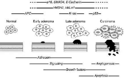
Figure 10. Basic outline of the adenoma to carcinoma sequence. Taken from Earnhead et al., 2002. The
time-line in which key genes may be affected is shown above the histological stages of disease during which they are thought to occur.
The sequential process of gene mutations and epigenetic alterations is widely believed to drive the initiation and progression of benign adenomas to malignant adenocarcinomas because these mutations affect signaling pathways that regulate hallmark behaviors of cancer. [Hanahan et al., 2000. Fearon et al., 1990] The earliest genetic change associated with adenomatous polyps is most frequently mutation and/or loss of the APC gene. The exact sequence of commonly acquired genetic changes accumulated subsequent to inactivation of APC is variable. K-ras mutations are found in ~50% of colorectal cancers and are thought to be relatively early events that correlate histologically with early to late adenomas. There is good evidence to suggest that p53 mutations occur more frequently in high-grade dysplastic polyps and are thought to mark the transition from adenoma to carcinoma. [Fearon E.R. et
al., 1990.Rodrigues N.R. et al., 1990]
These mutations create a clonal growth advantage that leads to the outgrowth of progressively more malignant cells, which ultimately manifests itself as invasive adenocarcinoma. The acquisition of these mutations is facilitated by the loss of genomic stability, which appears to be a key molecular step in cancer formation. [Lengauer et al.,
1998. Grady et al., 2004. Gollin et al., 2005]
The loss of genomic stability and resulting gene alterations are key molecular pathogenic steps that occur early in tumorigenesis; they permit the acquisition of a sufficient number of alterations in tumor suppressor genes and oncogenes that transform cells and promote tumor progression. Three predominant forms of genomic instability that have been identified in
17
colon cancer: chromosome instability (CIN), microsatellite instability (MSI) and CpG island methylator phenotype.
The first form is the 'chromosomal instability’ (CIN) with karyotype changes that occurs in 80-85% of colorectal carcinomas;
Microsatellite instability (MSI) is a hypermutable phenotype caused by the loss of DNA mismatch repair activity and it is characterized by deletions and amplifications of short sequences of nucleotides. MSI is detected in about 15% of all colorectal cancers; 3% are of these are associated with Lynch syndrome and the other 12% are caused by sporadic, acquired hypermethylation of the promoter of the MLH1 gene, which occurs in tumors with the CpG island methylator phenotype [Sheffer et al., 2009].
The third form is the CpG island methylator phenotype (CIMP) which was originally grouped together with MSI tumors. The islands are CpG rich regions within the genome and especially found in promoter sequences. In carcinogenesis, methylation of CpG islands (so-called type C methylation) leads to transcriptional silencing of genes involved in tumor suppression, apoptosis, DNA repair, and cell-cycle control. [Goel et al., 2007] Genes that are frequently affected by this non-covalent epigenetic modification are p16, MGMT, and hMLH1.
MSI tumours are usually euploid, while CIN tumours are aneuploid; these genetic differences are reflected in a different pathological and clinical behaviour.
1.2.1 Inherited predisposition to colorectal cancer
Inherited forms of CRC account for as much as 30% of all CRC cases and between 2% to 5% of all colon cancers arise in the setting of well defined inherited syndromes, including Lynch syndrome (also called hereditary nonpolyposis colorectal cancer [HNPCC]), familial adenomatous polyposis (FAP), MUTYH-associated polyposis (MAP), and certain hamartomatous polyposis conditions. All of these conditions are inherited, autosomal dominant disorders, except MAP, which is autosomal recessive.
Colorectal cancer results from the progressive accumulation of genetic and epigenetic alterations that lead to the transformation of normal colonic epithelium to colon
18
adenocarcinoma, which is called the polyp → cancer progression sequence (Fig. 11). [Grady
et al., 2008]
The tumor initiates from a normal colonocyte stem cell that has sustained genetic damage over time due to the local environment or has inherited a germline genetic mutation. The damaged DNA drives tumor progression up to the formation of carcinoma. In FAP, tumor initiation is accelerated with the inheritance of a germline APC mutation; in Lynch syndrome, tumor initiation might be normal to slightly accelerated, but the true driving force to the tumor progression is due to the hypermutable phenotype that occurs with loss of DNA MMR.
Figure 11. Depiction of colorectal tumor progression in sporadic and high-risk genetic syndromes. Taken from Grady et al., 2008.
1.2.2 Lynch Syndrome (LS)
The syndrome accounts for 2%–4% of all CRCs [Hampel et al., 2008] and individuals with Lynch syndrome are predisposed to various types of cancers, especially colon-rectum and endometrial, [Lynch et al., 2003] other cancers associated with Lynch syndrome are gastric, ovarian, biliary, urinary tract, small bowel, brain and pancreatic tumors. Although affected individuals can develop colonic adenomas with greater frequency than the general population, polyposis is rare. The lifetime CRC risk is estimated to be 50%–80%. [Stoffel et
19
al., 2005] Histologically, cancers are often poorly differentiated, mucinous, and have large
numbers of tumor-infiltrating lymphocytes.
Lynch syndrome is the result of a autosomal dominant heterozygous germline mutations in a class of genes involved in DNA MMR, including hMSH2, hMLH1, hMSH6, and hPMS2. The MMR system is necessary for maintaining genomic stability by correcting single-base mismatches and insertion-deletion loops that form during DNA replication. Mutations in hMSH2 and hMLH1 account for the up to 90% of Lynch syndrome cases; mutations in hMSH6 account for approximately 10% and mutations in hPMS2 are detected on rare occasions. [Rustgi 2007. Kastrinos et al., 2009. Peltomaki P et al., 2004] They are also characterized by a high level of microsatellite instability (MSI-H), a molecular phenotype that is a direct consequence of impaired MMR activity. Of interest is that colon cancers with MSI-H overall have a better prognosis compared to those without MSI. [Ribic et al., 2003] Several tools are available to assist clinical diagnosis of Lynch syndrome, including analyses of family histories, tumor testing, mutation prediction models, and genetic testing.
1.2.3 MSI AS A MARKER OF DEFECTIVE MMR
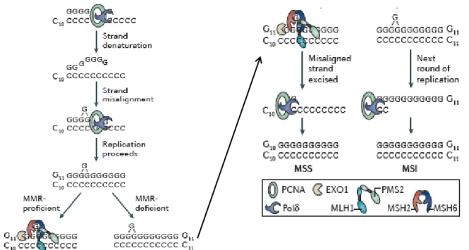
The human DNA MMR system is an evolutionarily conserved system that involves a number of proteins that recognize and direct repair of nucleotide mismatches and is responsible for maintaining replicative fidelity of DNA. [Marra et al., 1996. Fishel R. 1998] Parsons and colleagues [Parson et al., 1993] demonstrated that LS cancer cells lacked the ability to repair small insertions or deletions in tandem repeat sequences, as well as single-base mispairs, which is consistent with defective MMR (Fig. 12). These findings provided the first evidence that LS was a disease of defective MMR. Approximately 90% of Lynch syndrome-associated CRCs will have MSI-H and loss of MMR protein expression it was quickly shown that inactivation of members of the human DNA MMR gene family was the cause of microsatellite unstable colorectal cancer, on the contrary mechanism(s) responsible for causing CIN and CpG island methylator phenotype (CIMP) remain to be identified.
Following the recognition that genome-wide MSI was a key characteristic of LS tumours. Thus, MSI is a marker for loss of DNA MMR activity. In the analysis of tumor DNA, it has become clear that in some tumors, genetic sequences with short repetitive nucleotide sequences such as An or CAn (termed microsatellites, where n is the number of repeats) can
20
individual. [Grady and Carethers 2008] A microsatellite may lengthen in a daughter cell if there is nucleotide-pairing slippage (looping) along the newly synthesized strand during DNA synthesis or may shorten if the template strand microsatellite has slippage during DNA replication.This alteration in microsatellite length in genomic DNA defines MSI.Although the DNA MMR system recognizes and directs repair of single nucleotide mispairs (see the following text), this aspect is not usually tested when examining tumors for MSI. Multiple point mutations as well as MSI are observed in these tumors, providing the basis for the term “hypermutable” phenotype, and this increase in the spontaneous mutation rate appears to be the mechanism for the apparent rapid neoplastic progression in Lynch syndrome and sporadic tumors that exhibit MSI. [Aaltonen et al., 1993; Tsao et al., 2000]
Figure 12. Molecular mechanism of MSI, taken from Henry T. Lynch et al., 2015. During replication of
tandem repeat sequences (C10), DNA strand denaturation may occur, resulting in strands reannealing ‘out of register’ (that is, becoming misaligned). This may lead to the addition (or subtraction) of one or more nucleotides during replication (G11). Moreover, in the absence of MMR activity, the extra nucleotide remains. During the next round of DNA replication, the G11 strand becomes the template strand. Successful replication of this errant strand results in permanent fixation of the additional nucleotide and the generation of a new allele (C11). EXO1, exonuclease 1; MSI, microsatellite instability; MSS, microsatellite stability; PCNA, proliferating cell nuclear antigen; Polδ, DNA polymerase-δ.
21
The normal colonic epithelium in carriers of a heterozygous germline mismatch repair (MMR) gene mutation is MMR-proficient and thus microsatellite stable (MSS). Hemminki [Hemmininki et al., 1994] showed that loss of the remaining wild-type allele of the mutated MMR gene was a feature of LS tumours, which is consistent with Knudson’s [Knudson 1985] two-hit model of carcinogenesis, according to this the combination of a germline mutation in an MMR gene with inactivation of the remaining normal allele, results in loss of MMR function and accumulation of mutations in microsatellites. The loss of function of both alleles in somatic cells results in loss of MMR function and the establishment of a mutator phenotype [Fishel and Kolodner 1995].
The idea of a mutator phenotype was first proposed in 1974 by Loeb who argued that defects in DNA replication or repair would enhance the mutation frequency in cancer cells and increase the chances of a mutation in an important oncogene or tumour suppressor gene. Loss of MMR would result in DNA polymerase errors remaining intact. Upon the next round of DNA replication, an incorrectly inserted base or an extra repeat sequence bulge would be replicated and permanently fixed in the genome (Fig. 13). This phenomenon underlies the presence of MSI and the several hundred-fold increase in mutation frequency observed in MMR-deficient cells85.
The recognition that microsatellite sequences were particularly susceptible to a mutator phenotype led researchers to search for mutations within tandem repeat sequences in the coding region of cancer genes. Further evidence for a mutator phenotype in MMR-deficient cancer cells came recently from The Cancer Genome Atlas (TCGA) colon cancer study, which showed that MMR-deficient tumours contain hundreds to thousands of somatic point mutations, including both frameshift and nucleotide substitutions, well in excess of those observed in MMR-proficient tumours. [Cancer Genome Atlas Network 2012] This accelerated accumulation of mutations in MMR-deficient cells may underlie the rapid progression observed in LS tumours .
22
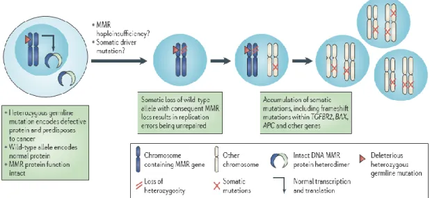
Figure 13. CRC development in individuals with LS , taken from Henry T. Lynch et al., 2015. A schematic
representation of the molecular mechanism of development of colorectal cancer (CRC) via pre-malignant colorectal polyps in individuals with Lynch syndrome (LS)
1.2.5 FAMILIAL ADENOMATOUS POLYPOSIS (FAP)
Familial adenomatous polyposis (FAP) is the second-most common inherited CRC syndrome with a prevalence of 1 in 10,000 individuals. Characteristic features of FAP include the development of hundreds to thousands of colonic adenomas, beginning in early adolescence, and inevitable CRC in untreated individuals. The genetic basis for FAP lies in germline (inherited) mutation of the adenomatous polyposis coli (APC) genewhich encodes a tumor suppressor that is part of the WNT signaling pathway (OMIM 175100). Approximately one-quarter of all cases are caused by new mutations that maintain the incidence of FAP [Bisgaard et al., 1994], despite the strong selective disadvantage of the disease. Germline mutations in the APC gene have been demonstrated in most FAP patients. [Cottrell et al.,
1992] The vast majority (95%) of APC mutations are nonsense or frameshift mutations that
result in a truncated protein product with abnormal function.
1.2.6 MUTYH-ASSOCIATED POLYPOSIS (MAP)
MAP is characterized by the presence of adenomatous polyposis of the colorectum and an increased risk of CRC. MAP is caused by biallelic mutations in MUTYH (also referred to as
23
MYH). The MUTYH gene product is part of the base-excision repair pathway, which is involved in defending against oxidative DNA damage. Functionally, MUTYH helps prevent G:C to T:A transversions caused by oxidative stress to highly mutagenic DNA bases. Colonic polyposis typically occurs by the time patients reach their 40s although polyps and cancer can occur at earlier ages. Biallelic MUTYH mutations have also been found in individuals with early onset CRC and few-to-no polyps [Cleary et al., 2009].
24
1.3 ANTICANCER ACTIVITY OF THE COPPER COMPLEXES
Copper assumes a critical role in various biochemical reactions including signaling, catalytic, regulatory and structural functions, for this reason is fundamental importance is to maintain the correct concentration inside the cells (free divalent metals approximatively 10-18 M) in order to guarantee normal physiological functions.
In humans, disruption of this tightly regulated cellular copper homeostasis affects normal tissue development and a growing number of reports indicate an alteration of essential metal ion homeostasis (metallostasis) in tumors. [Harrison et al., 2000.
Robinson and Winge 2010] In particular, copper is crucial to the angiogenic process that sustains tumor and metastasis development, and its sequestration underlies an anticancer strategy aimed at preventing establishment of the tumor.
Alternatively, copper complexes have shown antibacterial and anticancer activity, whereby the organic component (the ligand) is responsible for directing the metal to different molecular targets [Tardito et al., 2012]. For this reason, organic-copper compounds are systems of great interest in the field of inorganic and bioinorganic chemistry. Several small molecules able to bind metals, such as copper and zinc, have been proposed as anticancer agents [Ding et al., 2005. Ding and Lind 2009]. It has been suggested the such molecules can be distinguished in two classes: metal chelators able to remove metals from biologically active site, such as TPEN (Zn), and metal ionophores capable of transferring multiple metal ions across biological membranes such as hydroxyquinolines (Zn, Cu, Pb) [Ding et al., 2005. Ding and Lind 2009].
The effects of “metal chelator” can be contrasted by an increase in metal concentration while the metal-ionophore’s effect is potentiated in manner metal-dose-dependent by increasing the metal concentration inside the cells. In the last years some researchers are investigating the possibility to hint the homeostasis of transition metals inside the cells to kill cancer cells. In the present study we wanted to investigate if the copper dyshomeostasis induced by metal chelators and ionophores (compounds used are reported in Table n1 ) can address the tumor cells to programmed death and if the expression of components of the copper homeostasis functional network can modify the sensitivity to the cytotoxicity of copper and copper-binding compounds.
25 Table n1
Clioquinol (5-chloro-7-iodo-hydroxyquinoline, CQ) is a member of 8-hydroxyquinoline family with metal-ionophore function, it is known to exhibit a variety of biological activities such as antibacterial [Margalioth et al., 1983] and anticancer [Dìez et al., 1989] activities. CQ displays an anticancer effect in vitro [Mao
and Schimmer 2008. Mao et al., 2009] and in vivo [Chen et al., 2007] preclinical
models. CQ induces apoptosis in breast cancer cells through a caspase-dependent apoptotic pathway. [Daniel et al., 2005. Ding et al., 2006] Moreover, Clioquinol induces apoptosis in leukemia and myeloma cells by inhibiting histone Deacetylase activity. [Cao et al., 2013] In addition, Zhai and collaborators (2010) [Zhai et al.,
2010] demonstrated that both 8-OHQ and CQ/copper complexes are able to inhibit the
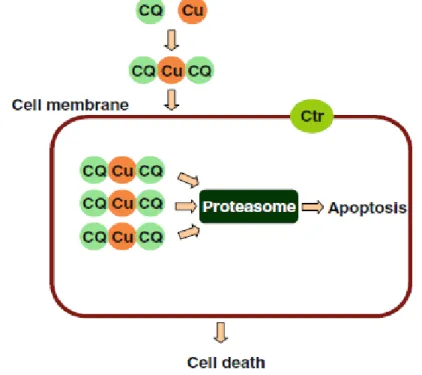
proteasome activity resulting in proliferation suppression and apoptosis in cultured breast cancer cells before cell death (Fig. 14). Recently a new mechanism, called parapoptotic, was described in Hela cells treated with the CQ-copper complex at 14
Clioquinol CQ
5-chloro-7-iodo-8 hydroxyquinoline
Chloro-hydroxyquinoline ClHQ
5-chloro-8-hydroxyquinoline
Hydroxyquinoline OHQ
8-hydroxyquinoline
TPEN
N,N,N’,N-tetrakis (2-pyridylmethyl)
ethylenediamine
Cl I H Cl H H26
µM. [Sperandio et al., 2000. Tardito et al., 2012] This phenomena is characterized by a massive cytoplasmic vacuolization due to accumulation of unfolded proteins in endoplasmic reticulum. [Sperandio et al., 2000] Conversely, the activation of the caspase-3 during the apoptotic process was observed in Hela Cells treated with CQ at the highest concentration 50 µM. [Tardito et al., 2012]
Figure 14. Probable model for clioquinol-induced apoptosis. The CQ binds the extracellular copper
and the copper-clioquinol complex induces apoptosis by the inhibition of proteasome activity.
On the contrary of 8-hydroxyquinoline derivatives, TPEN (N,N,N’,N-tetrakis (2-pyridylmethyl) ethylenediamine) is a cell permeable, high-affinity zinc and copper chelator. Different studies demonstrated that TPEN, at concentration higher than 1 µM induces apoptosis. In particular, was demonstrated that exposure of PC3 and DU 145 prostate cancer cells to TPEN (2-20 µM) activated caspase 3/7 activity. [Carrawayet al., 2012] Same results was obtained in cultured hippocampal neurons through
inhibition of ERK signalling pathway and activation of caspase-3. [Pang et al., 2013] Although TPEN can chelate all the endogenous transition metals such as zinc, iron, and copper, TPEN-induced apoptosis is most likely caused by chelating and thus depleting intracellular zinc, and possibly copper. This conclusion was deduced from the result that addition of equimolar copper or zinc, but not iron, blocked TPEN-induced apoptosis, and from the affinities of metals with TPEN (copper > zinc >iron). [Hyunet al., 2001] Recently it has been demonstrated that, in HCT116 colorectal cells,
27
TPEN induces cell death by chelating intracellular copper to produce TPEN-copper complex that engage in redox cycling and selectively induce the cell death. [Fatfat et
al., 2014] In particular TPEN’s ability to extract essential metals, particularly copper
from mitochondrial electron carriers facilitated the generation of superoxide and H2O2.
1.4 HIGH DENSITY OLIGONUCLEOTIDE ARRAYS
The microarray technology is based on the hybridization between target nucleic acids and probe nucleic acids (we call “target” the nucleic acid that is the object of the analysis and “probe” the known nucleic acid that is used as a tool for the analysis). The peculiarity of microarray is that millions of hybridization reactions to different probes are performed simultaneously. This technology is evolving rapidly and different platforms and several different solutions to processing and labeling the biological samples are commercial available. The Affymetrix GeneChips platform is most frequently used by scientists to detect practically every known human gene that is expressed and eventually turned into a protein [Auer
et al., 2009]. This platform helps researchers learn more about different diseases such as heart diseases, mental illness, infectious disease and especially the study of cancer. Until recently, different types of cancer have been classified on the basis of the organs in which the tumors develop. Now, with the evolution of microarray technology based on GeneChip probe, it will be possible for the researchers to further classify the types of cancer on the basis of the patterns of gene activity in the tumor cells. This will tremendously help the pharmaceutical community to develop more effective drugs as the treatment strategies will be targeted directly to the specific type of cancer.
The highly-automated manufacture process used by Affymetrix, combines chemistry and photolithography to build vertically (on each feature of the chip’s quartz surface) specific 25 nucleotides-long DNA probes, in a predetermined spatial orientation. The process is miniaturized (a typical glass chip is about 1.28 cm2 in size) to generate high-density arrays of oligonucleotide probes.
The GeneChip array is high-resolution array, is formed by approximately 500 thousand probe cells with size of 5 μm2 each, contains >6.0 million distinct probes covering coding and non-coding
transcripts. 70% of the probes on this array cover exons for coding transcripts, and the remaining 30% of probes on the array cover exon-exon splice junctions and non-coding transcripts [Affymetrix] (Fig.15). On average, 109 probes per gene are used. Since oligonucleotide probes are synthesized in known locations on the array, the hybridization patterns and signal intensities
28
can be interpreted in terms of gene identity and relative expression levels by Affymetrix GeneChip Operating Software.
Figure 15. Microarray Affymetrix Chips
1.4.1 THE GENECHIP HUMAN TRANSCRIPTOME ARRAY 2.0
The entire procedure can be subdivided in five main steps: A) Extraction of RNA;
B) Preparation and labeling of target cDNA;
C) Hybridization of target cDNA to oligonucleotide probes on microarray surface; D) Fluorimetric scan of the array;
E) Processing of hybridization signals and analysis of gene expression.
The first phase is the extraction of RNA form the biological sample. In this training the starting biological samples is a frozen colon cancer sample. We will extract total RNA by a method based on adsorption to a silica gel matrix.
29
The second phase is the preparation and labeling of target nucleic acid. The target nucleic acid is the nucleic acid that we want to analyze in our procedure. In this case we want to determine the levels of entire repertoire of cellular mRNAs and a large number of non-coding RNAs. However we must convert RNA in a form useful for the hybridization reaction. The protocol depends on the type of microarray used. The procedure for “Human Transcriptome Array 2.0” is summarized in the following in Figure 16.
Assay Workflow
Figure 16. Human Transcriptome Array 2.0 workflow.
In this protocol total RNA is first reverse transcribed to double-cDNA. The cDNA is converted in antisense cRNA by in vitro transcription. cRNA is then reverse-transcribed in single strand sense cDNA. Sense cDNA is then fragmented and labeled with biotin and
Sense-strand cDNA 5’ 3’ 5’ 3’ 3’ 5’ 3’ 3’ 5’ 5’ cDNA fragments 5’ 5’ B B 5’ B 5’ B Chip
Hybridization of labeled cDNA fragments to the array cDNA fragments labeled by
a reagent linked to biotin
from sense-strand cDNA to Array Hybridization
Sense-strand cDNA 5’ 3’ 5’ 3’ 3’ 5’ 3’ 3’ 5’ 5’ cDNA fragments 5’ 5’ B B 5’ B 5’ B Chip
Hybridization of labeled cDNA fragments to the array cDNA fragments labeled by
a reagent linked to biotin
from sense-strand cDNA to Array Hybridization
mRNA 5’ 3’ Double-strand cDNA 3’ 5’ 5’ 3’ cRNA 3’ 5’ Sense-strand cDNA 5’ 3’
30
represents the target DNA. In the third phase labeled target cDNAs are hybridized to oligonucleotide probes on the microarray surface. The chip probe array consists of a square glass (1.28 cm for side) mounted in a plastic cartridge. The array of oligo probes is fixed on the inner glass surface. A plastic chamber is under the glass and act a reservoir where hybridization occurs. After loading microarrays are incubated in an hybridization oven at 45°C for 16 hours with rotation at 60 rpm. During incubation sense target cDNAs will hybridize to the corresponding antisense oligoprobes. Then the Fluidic station will perform the staining to amplify the signal by three consecutive incubations: (the first incubation will be with streptavidin-phycoerythrin, the second incubation with biotinylated antibody against streptavidin and the third again with streptavidin-phycoerythrin). The fluidics station mix the staining solution by alternately draining and filling the cartridge. At the end the fluidic station expel the staining solution to the waste line and fill the cartridge with holding buffer for the fluorimetric scan by GeneChip Scanner 3000, that is an epifluorescent confocal microscope that
uses a solid‐state YAG laser (532 nm) to excite phycoerythrin bound to hybridized nucleic acids at 1.56 μm pixel resolution (www. affymetrix. com). In the fourth phase raw fluorescence signals are processed in order to perform an analysis of gene expression.
1.4.2 PROCESSING OF HYBRIDIZATION SIGNALS
AND ANALYSIS OF GENE EXPRESSION
The software AGCC ( Affymetrix GeneChip Command Console) represents the fluorescence intensity values from each pixel on the array in a grayscale or pseudocolor mode and creates Image data files (.DAT). Then the AGCC software superimposes a grid on the image to delineate the probe cells (features) and summarizes probe cell intensity data (CEL file generation).
The Expression Console software will use CEL files to create summarized expression values (.CHP file) by the Gene Level-SST-RMA analysis, that looks at the expression of the gene overall, giving us a RMA (Robust Multiple-Array) value for each Probe Set perfectly matched. Finally dedicated software will perform comparison of gene expression between different samples.
For instance, “Transcriptome Analysis Console” (TAC) by Affymetrix performs statistical analysis to obtain a list of differentially expressed genes and alternative splicing events. It provides the visualization of genes, exons, junctions and transcript isoforms. TAC software reports a Fold Change number (FC) that describes how much the signal changes from an first
31
group (for example CRC samples) to a second group (for example Mu samples). Moreover it gives us the relative FDR (false discovery rate) control, a statistical method used to reduce the number of false positives and to increase the chances of identifying all the differentially expressed genes.
The Transcriptome Analysis Console uses rma.alt-splice-sst.chp files, generated by Expression Console, for exon-level analysis estimating the alternative splicing events. The Splicing Index algorithm (SI) normalizes the exon and junction expression values by the level of gene expression and creates a ratio of normalized signal: The Splicing Index value. This value shows the normalized fold change (in linear space) of Condition1 vs. Condition2. High value of SI indicates that a particular Probe Selection Region (PSR) is preferentially included in a condition, but not in the other.
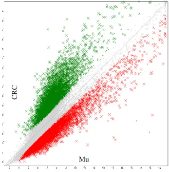
TAC software provides a series of Differential Expression Analysis Graphics such as: Scatter Plot, Chromosome Summary, WikiPathways and Hierarchical Clustering. The Scatter Plot (Fig. 17), for example, is a standard scatter plot graph of the comparison between two conditions. The two conditions that are compared are reported respectively in the Y and in the X axis. In this case we compare colon cancer samples with their matched normal colon mucosa. The gray point are the ones filtered out by the table, because their differential value between cancer and normal tissue is lower that a predetermined threshold (2 fold difference). The green points are genes up-regulated in colon cancer. The red points are genes down-regulated in colon cancer.
Figure 17. Scatter Plot Graph
CRC
AIM OF THE WORK
Copper, a catalytic cofactor required for the normal function of many enzymes involved in biological processes, is highly cytotoxic when in excess. Therefore its homeostasis and distribution is strictly regulated by a network of transporters (SLC31A1, SLC31A2) and intracellular chaperones (ATOX1, CCS and COX11) encoded by a group of genes that can be collectively called Copper-Homeostasis Genes (CHGs). Recent advances reveal that an alteration of copper homeostasis is very frequent in tumors supporting the hypothesis that can play an essential role in cancer angiogenesis.
The mutational and gene expression profiles associated to cancer are excellent analysis tools to identify target genes sensitive to drug therapy. Therefore, a first aim of this work was to investigate the presence of somatic mutations in CHGs in colorectal cancer, consulting the public database Cancer Genome Atlas Network. Such analysis, performed on 228 colorectal tumor samples, has revealed that inactivating mutations are extremely rare in CHGs.
The collection of whole transcriptome profiles by oligonucleotide microarrays was the second aim of the thesis. CHGs mRNA levels have been measured in 37 colorectal carcinoma samples and in their matched normal colonic mucosae. The transcriptome analysis offers the most advanced and comprehensive gene expression profiling tool for whole-transcript coverage available on any microarray platform. In this thesis, the use of the last Human Transcriptome Array (HTA 2.0, Affymetrix) allowed to analyze simultaneously 40.000 coding transcripts and 20.000 non-coding transcripts and to reveal variations of mRNA levels between colorectal cancer and normal colonic mucosa.
The alternative splicing is a biological phenomenon that determines the generation of several distinct transcripts, and consequently proteins, from a single gene and these alternative transcripts affect phenotypes. The transcriptome analysis by the last generation on oligonucleotide microarrays gives a possibility to analyse gene expression levels and single exon expression levels. In the 37 human colorectal samples, the exon-level expression analysis has been performed for the CHGs, such as SLC31A1 and SCO1, more expressed in colorectal cancer samples in comparison to normal colonic mucosa, in order to estimate the presence of alternative transcripts prevalent in a condition respect to other.
A third aim of the thesis was the evaluation of the role of the copper chaperone ATOX1 in sensitivity to copper chelating or ionophore drugs. In the last years, the research has been
33
focused on the possibility to use a class of a drugs, called “metal ionophores and chelators” with the aim to create an intracellular copper dyshomeostasis. “Metal ionophores” are molecules able to transfer a single metal ions across biological membranes in order to increase intracellular free metals while, in opposite sense, “metal chelators” are an another class of molecules that remove copper from biological active site in the intracellular proteins .
In this thesis we have analyzed the effects of copper addition on the toxic effects of some copper binding compounds, in particular, the ionophore copper-ionophore 5-chloro-8-hydroxyquinoline (ClHQ) and the copper chelator (N,N,N'N',-tetrakis (2- pyridylmethyl)ethylenediamine (TPEN) in two human colorectal cancer cell lines (Caco-2 and HT29).
ATOX1 expression was attenuated by treatment with short interfering RNAs in a colorectal cell line, Caco-2, showing a strong chromosomal aneuploidy, but a normal copy number and gene expression for ATOX1. Indeed, ATOX1 silencing enhanced the toxic effects of copper-ClHQ complexes and TPEN in Caco-2 cells confirming that the inhibition of copper chaperone Atox1 could be a good strategy to attenuate the cancer cell proliferation and to increase the anticancer effects of some copper binding drugs.
34
2. Material and methods
2.1. Human Cancer Cell Cultures
The following human cancer cell lines have been used: Human colon adenocarcinoma Caco-2
Human colon adenocarcinoma HT29 Human colon cancer HCT116
Human mammary adenocarcinoma MCF7 Human prostate cancer PC3
Human colon cancer Caco-2 (ATCC number: HTB-37) and HT29 (ATCC number: HTB-38) cell lines were maintained in D-MEM medium (Dulbecco's Modified Eagle Medium 1X; GIBCO, COD. 31965-023 containing 4.5g/L of D-glucose), supplemented with 10% FBS (Fetal Bovine Serum) and 100 U/ml of antibiotics (Penicillin-Streptomycin). The HCT116 (ATCC number: CCL-247) cell line was cultured in McCoy 5A+ (McCoy’s 5A Medium 1X GIBCO, COD. 36600021 containing 3 g/L of D-glucose) supplemented with 10%FBS (Fetal Bovine Serum), 1.5 mM L-glutamine and 2200 mg/L of sodium bicarbonate. Human mammary adenocarcinoma MCF7 (ATCC: HTB-22) were grown in Dulbecco's MEM (DMEM), 1.0 g/l D-glucose. Each medium was supplemented with 10% (vol/vol) heat-inactivated fetal bovine serum, 2mM L-Alanyl-L-Glutamine, penicillin-streptomycin (50 units-50 g for ml). Human prostate cancer cells PC3 (ATCC number: CRL-1435) were grown in DMEM/F12 (GIBCO, Cod. 21331) supplemented with 10%FBS (Fetal Bovine Serum). The cell cultures were grown in dishes (100 mm) and incubated at 37 °C in humidified atmosphere with 5% of CO2 and 95% of air. The culture medium was changed twice a week.
35
2.2 RNA Interference
Caco-2 cells were plated on 96/6-well plates (Nunclon TM Microwell TM" (Nunc)) in D-MEM with 10% FBS(fetal bovine serum) and 100 U/ml of antibiotics (Penicillin-Streptomycin). At 60% confluence, 25nM of small interfering RNA (siRNA) targeted to the copper chaperone Atox-1 was added, according to manufacturer’s guidelines, in transfection medium (Opti-MEM I, Ca.Nu. 51985-026 Gibco®) containing the transfection reagent, Lipofectamine™ RNAiMAX (Cat. 13778 INVITROGEN) for 24 and 48h. Nonspecific siRNA was used as control under identical conditions. The silencing of Atox1(NM_004045) was performed with small interfering RNA (siRNA), this molecule and its scramble were designed using siRNA Design Tool (MWG).
The targeted sequence of Atox1 and its scramble were as follows:
siRNAAtox1 206 MWG: CAAGAAGGUCUGCAUUGAA (19bp) scrRNAAtox 206 MWG: CAACGGUACGGAUUAAUAG (19bp)
The siRNA206 was designed on exon 3 to 110 bp from ATG site and to 96bp from TAG site (Fig.18).
Figure 18. ATOX1 mRNA sequence. NCBI Reference Sequence:NM_004045.3
Highlighted in black the primers for quantitative amplification by PCR.
The cell line Caco-2 was transfected with siAtox1 and scrAtox1 following the forward transfection procedure by protocol Stealth ™ RNAi or siRNA by Lipofectamine™ RNAiMAX reagent (Cat. 13778 INVITROGEN). The cells were seeded on six-well plastic plates at a density of 7.0x10⁴ cells/well, for the RNA extraction that was performed using the commercial RNeasy
Mini Kit (Qiagen), and on ninety-six well plastic plate at a density of 2.5-3x10³ cells/well, to test
cell viability by MTT (3-(4,5 dimethylthiazol-2-yl)-2,5-diphenyltetrazolium bromide) colorimetric assay.
36
2.3 MTT Proliferation Assay
Caco-2 cell line (2.5-3x10³ cells/0.33cm2) were plated in a 96-well microplates "Nunclon TM
Microwell TM" (Nunc) and were incubated at 37 °C. After 24 hours cells (60% confluence) were transfected with 25nM siRNA or scrRNA in 100 μl Opti-Mem (Opti-MEM I, Cat. No. 51985-026 Gibco®) containing the transfection reagent, Lipofectamine™ RNAiMAX (Cat.No. 13778 Invitrogen, Carlsbad CA) for 10 hours. At 24 hours from transfection (time line experimental condition) cells were treated with Copper(II) nitrate, [Cu(NO3)2] at increasing concentrations
(0.01-0.1-1-10-50-100 μM) and/or with the copper-ionophore 5-chloro-8-hydroxyquinoline (ClHQ, Sigma Aldrich Inc) and/or the copper chelator (N,N,N'N',-tetrakis (2- pyridylmethyl)ethylenediamine (TPEN), Santa Cruz Biotechnology). Short incubations (2 hours) with copper and/or drugs were performed in 100 μl of PBS (Phosphate-buffered saline) supplemented with 0.9 mM Calcium Chloride (FLUKA Cat. No. 21014), 0.5 mM Magnesium Chloride (MERCK Cat. No. TA575835) and 5.5 mM D-glucose (SIGMA Cat. No. G-7528). This copper complexes were been premixed and subsequently were added to the cells. After 2 hours of incubation the PBS was removed and replaced by fresh standard growth medium (DMEM+10% FBS). Cells treated with 0.1% of DMSO were used as controls.
Time-line for silencing experimental condition
Microplates were incubated at 37 °C in humidified atmosphere of 5% CO2, 95% air and the
cellular growth inhibition and/or the cellular cytotoxicity has been valuated after 48 hours from the treatment by colorimetric assay based on the use of tetrazolium salt MTT (3 (4,5 dimethylthiazol-2-yl),-2,5-diphenyl tetrazolium bromide) [Mosmann 1983]. Solubilization of the converted purple formazan dye was accomplished by adding 150ul/well of DMSO
CELL PLATING IN GROWTH MEDIUM
TRANSFECTION WITH SI/SCR ATOX1 IN OPTI-MEM FOR 10 HOURS. AFTER 10 HOURS OF SILENCING : *REMOVING OF OPTI-MEM AND GROWTH MEDIUM ADDITION *READING OF T0 REMOVING OF D-MEM AND PBS ADDITION TREATMENT WITH COPPER CPMPLEXES FOR 2H CHANGING PBS WITH GROWTH MEDIUM READING PLATES AT 24H FROM TREATMENTS READING PLATES AT 48H FROM TREATMENTS 1 DAY 3 DAY 2 DAY 4 DAY 5 DAY
37
(dimethyl sulfoxide). The results were read on a multiwell scanning spectrophotometer (Multiscan reader), using a wavelength of 570 nm.
Each value was the average of 8 wells (standard deviations were less than 10%). The IC50
values were calculated by the nonlinear regression analysis, using the GraphPad Prism 6.0 software. Dose-response curves were obtained by the equation: log (inhibitor) vs. normalized response and the statistically significant results have been calculated by GraphPad Prism 6.0 software that has reported "95% Confidence Intervals" values. The cytotoxicity effect was calculated according to NCI when the optical density of treated cells was lower than the T0 value using the following formula: 100x (T-T0)/T0<0. (T is the optical density of the test well after a 24-48 h period of exposure to test compound; T0 is the optical density at time zero). When the optical density of treated cells was lower than the T0 value the following formula was used: 100 x (T-T0)/T0. The percentage of controls was calculate to determine cell viability and proliferative effect after a 24-48 h period of exposure to test compound.
2.4 Analyze of RNA expression by qRT-PCR
2.4.1 Purification of total RNA and reverse transcription
Caco-2 cells were plated on six-well plates at a density of 7.0x10⁴ cells/well and siRNA transfection was performed as described in the previous section. Total RNA was extracted from control cells and treated cells with siAtox1 of colorectal carcinoma cell line (Caco-2), the extraction was performed using the commercial RNeasy Mini Kit (Qiagen) according to manufacturer’s recommendations. Total RNA recovered from each sample (Ctrl, siAtox, scrAtox1) was quantified by means of a double reading performed by the NanoDrop spectrophotometer. Reverse transcription was performed using total RNA, RNaseH reverse transcriptase (Superscript II, Gibco BRL, Life Technologies, Gaithersburg, MD) and random primer hexamers.
2.4.2 Quantitative Real Time –PCR
Quantitative real-time PCR analysis was performed using StepOne™ Real-Time PCR System by Applied Biosystems(Applied Biosystems, Foster City, CA,USA). Three sequence-specific oligonucleotides (Table 2) were designed using the qPCR Probe design software (Eurofins MWG Synthesis GmbH, Ebersberg) based on the sequence of target genes: Atox1, CCND1 (NCBI