Original Paper
© 2017 The Author(s) Published by S. Karger AG, Basel
Gene Expression and Apoptosis Levels
in Cumulus Cells of Patients with
Polymorphisms of FSHR and LHB
Undergoing in Vitro Fertilization Program
Liana Boscoa Giovanni Ruvolob Claudio Luparelloa Stefania FerraricDomenico Valeriod Daniele Santie Paola Piombonif Elena Sarcinag
Monica Lispih Maria Carmela Roccheria
aUniversità degli Studi di Palermo, Dipartimento di Scienze e Tecnologie Biologiche, Chimiche e
Farmaceutiche (STEBICEF), Palermo, bCentro di Biologia della Riproduzione, Palermo, cFondazione
IRCCS Ca’ Granda Ospedale Maggiore Policlinico, Infertility Unit, Milano, dIstituto di Ricerche Genetiche
(IRG), Napoli,eUniversità di Modena e Reggio Emilia, Dipartimento di Scienze Biomediche, Metaboliche
e Neuroscienze, Unità di Endocrinologia, Modena, fUniversità di Siena, Dipartimento di Medicina
Molecolare e dello Sviluppo, Siena, Centro Diagnosi e Terapia Sterilità di Coppia, Policlinico S. Maria alle Scotte , Siena, gCentro Medico San Luca, Bari, hMerck Serono S.p.A, Italia
Key Words
Cumulus cells • Polymorphism • FSHR • LH • Apoptosis • Gene expression
Abstract
Background/Aims: FSH receptor (FSHR) Ala307Thr and Asn680Ser and LHβ chain (LHB)
Trp28Arg and Ile35Thr polymorphisms affect the response to pharmacological ovarian stimulation with r-FSH in women undergoing assisted reproductive treatment (ART). Here, we evaluated the expression level of selected genes involved in follicle maturation and the possible onset of apoptosis in cumulus cells of patients with single and double FSHR and
LHB polymorphisms, as potential markers of oocyte competence. Methods: Cumulus cells
from 36 stimulated patients were collected and SNP genotyping performed by PCR. Gene expression was evaluated through real-time PCR, and apoptosis estimated via TUNEL assay, and cleaved caspase-3 and pAKT immunostaining. Results: The cumulative data show significant correlations indicating that the genetic alteration of FSHR and/or LHB genes may lead to perturbations of the signaling network programmed to granulosa cell survival and follicle development. Notably, when double heterozygotes were compared to the rest of the patients, a higher level of apoptosis in terms of both DNA fragmentation index and amount of active caspase-3 was observed in cumulus cells. Conclusions: These results may help to define personalized stimulation protocols in ART programs, to increase the success rate of ICSI procedures in accordance with the polymorphic condition of the individual patient.
Liana Bosco Università di Palermo, Dipartimento di Scienze e Tecnologie Biologiche
Chimiche e Farmaceutiche (STEBICEF), Edificio 16, Viale delle Scienze Palermo (Italia) E-Mail [email protected]
Introduction
It is well-known that gonadotropins are key protagonists in folliculogenesis and specific polymorphisms of the genes encoding for such hormones and their receptors may influence the growth of follicles and oocytes, also in relation to the pharmacological stimulation of ovulation. Some polymorphisms of the receptors for FSH (FSHR) and of the β subunit of LH (LHB) have been extensively studied [1] and, in some cases, it was possible to establish that the presence of specific allelic combinations should be taken into consideration for personalized stimulation protocol [2, 3]. Regarding FSHR, the Ala307Thr (A307T) and Asn680Ser (N680S) polymorphisms located in exon 10 and resulting in aminoacidic substitutions in the intracellular FSH binding- and extracellular adenylate cyclase coupling domains of the receptor, respectively, are the most commonly found. Being in linkage disequilibrium, these polymorphisms may give rise to two different combinations of receptor isoforms, i.e. A/S and T/N [4]. Previous studies demonstrated a prominent interrelation between FSHR genotype and FSH-induced ovarian response, which may affect the efficacy of the stimulation protocol. As an example, in vitro fertilization (IVF) patients, in the case of S/S combination, show both an increased FSH baseline level and a resistance for ovarian stimulation thereby needing a higher amount of FSH administration or a higher starting dose [5].
Also the LHB variants appear to regulate the follicular growth and maturation. It is known that LH-triggered up-regulation of androgens may influence follicular metabolism promoting the acquisition of FSH sensitivity [6]. The two more studied polymorphisms of
LHB are represented by the missense nucleotide substitutions in exon 2 Trp28Arg (W28R)
and Ile35Thr (I35T), which are often found to occur concurrently, that generate an extra glycosylation signal in the protein subunit thereby affecting protein folding and hormonal activity [7]. Owing to the lower bioactivity of mutated LH, the carriers of such polymorphic variant of LHB, named v-βLH, necessitate higher r-FSH administration during controlled ovarian stimulation [8]. No studies have been designed so far relating the polymorphic variants of FSHR and LHB with oocyte competence.
The efficacy of IVF treatments is evaluated considering the rate of healthy children born, nonetheless reproductive biology provides diverse means to estimate the comparative success of an IVF protocol. Among the non-invasive tools, it is possible to include specific assays on cumulus cells; these cells are in close molecular communication with the female gamete and therefore represent an interesting investigative instrument in embryology [9]. Both the expression levels of some components of intracellular survival pathways and the apoptosis rate can be assessed in cumulus cells and utilized as potential markers of oocyte competence. The amounts of the mRNAs for the epidermal growth factor (EGF)-like factors amphiregulin (AREG) and epiregulin (EREG) have been shown to increase in gonadotropin (especially LH)-treated primary human granulosa cells [10]. The protein products, once released from the cell membrane by proteolysis, can bind the EGF receptor (EGFR) and induce cumulus expansion and oocyte maturation via mitogen-activated protein kinases (MAPK)-1 and -3 pathways [11, 12]. Also, EGFR expression levels have been correlated to the maturative capacity of oocytes [13]. In addition, the expression levels of gonadotropin receptors such as FSHR and luteinizing hormone/human chorionic gonadotropin receptor (LHCGR), the latter being an adenylate cyclase activator, have been proven to regulate key events such as follicular growth, dehiscence and the activity of corpus luteum via promotion of EGF-like factor accumulation [12, 14]. Among the molecules implicated in signal transduction, phospho-AKT (pAKT) is a well-known activated serine/threonine kinase involved not only in life/death decisions but also in the control of metabolism and differentiation, as shown in both normal and pathologic cell systems (e.g [15, 16]). In the ovary, it is known that Kit . ligand induces maintenance of primordial follicular pools via phosphatidylinositol 3-kinase (PI3K)/Akt signalization, whereas Phosphatase and Tensin Homolog (PTEN)-induced Akt dephosphorylation in granulosa cells can be observed during terminal follicular growth [17, 18].
Concerning the evaluation of the onset of apoptosis, the terminal deoxynucleotidyl transferase (TdT) dUTP nick-end labeling (TUNEL) assay, which provides information on DNA fragmentation at single cell level [19], has been widely used to quantitate the number of apoptotic cells, including those present in oocyte, cumulus cell, and spermatozoa preparations [20-23]. This analysis has been also supplemented by parallel immunohistochemistry with anti-cleaved caspase-3, thereby evaluating the extent of activation of the ubiquitously-expressed “effector” enzyme, which is a widely-acknowledged sensitive tool for the in situ detection of apoptotic [24].
In this study, we wanted to check whether a correlation exists in the extent of gene expression and/or apoptosis markers from cumulus cells of oocytes among patients with specific gonadotropin polymorphisms, after ovarian stimulation with r-FSH. Our work hypothesis is that the presence of specific polymorphisms of FSHR and LHB genes might be reflected in an alteration of specific pathways influencing the oocyte competence. In particular, the following four SNPs have been taken into consideration: for FSHR, those identified as A307T and N680S; for LHB, those identified as W28R and I35T.
Materials and Methods Study design and patients
A longitudinal, prospective clinical trial was carried out in three assisted reproductive treatment (ART) Centres in Italy (Palermo, Siena and Bari).
Thirty-six women attending the ART clinic because of fertility problems, were consecutively recruited according to the following inclusion and exclusion criteria. The inclusion criteria were: age < 38 years, normal FSH basal level (<12 UI/mL), body mass index (BMI) <28 kg/m2, normoresponder patients with a
minimum of 6 oocytes collected at pick-up. Only one inclusion criterion for the male partner was provided, i.e. a motile sperm count higher than 4x10⁶ per ejaculate. Women who underwent multiple attempts of
assisted reproduction were included in the study. The occurrence of endometriosis, even if non-overt, in r-FSH-treated women was considered as an exclusion criterion. Such inclusion and exclusion criteria were chosen to minimize bias and help ensure the comparability of the individuals analyzed, so that the results of our study may be related mostly to polymorphism-related conditions.
Ovarian stimulation
All women recruited were similarly treated with a GnRH agonist Buserelin (Suprefact, Sanofi-Aventis, Italy, 0.2 ml/day) administration, starting on day 21 of the previous cycle. Administration of 150–225 IU r-FSH/day (Gonal-f, Merck, Rome, Italy) was started at day 8 after GnRH agonist treatment and the follicular growth was monitored every two days using ultrasound and serum estradiol E2 levels, starting on day 6 of stimulation, modifying the dose of r-FSH as a consequence. The dose of 10, 000 IU of hCG (Ovitrelle; Merck, Rome, Italy) was administered when at least 3 follicles showed a diameter ≥ 18 mm.
Extraction of genomic DNA and genotyping of FSHR and LHB single nucleotide polymorphisms (SNPs) Genomic DNA was obtained from samples of whole blood with MagNA LC 2.0 instrument (Roche, Monza, Italy). The genotyping of SNPs was performed by PCR amplification in the presence of 250 nM of each primer, 2.5 U of AmpliTaq Gold DNA polymerase and 180 ng DNA in an Applied Biosystem 2720 thermocycler (Life Technologies). The PCR primers were designed using the Primer3 software available at http://biotools.umassmed.edu/bioapps/primer3_www.cgi.
The primers used for detection of rs6165 (polymorphism A307T) and rs6166 (polymorphism N680S) FSHR SNPs were: FSHR_A307T_F (5’-AAGCAGCATTAACCCTTGAG-3’), FSHR_A307T_R (5’-TCTTCACATGGGTTGAATGC-3’), FSHR_N680S_F (5’-ATTTCTGCCTCCCTCAAGGT-3’) and FSHR_N680S_R (5’-GAAGCACTGTCAGCTCTTTGTG-3’). The expected sizes of the two amplification products were 427 e 440 bp, respectively. The primers used for detection of rs1800447 (polymorphism W28R) and rs34349826 (polymorphism I35T) LHB SNPs were LHB_EX2_F (5’-AGGGTGGGGATCTGAAATG-3’) and LHB_EX2_R (5’-TGAGCTCCCAAGCTGACC-3’). The expected size of the amplification product was 557 bp. Thermocycler conditions were as follows: 94° C for 8 min., 2 cycles of 94° C for 1 min., 55° C for 1 min. and 72° C for 1 min.,
33 cycles of 94° C for 30 sec., 55° C for 30 sec. and 72° C for 30 sec., with a final extension at 72° C for 7 min. The PCR products were visualized on a 2% agarose gel stained with 50 µg/ml of ethidium bromide under UV light and their size compared with a 100 bp DNA ladder (Invitrogen).
For restriction fragment length polymorphism (RFLP) analysis, the amplification products were digested with Fermentas FastDigest restriction enzymes as follows: FSHR-A307T with Eam1105I (37°C, 1 h.); FSHR-N680S with BseNI (65°C, 1 h.); LHB-Ex2 with NcoI (37°C ,1 h.) to detect W28R polymorphism and with FokI (37°C, 1 h.) to detect I35T polymorphism. The digestion products were checked by 2% agarose electrophoresis and ethidium bromide stain and their size compared with a 100 bp DNA ladder (Invitrogen). The digestion patterns of the different restriction assays were as follows: (i) FSHR-A307T/Eam1105I: 285 bp + 138 bp for A/A genotype; 254 bp + 138 bp + 31 bp for T/T genotype and 285 bp + 254 bp + 138 bp + 31 bp for A/T genotype. (ii) FSHR-N680S/BseNI: 279 bp + 157 bp for S/S genotype; 440 bp for N/N genotype and 440 bp + 279 bp + 157 bp for S/N genotype. (iii) LHB-W28R/NcoI: 275 bp + 182 bp + 100 bp for W/W genotype; 375 bp + 182 bp for R/R genotype and 375 bp + 275 bp + 182 bp + 100 bp for W/R genotype. (iv) LHB-I35T/FokI: 273 bp + 160 bp + 81 bp + 43 bp for I/I genotype; 316 bp + 160 bp + 81 bp for T/T genotype and 316 bp + 273 bp + 160 bp + 81 bp + 43 bp for I/T genotype.
The PCR fragments were also be checked by modified Sanger’s sequencing reactions to ensure the correct match between genotype and RFLP.
Preparation of cumulus cells
Sample preparation was performed as reported by [20]. Briefly, after ovum pick-up, the cumulus-oocyte complexes were incubated with 80 IU hyaluronidase/ml (Medicult, Jyllinge, Denmark; Irvine Scientific, California, USA) for 1 min and cumulus cells were then mechanically released by gently pipetting with a 170 micron denuding pipette. Cumulus cells, deriving from the patient-specific pool of available COCs, were divided into two parts: one was transferred in a 1.5 ml vial containing 500 µl of TRI reagent™ (Sigma, St.Louis, MO/USA) and stocked at -80°C for gene expression analysis, whereas the other was collected in a test tube (Falcon, Franklin Lakes, NJ) containing 2 mL of medium with HEPES (SAGE IVF, Trumbull, CT/USA; Irvine Scientific, California, USA) and centrifuged twice for 7 min. at 800 rpm and then fixed in 3.7% paraformaldehyde for 1 h. Then, cells were centrifuged for 7 min. at 2000 rpm and suspended in PBS after the removal of the supernatant. The cytospin method was used to mount cells on polylysine-coated glass slides to perform in situ immunocytochemistry and TUNEL assay. Oocytes were transferred to fertilization medium (SAGE IVF; Irvine Scientific, California, USA) and incubated at 37°C and 6% CO2, until intracytoplasmic sperm injection (ICSI).
Gene expression assay
RNA extraction was performed with TRI reagent™ according to manufacturer’s instructions starting from 500 μl thawed aliquots and resuspending extracted RNA in 25 μl of diethyl pyrocarbonate (DEPC)-treated water. One µg of RNA was reverse transcribed with the GoScript Reverse Trascription System (Promega Italia, Milan, Italy) according to manufacturer’s instructions and gene expression analysis was performed using Real-time PCR. Briefly, a protocol with TaqMan® Gene Expression Master Mix and Assay
on Demand on a 7700 Real-time PCR System (Applied Biosystems Italia, Monza, Italy) was used with the following cycle characteristics: 2 minutes at 50°C, 10 minutes at 95°C and 40 cycles with 15 second at 95°C and 1 minutes at 60°C. The expression levels of LHCGR (Hs00896336_m1), FSHR (Hs00174865_m1), EREG (Hs00914313_m1), EGFR (Hs01076078_m1), AREG (Hs00950669_m1) and HPRT1 (Hs99999909_m1) genes, the latter as endogenous control, were investigated. Data were analyzed using the comparative Ct method, where Ct is defined as the cycle number in which fluorescence first crosses the threshold. ΔCt was found by subtracting the endogenous gene Ct values from the values of the target genes. The result was applied to the term 2(-ΔCt). The personnel involved in the procedure were blinded to the IVF treatment and
genetic analyses outcomes.
Fluorescent in situ TUNEL assay
In order to assess the extent of DNA apoptotic fragmentation [25], cumulus cells were washed in PBS and permeabilized at 4°C in 0.1% Triton X100 plus 0.1% sodium-citrate in PBS at 4°C, and then washed three times in PBS at room temperature. Subsequently, cells were incubated for 60 min. at 37°C in a humidified chamber in 50 µl of a mixture containing 5 µl of nucleotide mix, 1 µl of TdT enzyme, and 45 µl of equilibration buffer (DeadEndFluorometric TUNEL System, Promega Italia, Milan, Italy). Negative
control was made by incubating the cells with the same mixture without the TdT enzyme, whereas positive control another by treating cells briefly with 10 U/ml DNAse I. The reaction was blocked with SSC, followed by exhaustive washing in PBS. The cumulus cells were counterstained with propidium iodide (1µg/ml) and observed under an Olympus BX 50 microscope equipped with a reflected light fluorescent attachment (Olympus), and a 20× 0.40 objective.
Immunofluorescence in situ assays
For immunodetection, cells were washed in PBS and permeabilized for 10 minutes at 4°C in a solution of 0.1% Triton X-100 plus 0.1% sodium citrate in PBS. Then, cells were washed three times in PBS and incubated overnight at 4°C with anti-pAKT polyclonal antibody (1:50 dilution, Santa Cruz Biotechnology, Santa Cruz, CA, USA), and anti-cleaved caspase-3 polyclonal antibody (1:50 dilution, Cell Signaling Technology, Danvers, MA/USA) dissolved in 3% BSA in PBS. The primary antibody was omitted in negative controls. After three rinses with PBS, incubation with the secondary antibody, anti-rabbit IgG (whole molecule) F(ab)2 fragment-Cy3 (1:50 dilution, Sigma), was performed for 1 h. The cumulus cells were counterstained for 10 min. with Hoechst 33342 (Invitrogen), mounted in 10 μl DABCO solution (deionized H2O, 1 M Tris-HCl pH 8, 2 mM DABCO, glycerol) and observed under an Olympus BX 50 microscope equipped with a reflected light fluorescent attachment (Olympus), and a 20×0.40 objective. Densitometric analysis of fluorescent signals was performed using NIS-Elements BR 3.10 image analyzer software (Nikon) as reported by [26].
Statistical Analysis
This is an exploratory analysis and genotype frequencies of SNPs and Hardy-Weinberg equilibrium were evaluated by SNPStats software [27] available at http://bioinfo.iconcologia.net/SNPstats.
Each parameter was considered according to genotypes of each SNP evaluated. Kolmogorov-Smirnov test was used for evaluation of variable distribution and differences for continuous variables among groups were evaluated performing univariate ANOVA, for variables normally distributed, and Kruskal-Wallis or Mann-Whitney test for not-normally distributed ones. Dunnet test was used as post-hoc test. Spearman’s coefficient regression was selected because the normal distribution of parameters was not requested for this evaluation. Moreover, the same analyses were repeated comparing women with SNP heterozygosis on both FSHR and LHB with all other women.
Statistical analysis was performed using the ‘Statistical Package for the Social Sciences’ software for Macintosh (version 20.0; SPSS Inc., Chicago, IL). Statistical significance was considered for p-values <0.005.
Results
Patients: clinical characteristics, r-FSH dosage and treatment response
Thirty-six women were recruited with a mean age of 34.36±2.60 years and a mean BMI of 22.05±2.91 kg/m2. Both age and BMI were normally distributed and did not differ among genotypes, considering FSHR p.A307T (p=0.384 and p=0.682, respectively), FSHR p.N680S (p=0.384 and p=0.682, respectively), LHB p.W28R (p=0.956 and p=0.372, respectively) and LHB p.I35T (p=0.956 and p=0.372, respectively).
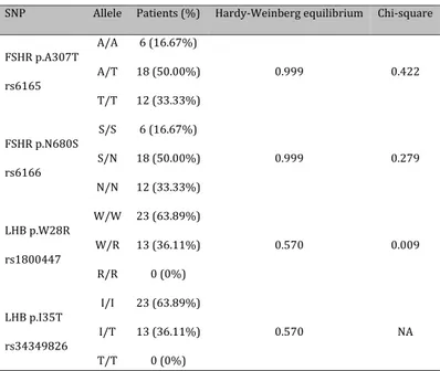
Table 1. Genotype frequencies
SNP Allele Patients (%) Hardy-Weinberg equilibrium Chi-square FSHR p.A307T rs6165 A/A 6 (16.67%) 0.999 0.422 A/T 18 (50.00%) T/T 12 (33.33%) FSHR p.N680S rs6166 S/S 6 (16.67%) 0.999 0.279 S/N 18 (50.00%) N/N 12 (33.33%) LHB p.W28R rs1800447 W/W 23 (63.89%) 0.570 0.009 W/R 13 (36.11%) R/R 0 (0%) LHB p.I35T rs34349826 I/I 23 (63.89%) 0.570 NA I/T 13 (36.11%) T/T 0 (0%)
The total r-FSH dosage, although not normally distributed, did not differ among genotypes considering each genotype, such as FSHR p.A307T (p=0.783), FSHR p.N680S (p=0.783), LHB p.W28R (p=0.471) and LHB p.I35T (p=0.471). The response to FSH treatment was evaluated by the number of retrieved oocytes, which did not differ among genotypes, considering FSHR p.A307T (p=0.975), FSHR p.N680S (p=0.975), LHB p.W28R (p=0.709) and LHB p.I35T (p=0.709). Moreover, no differences were observed between women showing heterozygosity in all SNPs (p=0.796).
SNP genotyping
Four SNPs were genotyped and their allele frequencies were shown in Table 1. Genotypic distribution of polymorphisms was consistent with the Hardy-Weinberg equilibrium. No differences were observed when comparing FSHR p.A307T and FSHR p.N680S SNPs to general population using the International HapMap project. Conversely, LHB p.W28R SNP frequency significantly differed from that of general population (p=0.009), even if the only data available in literature refer to Utah residents with Northern and Western European origin. Concerning LHB-I35T SNP frequency, no data in the general population were available. The most frequent haplotype, with a cumulative frequency of 52.24%, was the following:
FSHR p.A307T-p.S680N, p.T307-p.N680, LHB p.W28-p.I35, p.W28R-p.I35T.
Genotype distributions and combinations found in the group of patients studied are shown in Fig. 1. According to genotype frequencies, four models were generated: codominant, dominant, recessive and over-dominant.
Haplotype analysis: clinical characteristics, oocyte retrieval and zygote numbers
No significant differences among haplotypes were found for age (p=0.110), BMI (p=0.089) and r-FSH dosage (p=0.490). The number of arrested metaphase I (MI) oocytes retrieved was
Fig. 1. Schematic representation of the dis-tribution and combination of the genotypes which have been the object of the reported study.
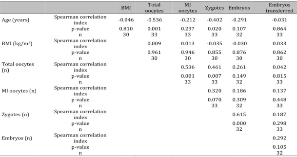
Table 2. Correlation analysis of women’s clinical parameters
BMI oocytes Total oocytes MI Zygotes Embryos transferred Embryos Age (years) Spearman correlation index -0.046 -0.536 -0.212 -0.402 -0.291 -0.031
p-value 0.810 0.001 0.237 0.020 0.107 0.864
n 30 33 33 33 32 33
BMI (kg/m2) Spearman correlation
index 0.009 0.013 -0.035 -0.030 0.033
p-value 0.961 0.946 0.855 0.876 0.862
n 30 30 30 30 30
Total oocytes
(n) Spearman correlation index 0.536 0.461 0.261 0.042
p-value 0.001 0.007 0.149 0.815
n 33 33 32 33
MI oocytes (n) Spearman correlation index 0.320 0.186 0.137
p-value 0.070 0.309 0.448
n 33 32 33
Zygotes (n) Spearman correlation index 0.615 0.187
p-value 0.000 0.298
n 32 33
Embryos (n) Spearman correlation index 0.292
p-value 0.105
significantly different considering all SNPs evaluated. In particular, the highest number of MI oocytes was obtained when the following haplotypes were found: FSHR p.A307T-p.N680S, p.A307-S680, LHB p.W28R-p.I35T, p.R28-T35 (3.77; 95% CI: 1.63 – 5.92, p=0.002). More in detail, the highest number of MI oocytes was retrieved when a heterozygous haplotype was present in both LHB p.W28R and LHB p.I35T (2.10; 95% CI: 0.14 – 4.05, p=0.044). Similarly, the number of recorded zygotes were the highest in the same haplotypes: FSHR p.A307T-p. N680S, p.A307-S680, LHB p.W28R-p.I35T, p.R28-T35 (1.85; 95% CI: 0.09 – 3.62, p=0.047).
Haplotype analysis: gene expression, apoptosis and pAKT levels in cumulus cells
After oocyte pick-up and decoronization, the pool of cumulus cells obtained from mini-mum six oocytes for each patient was submitted to evaluation of the expression levels of genes encoding for EGFR, EGFR ligands and gonadotropin receptors. In addition, their DFI, and both cleaved caspase-3 and pAKT accumulation were estimated.
The expression of EGFR was significantly different considering the haplotypes of all SNPs evaluated. In particular, the highest expression was found when the following haplotypes were found: FSHR p.A307T-p.N680S, p.T307-N680, LHB p.W28R-p.I35T, p.R28-p. T35 (0.18; 95% CI: 0.03 – 0.34, p=0.025). Moreover, the lowest levels were found when the following haplotype was present: FSHR p.A307T-p.N680S, p.A307-S680, LHB p.W28R-p. I35T, p.R28-T35 (-0.17; 95% CI: -0.034 – -0.01, p=0.049).
The expression of AREG was significantly different considering the haplotypes of all SNPs evaluated. In particular, the highest expression was found when the following haplotypes were found: FSHR p.A307T p.N680S, FSHR p.A370-S680, LHB p.W28R p.I35T and
LHB p.R28-p.T35 (3588.96; p<0.001). More in detail, the expression of AREG was highest in
the recessive model (T/T-A/T vs. A/A) of FSHR p.A307T (4066.07; 95% CI: 663.44 – 7468.70,
p=0.026), as well as in the recessive model (N/N-S/N vs. S/S) of FSHR p.N680S (4066.07;
95% CI: 663.44 – 7468.70, p=0.026).
No significant differences among models were found for expression of LHCGR (p=0.059),
FSHR (p=0.280) and EREG (p=0.074).
The percentage of apoptotic cells, as evaluated by DFI via TUNEL assay, was not significantly different among haplotypes (p=0.099). However, the highest percentage of apoptotic cells (11.06; 95% CI: 2.54-19.58, p=0.016) was found in the over-dominant model for both FSHR p.A307T (T/T-A/A vs. A/T) and FSHR p.N680S (N/N-S/S vs. S/N).
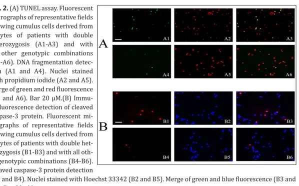
Fig. 2. (A) TUNEL assay. Fluorescent micrographs of representative fields showing cumulus cells derived from oocytes of patients with double heterozygosis (A1-A3) and with all other genotypic combinations (A4-A6). DNA fragmentation detec-tion (A1 and A4). Nuclei stained with propidium iodide (A2 and A5). Merge of green and red fluorescence (A3 and A6). Bar 20 µM.(B) Immu-nofluorescence detection of cleaved caspase-3 protein. Fluorescent mi-crographs of representative fields showing cumulus cells derived from oocytes of patients with double het-erozygosis (B1-B3) and with all oth-er genotypic combinations (B4-B6). Cleaved caspase-3 protein detection
(B1 and B4). Nuclei stained with Hoechst 33342 (B2 and B5). Merge of green and blue fluorescence (B3 and B6). Bar 20 µM.
The percentage of cleaved caspase-3 was significantly different considering the haplotypes of all SNPs evaluated. In particular the highest values were found when the following haplotypes were found: FSHR p.A307T-p.N680S, FSHR p.A307-S680, LHB p.W28R-p. I35T, LHB p.R28-p.T35 (2.74; 95% CI: 0.75-4.72; p=0.010). More in detail, the percentage of cleaved caspase-3 was highest in the dominant model (T/T vs. A/T-A/A) of FSHR p.A307T (1.87; 95% CI: 0.24-3.50, p=0.031), as well as in the dominant model (N/N vs. S/N-S/S) of
FSHR p.N680S (1.87; 95% CI: 0.24-3.50, p=0.031).
No significant differences among models were found for oocyte number (p=0.140) and pAKT intracellular accumulation (p=0.160).
Correlation analysis
As reported in Table 2, considering the clinical parameters, women’s age showed a significant inverse correlation to total oocytes retrieved (p=0.001) while the number of total oocytes was directly related to that of MI oocytes (p=0.001) and of zygote number (p=0.007). Moreover, zygote number was directly related to embryo number (p<0.001).
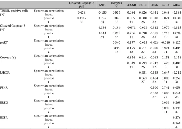
The number of apoptotic cells was directly related to FSHR and EREG expression (p<0.001 and p=0.010, respectively) (Table 3). The number of oocytes was directly related to LHCGR expression (p=0.049) and to pAKT accumulation (p=0.036) (Table 3). On the contrary, the amount of cleaved caspase-3 was not related to the expression of the receptor genes evaluated (Table 3). Considering gene expression levels, LHCGR was directly related to
EGFR (p<0.001) whereas FSHR was directly related to EREG (p<0.001), EGFR (p<0.001) and AREG (p=0.040) (Table 3).
When matching all these parameters together, as reported in Table 4, the number of MI oocytes showed a significant positive correlation to the amount of cleaved caspase-3 (p=0.046), the percentage of TUNEL-positive cells (p=0.014), FSHR expression (p=0.045) and EREG expression (p=0.010). In addition, the expression of LHCGR showed significant negative correlation to both zygotes (p=0.048) and embryo number (p=0.037).
Table 3. Correlation analyses of gene expression of cellular data
Cleaved Caspase-3 (%) pAKT Oocytes (n) LHCGR FSHR EREG EGFR AREG TUNEL positive cells
(%) Spearman correlation index 0.433 -0.150 0.036 0.034 0.826 0.451 0.043 -0.038
p-value 0.0112 0.396 0.843 0.855 0.000 0.010 0.824 0.838
n 33 34 33 31 26 32 30 32
Cleaved Caspase-3
(%) Spearman correlation index 0.036 0.194 -0.071 -0.026 0.342 0.070 -0.025
p-value 0.840 0.279 0.706 0.898 0.055 0.713 0.896
n 34 33 31 26 32 30 31
pAKT Spearman correlation index 0.340 0.277 -0.023 -0.026 -0.018 0.125
p-value .036 0.125 0.911 0.888 0.924 0.495
n 34 32 27 33 31 32
Oocytes (n) Spearman correlation index 0.354 0.214 0.013 0.151 -0.154
p-value 0.049 0.293 0.942 0.426 0.409
n 31 26 32 30 31
LHCGR Spearman correlation index 0.451 0.128 0.647 -0.212
p-value 0.063 0.484 0.000 0.252
n 27 32 31 31
FSHR Spearman correlation index 0.900 0.742 0.659
p-value 0.000 0.000 0.040
n 27 27 26
EREG Spearman correlation index 0.038 0.269
p-value 0.838 0.137
n 31 32
EGFR Spearman correlation index 0.276
p-value 0.140
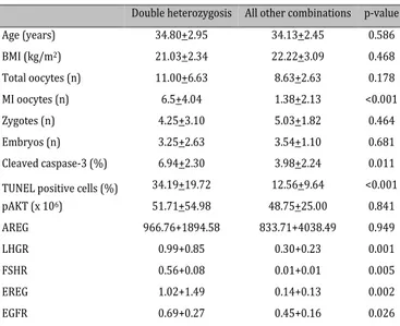
Finally, we focused our atten-tion on the differences between women with double heterozy-gosis, i.e. on both FSHR and LHB (5/13.89%), and the rest of the patients analyzed (31-86.11%). As shown in Table 5, age and BMI were not significantly different between these two further sub-groups. As regards the endpoints, the results obtained with TUNEL assay and immunostaining of cleaved-caspase-3 were signifi-cantly different between groups (see panels in fig.2), whereas pAKT and the oocyte number did not differ. Noteworthy, gene expression was significantly dif-ferent between groups for LHGR,
FSHR, EREG and EGFR, but not for AREG.
Table 4. Global correlation analysis
pAKT Caspase-3 Cleaved
(%)
TUNEL positive
cells (%) AREG LHCGR FSHR EREG EGFR Age (years) Spearman correlation index -0.216 0.042 -0.183 0.353 -0.062 -0.011 -0.240 0.049
p-value 0.213 0.812 0.301 0.055 0.738 0.955 0.179 0.793
n 33 32 32 32 32 27 33 31
BMI (kg/m2) Spearman correlation
index -0.106 -0.235 -0.171 -0.004 0.126 0.230 0.140 0.174
p-value 0.577 0.219 0.374 0.985 0.515 0.280 0.462 0.376
n 30 29 29 29 29 24 30 28
total oocytes
(n) Spearman correlation index -0.140 0.135 0.199 -0.086 0.155 0.101 0.098 -0.018
p-value 0.437 0.461 0.275 0.640 0.397 0.616 0.588 0.924
n 33 32 32 32 32 27 33 31
MI oocytes
(n) Spearman correlation index -0.240 0.262 0.430 0.081 0.253 0.732 0.530 -.160
p-value 0.178 0.046 0.014 0.659 0.196 0.045 0.010 0.389
n 33 32 32 32 32 27 33 31
Zygotes (n) Spearman correlation index 0.229 0.015 -0.061 -0.093 -0.551 0.079 -0.041 -0.234
p-value 0.200 0.934 0.741 0.611 0.048 0.694 0.819 0.205
n 33 32 32 32 32 27 33 31
Embryos (n) Spearman correlation index 0.098 0.032 0.025 -0.144 -0.375 0.277 -0.026 -0.018
p-value 0.595 0.864 0.894 0.438 0.037 0.125 0.888 0.924
n 32 31 31 31 31 26 32 30
Table 5. Correlation analysis in women showing double hetero-zygosis vs. all other combinations
Double heterozygosis All other combinations p-value
Age (years) 34.80+2.95 34.13+2.45 0.586 BMI (kg/m2) 21.03+2.34 22.22+3.09 0.468 Total oocytes (n) 11.00+6.63 8.63+2.63 0.178 MI oocytes (n) 6.5+4.04 1.38+2.13 <0.001 Zygotes (n) 4.25+3.10 5.03+1.82 0.464 Embryos (n) 3.25+2.63 3.54+1.10 0.681 Cleaved caspase-3 (%) 6.94+2.30 3.98+2.24 0.011
TUNEL positive cells (%) 34.19+19.72 12.56+9.64 <0.001
pAKT (x 106) 51.71+54.98 48.75+25.00 0.841 AREG 966.76+1894.58 833.71+4038.49 0.949 LHGR 0.99+0.85 0.30+0.23 0.001 FSHR 0.56+0.08 0.01+0.01 0.005 EREG 1.02+1.49 0.14+0.13 0.002 EGFR 0.69+0.27 0.45+0.16 0.026
Multivariate regression analyses
Considering a single gene expression as dependent variable and the other data available as independent variables, the following models were generated (p<0.001). FSHR expression was predicted by those of EREG (p=0.021) and EGFR (p=0.040). EREG expression was predicted by those of AREG (p=0.039) and FSHR (p=0.021). AREG expression was predicted by that of EREG (p=0.039).
Considering the percentage of apoptotic cells as dependent variable and the other data available as independent variables, one model was generated (p=0.001). In particular, the number of apoptotic cells expression was predicted by the percentage of cleaved caspase-3 (p=0.011).
No significant models were generated considering the expression of LHCGR (p = 0.104) and EGFR (p=0.094), as well as the accumulation of cleaved caspase-3 (p=0.271) and pAKT (p=0.950) and the number of oocytes number (p=0.575) as dependent variables.
Discussion
It is known that the FSHR and LHB variants under study are differently distributed in the population. In particular, the T/N and A/S SNPs of FSHR are more frequent and account for 60 and 40%, respectively, differently from the rarer A/N and T/S variants which are found sporadically and restricted within specific ethnical groups [28]. Also the v-βLH variant of LHB is widely diffused with a maximum incidence in Northern Finland population (41.9%) and a carrier frequency of about 14% in Italy where this study was carried out [29]. In vitro functional analyses have shown that the FSHR isoforms are endowed with equivalent FSH-binding and cAMP-producing activity, differently from v-βLH variant that, if compared with wild type LH, displays enhanced bioactivity in vitro and lower half-life in
vivo which may be compensated by the higher basal activity of its gene promoter [30-32].
Studies focused on the possible relationship between either polymorphism and ovarian markers have shown a different response to ovarian stimulation by carriers of FSHR SNPs. In particular, Ser680 variant is associated to increased resistance of granulosa cells to FSH action, enhanced levels of basal FSH and the need of higher doses of FSH administration to obtain a good response in terms of follicular growth when the patients undergo FIVET. The phenotypic Asn680 polymorphism, on the contrary, confers to carriers an advantage in terms of ovarian responsivity to exogenous FSH, being associated with low basal FSH levels and a better response to exogenous FSH administration, thereby resulting in a higher percentage of clinical pregnancies after embryo transfer [33-36]. The polymorphisms in positions 307 and 680 appear to be correlated also with the degree of severity of iatrogenic ovarian hyperstimulation syndrome, and the presence of Asn680 genotype may increase the risk of developing the pathology due to excessive FSH stimulation [37]. Regarding the LHB variant, in the past two decades several studies have found a correlation between v-LHβand both fertility and menstrual problems. More recently, Alviggi et al [8]. have confirmed that v-LHβpolymorphism produces a less active form of the hormone which is not able to support satisfactorily FSH activity during controlled ovarian stimulation, and that the v-LHβ carriers experience a considerable decrease of the number of transferred embryos, thereby highlighting the essential role of LH/FSH cooperation in the latest stages of follicle maturation.
In our study, we have analyzed the expression of selected markers of oocyte competence and apoptosis-linked cellular features in cumulus cells obtained from follicles of women bearing polymorphisms in hormone (LHB) and hormone receptor (FSHR) genes. Among the marker genes tested, EGFR appears to undergo the more varying modifications with respect to the specific FSHR SNP, when coupled to p.R28-p.T35 LHB aplotype, being up-regulated in p.T307-N680 carriers and down-up-regulated in p.A307-S680 carriers, whereas
AREG expression was always found increased in the recessive models of FSHR. It is known
that FSH triggers the expression of both EGFR, thereby stimulating EGF sensitivity, and
LHCGR which, in turn, elicits the production of the EGF-related peptides, such as AREG
and EREG, and the accumulation of EGFR on the plasmalemma of granulosa cells [38, 39]. In the mouse, the LH-mediator AREG is responsible of the expression of several genes controlling follicular growth and steroidogenesis and also of its own synthesis by paracrine stimulation [40]. Similarly, AREG has been found to induce follicle maturation in humans [41] and its protective anti-apoptotic effect has been acknowledged in diverse human cell models (e.g [42, 43].). Therefore, consistent with the observed concomitant changes of the expression levels of most marker genes analyzed, it is plausible that the genetic alteration of FSHR and/or LHB genes may lead to perturbations of the signaling network programmed to granulosa cell survival and follicle development, ultimately altering oocyte quality via a
failure in the synchronization of nuclear and cytoplasmic maturity. This hypothesis is further corroborated by results obtained testing the survival/apoptosis markers. Previous data had shown DNA fragmentation in human unfertilized oocytes after ICSI, and the positivity to caspase-3 immunoassay suggested the occurrence of apoptosis. The high percentage of unfertilized oocytes bearing DNA fragmentation was correlated with fertilization failure [9]. In addition, Ruvolo et al [20]. demonstrated that preservation of cumulus cells from apoptotic processes inhibited the onset of programmed death in the oocytes; hence, the apoptosis rate in cumulus cells was used as an indicator of oocyte quality. More recently, the occurrence of the apoptotic pathway was analyzed in cumulus cells of single oocytes, focusing on the ability of the embryo to reach the blastocyst stage. The obtained data showed that in these cells the apoptosis rate was significantly lower and the pAKT/TUNEL ratio was higher than in cumulus cells of arrested embryos, indicating a reverse correlation between DNA fragmentation and pAKT accumulation. These two parameters were suggested to be molecular markers of oocyte competence, to be evaluated as a prognostic pattern of blastocyst formation [44]. In the present study, the percentage of TUNEL-positive cells and, more prominently, the quantitation of cleaved caspase-3 appear to be the more robust markers to predict low developmental potential of the oocyte. These two parameters, in fact, appear to be consistently associated with the number of MI oocytes, i.e. those lacking germinal vesicle and polar body; interestingly, TUNEL positivity was also significantly correlated with FSHR and EREG up-regulation in cumulus cells, and this result may be interpreted as an attempt of granulosa cells to oppose to the onset of programmed cell death, although with no final positive outcome. On the other hand, the estimation of the amount of active AKT appeared not to be a significant marker in the model system under study.
It is worth mentioning that, to the best of our knowledge, our study represents the first report in which carriers of double heterozygosis were included and analyzed. Interestingly, when these women were compared to the rest of the patients a higher level of apoptosis in terms of both DFI and amount of active caspase-3 has been observed in cumulus cells. The analysis of clinical data between this group and the other genotypic combinations showed also statistically significant differences concerning the prominent increase of the number of immature oocytes in metaphase I, and the overexpression of EGFR, EREG, FSHR and LHCGR as molecular markers. The reasons for expression specificity of EGF-like peptides (i.e. EREG and not AREG up-regulation as in the analysis of the whole group of patients) is unclear at present but may reflect non-redundant biological roles played by AREG and EREG in the ovarian tissue, as suggested by [45, 46] in other histotypes, which are still to be elucidated. Nonetheless, if this result is further validated, EREG expression level may be considered a specific molecular signature associated to the state of double heterozygous carrier. In light of the present data, we expect that patients with double heterozygosity will produce oocytes with a reduced competence after ovarian stimulation with r-FSH, and therefore these results could be used to define personalized stimulation protocols in ART programs, with the final aim of increasing the success rate of ICSI procedures in accordance with the polymorphic condition of the individual patient.
Acknowledgements
This work was supported by a research grant from Merck Serono S.p.A, Italy. We thank Dr. Alessio Paffoni for help with gene expression assay and Dr. Alberto Ferrigno for his assistance in preparing the figures.
Disclosure Statement
The manuscript is original work that has not been submitted to and is not under consideration for publication by another journal. We confirm that all the listed authors have
participated actively in the study and have seen and approved the submitted manuscript. Monica Lispi is employed at Merck Serono company; all others authors declare that they have no competing interests
References
1 Laven JS, Mulders AG, Suryandari DA, Gromoll J, Nieschlag E, Fauser BC, Simoni M: Follicle stimulating hormone receptor polymorphisms in women with normogonadotropic anovulatory infertility. Fertil Steril 2003;80:986-992.
2 Loutradis D, Patsoula E, Minas V, Koussidis GA, Antsaklis A, Michalas S, Makrigiannakis A: FSH receptor gene polymorphisms have a role for different ovarian response to stimulation in patients entering IVF/ ICSI-ET programs. J Assist Reprod Genet 2006;23:177-184.
3 Casarini L, Santi D, Marino M: Impact of gene polymorphisms of gonadotropins and their receptors on human reproductive success. Reproduction 2015;150:R175-184.
4 Qin X, Ma L, Yang S, Zhao J, Chen S, Xie Y, Wang J, Li T, He Y, Peng Q, Deng Y, Li S, Qin A: The Asn680Ser polymorphism of the follicle stimulating hormone receptor gene and ovarian cancer risk: a meta-analysis. J Assist Reprod Genet 2014;31:683-688.
5 Perez Mayorga M, Gromoll J, Behre HM, Gassner C, Nieschlag E, Simoni M: Ovarian response to FSH depends on the FSH receptor genotype. J Clin Endocr Metab 2000;85:3365-3369.
6 Durnerin CI, Erb K, Fleming R, Hillier H, Hillier SG, Howles CM, Hugues JN, Lass A, Lyall H, Rasmussen P, Thong J, Traynor I, Westergaard L, Yates R: Luveris Pretreatment Group. Effects of recombinant LH treatment on folliculogenesis and responsiveness to FSH stimulation. Hum Reprod 2008;23:421-426. 7 Du JW, Xu KY, Fang LY, Qi XL: Association between mutations of the luteinizing hormone β subunit and
female infertility. Mol Med Rep 2012;5:473-476.
8 Alviggi C, Pettersson K , Longobardi S, Andersen CY, Conforti A, De Rosa P, Clarizia R, Strina I, Mollo A, De Placido G, Humaidan P: A common polymorphic allele of the LH beta-subunit gene is associated with higher exogenus FSH consumption during controlled ovarian stimulation for assisted reproductive technology. Reprod Biol Endocrinol 2013;11:51.
9 Bosco L, Ruvolo G, Morici G, Manno M, Cittadini E, Roccheri MC: Apoptosis in human unfertilized oocytes after intracytoplasmic sperm injection. Fertil Steril 2005;84:1417-1423.
10 Freimann S, Ben-Ami I, Dantes A, Ron-El R, Amsterdam A: EGF-like factor epiregulin and amphiregulin expression is regulated by gonadotropins/cAMP in human ovarian follicular cells. Biochem Biophys Res Commun 2004;324:829-834.
11 Lorenzo PL, Liu IK, Illera JC, Picazo RA, Carneiro GF, Illera MJ, Conley AJ, Enders AC, Illera M: Influence of epidermal growth factor on mammalian oocyte maturation via tyrosine-kinase pathway. J Physiol Biochem 2001;57:15-22.
12 Park JY, Su YQ, Ariga M, Law E, Jin SL, Conti M: EGF-like growth factors as mediators of LH action in the ovulatory follicle. Science 2004;303:682-684.
13 Guzman L, Adriaenssens T, Ortega-Hrepich C, Albuz FK, Mateizel I, Devroey P, De Vos M, Smitz J: Human antral follicles <6 mm: a comparison between in vivo maturation and in vitro maturation in non-hCG primed cycles using cumulus cell gene expression. Mol Hum Reprod 2013;19:7-16.
14 Lei ZM, Mishra S, Zou W, Xu B, Foltz M, Li X, Rao CV: Targeted disruption of luteinizing hormone/human chorionic gonadotropin receptor gene. Mol Endocrinol 2001;15:184-200.
15 Song G, Ouyang G, Bao S: The activation of Akt/PKB signaling pathway and cell survival. J Cell Mol Med 2005;9:59-71.
16 Librizzi M, Chiarelli R, Bosco L, Sansook S, Gascon JM, Spencer J, Caradonna F, Luparello C: The histone deacetylase inhibitor JAHA down-regulates pERK and global DNA methylation in MDA-MB231 breast cancer cells. Materials 2015;8:7041-7047.
17 Goto M, Iwase A, Ando H, Kurotsuchi S, Harata T, Kikkawa F: PTEN and Akt expression during growth of human ovarian follicles. J Assis Reprod Genet 2007;24:541-546.
18 Sánchez F, Smitz J. Molecular control of oogenesis. Biochim Biophys Acta 2012;1822:1896-1912.
19 Crowley LC, Marfell BJ, Waterhouse NJ: Detection of DNA fragmentation in apoptotic cells by TUNEL. Cold Spring Harb Protoc 2016;2016:pdb.prot087221.
20 Ruvolo G, Bosco L, Pane A, Morici G, Cittadini E, Roccheri MC: Lower apoptosis rate in human cumulus cells after administration of recombinant luteinizing hormone to women undergoing ovarian stimulation for in
vitro fertilization procedures. Fertil Steril 2007;87:542-546.
21 Ruvolo G, Fattouh RR, Bosco L, Brucculeri AM, Cittadini E: New molecular markers for the evaluation of gamete quality. J Assist Reprod Genet 2013;30:207-212.
22 Ruvolo G, Roccheri MC, Brucculeri AM, Longobardi S, Cittadini E, Bosco L: Lower sperm DNA fragmentation after r-FSH administration in functional hypogonadotropic hypogonadism. J Assist Reprod Genet
2013;30:497-503.
23 Bosco L, Roccheri MC, Martino C, Chiarelli R, Lispi M, Ruvolo G: Apoptosis rate in cumulus cells as possible molecular biomarker for oocyte competence. EuroMediterranean Biomedical Journal 2017;12:51-56. 24 Crowley LC, Waterhouse NJ: Detecting cleaved caspase-3 in apoptotic cells by flow cytometry. Cold Spring
Harb Protoc 2016;2016:pdb.prot087312.
25 Gavrieli Y, Sherman Y, Ben-Sasson SA: Identification of programmed cell death in situ via specific labeling of nuclear DNA fragmentation. J Cell Biol 1992;119:493-501.
26 Choi SK, Galán M, Kassan M, Partyka M, Trebak M, Matrougui K. Poly(ADP-ribose) polymerase 1 inhibition improves coronary arteriole function in type 2 diabetes mellitus. Hypertension 2012;59:1060-1068. 27 Solé X, Guinó E, Valls J, Iniesta R, Moreno V: SNPStats: a web tool for the analysis of association studies.
Bioinformatics 2006;22:1928-1929.
28 Simoni M, Nieschlag E, Gromoll J: Isoforms and single nucleotide polymorphisms of the FSH receptor gene: implications for human reproduction. Hum Reprod Update 2002;8:413-421.
29 Nilsson C, Jiang M, Pettersson K, Iitiä A, Mäkelä M, Simonsen H, Easteal S, Herrera RJ, Huhtaniemi I: Determination of a common genetic variant of luteinizing hormone using DNA hybridization and immunoassays. Clin Endocrinol (Oxf) 1998;49:369-376.
30 Haavisto AM, Pettersson K, Bergendahl M, Bergendahl M, Virkamäki A, Huhtaniemi I: Occurrence and biological properties of a common genetic variant of luteinizing hormone. J Clin Endocrinol Metabol 1995;80:1257–1263.
31 Jiang M, Pakarinen P, Zhang FP, El-Hefnawy T, Koskimies P, Pettersson K, Huhtaniemi I: A common polymorphic allele of the human luteinizing hormone beta-subunit gene: additional mutations and differential function of the promoter sequence. Hum Mol Genet 1999;8:2037-2046.
32 Desai SS, Roy BS, Mahale SD: Mutations and polymorphisms in FSH receptor: functional implications in human reproduction. Reproduction 2013;146:R235-R248.
33 Behre HM, Greb RR, Mempel M, Sonntag B, Kiesel L, Kaltwasser P, Seliger E, Ropke F, Gromoll J, Nieschlag E, Simoni M: Significance of a common single nucleotide polymorphism in exon 10 of the follicle-stimulating hormone (FSH) receptor gene for the ovarian response to FSH: a pharmacogenetic approach to controlled ovarian hyperstimulation. Pharmacogen Gen 2005;15:451-456.
34 Greb RR, Grieshaber K, Gromoll J, Sonntag B, Nieschlag E, Kiesel L, Simoni M: A Common single nucleotide polymorphism in exon 10 of the human follicle stimulating hormone receptor is a major determinant of length and hormonal dynamics of the menstrual cycle. J Clin Endocrinol Metab 2005;90:4866-4872. 35 Jun JK, Joon JS, Ku SY, Min Choi Y, Hwang KR, Park SY, Hoon Lee G, Don Lee W, Hyun Kim S, Gu Kim J, Young
Moon S: Follicle-stimulating hormone receptor gene polymorphism and ovarian response to controlled ovarian hyperstimalation for IVF-ET. J Hum Genet 2006;51:665-670.
36 Loutradis D, Patsoula E, Minas V, Koussidis GA, Antsaklis A, Michalas S, Makrigiannakis A: FSH receptor gene polymorphisms have a role for different ovarian response to stimulation in patients entering IVF/ ICSI-ET programs. J Assist Reprod Genet 2006;23:177-184.
37 Gromoll J, Simoni M: Genetic complexity of FSH receptor function. Trends Endocrinol Metab 2005;16:368-373.
38 Gloaguen P, Crépieux P, Heitzler D, Poupon A, Reiter E: Mapping the follicle-stimulating hormone-induced signaling networks. Front Endocrinol (Lausanne) 2011;2:45.
39 El-Hayek S, Demeestere I, Clarke HJ: Follicle-stimulating hormone regulates expression and activity of epidermal growth factor receptor in the murine ovarian follicle. Proc Natl Acad Sci USA 2014;111:16778-16783.
40 Nautiyal J, Steel JH, Rosell MM, Nikolopoulou E, Lee K, Demayo FJ, White R, Richards JS, Parker MG: The nuclear receptor cofactor receptor-interacting protein 140 is a positive regulator of amphiregulin expression and cumulus cell-oocyte complex expansion in the mouse ovary. Endocrinology 2010;151:2923-2932.
41 Ben-Ami I, Komsky A, Bern O, Kasterstein E, Komarovsky D, Ron-EI R: In vitro maturation of human germinal vescicle-stage oocytes: role of epidermal growth factor-like growth factor in the culture medium. Hum Reprod 2011;26:76-81.
42 Hurbin A, Dubrez L, Coll JL, Favrot MC: Inhibition of apoptosis by amphiregulin via an insulin-like growth factor-1 receptor-dependent pathway in non-small cell lung cancer cell lines. Ann NY Acad Sci 2003;1010:354-357.
43 Perugorria MJ, Latasa MU, Nicou A, Cartagena-Lirola H, Castillo J, Goñi S, Vespasiani-Gentilucci U, Zagami MG, Lotersztajn S, Prieto J, Berasain C, Avila MA: The epidermal growth factor receptor ligand amphiregulin participates in the development of mouse liver fibrosis. Hepatology 2008;48:1251-1261.
44 Ruvolo G, Roccheri MC, Chiarelli R, Matranga D, Manno M, Bosco L: A new strategy in selecting oocytes using cumulus cells analysis of specific molecules of the apoptotic pathway, according to the ability to reach blastocyst stage. Human Reprod 2015;30:i88.
45 Pastore S, Mascia F, Mariani V, Girolomoni G: The epidermal growth factor receptor system in skin repair and inflammation. J Invest Dermatol 2008;128:1365-1374.
46 Gregorieff A, Liu Y, Inanlou MR, Khomchuk Y, Wrana JL: Yap-dependent reprogramming of Lgr5(+) stem cells drives intestinal regeneration and cancer. Nature 2015;526:715-718.