Main area: Pharmacology and Therapeutics Secondary areas: Nutrition and Bromatology Biochemistry and Molecular Biology
FINAL DEGREE PROJECT
JUNE 2018
Paula Àlvarez Lorenzo Universitat de Barcelona
Facultat de Farmàcia i Ciències de l’Alimentació
ALZHEIMER’S DISEASE AS
A METABOLIC
PATHOLOGY: NEW
APPROACHES
Integration of the areas 1. Introduction...…...…...2 2. Objectives.…...…...3 3. Methods.…...…...3 4. Results.…...…...4 4.1. Dementia.…...4 4.1.1 Risk factors.…...4 4.1.1.1. Age 4.1.1.2. Ethnicity 4.1.1.3. Family history 4.1.1.4. Others 4.1.2. Types of dementia.…...5 4.1.2.1. Alzheimer’s disease 4.1.2.2. Vascular dementia 4.1.2.3. Mixed dementia 4.1.2.4. Dementia with Lewy bodies 4.1.2.5. Frontotemporal dementia (including Pick’s disease) 4.2. Alzheimer’s disease etiology.…...7
4.2.1. Age.…...7
4.2.2. Gender.…...7
4.2.3. Genetic inheritance.…...8
4.2.4. Health and lifestyle.…...9
4.2.4.1. Insulin resistance and type 2 diabetes 4.2.4.2. Diet 4.3. Pathophysiological bases of Alzheimer’s disease.…...11
4.3.1. “Amyloid hypothesis” .…...11
4.3.2. Metabolic syndrome.…...14
4.3.3. Brain energy deficit hypothesis.…...15
4.4. Current Alzheimer’s disease treatment.…...16
4.4.1. Acetylcholinesterase inhibitors.…...16 4.4.1.1. Donepezil
4.4.1.3. Rivastigmine 4.4.1.4. Galantamine
4.4.2. NMDA antagonist: memantine.…...18 4.4.3. New perspectives.…...18
4.4.3.1. Solanezumab 4.4.3.2. Tideglusib 4.4.3.3. Lithium
4.4.3.4. Metabolic treatment for AD
4.5. A different treatment approach to Alzheimer’s disease.…...20 5. Conclusions.…...24 6. Bibliography.…...26
AcAc: Acetoacetate
ACE: Angiotensin converting enzyme ACh: Acetylcholine
AD: Alzheimer’s disease
AMPA: Alpha-amino-3-hydroxy-5-methyl-4-isoxazolepropionic acid ApoE: Apolipoprotein E
APP: Amyloid precursor protein
ARIAs: Amyloid-related imaging abnormalities ATP: Adenosine triphosphate
Aβ or Abeta: Amyloid beta BACE: Beta secretase BBB: Blood-brain barrier
CMRgIc: Cerebral metabolic rate for glucose CNS: Central nervous system
DMTs: Disease-modifying therapies ECE-1: Endothelin-converting enzyme-1 FDA: Food and drug administration GABA: Gamma-amino butyric acid GLUTs: Glucose transporters
GSK3β: Glycogen synthase kinase 3 β HDL: High-density lipoprotein
HIV: Human immunodeficiency virus IDE: Insulin degrading enzyme IRS-1: Insulin receptor substrate-1 KBs: Ketone bodies
KD: Ketogenic diet
LCHF: Low carbohydrate high fat LDL: Low-density lipoprotein
MAP: Microtubule-associated protein MCI: Mild cognitive impairment
MetS: Metabolic syndrome MOAs: Mechanisms of action
MUFA: Monounsaturated fatty acids
NCEP ATP III: National Cholesterol Education Program Third Adult Treatment Panel NEP: Neprilysin
NMDA: N-methyl-D-aspartate NFTs: Neurofibrillary tangles PHFs: Paired helical filaments PI3K: Phosphoinositide 3-kinase PUFA: Polyunsaturated fatty acids ROS: Reactive oxygen species βHB: β-hydroxybutyrate
Alzheimer’s disease (AD) is the most common cause of dementia and it has been estimated that the number of people suffering it is going to keep growing. AD is a chronic neurodegenerative disease that drives to a loss of connection between nerve cells and finally to their death. Although people are unlikely to experience the condition in exactly the same way, some symptoms like memory loss, confusion and difficulty with language are very common. The etiology of this disease is heterogeneous and wide since a lot of risk factors have been described, including modifiable lifestyle-related factors like diet. Different pathophysiological mechanisms have been hypothesized to explain AD origin and development. The most known one is the “amyloid hypothesis”, but some data support that the onset of the disease might be related to a brain metabolism abnormality. The decreased metabolic activity may increase amyloid beta (Aβ) deposition as a secondary response, and this would lead to the death of neurons causing dementia. At present, the available AD treatments show only modest symptomatic benefits, so a different approach has been considered: a ketogenic diet (KD). It is a high saturated fat/low carbohydrate diet that increases ketogenic bodies. By doing this, the brain can obtain energy from a different source and not only glucose.
La malaltia d’Alzheimer és la causa més comú de demència i s’estima que la incidència seguirà augmentant. És una malaltia crònica neurodegenerativa que desencadena una pèrdua de connexió entre les cèl·lules nervioses i la mort d’aquestes. Tot i la varietat interindividual en la simptomatologia, s’han descrit símptomes comuns com la pèrdua de memòria, la confusió o la dificultat en el llenguatge. L’etiologia de malaltia d’Alzheimer és heterogènia i àmplia ja que s’han descrit molts factors de risc, entre els quals es troben factors modificables com la dieta. Pel que fa a les bases fisiopatològiques, la hipòtesi més establerta és l’amiloide. Tot i això, diferents estudis conclouen que l’inici de la malaltia és realment una disfunció en el metabolisme cerebral. Aquesta disminució en l’activitat metabòlica del cervell provocaria una acumulació de la proteïna beta amiloide (Aβ) com a resposta secundària i això comportaria la mort de les neurones i la demència. Els tractaments actuals de la malaltia només milloren lleugerament els símptomes i per això s’ha proposat un nou enfocament: la dieta cetogènica. Aquesta dieta és alta en greixos saturats i baixa en carbohidrats i això provoca la formació de cossos cetònics. Aquestes molècules proporcionen al cervell una font d’energia diferent a la glucosa.
- 1 -
INTEGRATION OF THE AREAS
Mental diseases, specially dementia, primarily afflict the elderly, and since we are an aging society, they have an important role. Most of these diseases are progressive so the symptoms get worse as the disease goes on. This kind of disorders should concern us because they affect the society in different aspects, from the relatives who have a mentally ill person in the family to the country’s economy. Furthermore, there is a huge need of effective treatments for these pathological conditions. This project is focused on Alzheimer’s disease (AD).
Pharmacology is the science that studies how the chemical agents, both natural and synthetic, affect biological systems. It is often described as a bridge science because it includes knowledge and skills from a number of basic science disciplines including physiology or molecular biology. Due to the vast need of new treatments for AD and taking into account the objectives set below, the principal area of study is Pharmacology and Therapeutics.
Biochemistry and Molecular Biology has been integrated in the development of this project because in order to suggest a treatment we should first understand what is happening at a molecular level. Pathophysiological bases of dementia and the molecular mechanisms that the brain uses to get energy are going to be considered in the present work.
The other secondary study area is Nutrition and Bromatology. This field is also known as the science of the food. It has an important role in this project because it has been established that our lifestyle, including our diet, has an enormous impact in AD. The effects of diet in aging and neurodegeneration are becoming well-established and an early feature of AD is region-specific declines in brain glucose metabolism. Based on this knowledge, a new approach in AD treatment is going to be suggested.
- 2 -
1. INTRODUCTION
1.1. ALZHEIMER’S DISEASE GENERALITIES
The most common cause of dementia is Alzheimer’s disease (AD). This dementia involves the parts of the brain that control thought, memory and language, so it causes problems with memory, thinking and behaviour. AD is a progressive disease and this means that it begins slowly and the symptoms gradually get worse over time (4). During the course of the disease, proteins build up in the brain to form structures called “plaques” and “tangles”. This leads to the loss of connections between nerve cells, and eventually to their death and loss of brain tissue. Furthermore, people with AD also have a shortage of some important neurotransmitters so the transmit of information in the brain is reduced (3).
It has been proposed that AD arises from the inability of certain parts of the brain to get enough energy from glucose. This disability can be compensated during decades until the point when the signs and symptoms become evident (5). The most common symptom pattern begins with losing the abilityto remember new information, but as damage spreads, individuals experience other difficulties. The following are warning signs of AD (6):
• Memory loss that disrupts daily life.
• Challenges in planning or solving problems.
• Difficulty completing familiar tasks at home, at work, or at leisure. • Confusion with time or place.
• Trouble understanding visual images and spatial relationships. • New problems with words in speaking or writing.
• Misplacing things and losing the ability to retrace steps. • Decreased or poor judgment.
• Withdrawal from work or social activities. • Changes in mood and personality.
As discussed below, the main problem regarding the treatment of AD is that when the first clinical manifestations appear, the pathogenesis of the disease is already in an advanced stage.
- 3 -
2. OBJECTIVES
Alzheimer’s disease has a huge repercussion and it seems that it will continue like this, since the number of people affected grows every day. Despite of recent progress in understanding the neurobiology and pathophysiology of AD, no resolutive pharmacological treatments but only a few symptomatic drugs are currently available to patients. This lack of progress in AD treatment is disappointing given the enormous emotional, psychological and economic costs involved for patients and their caregivers. Due to the importance of this type of dementia in our society, the objectives of this final degree project are:
1. To review the pathophysiological bases of dementia, mainly AD.
2. To describe AD etiology, paying special attention to the importance of the diet. 3. To study the molecular mechanisms that the brain uses to get energy.
4. To analyse the link between the diet and AD, focusing on the role of carbohydrates. 5. To review the current treatments and suggest new approaches in AD treatment.
3. METHODS
In order to achieve the objectives set before, an exhaustive research has been carried out. This bibliographical research includes primary, secondary and tertiary sources and took place from February 2018 to May 2018. The methodology mainly involves the analysis of scientific articles and their subsequent synthesis. The most used databases have been PubMed and Scifinder, and only free access articles facilitated by the Centre de Recursos per a l’Aprenentatge i la Investigació (CRAI) could have been consulted and included.
First of all, a general search about dementia and AD was done. The aim was to be able to have a general knowledge about it, including types of dementia, risk factors and AD etiology data. As AD is the most frequent type of dementia, the work is focused on it. Secondly, a research about the pathophysiological bases of AD was done. The main keywords used during this search were “amyloid”, “hypothesis”, “metabolic syndrome”, “brain” and “energy deficit”.
Last but not least, the bibliographical research was focused on the relationship between the diet, and the onset of the AD. Some scientific articles found in the
- 4 -
databases mentioned before were analysed. So as to enrich the information, an interesting book has been included in the bibliographical sources: Antídoto para el Alzheimer, written by Amy Berger in 2017.
4. RESULTS
4.1. DEMENTIA
4.1.1. RISK FACTORS
Each type of dementia has its own risk factors, but some of these factors are shared:
4.1.1.1. Age
It is the most significant risk factor for dementia. It is very unusual to see dementia in people under their 60s and it becomes very common in people older than 80 years old. Dementia affects one in six people between 80 and 85 years old, one in three above 85 years, and almost half of the people over 90 years old.
4.1.1.2. Ethnicity
People from certain ethnic communities are at higher risk of dementia than others. South Asian people (India and Pakistan, for example) seem to develop dementia more often than white Europeans. In this case we are talking specifically about vascular dementia. This higher risk can be explained by the fact that they are well known to be at a higher risk of stroke, heart disease and diabetes. Higher risk is likewise associated to people of African or African-Caribbean origin since they are known to be more prone to diabetes and stroke (7).
4.1.1.3. Family history
The genes’ role in the development of dementia is not yet fully understood, but there are some types of dementia that have a heritable component. That is to say that if the person has a close relative who has or had suffered AD, he or she will have more chances to develop it. The risk increases when the relative developed AD at a young age (less than 70 years old). Some studies have shown that a reason for cerebral amyloid deposition, which defines AD at the molecular level, is the APOE ε4 genotype, widely explained below in section 4.2.3 (8).
- 5 -
4.1.1.4. Others
High blood pressure, smoking and diabetes may be risk factors for dementia. Having cardiovascular disease or type 2 diabetes increases a person’s risk of developing dementia by up to two times. There is also some evidence that depression could be a risk factor, but the relationship between dementia and depression is far from clear yet (9). Other medical conditions such as Parkinson’s disease, multiple sclerosis, human immunodeficiency virus (HIV) or Down’s syndrome also increase a person’s risk of dementia. Moreover, lifestyle factors have also been implicated in dementia. The main lifestyle risk factors would be physical inactivity, smoking, unhealthy diet, excessive alcohol and head injuries. It seems that people who remain active, socially connected and mentally engaged are less likely to develop a dementia (10).
4.1.2. TYPES OF DEMENTIA
There are a large number of conditions which cause the symptoms of dementia and dementia affects people differently. There are a lot of factors that determine the impact of dementia on the person, so two people might have the same type of dementia, but show different symptomatology. The most common types of dementia are drawn below:
4.1.2.1. Alzheimer’s disease
AD is the most common cause of dementia and it is a progressive disease. Day-to-day memory is the problem that often draws our attention, but the symptomatology might also include difficulties in finding the right words, making decisions, solving problems or perceiving things in three dimensions. AD etiology and pathophysiology will be explained in more detail in sections 4.2 and 4.3. In order to improve the diagnosis, the current criteria identifies three stages of AD (6):
a) Preclinical AD or presymptomatic stage
At this stage, individuals have measurable changes in the brain, cerebrospinal fluid, and/or blood (biomarkers) that indicate the earliest signs of disease, but they have not yet developed symptoms such as memory loss (6).
b) Mild cognitive impairment due to AD
Since AD is a slow, progressive disorder, with no fixed events that define its onset, it is particularly challenging for clinicians to identify transition points for individual patients. This predementia phase of AD is referred as mild cognitive impairment (MCI)
- 6 -
(11). People with MCI may suffer some forgetfulness or weaknesses that we might think are not pathological, but they can be indicative of the problems that the brain is having to obtain energy. One of the first characteristics of this cognitive impairment is the decrease of the cerebral metabolic rate for glucose (CMRglc) (12). People with AD showed reduced CMRglc and some researchers even assure that this is the most prevalent abnormality in this disease. This decreased use of the brain fuel mainly takes place in the areas involved in memory and learning while the ones involved in visual processing and sensory motor skills are not affected. That means that the person is going to have his cognitive skills modified, but he or she will be able to walk, see, etc (5).
c) Dementia due to AD
This stage is characterized by memory, thinking, and behavioural symptoms that impair a person’s ability to function in daily life and that are caused by AD-related processes.
4.1.2.2. Vascular dementia
This dementia is characterised by a decrease in thinking skills caused by conditions that block or reduce blood flow to the brain. This inadequate blood flow can damage and eventually kill cells anywhere in the body, but as the brain has one of the body’s richest networks of blood vessels, it is especially vulnerable. Its symptoms may be most obvious when the person has a major stroke. Vascular dementia is considered the second most common cause of dementia, accounting for 10% of cases (13).
4.1.2.3. Mixed dementia
It refers to the person who has more than one type of dementia. Consequently, the person might have a mixture of the symptoms of those types of dementia. For example, it is quite common to have both Alzheimer’s disease and vascular dementia together.
4.1.2.4. Dementia with Lewy bodies
In this particular type of dementia, the death of brain cells is due to the formation of tiny abnormal structures inside the brain cells called Lewy bodies. Some of the symptoms include alertness, hallucinations and difficulties in judging distances. This dementia is closely related to Parkinson’s disease and sometimes people having
- 7 -
dementia with Lewy bodies share some symptoms with this disease, including difficulty with movement (14).
4.1.2.5. Frontotemporal dementia (including Pick’s disease)
The damaged parts of the brain are the front and side ones. Brain cells are also dead because of the formation of an abnormal mass of proteins inside them. At first sight, the most obvious signs may be changes in personality and behaviour (14).
Pick’s disease was first described by Czech neurologist and psychiatrist Arnold Pick in 1892. It is known especially for the aphasia it causes. This can distinguish it from other types of frontotemporal dementia, in which behaviour problems and personality changes are often a primary first symptom. Like Alzheimer’s disease, Pick’s disease is currently untreatable (15).
4.2. ALZHEIMER’S DISEASE ETIOLOGY
As mentioned before, AD is commonly suffered by people after the age of 65, but people under this age can also develop it. In this case, it is called early-onset AD. Since AD represents approximately the 70% of the dementia cases, this project will be focused in this specific dementia. Below are the main risk factors explained in more detail.
4.2.1. AGE
Like for the other types of dementia, age is the greatest risk factor for AD. Above 65 years old, a person’s risk of developing Alzheimer’s disease doubles approximately every five years. This leads to the fact that one in six people over 80 years old have dementia (4).
4.2.2. GENDER
Female sex may also be a risk factor in developing AD, despite the reasons are not clear. There are about twice as many women as men over 65 years old with AD, regardless of their longevity. It is well-known that young females’ mitochondria are protected against amyloid-β toxicity because they generate less reactive oxygen species and release less apoptogenic signals than males’ mitochondria. But this advantage is lost in old females, who don’t have this protection due to the decrease of estrogen levels (16).
- 8 -
4.2.3. GENETIC INHERITANCE
Genetics also take part in our health. The strongest genetic risk factor is Apolipoprotein E (ApoE), specifically APOEε4, one of the alleles of the gene that codifies for the molecule apolipoprotein E. This molecule transports through the blood stream the hydrophobic substances that cannot be transport by themselves (vitamins A, D, E, K, cholesterol and fatty acids). As the major component of high-density lipoprotein-like (HDL-like) particles in the brain, ApoE facilitates the transfer of cholesterol and phospholipid between cells. In the receptor-mediated endocytosis of HDL-like particles through low-density lipoprotein (LDL) receptor family, ApoE performs as a ligand. Each ApoE isoform has a different structure and function. ApoE2 has only 1-2% of binding affinity to LDL receptor (LDLR), compared with ApoE3 and ApoE4. The structural difference is also attributed to the differential ApoE cleavage (17).
APOEε4 carriers have more risk to develop AD because the genotype affects the clearance and aggregation of Aβ. Carriers have even more risk if they have the two copies. However, this gene doesn’t cause AD, and it’s not essential to develop the dementia. Emerging data suggest that ApoE may also affect AD pathogenesis through amyloid β-independent pathways (18–20) (figure 1). ApoE affects cerebral vascular function by affecting cerebral blood flow, neuronal-vascular coupling and blood-brain barrier (BBB). It also has a role in the metabolism of lipid and glucose and synaptic plasticity (17).
- 9 -
Furthermore, APOEε4 genotype is not associated with insulin resistance, which suggests that genotype and glycemic impairment may represent independent risk factors for disease conversion (8).
4.2.4. HEALTH AND LIFESTYLE
Some medical conditions such as diabetes, stroke and heart problems are known to be risk factors for dementia. People who adopt a healthy lifestyle, especially from mid-life onwards, are less likely to develop AD. This healthy lifestyle includes physical exercise, healthy weight, not smoking, eating a healthy balanced diet and drinking alcohol only in moderation.
4.2.4.1. Insulin resistance and type 2 diabetes
Insulin resistance and type 2 diabetes are associated with cognitive decline and increased risk for AD. The prevalence of both insulin resistance and AD increase with age, and more than half of individuals older than 65 years have either diabetes or prediabetes. Studies suggest that diabetes may accelerate conversion from mild cognitive impairment to AD (21). It has been established that diabetics have up to a 65% increased risk of developing AD (22). Insulin is one of the key mediators of metabolic homeostasis, since this hormone regulates glucose, energy and lipids. After a meal, elevated glucose provokes that the pancreas releases insulin which stimulates muscle and adipocytes to take up and utilize glucose, and this way plasma glucose levels are reduced. Insulin has three mainly targets: the liver, the muscle and the adipose tissue. This hormone reduces the gluconeogenesis in the liver, increases glucose uptake by the muscle and reduces lipolysis so the plasma level of free fatty acids is reduced. But when there is insulin resistance, it is the other way around: glucose production is increased, glucose uptake is reduced and free fatty acids are also increased.
For a long time, it was assumed that insulin signalling has only these peripheral actions, but now it is widely accepted that insulin has specific effects in the brain. These actions are metabolic, neurotrophic, neuromodulatory and neuroendocrine functions (23). Under physiological conditions, insulin binding to its receptor triggers phosphorylation of insulin receptor substrate-1 (IRS-1). Through phosphoinositide 3-kinase (PI3K) activation, glycogen synthase 3-kinase 3 β (GSK3β) is inactivated and so is the phosphorylation of protein tau, a neuronal microtubule-associated protein found
- 10 -
in axons. This protein is responsible for the polymerization and stabilization of microtubules. When there is insulin resistance, GSK3β is activated and the hyperphosphorylation of tau is induced. This leads to the formation of neurofibrillary tangles (NFTs) and the subsequent cognitive dysfunction (23,24). The importance of tau and its phosphorylation in the formation of NFTs will be explained in more detail in section 4.3.1.
Insulin resistance has also been associated with an increased amyloid β (Aβ) deposition in the brain, one of the hallmarks of AD. Insulin increased insulin degrading enzyme (IDE) expression (25), but in an insulin resistance state, the activity of this enzyme is reduced. This would drive to an increased level of Aβ and cognitive abnormalities (26,27).
Therefore, the link between insulin and AD might be related to the role of insulin in brain metabolism and plasticity. There are insulin receptors in brain regions involved in memory function, and abnormalities in central nervous system (CNS) insulin signalling drive to cognitive alterations. Studies in animal models of insulin resistance have concluded that it exacerbates Aβ and tau phenotypes including hyperphosphorylated tau, which are the characteristic alterations in neurodegenerative processes (26,28– 34).
4.2.4.2. Diet
Although it is well known that some lifestyle-related factors are associated with cognitive decline, diet in particular has become the object of intense research in relation to neurodegenerative diseases (35). Recent findings demonstrate that maintaining a healthy diet may impact on many of the possible risk factors for cognitive decline and the model to follow seems to be the Mediterranean diet (MeDi) (36). It is thought that dietary factors that affect AD risk are those that have an effect on the amyloid cascade (37). That is why our diet choice has a key role in our cerebral health (5). Thus, it has been shown that diets rich in cholesterol and saturated fats increased the deposition of Aβ and the risk of developing AD (38,39). However, these diets were not low-carbohydrate diets because cholesterol was added to the diet without reduction of other components. In fact, it has been recently demonstrated that a diet rich in saturated fats and low in carbohydrates can actually reduce levels of
- 11 -
Aβ (40). This diet induces hepatic production of ketone bodies and, as explained in section 4.5, it is known as a ketogenic diet (KD).
On the other hand, epidemiological evidence has suggested a possible association among fish consumption, monounsaturated fatty acids (MUFA) and polyunsaturated fatty acids (PUFA) (particularly, n-3 PUFA) and reduced risk of cognitive decline, AD and dementia (36). On the contrary, high carbohydrate intake might cause AD by two mechanisms (41):
1. By disturbing lipid homeostasis and compromising the integrity of cellular membranes. This would cause a decrease in membrane proteins activity such as glucose transporters, leading to a reduction in the utilization of glucose as a source of energy in the brain.
2. By inducing mild chronic elevated insulin signalling, which accelerates cellular damage. High carbohydrate diets are well known to stimulate insulin signalling and chronic stimulation of this pathway may lead to an alteration of lipid metabolism. This drives to inappropriate lipid environments in neurons, mis-cleavage of amyloid precursor protein (APP) and the resulting inhibition of cellular trafficking, and increasing the risk of developing AD (40). Moreover, it has been shown that apoE4 and high carbohydrate diets alter lipid metabolism in a similar manner, reducing lipoprotein lipase activity and thus increasing triglyceride-rich lipoprotein levels, which suggests a common mechanism for the etiology of AD (41).
4.3. PATHOPHYSIOLOGICAL BASES OF ALZHEIMER’S DISEASE
4.3.1. “AMYLOID HYPOTHESIS”
Amyloid beta is a group of peptides generated as cleavage products from 36 to 43 amino acids and with a molecular mass of approximately 4kDa. They derive from the amyloid precursor protein (APP), a transmembrane protein. The “amyloid hypothesis” is the most influential model of the pathology of AD proposed over the last 25 years and, according to it, accumulation of Aβ in the brain is the primary influence driving AD pathogenesis.
- 12 -
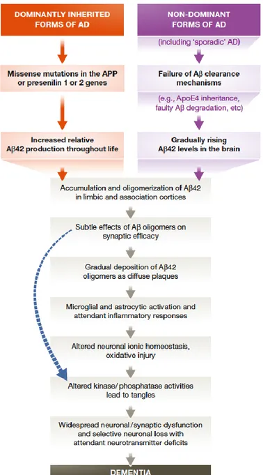
Figure 2. The sequence of pathogenic events leading to AD (42)
The deposition of Aβ in brain is one of the characteristics of a pathologic brain because it drives to a lack of communication between the neurons since the synapses are altered. Normal amounts of Aβ in healthy brains don’t modify the cognitive function. Actually, Aβ depresses synaptic activity and without this depression, synaptic activity could become excessing, leading to excitotoxicity. Data suggest that Aβ has important physiological functions and, further, that it should only be regarded as toxic when its production and degradation are imbalanced (43). It has been established that this deposition could be due to an overproduction of Aβ or to a lack of degradation of it. Last studies suggest that the second hypothesis is more likely (43). Several proteases
- 13 -
are shown to degrade Aβ including insulin degrading enzyme (IDE), angiotensin converting enzyme (ACE), neprilysin (NEP) and endothelin-converting enzyme-1 (ECE-1). So, several proteases are involved in Aβ degradation in vitro, but the two major enzymes in vivo are IDE and NEP. IDE has been shown to degrade Aβ and to influence brain Aβ levels in vivo. Since extracellular Aβ aggregation is one of the hallmarks of AD pathology according to the amyloid hypothesis and Aβ is not found in cytosol, extracellular IDE may play a key role in the clearance of Aβ and can relate to the AD pathology (5,44).
Several peptides have been highlighted as substrates of IDE, including insulin, insulin-like growth factor I and II, amylin, Aβ and others. Under physiological conditions, IDE is secreted at high levels from the microglial cells and degrades Aβ extracellularly. This activity has shown to be lower in AD brains. As IDE has different substrates in vivo with different affinities, the imbalance of the substrates could affect the degradation process by IDE and, consequently, the pathogenesis of AD. As mentioned before (pg 12), it has been suggested that insulin resistance leads to reduced IDE activity. This could be explained because insulin resistance leads to compensatory hyperinsulinemia and therefore to increased brain insulin level. Due to substrate competition, increased insulin would inhibit the ability of IDE to degrade Aβ properly, causing Aβ neurotoxicity and AD (5,25).
Another histopathological hallmark of AD is neurofibrillary tangles (NFTs). These NFTs are insoluble twisted fibers found inside the neurons, made up of aggregates of tau protein. Tau was first discovered as a microtubule-associated protein (MAP) that stimulates tubulin assembly into microtubules in the brain. There was not much research interest in tau protein until a decade later, when it was found to make up the paired helical filaments (PHFs) of these NFTs in AD brains. Tau is modified post-translationally by several ways in both normal and pathological conditions (phosphorylation, glycosylation, ubiquitination, glycation, polyamination, nitration, truncation and aggregation). But the major component of PHFs in AD is abnormally hyperphosphorylated tau. The normal level of tau phosphorylation is a consequence of dynamic regulation of tau kinases and tau phosphatases. However, the causes leading to abnormal hyperphosphorylation of tau are still not fully understood (45). It has been known that inherited mutation in APP and its protease (presenilin) cause
early-- 14 early--
onset Aβ deposition and this is followed by accumulation of tangles containing filaments of wild-type tau (46,47). In contrast, mutations in the tau gene lead to a form of frontotemporal dementia without Aβ accumulation. So, Aβ accumulation can lead to progressive tau deposition and its phosphorylation (48), but the converse has not been clearly demonstrated in humans (42).
Tau O-GlcNAcylation was found to be decreased in AD brain, and this decrease was correlated to tau hyperphosphorylation. This acylation is directly regulated by glucose metabolism: impaired glucose metabolism may cause decreased tau O- GlcNAcylation which, in turn, leads to hyperphosphorylation of tau and neurofibrillary generation (45).
4.3.2. METABOLIC SYNDROME
Metabolic Syndrome (MetS) is considered a major public health problem and is one of the few clinical syndromes that affects a large portion of the general population which is potentially reversible by established interventions. The most commonly used definition for MetS in the United States is the one described by the National Cholesterol Education Program Third Adult Treatment Panel (NCEP ATP III), which is the presence of three or more of the following criteria:
• Abdominal obesity: waist >102 cm (>40 in) for men or >88 cm (>35 in) for women • Triglycerides ≥150 mg/dL
• HDL <40 mg/dL for men or <50 mg/dL for women
• Blood pressure ≥130/≥85 mmHg or current use of anti-hypertensive medications • Fasting glucose level ≥110 mg/dL
Most studies report that MetS and its components have a negative impact on cognition. However, findings may vary by sex (49). MetS could be one of the causes leading to MCI, the first stage of AD. Furthermore, MetS is often the result of long term insulin resistance due to an excessive consumption of some food components (especially refined carbohydrates) aggravated by the modern lifestyle stress, lack of sleep and poor quality of it, and insufficient physical activity (5).
All the indicators established by the NCEP ATP III might suggest that the body is not properly assimilating carbohydrates and it is reacting with abnormally high insulin or glucose levels. The main metabolic alteration linked with AD is the insulin resistance.
- 15 -
Insulin not only regulates glucose and lipid metabolism in the brain, but also plays an important role in learning and memory (50). As mentioned in section 4.2.4.1, insulin resistance causes GSK3β activation and the hyperphosphorylation of tau is induced. This leads to the formation of NFTs and the subsequent cognitive dysfunction (23,24). 4.3.3. BRAIN ENERGY DEFICIT HYPOTHESIS
Despite the brain represents only a 2 percent of the total body mass, it requires more than the 20 percent of the daily energy intake. The brain is one of the most metabolically active organs in the body. As a consequence, the food we consume has a direct impact on brain function. Glucose is the primary energy source for the brain, but in prolonged fasting, the brain uses ketone bodies, that can replace glucose perfectly. The average adult human brain metabolizes about 110 to 145 g/day of glucose. However, in AD there is a decline of a 17 to 24% in the cerebral metabolic rate for glucose (CMRglc) and low CMRglc has been correlated with low cognitive scores (51). AD is, above all, a brain fuel metabolism dysfunction, which means that it results from the brain’s inability to generate or use properly the energy (5). A bulk of studies have shown that the blood-brain barrier (BBB) is impaired in AD resulting in an altered expression of some transporters, including the down-regulation of glucose transporter. The primary fuel for the brain, as mentioned before, is glucose. It must be taken from the blood and transported across the BBB by the specific glucose transporters (GLUTs) due to the inability of neurons to synthesize or store it. In AD, the expression of these transporters is decreased and the brain has an energy deficit (1).
This explanation may indicate that the amyloid cascade hypothesis in not valid for sporadic AD, but that the formation of both, amyloidogenic derivatives and hyperphosphorylated tau protein is downstream the origin of this neurodegenerative disease. So, a decline in brain metabolic activity regardless of etiology, seems to be the underlying cause of cognitive decline and dementia (34,52,53). There is evidence that the reductions in the availability of both glucose/energy and insulin contribute to the formation of amyloidogenic derivatives and hyperphosphorylated tau protein. Decreased metabolic activity increases beta secretase (BACE) which, in turn, increases Aβ deposition as a secondary response. BACE is an enzyme involved in the cleavage of APP, resulting in the formation of Aβ. A study established that chronically induced cerebral hypoperfusion in rats resulted in increased BACE and Aβ and poorer learning
- 16 -
in a Morris water maze (54). To sum up, Aβ overproduction is believed to be a secondary phenomenon that may result in further clinical decline. According to this hypothesis, the goal would be to identify the factors that cause this decreased metabolic activity that leads to neuronal death, and then find ways to increase it. The interaction of different macronutrients, in particular fats and carbohydrates, is known to influence the metabolic state (36). It might be that the way the body normally copes with condition of low glucose availability could be exogenously applied to AD. This way might be the use of ketone bodies (KBs). KBs metabolism becomes the main source of alternative energy as a results of carbohydrate deficiency and increased availability of fatty acids from lipolysis. The two KBs that can replace glucose in the brain are β-hydroxybutyrate (βHB) and acetoacetate (AcAc). The lack of success in the therapy of AD moved the focus of research to functional dysregulation of brain metabolism, mitochondrial homeostasis and excitatory and inhibitory neuronal signaling (1). A possible “metabolic therapy” is widely explained in section 4.5.
4.4. CURRENT ALZHEIMER’S DISEASE TREATMENT
Alzheimer’s disease eventually leads to the death of affected individuals at an average of nine years after diagnosis. For the last three decades, the standard treatments for AD have been acetylcholinesterase inhibitors, highlighted below, together with other drugs to manage the mood disturbance, agitation and psychosis that usually appear in the later stages. In the recent years, memantine, also explained below, has been widely used. Although the efforts to find a suitable treatment, all of these drugs show modest symptomatic effects and it is believed that the major barrier to effective treatments is the lack of full understanding of the mechanism of AD (45).
There are two different groups of drugs approved in Spain by the competent administration, the Agencia Española de Medicamentos y Productos Sanitarios (AEMPS).
4.4.1. ACETYLCHOLINESTERASE INHIBITORS
All cholinesterase inhibitors work in a similar way, but one might suit a certain individual better than another, particularly in terms of side effects experienced. The mechanism of action of these drugs is by increasing the amount of Acetylcholine (ACh),
- 17 -
a neurotransmitter responsible, among others, of the memory capacity. These drugs increase ACh by inhibiting its degradation since they are acetylcholinesterase inhibitors, the enzyme responsible of ACh’s degradation. The improvement of cholinergic function is accomplished by this increase in ACh concentration. So the effect may lessen as the disease progresses and fewer cholinergic neurons remain functionally intact (55). Tacrine was the first acetylcholinesterase inhibitor approved to treat AD, but it was withdrawn from the market for its hepatotoxicity.
4.4.1.1. Donepezil
Donepezil may improve mental function and this includes the ability to think and remember, or slow the loss of these abilities in people who have AD. However, as mention before, it will not cure AD or prevent the loss of mental abilities (56). Donepezil is a reversible and non-competitive acetylcholinesterase inhibitor.
4.4.1.2. Rivastigmine
Rivastigmine is also a reversible and non-competitive cholinesterase inhibitor. But, unlike the others, rivastigmine inhibits both butyrylcholinesterase and acetylcholinesterase. Butyrylcholinesterase is mainly found outside the central nervous system (CNS) and the fact that this drug inhibits the two cholinesterase reduces its side effects. One important characteristic about this drug is that it does not use the cytochrome route to be eliminated and this is very useful for people who suffer different pathologies and receive different treatments.
4.4.1.3. Galantamine
This drug is a benzazepine. It is a cholinesterase inhibitor that has been used to reverse the muscular effects of gallamine triethiodide and tubocurarine, and has been studied as a treatment for AD and other CNS disorders. It is a reversible and competitive acetylcholinesterase inhibitor, but also
an allosteric modulator of the nicotinic receptor, which promotes the cholinergic activity. This mechanism of action is because galantamine is not structurally related to other acetylcholinesterase inhibitors (55) (figure 3). However, some side effects are common with Donepezil: leg cramps and urinary incontinence.
- 18 -
4.4.2. NMDA ANTAGONIST: MEMANTINE
Glutamate is another neurotransmitter, an endogenous chemical messenger which transmits signals across a chemical synapse. Glutamate is released in excessive amounts when brain cells are damaged by AD. This causes the brain cells to be damaged further. Memantine protects brain cells by blocking the effects of excess glutamate so the mechanism of action is different from that of donepezil, rivastigmine and galantamine. The neurotransmitter glutamate activates several classes of metabotropic receptors and three major types of ionotropic receptors: alpha-amino-3-hydroxy-5-methyl-4-isoxazolepropionic acid (AMPA), kainate and N-methyl-D-aspartate (NMDA). It is suggested that glutamate receptors, in particular of the NMDA type, are overactivated in a tonic rather than a phasic manner. Such continuous, mild, chronic activation ultimately leads to neuronal damage/death (57). Memantine has a great clinical efficiency and it is indicated in serious AD cases. It also has effects in other receptors like the dopaminergic and that is why neurologic and psychologic effects such as confusion or hallucinations could be observed.
4.4.3. NEW PERSPECTIVES
There have not been new drugs approved for the treatment of AD since 2003 (58), but new therapies are urgently needed to treat affected patients and to prevent, defer, slow the decline, or improve the symptoms of AD. It has been estimated that the overall frequency of the disease would be decreased by nearly 50% if the onset of the disease could be delayed by 5 years (59). Most candidate therapies are disease-modifying therapies (DMTs) (70% across all phases). This term refers to a drug that can modify or change the course of the disease. The remainder candidate therapies are symptomatic agents directed at cognitive enhancement (14%) or treatment of neuropsychiatric symptoms (13%) and agents with undisclosed mechanisms of action (MOAs) (2%) (59).
4.4.3.1. Solanezumab
Solanezumab is a humanized monoclonal antibody against Aβ and it was designed to increase the clearance from the brain of soluble Aβ. The results recently published showed that it does not significantly affect cognitive decline (60).
- 19 -
4.4.3.2. Tideglusib
This drug is an inhibitor of glycogen synthase kinase 3 (GSK3), the responsible of tau phosphorylation. A phase II trial of tideglusib established that it is acceptably safe, but produced no clinical benefit (61).
4.4.3.3. Lithium
Lithium inhibits site-specific AD-like tau phosphorylation in living neurons and it has been suggested that chronic lithium treatment similar to that prescribed for manic-depressive patients might slow down tau hyperphosphorylation in the brains of patients with AD (62). Furthermore, a systematic review and meta-analysis has indicated that lithium treatment may have beneficial effects on cognitive performance in subjects with MCI and AD (63).
4.4.3.4. Metabolic treatment for AD
AD neurodegeneration has been associated with different abnormalities like energy imbalance, dysregulated lipid and carbohydrate metabolism, cytokine mediated inflammation, increased oxidative stress, cell death and vascular degeneration. These problems are also present in type 2 diabetes so some scientifics support that AD may be managed by similar or identical therapeutic strategies. According to some researchers, therapeutic options for treating AD as a metabolic disorder are mainly three: lifestyle changes, anti-inflammatory/anti-oxidant measures and insulin signalling support. The first one mainly includes aerobic physical exercise, weight training and adopting a healthful diet. These measures that preserve insulin responsiveness would protect against cognitive impairment and neurodegeneration. There are epidemiological studies that suggest that individuals who chronically take anti-inflammatory or anti-oxidant drugs had lower risks for developing cognitive impairment and AD (64). The last therapeutic option is insulin signalling support. The brain needs insulin for different reasons such as to support metabolism, neuronal survival, synaptic plasticity or neuroprotection. Insulin could be administrated by different routes. Since insulin crosses the blood-brain barrier (BBB), various routes could modulate brain metabolism (34).
- 20 -
4.5. A DIFFERENT TREATMENT APPROACH TO ALZHEIMER’S DISEASE
As mentioned before, it has been hypothesized that Alzheimer’s disease is mainly a problem in the metabolism of the cerebral fuel and this leads the brain to a situation of energy deficit. That is why a metabolic strategy should be considered as a treatment. Specifically, for example, a change in the diet and lifestyle.
Ketogenic diet (KD)
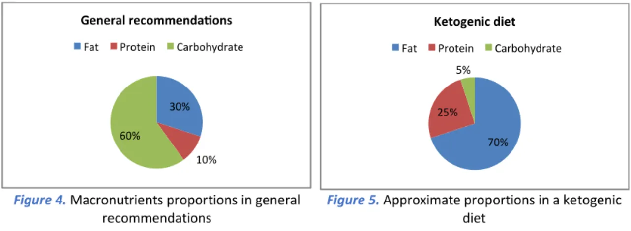
The KD is a high-fat, adequate-protein and low-carbohydrate diet. The fact that carbohydrates are nearly eliminated means that the body has minimal dietary sources of glucose (65). This diet is also known as keto diet, low carb diet, low carb high fat (LCHF) among others. Glucose is the easiest molecule for our body to convert and use as energy so it is the one that would be chosen over other sources. Since glucose is being used as a primary energy source, fats are not needed and are stored (66). This is what usually happens because according to most dietary guidelines (figure 4), the main components of a healthy diet are carbohydrates. These guidelines were developed as part of the Strategy for Nutrition, Physical Activity and the Prevention of Obesity (NAOS Strategy) of the Spanish Agency for Consumer Affairs, Food Safety and Nutrition of the Spanish Ministry of Health, Social Services and Equality. However, as mentioned before, these are not the recommendations in a ketogenic diet. The nutrient intake in a KD should be something around 70% of fats, 25% of proteins and 5% of carbohydrates (figure 5).
By lowering the intake of carbohydrates, the body is induced into a state known as ketosis or ketogenesis, which is a natural process the body initiates to help us survive when food intake is low. During this state, we produce ketones or ketone bodies,
70% 25%
5%
Ketogenic diet
Fat Protein Carbohydrate
Figure 5. Approximate proportions in a ketogenic
diet
Figure 4. Macronutrients proportions in general
- 21 -
which are produced from the breakdown of fats in the liver. The three ketone bodies are acetoacetate (AcAc), β-hydroxybutyrate (βHB) and acetone; and they are produced from acetyl-CoA in the mitochondrial matrix of liver cells. The aim of this diet is to force the body into this metabolic state. KD diet was developed to mimic a starvation response in animals without reducing calories to harmful levels (40). Ketones are supposed to be a second line fuel for our body, but this is just said because glucose is the most favourite fuel for our body. However, ketone bodies are a more efficient fuel than glucose. Furthermore, ketosis induces a moderate uncoupling state and less oxidative efficiency compared to glucose oxidation (67). During starvation, ketone bodies are the major energy source (75%) for the brain.
KD was set up in the first decade of the last century to treat refractory epilepsy and then applied with success to treat other neurological diseases. The proved effectiveness of the KD in childhood refractory epilepsy and the early metabolic dysfunction observed in MCI, paved the way to evaluate the possible therapeutic role of dietary metabolic approaches to AD and other neurodegenerative diseases (1). There are different types of ketogenic diets. The classic KD was designed in 1923 for the treatment of epilepsy by Dr. Russell Wilder and it approximately results in 90% of total calories intake from fat, 6% from protein and 4% from carbohydrates. As it is very difficult to maintain this KD, some variants of the KD have been published. The main alternative one is the Medium-Chain Triglyceride ketogenic diet (MCTKD), which is characterised for less lipid energy intake (45%) and this fat is mainly short fatty acids. These shorter fatty acids have a quick metabolism that results in more efficient generation of ketone bodies allowing to assume a greater proportion of carbohydrates (1), but it is still limited to approximately 29% of energy (68). Medium-chain fatty acids are able to cross the BBB and provide an alternative energy source for brain neurons and astrocytes.
The neuroprotective effects of ketone bodies have been associated with different mechanisms (1):
• Increasing intracellular adenosine triphosphate (ATP) availability
• Reducing reactive oxygen species (ROS) generation by mitochondrial complex I • Inhibiting mitochondrial permeability transition
- 22 -
• Altering metabolism of neurotransmitters such as glutamate and gamma-amino butyric acid (GABA)
• Activating energy-sensing signalling pathways
AD etiopathogenesis has been linked to oxidative stress, neuroinflammation, mitochondrial impairment, hypometabolism and BBB disruption (69–71). Therefore, it is believed that all these mechanisms set before would be effective to treat AD.
The findings of recent investigations have suggested that caloric restriction prevents age-related neuronal damage and may be useful in the prevention and treatment of AD (72). Animal models provide insight into the underlying mechanisms of many diseases, included AD, and they have been useful in the preclinical evaluation of potential treatments. The results obtained in animal models vary. Some data suggest that KD may play an important role in enhancing motor performance in mice, but have minimal impact on the phenotype of murine models of amyloid or tau deposition (73). Another study with aged dogs, a natural model of amyloidosis, suggested that short-term administration of medium-chain triglycerides improves energy metabolism in the aged canine brain (74). Furthermore, some clinical trials have been done to analyse the efficacy of ketone bodies in patients with AD. For example, the effects of an increase in plasma ketone body levels on cognitive functions were investigated in older adults with memory disorders. It was observed that this increase was moderated by ApoE genotype. In APOEε4 carriers, levels continued to rise, while levels in ApoE4 non-carriers remained constant. On cognitive testing, medium chain triglycerides (MCT) treatment facilitated performance on the Alzheimer’s Disease Assessment Scale-Cognitive Subscale (ADAS-cog) for ε4− subjects, but not for ε4+ subjects. Thus, higher ketone values were associated with greater improvement in paragraph recall with MCT treatment relative to placebo across all subjects (75). In another study, ketosis was induced by a diet very low in carbohydrates and the findings suggested that this diet can improve memory function in older adults with increased risk for AD (76).
According to the data we have access to, a dietary intervention represents a relatively safe and available method to combat AD that deserves further clinical investigations.
- 23 -
Possible complications of a ketogenic diet
A KD should be established for a long period. However, the restrictions in the diet might make people think that this diet is difficult to achieve. Therefore, an important complication to face in a KD treatment is the lack of adherence.
A lot of the food recommended in a KD provide us cholesterol such as eggs, butter or animal protein. Most of this food has also choline, an essential nutrient for acetylcholine production which, as mentioned before, plays a key role in the memory and learning. However, this food is also rich in cholesterol, which may lead to hyperlipidemia. Previous studies have indicated that hyperlipidemia may be a risk factor for dementia and AD. Thus, some results suggested a link between high midlife cholesterol levels and increased dementia risk, but later studies found no association between midlife cholesterol and subsequent dementia (77). Cholesterol is a chemical essential compound for the cell membranes and hormones like estrogen and testosterone. The 80% of this substance is generated in the liver and the rest comes from our diet. After a meal, cholesterol in the diet is absorbed through the small intestine and metabolized and stored in the liver. As the body requires cholesterol, the liver releases it. High blood cholesterol is usually caused by lifestyle factors, such as diet, in combination with our genes. Untreated high blood cholesterol can build up in deposits called plaque in the blood vessels, known as atherosclerosis. If this high level becomes chronic or uncontrolled, it can cause serious complications including carotid artery disease, coronary heart disease (angina or heart attack), peripheral artery disease or stroke (78). So, although we know that cholesterol is essential in our body, we should be aware about the possible complications of chronic high cholesterol consumption and determine plasma cholesterol levels regularly, especially in people above their 40s.
Furthermore, it is important to mention what ketoacidosis is. Diabetic ketoacidosis is a pathological metabolic state characterized by extreme ketosis which cannot be adequately regulated due to a lack of insulin in patients with type 1 diabetes. The resulting pH imbalance may be fatal. However, this metabolic abnormality is not possible in nutritional ketosis (“physiological ketosis”) induced by KD. The evaluation of side effects of ketogenic diets in patients with AD is difficult due to the small number of available clinical trials, but major adverse reactions have not been reported
- 24 -
in the literature. In some studies, transient gastrointestinal effects like diarrhea, dyspepsia and flatulence were reported in patients receiving a formulation of medium chain triglycerides (79,80). Investigations assess the supplementation of the normal glucose supply with ketone bodies which are produced by the body during glucose deprivation and can be metabolized by the brain when glucose utilization is impaired (81).
5. CONCLUSIONS
Alzheimer’s disease is a complex disorder, influenced by a number of different factors. That’s why I strongly believe that a multifactorial approach would be needed. The drugs that we have nowadays are treating the neurotransmitters deficit, because the memory problem is mainly caused by this lack of acetylcholine. By this mechanism, these drugs just control the symptoms of the disease, managing behavioural manifestations and slowing down their progression. However, these drugs are not a cure for AD, and we are not treating the onset of the dementia.
According to all the research done about the pathogenesis of the AD, one of the mechanisms leading to this type of dementia is hyperinsulinemia and impaired glucose metabolism. To summarise, this chronic condition would provoke:
• Aβ accumulation because, as explained in section 4.3.1, IDE degrades both insulin and Aβ. Under a situation of hyperinsulinemia, Aβ degradation by IDE would be reduced.
• Neurofibrillary tangles, caused by impaired glucose metabolism which leads to a decrease of tau-O-GlyNAcylation that causes an hyperphosphorylation of tau. • Metabolic Syndrome that would end with a mild cognitive impairment and its
subsequent dementia.
Having plasma glucose levels under control does not assure that there is not any abnormality because the problem may be also the hyperinsulinemia. Our body generates insulin in order to reduce the glucose in our blood. So, the best way to reduce the amount of blood glucose and, as a consequence, correct the hyperinsulinemia, is reducing the quantity of glucose in our diets. As said before, glucose is not the only way of obtaining energy for our body and it is not the most
- 25 -
effective. Ketones can provide approximately 40-60% of the energy that our brain needs (5). A promising treatment for AD is a change in the diet that implies a decrease in carbohydrate intake which would subsequently reduce the hyperinsulinemia. Moreover, in AD brain there is a situation of energy deficit due to a reduced ability to obtain glucose from plasma. A diet low in carbohydrate and high in lipids would generate ketones and the brain would be able to obtain energy from this source. However, we should be aware of the complications of this diet and make sure that it will be used under medical supervision.
- 26 -
6. BIBLIOGRAPHY
1. Pinto A, Bonucci A, Maggi E, Corsi M, Businaro R. Oxidant and Anti-Inflammatory Activity of Ketogenic Diet: New Perspectives for Neuroprotection in Alzheimer’s Disease. Antioxidants [Internet]. 2018;7(5):63. Available from: http://www.mdpi.com/2076-3921/7/5/63
2. World Health Organization. Dementia [Internet]. [cited 2017 Nov 26]. Available from: http://www.who.int/mediacentre/factsheets/fs362/en/
3. Alzheimer’s Disease International. About dementia [Internet]. [cited 2018 Mar 14]. Available from: https://www.alz.co.uk/about-dementia
4. Alzheimer’s association. What is Alzheimer’s? [Internet]. [cited 2018 Mar 14].
Available from:
https://www.alz.org/alzheimers_disease_what_is_alzheimers.asp 5. Amy Berger. Antídoto para el Alzhéimer. Editorial Sirio; 2017. 442 p.
6. Association A. 2012 Alzheimer’s disease facts and figures. Alzheimer’s Dement
[Internet]. 2012;8(2):131–68. Available from:
http://dx.doi.org/10.1016/j.jalz.2012.02.001
7. Alzheimer’s Society. Risk factors for dementia. 2016;(April).
8. Morris JK, Vidoni ED, Wilkins HM, Archer AE, Burns NC, Karcher RT, et al. Impaired fasting glucose is associated with increased regional cerebral amyloid. Neurobiol Aging [Internet]. 2016;44:138–42. Available from: http://dx.doi.org/10.1016/j.neurobiolaging.2016.04.017
9. Muliyala KP., Varguese M. The complex relationship between depression and dementia. Ann Indian Acad Neurol [Internet]. 2010; Available from: https://www.ncbi.nlm.nih.gov/pmc/articles/PMC3039168/
10. Baumgart M, Snyder HM, Carrillo MC, Fazio S, Kim H, Johns H. Summary of the evidence on modifiable risk factors for cognitive decline and dementia: A population-based perspective. Alzheimer’s Dement [Internet]. 2015;11(6):718– 26. Available from: http://dx.doi.org/10.1016/j.jalz.2015.05.016
11. Albert MS, Dekosky ST, Dickson D, Dubois B, Feldman HH, Fox NC, et al. The diagnosis of mild cognitive impairment due to Alzheimer’s disease: Recommendations from the National Institute on Aging-Alzheimer’s Association
- 27 -
workgroups on diagnostic guidelines for Alzheimer’s disease. Alzheimer’s Dement [Internet]. 2011;7(3):270–9. Available from: http://dx.doi.org/10.1016/j.jalz.2011.03.008
12. Frank R, Frackowiak R, Jack C, Jagust WJ. The Use of MRI and PET for Clinical Diagnosis of Dementia and Investigation of Cognitive Impairment: A Consensus Report. (1):1–15.
13. Alzheimer’s association. Vascular Dementia [Internet]. [cited 2018 Mar 14]. Available from: https://www.alz.org/dementia/vascular-dementia-symptoms.asp
14. Alzheimer’s Society. What is dementia? 2017;(January).
15. Alzheimers.net [Internet]. [cited 2018 Apr 13]. Available from: https://www.alzheimers.net/what-is-picks-disease/
16. Lloret A, Vi J. Why Women Have More Alzheimer’s Disease Than Men: Gender and Mitochondrial Toxicity of Amyloid- β Peptide. Journal of Alzheimer’s Disease. 2010;20:527–33.
17. Liao F, Yoon H, Kim J. Apolipoprotein E metabolism and functions in brain and its role in Alzheimer’s disease. Curr Opin Lipidol. 2017;28(1):60–7.
18. Wang MO, Vorwald CE, Dreher ML, Mott EJ, Cinar A, Mehdizadeh H, et al. The Role of APOE in Cerebrovascular Dysfunction. Acta Neuropathol. 2016;27(1):138–44.
19. Tai LM, Ghura S, Koster KP, Liakaite V, Maienschein-Cline M, Kanabar P, et al. APOE-modulated Aβ-induced neuroinflammation in Alzheimer’s disease: Current landscape, novel data, and future perspective. J Neurochem. 2015;133(4):465–88.
20. Kim J, Yoon H, Basak J, Kim J. Apolipoprotein E in Synaptic Plasticity and Alzheimer’s Disease: Potential Cellular and Molecular Mechanisms. Mol Cells
[Internet]. 2014;37(11):767–76. Available from:
http://www.molcells.org/journal/view.html?doi=10.14348/molcells.2014.0248 21. Xu W, Caracciolo B, Wang H, Winblad B, Ba L, Qiu C. Impairment to dementia in
people with diabetes. Diabetes. 2010;59(November).
22. Dineley K, Jahrling J, Denner L. Insulin Resistance in Alzheimer’s Disease. Neurobiol Dis. 2015;72:92–103.
- 28 -
23. Bedse G, Di Domenico F, Serviddio G, Cassano T. Aberrant insulin signaling in Alzheimer’s disease: Current knowledge. Front Neurosci. 2015;9(MAY):1–13. 24. Medsker B, Forno E, Simhan H, Juan C, Sciences R. Is Alzheimer’s disease a Type
3 Diabetes? A Critical Appraisal. Biochim Biophys Acta. 2016;70(12):773–9. 25. Qiao W, Folstein MF. Insulin, insulin-degrading enzyme and amyloid β peptide in
Alzheimer’s disease: review and hypothesis. Neurobiology of aging. 2006;27:190–8.
26. Ho Lap, Qin W, Pomp P, Xiang Z, Wang J, Zhao Z, et al. Diet-induced insulin resistance promotes amyloidosis in a transgenic mouse model of Alzheimer’s disease. FASEB J [Internet]. 2004 Mar 19;18(7):902–4. Available from: https://doi.org/10.1096/fj.03-0978fje
27. Zhao L. Insulin-Degrading Enzyme as a Downstream Target of Insulin Receptor Signaling Cascade: Implications for Alzheimer’s Disease Intervention. J Neurosci
[Internet]. 2004;24(49):11120–6. Available from:
http://www.jneurosci.org/cgi/doi/10.1523/JNEUROSCI.2860-04.2004
28. Lester-Coll N, J Rivera E, J Soscia S, Doiron K, R Wands J, M de la Monte S. Intracerebral streptozotocin model of type 3 diabetes: Relevance to sporadic Alzheimer’s disease. Vol. 9, Journal of Alzheimer’s disease : JAD. 2006. 13-33 p. 29. Li Z, Zhang W, Sima AAF. Alzheimer-Like Changes in Rat Models of Spontaneous
Diabetes. Diabetes [Internet]. 2007 Jul 1;56(7):1817 LP-1824. Available from: http://diabetes.diabetesjournals.org/content/56/7/1817.abstract
30. Masciopinto F, Di Pietro N, Corona C, Bomba M, Pipino C, Curcio M, et al. Effects of long-term treatment with pioglitazone on cognition and glucose metabolism of PS1-KI, 3xTg-AD, and wild-type mice. Cell Death Dis [Internet]. 2012;3(12):e448-10. Available from: http://dx.doi.org/10.1038/cddis.2012.189 31. Qu Z, Jiao Z, Sun X, Zhao Y, Ren J, Xu G. Effects of streptozotocin-induced
diabetes on tau phosphorylation in the rat brain. Brain Res [Internet]. 2011 Apr 6 [cited 2018 May 15];1383:300–6. Available from: https://www.sciencedirect.com/science/article/pii/S0006899311001922?via%3 Dihub
32. James L. Searcya, Jeremiah T. Phelpsa, Tristano Pancania, Inga Kadishb, Jelena Popovica, Katie L. Andersona, Tina L. Beckettc, Michael P. Murphyc, Kuey-Chu
- 29 -
Chena, Eric M. Blalocka, Philip W. Landfielda NMP and OT. Long-Term Pioglitazone Treatment Improves Learning and Attenuates Pathological Markers in a Mouse Model of Alzheimer’s Disease. J Alzheimers Dis. 2013;30(4):943–61. 33. Takeda S, Sato N, Uchio-Yamada K, Sawada K, Kunieda T, Takeuchi D, et al.
Diabetes-accelerated memory dysfunction via cerebrovascular inflammation and A deposition in an Alzheimer mouse model with diabetes. Proc Natl Acad Sci [Internet]. 2010;107(15):7036–41. Available from: http://www.pnas.org/cgi/doi/10.1073/pnas.1000645107
34. De la Monte SM. Insulin Resistance and Neurodegeneration: Progress Towards the Development of New Therapeutics for Alzheimer’s Disease. Drugs [Internet].
2017 Jan;77(1):47–65. Available from:
http://www.ncbi.nlm.nih.gov/pmc/articles/PMC5575843/
35. Caracciolo B, Xu W, Collins S, Fratiglioni L. Cognitive decline, dietary factors and gut-brain interactions. Mech Ageing Dev [Internet]. 2014;136–137:59–69. Available from: http://dx.doi.org/10.1016/j.mad.2013.11.011
36. Solfrizzi V, Panza F, Frisardi V, Seripa D, Logroscino G, Imbimbo BP, et al. Diet and Alzheimer’s disease risk factors or prevention: the current evidence. Expert Rev Neurother [Internet]. 2011;11(5):677–708. Available from: https://doi.org/10.1586/ern.11.56
37. Luchsinger JA, Noble JM, Scarmeas N. Diet and Alzheimer’s disease. Curr Neurol Neurosci Rep [Internet]. 2007 Sep;7(5):366–72. Available from: https://doi.org/10.1007/s11910-007-0057-8
38. Refolo LM, Pappolla MA, Malester B, LaFrancois J, Bryant-Thomas T, Wang R, et al. Hypercholesterolemia Accelerates the Alzheimer’s Amyloid Pathology in a Transgenic Mouse Model. Neurobiol Dis [Internet]. 2000 Aug 1 [cited 2018 May
24];7(4):321–31. Available from:
https://www.sciencedirect.com/science/article/pii/S0969996100903048?via%3 Dihub
39. George AJ, Holsinger RMD, McLean CA, Laughton KM, Beyreuther K, Evin G, et al. APP intracellular domain is increased and soluble Aβ is reduced with diet-induced hypercholesterolemia in a transgenic mouse model of Alzheimer disease. Neurobiol Dis [Internet]. 2004 Jun 1 [cited 2018 May 24];16(1):124–32.
- 30 -
Available from:
https://www.sciencedirect.com/science/article/pii/S096999610400021X?via%3 Dihub
40. Van Der Auwera I, Wera S, Van Leuven F, Henderson ST. A ketogenic diet reduces amyloid beta 40 and 42 in a mouse model of Alzheimer’s disease. Nutr Metab. 2005;2:1–8.
41. Henderson ST. High carbohydrate diets and Alzheimer’s disease. Med Hypotheses. 2004;62(5):689–700.
42. Selkoe DJ, Hardy J, Selkoe D, Hardy J. The amyloid hypothesis of Alzheimer’s disease at 25 years. EMBO Mol Med. 2016;8(6):595–608.
43. Pearson HA, Peers C. Physiological roles for amyloid β peptides. J Physiol. 2006;575(1):5–10.
44. Liu Z, Zhu H, Fang GG, Walsh K, Mwamburi M. Characterization of Insulin Degrading Enzyme and other Aβ Degrading Proteases in Human Serum: a Role in Alzheimer’s disease? J Alzheimers Dis. 2013;29(2):617–38.
45. Manuscript A. Hyperphosphorylation of Microtubule-Associated Protein Tau: A Promising Therapeutic Target for Alzheimer Disease. Curr Med Chem. 2009;15(23):2321–8.
46. Bateman RJ, Xiong C, Benzinger TLS, Fagan AM, Goate A, Fox NC, et al. Clinical and Biomarker Changes in Dominantly Inherited Alzheimer’s Disease. N Engl J Med [Internet]. 2012;367(9):795–804. Available from: http://www.nejm.org/doi/abs/10.1056/NEJMoa1202753
47. Lemere CA, Blusztajn JK, Yamaguchi H, Wisniewski T, Saido TC, Selkoe DJ. Sequence of Deposition of Heterogeneous Amyloid β-Peptides and APO E in Down Syndrome: Implications for Initial Events in Amyloid Plaque Formation. Neurobiol Dis [Internet]. 1996 Feb 1 [cited 2018 May 27];3(1):16–32. Available from:
https://www.sciencedirect.com/science/article/pii/S0969996196900030?via%3 Dihub
48. A. Armstrong R. A critical analysis of the ‘amyloid cascade hypothesis’. Folia Neuropathol [Internet]. 2014;3(3):211–25. Available from: http://www.termedia.pl/doi/10.5114/fn.2014.45562