Department of Psychology
Electrocortical underpinnings of error monitoring
in health and pathology
Candidate: Rachele Pezzetta
FINAL DISSERTATION
International Doctor of Philosophy course in
Psychology and Social Neuroscience
Supervisor:
Professor Salvatore Maria Aglioti
Sapienza University of Rome
Reviewers:
Professor Antonino Vallesi
University of Padua
Dr. Giovanni Vecchiato
Electrocortical underpinnings of error monitoring
in health and pathology
CHAPTER 1 GENERAL INTRODUCTION --- 5
THE NEUROPHYSIOLOGY OF PERFORMANCE MONITORING. WHAT DO WE MEAN WITH ‘ERROR’? --- 5
NEUROPHYSIOLOGICAL SIGNATURES IN ERROR-MONITORING --- 7
ERROR SIGNATURES IN AGING AND PATHOLOGY AND THE ROLE OF THE NEUROTRANSMITTERS --- 11
OUTLINE OF THE THESIS --- 13
CHAPTER 2 PREDICTIVE MONITORING OF ACTIONS, EEG RECORDINGS IN VIRTUAL REALITY --- 17
CHAPTER 3 ERROR, RATHER THAN ITS PROBABILITY, ELICITS SPECIFIC ELECTROCORTICAL SIGNATURES: A COMBINED EEG IMMERSIVE VIRTUAL REALITY STUDY OF ACTION OBSERVATION --- 22
SUPPLEMENTARY MATERIAL OF CHAPTER 3 --- 47
CHAPTER 4 ALTERED EEG DYNAMICS OF ACTION AND ERROR MONITORING IN IDEOMOTOR APRAXIA --- 50
SUPPLEMENTARY MATERIAL OF CHAPTER 4 --- 70
CHAPTER 5 ERROR MONITORING IN PARKINSON’S DISEASE: PRELIMINARY EEG DATA --- 78
SUPPLEMENTARY MATERIAL OF CHAPTER 5 --- 100
CHAPTER 6 GENERAL DISCUSSION --- 105
ERRORS AND EXPERIENCE --- 105
THE MULTIFACED FEATURES OF THE ERROR-MONITORING SYSTEM --- 107
WHY USE THE VIRTUAL REALITY INSTEAD OF 2D SCENARIOS? --- 110
LIMITS, FUTURE DIRECTIONS AND ONGOING PROJECTS --- 111
APPENDIX --- 116
CHAPTER 7 PREDICTING THE FATE OF BASKETBALL THROWS: AN EEG STUDY IN WHEELCHAIR BASKETBALL PLAYERS --- 118
REFERENCES --- 135
OVERVIEW OF PUBLICATIONS RELATED TO THE PHD PROGRAM --- 158
OVERVIEW OF PUBLICATIONS NOT RELATED TO THE PHD PROGRAM --- 158
OVERVIEW OF PUBLICATION STATUS OF CHAPTERS IN THESIS --- 159
‘You must learn from the mistakes of others. You can’t possibly live long enough to make them all yourself’ [Samuel Levenson, 1911 – 1980 comedian]
Chapter 1 General introduction
‘The capacity for cognitive control is perhaps the most distinguishing characteristic of the human behavior […] Understanding the mechanisms that underlie our capacity for cognitive control seems essential to unravelling the mystery of why […] we are capable of intelligent, goal-directed behavior’. [Jonathan D.Cohen, The Wiley Handbook of Cognitive Control, 2017]
The neurophysiology of performance monitoring. What do we mean with ‘error’?
The term error refers to an event in which action performance fails, and its outcome is worse than expected. A concept strictly related to error is that of associative learning, a mechanism that enables animals and humans to anticipate the occurrence of important events as well as to learn from unsuccessful outcomes how to optimize the behavior. These mechanisms are clearly important from an evolutive perspective (Stout, 2010), but also in everyday life. We can just think of the importance of an adaptive behavior in our daily actions, as when we need to prepare a cup of coffee. The actions to achieve this goal might lead to mistakes. We may have difficulties in opening the moka pot because we are rotating it in the wrong direction, or because we are suddenly distracted by our dog jumping in the kitchen, or again because we forgot the correct steps for preparing a good Italian coffee. In these scenarios it is necessary to detect the wrong behavior and eventually select contextual relevant information for creating future strategies. Indeed, learning arises when the actual outcome differs from the expected one and the brain respond to it, resulting in a prediction error (Kilner, Friston, Frith, 2007; Clark, 2013).
It is then clear how the ability to detect errors in one’s own actions, and in the actions of others, is central for flexible behavioral interactions with objects and people (Avenanti, Bolognini, Maravita,
Aglioti, 2007; Cavanagh & Frank, 2014; De Bruijn, Miedl, & Bekkering, 2011; Navarro-Cebrian, Knight, & Kayser, 2013; Ullsperger, Danielmeier, & Jocham, 2014; Panasiti, Porciello, Aglioti, 2017). Convergent evidence derived from studies in humans (Luu, Tucker, & Makeig, 2004; Hajcak, Moser, Yeung, & Simons, 2005) and primates (Tsujimoto, Shimazu, & Isomura, 2006) suggests that the brain communicate the need for control through a network centered upon the frontal regions of the brain (Miller, 2000). According to the detailed review written by Ullsperger and colleagues (2004), the brain structures involved in the process of performance monitoring and adaptive behavior comprise a wide network of frontal areas (i.e posterior medial frontal cortex, pMFC; lateral prefrontal cortex, LPFC; dorsolateral prefrontal cortex, DLPFC), sub-cortical regions, and the cerebellum (Ridderinkhof, Ullsperger, Crone, & Nieuwenhuis, 2004; Cohen, 2011; Ullsperger, Danielmeier, Jocham, 2014). These regions seem to sub-serve the synergic cooperation between several key functions like perceptual selection, inhibition of responses, programming of goal-directed action, selective attention and online adjustment, collectively referred to as: cognitive control (Botvinick et al., 2001; Ridderinkhof, Van Den Wildenberg, Segalowitz, & Carter, 2004; van Gaal, Ridderinkhof, Fahrenfort, Scholte, & Lamme, 2008; Ambrosini & Vallesi, 2016; Masina, Vallesi, Di Rosa, Semenzato, Mapelli, 2018). A crucial element of cognitive control is the error monitoring function, which operates when the brain detects a deviation of an action from the predicted goal or a conflict between two or more alternatives (Holroyd and Coles, 2002). From all the regions engaged in the performance monitoring, the anterior cingulate cortex (ACC) in particular - with its cytoarchitectonic and functional subdivision - has been largely addressed as a pivotal area involved in error-monitoring (Carter et al., 1998; Switck & Turken, 2002).
Specific electro-cortical markers in the time and time-frequency domains are associated with error monitoring. In the time domain, two cortical potentials are often associated with error processes: Error Related Negativity (ERN) and Error Positivity (Pe). The error processes seem to be highlighted
by specific signatures also in the frequency domain as the theta band (mid-frontal theta; 4-8Hz). A more exhaustive explanation of those markers from a neurophysiological perspective can be found later in the Chapter.
The electrical signatures of error detection (ERN/Pe/mid-frontal theta) have been described in the light of many different frameworks and theoretical models: error significance (Maier & Steinhauser, 2016), cognitive control (Ridderinkhof et al., 2004; Cavanagh & Frank, 2014), reinforcement learning (Holroyd & Coles, 2002; Holroyd, Hajcak, & Larsen, 2006), and rule violation (Krigolson & Holroyd, 2006; Tzur & Berger, 2007). Some studies showed that the mechanism that respond to errors appears to categorize outcomes in a binary manner as events that either do, or do not, indicate that the task goal has been achieved (Hajcak et al., 2005; Janssen, Poljac, & Bekkering, 2016). Other suggested how those signatures follow a graded rather than all-none modulation (Boldt & Yeung, 2015; Spinelli, Tieri, Pavone, Aglioti, 2017). Overall, previous studies suggest that error-related signatures are sensitive to a negative outcome (van Driel, Ridderinkhof, & Cohen, 2012; Wang et al., 2015). However, all these accounts are recently challenged by outcome-prediction interpretations that support valence-free positions, suggesting that error electrophysiological responses are modulated by the likelihood of occurrence related to unexpectedness (Oliveira, McDonald, & Goodman, 2007; Notebaert, Houtman, Van Opstal, Gevers, Fias & Verguts, 2009; Wessel, Klein, Ott, & Ullsperger, 2013) or a wrong prediction (Alexander & Brown, 2012; Hajihosseini & Holroyd, 2013; Donnarumma, Costantini, Ambrosini, Friston, Pezzulo, 2017), rather than the significance of the event itself (as the error significance account; Maier & Steinhauser, 2016).
Neurophysiological signatures in error-monitoring
As said, Electroencephalography (EEG) and Magnetoencephalography (MEG) studies demonstrated how committing an error in a goal-directed action elicits specific electrophysiological correlates.
In the time domain, the Error-related negativity (ERN) or Error Negativity (Ne) was discovered for the first time on errors in speed-response choice tasks (Falkenstein, Hohnsbein, Hoormann, Blanke, 1990; Gehring, Goss, Coles, Meyer, Donchin, 1990). It is a negative deflection peaking at around 80ms after an error is committed (ERN) and reaches its maximal amplitude on FCz and Cz electrodes. If a feedback is provided after an action, a Feedback Related Negativity (FRN) may be observed (Luck & Kappenman, 2011). Both ERN and FRN seem to be associated with the activity of a generic top-down monitoring mechanism (Holroyd & Coles, 2002; Holroyd, Nieuwenhuis, Yeung & Cohen, 2003; Cavanagh, Frank, Klein, & Allen, 2010; Gehring, Liu, Orr, & Carp, 2012; Ullsperger at al., 2014. Ozkan & Pezzetta, 2017). Usually – but not always - the ERN is followed by a positive deflection (Error positivity; Pe) with a diffuse scalp distribution but with maximal amplitude over the central-parietal region of the scalp (Shalgi, Barkan, & Deouell, 2009; Wessel, 2012). This component has many overlapping features with the widely known P300, normally elicited by salient and rare events (Falkenstein, Hohnsbein, Hoormann, & Blanke, 1991; Overbeek, Nieuwenhuis, & Ridderinkhof, 2005; Polich, 2007). Whether, on one side, the ERN is believed to reflect an initial and automatic brain response to errors, the Pe appears to be independent from the ERN (Leuthold & Sommer, 1999; De Gregorio et al., 2016) and possibly refers to other aspects of error-processing, such as error awareness, motivational significance, behavioural adaptation or context updating (Nieuwenhuis, Ridderinkhof, Blom, Band, & Kok, 2001; Overbeek et al., 2005).
Interestingly, observing an error committed by others elicits analogous error-related signatures (Miltner, Brauer, Hecht, Trippe, & Coles, 2003; van Schie et al., 2004; Bates, Patel, & Liddle, 2005; Panasiti, Pavone, & Aglioti, 2016). This parallel neural response indicates how observational learning rely on similar neural processes to action execution (van Schie et al., 2004) and may denote an adaptive mechanism that allows to learn from other’s behavior and establish effective social interactions (de Bruijn, Miedl, & Bekkering, 2011). A so-called observed ERN (oERN) has been found with similar topographic distribution and neural localization to the ERN, but with a delayed latency (between
50-400ms compared to the 50-100ms of the ERN). Similarly, also the observed late positivity (oPE) has been reported with overlapping characteristics with the Pe/P300 but with delayed peak amplitude (350-800ms compared to the 100-300ms of the Pe; Koban & Pourtois, 2014).
Recently, typical error signatures of vicarious error processing have also been investigated in a virtual scenario while embodying a fully virtual body in first person perspective (1PP; Padrao et al., 2016) compared to the third person perspective (3PP; Pavone et al., 2016). The term embodiment in virtual reality refers to the sensation of being inside, having and controlling a virtual body (Kilteni, Groten, Slater, 2012). In fact, when a virtual character is embodied in 1PP rather than 3PP (Petkova & Ehrsson, 2008; Slater, Perez-Marcos, Ehrsson, & Sanchez-Vives, 2009) a sensation of Ownership (i.e., the feeling that an artificial agent is part of the body of the observer; Tieri, Tidoni, Pavone, & Aglioti, 2015a; Fusaro, Tieri, & Aglioti, 2016) and Agency (i.e., the feeling of being able to control its movement; Tieri, Tidoni, Pavone, & Aglioti, 2015b) are reported. An association was found between the general sensation of embodiment and neural activity, showing how a greater sensation of Embodiment was correlated with greater neural response to observed errors, when those were committed in first person perspective (Pavone et al., 2016).
In the time-frequency domain, error monitoring has been primarily associated with Mid-frontal theta oscillations (4-8Hz; Cohen, 2011; Cavanagh, Zambrano-Vazquez, & Allen, 2012). At a cognitive level, modulations on this frequency range has been often found in paradigms which studied cognitive control, mental effort, and memory performance (Klimesh, 1999; Cavanagh & Frank, 2014). At a neurobiological level, theta has been implicated in synaptic mechanisms of learning, information flow, and inter-regional communication, suggesting entertaining a key role in the brain exchange of information (Fries, 2005; Fries 2015). An increasing number of studies are reporting theta power enhancement or theta synchronization after one commits an error or observe someone else committing a wrong action, as a signal for the need to control (Cohen et al., 2011). The question whether the ERN
might reflect the power increase or phase-locking of theta oscillations is still under debate. Current evidence indeed indicates that ERN activity may be generated from the phase resetting of ongoing theta activity in the prefrontal cortex (PFC) or may originate from a reorganization of ongoing oscillatory neural activity that might begin sometime prior to incorrect responses (Luu et al., 2004; Yeung, Bogacz, Holroyd, Nieuwenhuis & Cohen, 2007). Although over 1000 articles can be found on PubMed when searching ‘error-related negativity’, much is still unknown about the neural mechanisms that generate this component, as well as its relationship with the responses in the time- frequency domain.
In the error observation literature, there are less clear findings as only a few studies investigated the modulation of oscillatory activity during vicarious error processing (Wang et al., 2015; (Conejero, Guerra, Abundis-Gutirrez, & Rueda, 2016).
Apart from the theta oscillations, which play a primary role in the error monitoring processes, other frequencies have also a been investigated, both in error execution and error observation.
Many studies suggest that motor simulation is a behavioural mechanism crucially involved in attention orienting aimed at understanding the goals of other’s actions (Rizzolatti & Craighero, 2004). On this regard, a widely known manifestation of the observational network and the motor simulation process is the alpha (8-12 Hz) suppression among sensorimotor areas (Marshall, & Meltzoff, 2011; Meyer, Braukmann, Stapel, Bekkering, & Hunnius, 2016).
However, the alpha frequency band has also been associated with the general response to infrequent and novel stimuli, that often characterize errors, mainly localized in the posterior electrodes. Posterior alpha activity has in fact extensively been associated to a general reaction to novelty, and in particular with attention orientation to salient conditions (Carp & Compton, 2009; Mazaheri, Nieuwenhuis, Van Dijk, & Jensen, 2009; van Driel et al., 2012; Pavone et al., 2016; Wang et al., 2015). Even if less investigated in this context, also the delta (2-4 Hz) and beta (12-30 Hz) frequency bands seem to play a role in the general processes of performance monitoring. As recently pointed out by
Völker and colleagues (2018), delta activity has been considered a key mechanism in the error monitoring processes and - according to different studies - it has been liked with the generation of ERN/FRN and the Pe (Luu et al., 2004; Yordanova et al., 2004). Concerning the beta frequencies (12/15-30 Hz), not many studies have investigated those signatures in the error monitoring processes. Beta oscillations have usually been found in motor execution/observation tasks, characterized by a beta suppression during the action and post-movement rebound (Pfurtscheller, & da Silva, 1999). However, an interesting MEG study by Koelewijn et al. (2008) tested subjects while engaging in a motor observation task. Results showed that beta oscillations were modulated by the correctness of the observed actions, with greater rebound activity for erroneous rather than correct actions. It is important to underline that - beside their involvement with the observation, imagination and execution of actions - it is still not yet well understood the role of these beta oscillations in the brain, as so their relationship with the correctness of the actions.
Error signatures in aging and pathology and the role of the neurotransmitters
Cognitive control may be malfunctioning with the aging brain (Larson et al., 2016), such as slower brain reaction to errors, greater difficulty of inhibit intrusive and conflicting responses, that might or might not be also associated with reduced ERPs amplitude or reduced frequency power (Vallesi, Stuss, McIntosh, Picton, 2009; Porcaro, Balsters, Mantini, Robertson, & Wenderoth, 2019). However, thanks to compensatory strategies – i.e. overrecuritment of cortical activity or engagement of other brain regions -, the control mechanisms might be still functioning in an adaptive way (Vallesi, McIntosh, Stuss, 2010; Grady, 2012; Kopp, Lange, Howe, Wessel, 2014).
Neurophysiological evidence of alteration in the error-monitoring functioning has been found in patients with psychiatric disorders. Schizophrenic patients for example typically show an aberrant response to errors (Alain, McNeely, Christensen, West, 2002), compared to healthy adults. On the other side, patients with obsessive compulsive disorder (OCD) show an atypical overactivation of the
very same monitoring processes (Endrass et al., 2010). Investigating the functioning of the error monitoring system in psychiatric or neurologic population can help elucidate the complexity of the system itself.
Researches on neurologic patients revealed that people with prefrontal-cingulate lesions had a disrupted processing of error or novel/surprising events (Wessel et al., 2014). Furthermore, patients after traumatic or vascular brain injuries may report altered ability to perform routine gestures (apraxic patients) as well as they may have an impaired awareness of their own deficits – also known as anosognosia -, leading to inability to detect errors in behavior (Canzano et al., 2014). In patients with diagnosis of Apraxia (from the Greek ἀ- a- ("without") and πρᾶξις praxis (" action")), these deficits cannot be explained by motor/sensory deficits or difficulties in language compression (Zadikoff and Lang, 2005). Apraxia is usually the result of left frontal and parietal lesions, although also cases of apraxia after right brain damage have been reported. Studies showed that patients with lesions in the parietal areas have difficulties in predicting – through motor imagery – the time necessary to perform hand movements, suggesting how that area might be crucial in the storage and/or access of motor representation (Sirigu et al., 1996). In particular, the Ideomotor Apraxia (IA) is a disorder of higher order motor control characterize by the inability to voluntary mime tool use, or imitate gestures after command (Canzano, Scandola, Gobbetto, Moretto, D’Imperio & Moro, 2016). Nowadays, however, no many investigations have been conducted to study the cortical signatures of error monitoring in patients with ideomotor apraxia.
The strict relationship with neurochemical system and performance monitoring is another aspect that attracted the attention of the researchers. In particular, research has been focused on outlining a putative involvement of the dopamine (DA) neurons in the error processing and event coding, both with pharmacological studies on healthy populations, as well as analyses on populations with dopaminergic alterations (Jocham, Klein, & Ullsperger, 2011). Recent studies raised also the attention to a synergic role of other neurotransmitters including serotonine, norepinephrine, GABA
and adenosine, that altogether with the DA, they balance the complex architecture of the performance monitoring system. Concerning the DA, even if its contribution in the performance monitoring system is confirmed by pharmacological and cognitive studies, its precise role is not well understood yet (Jocham & Ullsperger, 2009; Cools & d’Esposito, 2011). At a neurophysiological level, the role of the dopamine is indirectly studied in Parkinson’s Disease (PD), a progressive pathology characterized by the degeneration of dopaminergic neurons in the substantia nigra pars compacta, which results in a depletion of dopamine that gradually extends to the limbic and neocortical regions, affecting the functional activity of the fronto-striato-thalamo-cortical circuits in the basal ganglia (Chaudhuri, Healy, & Schapira, 2006; Halliday, & McCann, 2010) and altered diffusion in the frontal lobes, including the supplementary motor area, the presupplementary motor area, and the cingulum (Kendi, Lehericy, Luciana, Ugurbil, & Tuite, 2008). Neuroimaging studies found that PD show an overall loss of gray matter in pre-frontal regions (Biundo et al., 2011) and develop dementia in up to 80% of cases (as attested by longitudinal studies; Aarsland, Zaccai, & Brayne, 2005; Dirnberger & Jahanshahi, 2013). A limited amount of studies has investigated the integrity of the error monitoring in the PD, reporting contrasting results. However, these are only partial results, and nowadays the consequences on the effects of the dopaminergic medication on the cognitive functioning in PD, such as performance monitoring, are not yet understood (Cools & d’Esposito, 2011; Seer, Lange, Georgiev, Jahanshahi, Kopp, 2016).
Outline of the thesis
It becomes clear from the literature described above (Chapter 1), that the error monitoring mechanisms play a fundamental role in signalling the need for cognitive control. Many studies already provided a consistent evidence on the existence of peculiar ways in which the brain signals this need through electrophysiological changes. However, the following set of empirical studies aims to gain
further insight into these complex processes by measuring brain activity changes in situations that alter the way one experience errors.
The second Chapter (Chapter 2) consists of a brief commentary that was made in response to an article on the brain activity to action errors. In this commentary we propose new possibilities to explore our topic of interest, by taking advantage of EEG and modern virtual reality facilities.
The thesis includes three EEG-VR studies: one on the error-mechanism in healthy participants (Chapter 3) and two studies on error monitoring system in pathological populations (Chapter 4, 5), as main parts of the core of the thesis. As a collateral project, in the Appendix, there is an EEG study on action observation in elite players (Chapter 7).
In the first study (Chapter 3), we investigated a very simple but fundamental question. As we saw in the introduction, error-related signatures are evoked when an error occurs. But it is not clear how much of this is due to the occurrence of a violation of the intended goal or simply to the observation of a rare – thus less predictable – event. To this aim, we used a paradigm developed in the former years in our laboratory (Pavone et al., 2016; Spinelli et al., 2017), characterized by a setup in immersive Virtual Reality (VR) and simultaneous EEG recording. Building on the previous findings, we designed an EEG-VR study in which we manipulated the probability of observing errors in actions. In another study (Chapter 4) we investigated how erroneous actions are experienced by people with brain damage and diagnosis of Apraxia. Apraxic patients are people with hemispheric lesions and defective awareness on a variety of aspects that cover perceptuo-motor, cognitive or emotional domains. This study was developed after the results obtained by Canzano and colleagues (2014) in a behavioral study in which apraxic patients were asked to imitate the actions executed by the experimenter and judge their correctness; results revealed that bucco-facial apraxic patients manifest a specific deficit in detecting their own gestural errors when they are explicitly asked to judge them.
With the present study we wanted to investigate apraxic brain’ response to action errors, while they embody an avatar in first person perspective (EEG-VR setup).
The third study (Chapter 5) investigates the integrity of the error-monitoring system in Parkinson’s Disease and the impact of the dopaminergic treatment in the brain response to errors. To this aim we used the proposed VR action-observation paradigm, in which Parkinson patients observed successful and unsuccessful reach-to-grasp actions in first person perspective while EEG activity was recorded; the same patients were tested while being under dopaminergic treatment and during a dopaminergic withdrawal state.
In another chapter we provide a critical overview of the findings of this work (General Discussion, Chapter 6).
In the last chapter, the Appendix (Chapter 7), there is a collateral project of another research line of the Laboratory, in which I have being involved. In this study we are investigating the cortical underpinning of elite players during observation of goal-directed actions, in their domain of expertise. We recorded the EEG activity of elite wheelchair basketball players while observing free-throws performed by paraplegic athletes. We expected their brain correlates to be different from novice players and to be able to easily discriminate whether a basketball shot would be successful or unsuccessful (project still ongoing).
Starting by the results obtained so far, that are leading us to understand a bit more of the complex architecture of the monitoring system, we are currently performing and designing new experiments, to deeper understand the way our brain processes the world outside and inside us. A brief summary of the ongoing and future studies can be found at the very end of the thesis.
Chapter 2 Predictive monitoring of actions, EEG recordings
in virtual reality
Abstract
Predictive monitoring of actions, EEG recordings in virtual reality. J Neurophysiol 119: 1254– 1256, 2018. First published December 20, 2017; doi:10.1152/jn.00825.2017. - Error-related negativity (ERN) is a signal that is associated with error detection. Joch and colleagues (Joch M, Hegele M, Maurer H, Müller H, Maurer LK. J Neurophysiol 118: 486–495, 2017) successfully separated the ERN as a response to online prediction error from feedback updates. We discuss the role of ERN in action and suggest insights from virtual reality techniques; we consider the potential benefit of self-evaluation in determining the mechanisms of ERN amplitude; finally, we review the oscillatory activity that has been claimed to accompany ERN.
Keywords: action effect monitoring; ERP; error observation; oscillations; virtual reality.
Error detection is a crucial ability for learning and implementing adaptive behavior. Several MEG/EEG studies have investigated the cortical signatures of error monitoring in a variety of tasks, ranging from the widely known speed-response tasks (i.e., Flanker task, go/no-go) to more finely tuned motor tasks such as grasping an object or hitting a target (Maier et al. 2012; Meyer et al. 2016). EEG studies indicate that execution errors as well as errors of observed action are indexed by specific electrocortical markers in the time and time-frequency do-mains. Specifically, in the time domain, when an error is detected but the result of an action is still explicitly unknown, an error-related negativity (ERN) is observed in the midfrontal regions. Moreover, when the external feedback about the performance is provided, a so-called feedback-related negativity (FRN) is elicited. As the ERN appears before the feedback, it may represent a signal of prediction error. However, this conclusion becomes less straightforward in complex tasks where many other “low level” (action execution related) factors can contribute to the generation of this negative event-related potential. In fact, despite the error prediction process itself, in many studies the participants could still execute corrections while the
action is in progress, or they could visually observe the trajectory that leads to the goal, which makes the contribution of these later factors to the ERN generation unclear. Furthermore, the temporal proximity of any external feedback overlapping with the error detection itself makes it difficult to pinpoint the neural bases of action outcome prediction.
In the target article, when the authors compared the mean amplitude in both the hit and error trials on electrode FCz, they detected a negative deflection between 200 and 350 ms following the ball release for the erroneous trials (ERN). They also found another deflection occurring between 1,000 and 1,200 ms as a reaction to feedback (FRN), also in the midfrontal regions. Whereas in their previous study Maurer et al. (2015) found a broad negative event-related potential (that was interpreted as the neural response to action effect monitoring of the ball flight trajectory), in the target article they observed no significant broad negative deflection in the interval between the ERN and the FRN. Therefore, the aforementioned confound was eliminated in the present work by Joch et al. (2017).
This efficiently verifies that the previously observed negativity was related to the action effect monitoring of the ball flight trajectory. The findings are important because they center on an issue in the error field that focuses on the action observation network (AON), since it involves both visual and proprioceptive information. As this study clarifies, the error signals can indeed continue to be generated due to action effect monitoring; by focusing on what happens in the brain when an action is produced before any immediate external feedback (as the ball trajectory represents), one is able to focus on the prediction of an error based on the pure action kinematics. When an action is observed (visual), the preexisting sensorimotor network (AON) is engaged, especially more robustly when the observed action is familiar. In a way, this resembles the information from the “memory” of a proprioceptive component of the very same action. Thus, if an individual is executing an action while observing the consequence visually, but not observing the outcome, it is possible to narrow the window in which the error signal occurs. Below we discuss how this quasi-virtual paradigm and the results pave the way to find out the fine nuances of error detection and even the prediction of an error.
We have three points to make about the implications of this study and the further opportunities it presents.
First, the target article paradigm emphasizes the distinction on whether the error signal codes the error prediction itself or the knowledge that an error has happened or whether it is a more general signal that occurs in both types of situations. Tellingly, the study is embedded in the context of the forward model of action prediction, which comprises the efferent in-formation derived from the muscles and the afferent information from the action trajectory. According to the forward model, a prediction is generated by integrating available sources. In this paradigm, the informative sources are 1) the throwing movement, which produces an efference copy, and 2) the visual and proprioceptive online information about the very same movement (Aglioti et al. 2008). Specifically, in the target article, the efferent copy is represented by the lever press (release of the ball), but it is still coupled with the observed trajectory until the movement of the press ends, even though the following observation is prevented. Therefore, as mentioned by the authors themselves, the online sensory in-formation here is both visual and proprioceptive. There are several ways to approach the issue, building on the target article. The intention to separate the efferent and afferent copies of error prediction lead us to propose an alternative use of the augmented reality techniques, coupled with motion tracking (for a similar paradigm, see Yazmir and Reiner 2016). With a motion tracking system (e.g., cyber gloves), it is possible to carry the movement of hands to the virtual environment without the need to utilize a lever as an intermediary apparatus. If the real-time movement can be translated into a virtual environment in another sensory prediction task, the visual cues from the virtual environment can be manipulated to create a scenario in which sensory information can be congruent with visual information and where the visual information is unavailable after the movement itself is learned by heart (similarly applications on animal studies, such as in Schwartz et al. 2004). In the unavailable visual information condition, it would be possible to observe whether there is a point in which the participants can detect their executed error. Thus, the pure effect of proprioceptive information on error
detection can be separated from the visual information that relates to the AON. By means of virtual reality, strong control over the environment and reliable sensory information can be obtained and new discoveries can be made regarding cortical signatures of error prediction in adaptation, such as in the cases of expertise, where the absence of efference copy (by means of a paradigm similar to the target article’s) is easily compensated due to the simulated action based on the learned domain-specific kinematics (Aglioti et al. 2008).
Second, the ERN reported by Joch et al. (2017) had a later latency and smaller amplitude compared with choice-reaction time tasks. The authors explain that task complexity leads to less accurate predictions. Collecting confidence ratings before the external feedback could also help address this issue. Di Gregorio et al. (2016) proposed a paradigm in which participants respond with one of the three options in a visual-color matching task. Each response was followed by a self-evaluation of performance: participants judged whether the response provided was correct or incorrect and, if incorrect, indicated which of the wrong targets they responded to. This allowed the authors to sort trials by the level of confidence on error commission and disentangle implicit and explicit correctness of the predictions. From the neuroelectrical point of view, this confidence about the prediction, and thus the level of aware-ness about the performed action, was previously linked to the error positivity (Pe, a positive signal that peaks approx. 100 –200 ms after an error) (Ridderinkhof et al. 2009). We think that in future research findings regarding a late positivity after ERN or FRN would clarify this point. This can create an informative picture of predictive processes and confirm whether the certainty of error prediction and the amplitude of ERN covary with the Pe or whether the two mechanisms underlying the two cortical potentials are independent.
Lastly, aside from the evidence based on ERPs, a growing literature based on time-frequency domain analyses reveal interesting results on the link between error prediction processes and brain oscillatory activity. For future research, we think that the current paradigm is well suited for analysis that aims to separate the phase-locked and non-phase-locked features of the theta-band activity, which may be
related to the ongoing events after an action. It is important to recognize that ERP components have a complex heterogeneous structure composed of frequency-specific oscillations (Yordanova et al. 2012). In the time-frequency domain, delta (2-4 Hz) together with frontal midline theta (4-8 Hz) has been considered a key mechanism in the generation of ERN and FRN. Moreover, extensive literature support theta involvement in behavioral and cognitive control (Cavanagh et al. 2009; Luu et al. 2004), so the oscillatory theta activity may be a consistent neurophysiologic marker of the mismatch (error) information by the monitoring system.
To conclude, the elegant paradigm in the target paper temporally localizes the action related ERN by obscuring the visual action effect monitoring and delaying the feedback. The authors’ paradigm and similar motor tasks could provide fruitful opportunities to disentangle the neuroelectrical patterns of error prediction processes in controlled virtual environments. The mentioned ERPs and the related oscillatory activity are useful to understand the neuropsychological mechanisms for coordinating different cognitive processes involved in predictive processes.
Chapter 3 Error, rather than its probability, elicits specific
electrocortical signatures: a combined EEG immersive virtual
reality study of action observation
‘Experience is the name everyone gives to their mistakes’ [Oscar Wilde ‘Lady Windermere’s Fan’, 1892]
And… in more recent years ... ‘The predictive brain may often function as a stubborn, rather than idealised scientist, failing to update predictions on the basis of sensory evidence’ [Yon, de Lange, & Press, Trends in Cognitive Science, 2018]
Abstract
Detecting errors in one’s own actions, and in the actions of others, is a crucial ability for adaptable and flexible behavior. Studies show that specific EEG signatures underpin the monitoring of observed erroneous actions (error-related negativity, error positivity, mid-frontal theta oscillations). However, the majority of studies on action observation used sequences of trials where erroneous actions were less frequent than correct actions. Therefore, it was not possible to disentangle whether the activation of the performance monitoring system was due to an error, as a violation of the intended goal, or to a surprise/novelty effect, associated with a rare and unexpected event. Combining EEG and immersive virtual reality (IVR-CAVE system), we recorded the neural signal of 25 young adults who observed, in first-person perspective, simple reach-to grasp actions performed by an avatar aiming for a glass. Importantly, the proportion of erroneous actions was higher than correct actions. Results showed that the observation of erroneous actions elicits the typical electrocortical signatures of error monitoring, and therefore the violation of the action goal is still perceived as a salient event. The observation of correct actions elicited stronger alpha suppression. This confirmed the role of the alpha-frequency band in the general orienting response to novel and infrequent stimuli. Our data provide novel evidence that an observed goal error (the action slip) triggers the activity of the performance-monitoring system even when erroneous actions, which are, typically, relevant events, occur more often than correct actions and thus are not salient because of their rarity.
NEW & NOTEWORTHY Activation of the performance-monitoring system (PMS) is typically investigated when errors in a sequence are comparatively rare. However, whether the PMS is activated by errors per se or by their infrequency is not known. Combining EEG-virtual reality techniques, we found that observing frequent (70%) action errors performed by avatars elicits electrocortical error signatures suggesting that deviation from the prediction of how learned actions should correctly deploy, rather than its frequency, is coded in the PMS.
Introduction
Detecting errors in one’s own actions, and in the actions of others, is a crucial ability for flexible behavioral interactions with objects and people (Avenanti et al. 2007; Cavanagh and Frank 2014; de Bruijn et al. 2011; Navarro-Cebrian et al. 2016; Panasiti et al. 2017; Ullsperger et al. 2014a). Studies in humans (Hajcak et al. 2005; Luu et al. 2004) and nonhuman primates (Tsujimoto et al. 2006) have shown that the monitoring of erroneous actions triggers specific EEG signatures that index neural activity in a network centered on the middle-frontal regions (Cohen, MX, 2004b; Cohen, MX, 2011; Ridderinkhof et al. 2004). Crucially, performance-monitoring signatures are elicited by committed and observed errors, which suggests the presence of a fundamental, adaptive mechanism that detects the deviation of an action from the predicted goal (Joch et al. 2017; van Schie et al. 2004). This mechanism is particularly relevant in sports (Abreu et al. 2012; Aglioti et al. 2008; Makris and Urgesi 2014; Proverbio et al. 2012; van Pelt et al. 2016) and music performances (Candidi et al. 2014; Panasiti et al. 2016), two domains that require fast detection of salient information. In the time domain, two event-related potentials (ERPs) have been extensively linked to error monitoring, namely, error-related negativity (ERN) and error positivity (Pe). The ERN is a negative deflection peaking around 80 ms after an error is committed (Luck and Kappenman 2011). The Pe is a sustained positive deflection that typically follows the ERN, with a diffuse distribution but with maximal amplitude over the central-parietal region of the scalp (Shalgi et al. 2009; Wessel et al., 2012). The
Pe has many overlapping features with the widely known P300 (Overbeek et al. 2005; Ridderinkhof et al. 2009). In addition to the morphology and the scalp distribution, both components are elicited in response to task-significant stimuli (e.g., low-probability targets) (Gehring et al. 2012; Overbeek et al. 2005; Polich 2007). However, the Pe has an additional property in that it reflects the motivational significance of a salient performance error (Ridderinkhof et al. 2009). In fact, whereas the ERN is believed to reflect an initial and automatic brain response to an error, the Pe likely reflects higher levels of error processing, such as error awareness, reorientation of the attention, behavioral adaptation, or context updating (Nieuwenhuis et al. 2001; Ridderinkhof et al. 2009). The ERN and the Pe appear to be two mechanisms that might be independent (Di Gregorio et al. 2016) and that characterize the complex performance monitoring system. In the time-frequency domain, error monitoring has been primarily associated with mid-frontal theta oscillations (4–8 Hz) that appear to increase when an error is committed (Cavanagh and Frank 2014; Cavanagh et al. 2012; Cohen 2011). Although a study suggested that ERN and theta activity are functionally linked and that the former may originate, at least partially, from the phase-locking of the latter (Luu et al., 2004), recent data simulations contradict this idea (Yeung et al. 2007). Another frequency band engaged in the general response to infrequent and novel stimuli, a property that often characterizes errors, is the alpha activity. The decreased power in this frequency band (generally 8–12 Hz) is often linked to the physiological reaction to novelty and the orientation effect toward salient conditions. This modulation of power in alpha activity has been found mainly in the middle-central (Pavone et al. 2016; Wang et al. 2015) and middle-posterior electrodes/sensors (Carp and Compton 2009; Mazaheri et al. 2009; van Driel et al. 2012). Whereas the original EEG characterization of the error monitoring system has been studied in relation to performed errors (Falkenstein et al. 1991; Gehring et al. 1993), subsequent studies also indicate that the detection of errors in the actions of others is indexed by specific electrocortical signatures, as well (Bates et al. 2005; Miltner et al. 2004; Panasiti et al. 2016; van Schie et al. 2004). In particular, studies report that the so-called observed ERN (oERN) and observed error positivity (oPe) have a topographic distribution and neural localization similar to the ERN and
Pe, respectively. However, the observation-related error-monitoring components exhibit smaller amplitude and delayed latency with respect to the execution-related components (Koban and Pourtois 2014; Koban et al. 2010; van Schie et al. 2004). The link between markers of error execution and observation in the time-frequency domain is less clear, because only a limited number of studies have investigated the modulation of oscillatory activity during vicarious error processing (Conejero et al. 2018; Pavone et al. 2016; Spinelli et al. 2018; Wang et al. 2015). In both execution and error-observation literature, error-related potentials and frontal midline theta activity have been associated with error significance (Maier and Steinhauser 2016; Nieuwenhuis et al. 2007), cognitive control (Cavanagh and Frank 2014; Cavanagh et al. 2009; Corbetta and Shulman 2002; Wokke et al. 2017), reinforcement learning (Holroyd and Coles 2002; Holroyd et al. 2006; Volpato et al. 2016), and rule violation (Arrighi et al. 2016; Krigolson and Holroyd 2006; Tzur and Berger 2007). Although certain studies suggest that error-related signatures are sensitive to outcome significance (van Driel et al. 2012; Wang et al. 2015), outcome-prediction accounts support valence-free interpretations. According to those accounts, error-related cortical responses are modulated by the likelihood of occurrence and its link with unexpectedness (Garofalo et al. 2017; Notebaert et al. 2009; Núñez Castellar et al. 2010; Oliveira et al. 2007; Wessel et al. 2014). Thus, a wrong prediction (Alexander and Brown 2011; Donnarumma et al. 2017; Hajihosseini and Holroyd 2013; Kilner et al. 2007), rather than the significance of the event itself (Maier and Steinhauser 2016; Maier et al. 2012), may account for the triggering of error-related signatures. To date, most research on action execution and observation has relied on tasks where error trials were the most infrequent events (Bates et al. 2005; Conejero et al. 2018; Miltner et al. 2004; Pavone et al. 2016; van Schie et al. 2004). This type of design makes it impossible to discern whether ERN amplitude and theta power are modulated by the goal violation or by the fact that the error is a rare event in a series. It is worth noting that error observation studies in which the same number of correct and erroneous actions were used did not find an oERN (de Bruijn et al. 2007; Panasiti et al. 2016) or found contrasting results (de Bruijn and von Rhein 2012; Kobza and Bellebaum 2013; Padrao et al. 2016; Wang et al. 2015). Also, most of the
previous studies were based on a speed-response choice task (Koelewijn et al. 2008; van Schie et al. 2004), and observed action errors were coded with respect to sequential frame pictures (de Bruijn et al. 2007) and thus considered as all-or-none events. In a minority of studies focused on continuous motor actions (Bekkering et al. 2009; Meyer et al. 2016), no analyses on error signals were provided. However, in the circumstances of daily life, actions and action errors occur along a continuum, because the environment requires us to constantly monitor and detect crucial information, often in the absence of explicit feedback about the instant at which the error is coded. To deal with such issues, we designed an EEG-immersive virtual reality task in which healthy participants observed an avatar perform successful (correct) or ineffective (erroneous) reach-to-grasp actions involving a glass. At variance with Pavone et al. (2016) and Spinelli et al. (2018), we reversed the proportion of erroneous trials. This meant that erroneous actions were the most frequent event (70% of cases) and that a successful grasp was rarely observed (30% of cases). This difference allowed us to disentangle the contribution error per se, rather than its rarity, in modulating the activity of the human performance monitoring system, and specifically in detecting errors in the continuous actions of a virtual agent. Finding greater ERN amplitude and greater oscillatory activity in theta after an erroneous action would suggest that these electrical events signal the detection of an error (as a divergent event compared with the intended goal) independently from its frequency. By contrast, finding increased ERN and theta power after correct actions would suggest that these electrical correlates code for rare, and thus less expected events. On the basis of the possible link between rare stimuli and alpha modulation, we predicted stronger parietal-occipital alpha suppression in the correct, less frequent outcome.
Methods and Materials Participants
Twenty-five participants (one left-handed) took part in the experiment. They had normal or corrected-to-normal visual acuity and reported no history of neurological or psychiatric diseases. All participants were naive as to the purposes of the experiment, signed the written informed consent, and received a compensation of €7.5 per hour. The experimental protocol was approved by the local Ethics Committee at the Fondazione Santa Lucia Research Hospital (Rome, Italy) and was conducted in accordance with the ethical standards of the 1964 Declaration of Helsinki. Data from two participants were discarded because of technical reasons; therefore, EEG analyses were conducted on a total sample of 23 participants (14 women; age: 25.22 ± SD 3.02 yr., mean ± SD). The appropriateness of our sample sizes was established using G*Power software (Faul et al. 2007), which indicated that 20 participants would be required to detect a medium effect with a power of 0.8 at an alpha of 0.05.
Apparatus, Stimuli, and Procedure
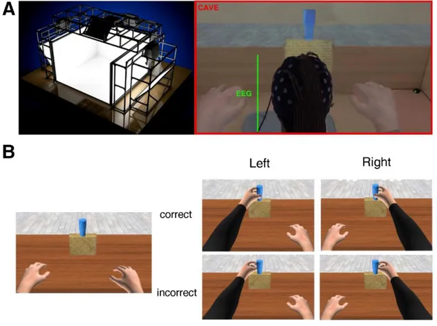
Participants sat in a cave automatic virtual environment (CAVE) with projectors directed to four walls of a room-sized cube (3m X 3 m X 2.5 m; Cruz-Neira et al. 1993). The virtual scenario consisted of a basic room with a table. At the center of the table, a yellow parallelepipedon was located with a blue glass on top of it. Participants observed one avatar in first-person perspective (1PP; see Fig. 1) seated on a chair in front of a table with its arms resting on the table. The glass was placed in the avatar’s peripersonal space at a distance of ~50 cm (Costantini et al. 2011). The avatar and the scenarios were created by means of Autodesk Maya 2011 and 3D Studio Max 2011, respectively. The kinematics of the avatar were implemented by HALCA library (Gillies and Spanlang 2010), and the experiment was performed in an immersive three-dimensional (3D) virtual environment with a real-size avatar drawn on a 1:1 scale and rendered in XVR 2.1 (Huang et al. 2013; Tecchia et al. 2014). Participants wore Nvidia stereo glasses in which 3D virtual images were alternately displayed on both eyes with a refresh rate of 60 Hz. Moreover, these stereo glasses were
interfaced with an Intersense 900 ultrasonic system (Thales Visionix) and constantly tracked the head position during the experiment.
Experimental Procedure
Before the beginning of the experiment, participants underwent a familiarization phase with the experimental setup, as well as a calibration phase within the virtual environment, which consisted of adapting the size of the virtual body to the real one. After this phase, a brief practice session (8 trials, 4 correct and 4 erroneous) occurred. Each participant was informed that the goal of the avatar’s movements was to reach and grasp the glass on the table and that the action might or might not be successful. The total number of trials per participant was 100, 70 of which were incorrect and 30 of which were correct. This is the same number of trials used in Pavone et al. (2016) but with the inverse proportion (70 correct and 30 incorrect). The total duration of our experiment was ~20 min. Participants were not informed about the probability of the different action outcome. At the onset of each trial, a sound signaled the beginning of the action. During the trial, participants observed the movement of the avatar’s right arm in 1PP. The kinematics of the movement were identical for the first 700 ms in both conditions and began to diverge in the last 300 ms. The trajectory deviation led to either a successful or unsuccessful grasp. The deviation from the to-be grasped object was identical in all the erroneous trials (Fig. 1). The sequence of correct and incorrect trials was randomized. After the end of the action, the avatar’s arm rested for 1,000 ms (± 50 ms) before a black screen appeared. During the intertrial interval (ITI), three different situations could occur: 1) in 10% of the trials, participants had to answer a catch question (“Did the arm take the glass?” (yes/no answer); 2) in 40% of the trials, an empty black screen was presented; and 3) in 50% of the trials, participants had to provide ratings concerning the sense of embodiment. When the first and third cases occurred, the black screen lasted until a vocal response was given, whereas when the second case occurred, the experimenter pressed a key to start the next trial, producing a variable ITI (mean duration: ~4,000 ms). To measure their sense of embodiment, participants were asked to verbally rate two questions
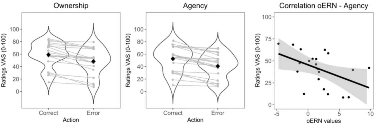
on a visual analog scale (VAS) from 0 to 100. One question was about their sense of ownership (“To what extent did you feel the arm was yours?”; 0 = no ownership to 100 = maximal ownership; Slater et al. 2010), and the other question was about their sense of agency (“How much did you feel in control of the arm?”; 0 = no control to 100 = maximal control; Fusaro et al. 2016; Tieri et al. 2015a, 2015b; Villa et al. 2018). The two questions were always presented together and in a randomized order. A total of 819 embodiment ratings for erroneous trials, and 351 embodiment ratings for correct trials, were collected across the whole sample, with each embodiment rating including the two questions on ownership and agency (2 participant ratings were missing due to technical issues during the saving of data and therefore were not included; 21 participants responded to the VAS in 50% of trials, and 4 responded to it in 30% of trials).
Statistical analyses were done using R software (R Core Team 2014). ERPs and time-frequency analyses were made using the erpR package (Arcara and Petrova 2014). All ANOVAs were performed using the ez package (Lawrence 2013). Analyses were performed using repeated-measures ANOVA, and Greenhouse-Geisser correction for nonsphericity was applied when appropriate. By estimating the effect size relative to the ANOVA test, we report the partial eta squared (η2p). Spearman correlations were executed, and Bonferroni corrections for multiple comparisons were applied when necessary. Practice trials were excluded from the analyses.
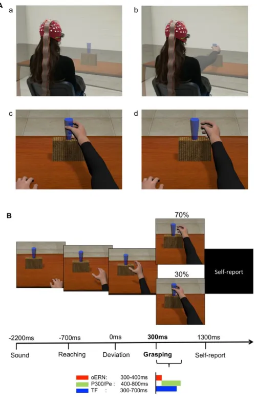
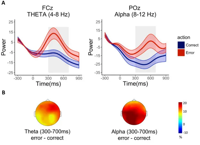
Figure 1. A: example of the experimental paradigm and setup. Top images show the participant in the 3-dimensional (3D) rendering of the virtual scenario (cave automatic virtual environment, CAVE), during which projectors are directed to the 4 walls of a room-sized cube; the participant observes a static situation before the beginning of the trial (a) and at the end of an action (b). Bottom images represent a snapshot from inside the CAVE. The participant is immersed in the virtual scenario. In first-person perspective, using 3D visualization glasses, the participant observes a real-size avatar’s arm, perceived as attached to the shoulder during a correct (c) and an erroneous (d) grasping. B: timeline of a single trial. The avatar’s action lasted 1,000 ms: the reaching
phase was equal for both types of movements and lasted 700 ms; the deviation phase in which the avatar’s arm-path deviation could occur and define an erroneous or correct action lasted 300 ms. The onset of the avatar’s limb-path deviation is set at 0 ms; the end of the avatar’s action occurs at 300 ms. The main EEG analyses (reported at bottom) have been conducted with intervals of interest chosen a priori. oERN, observed error-related negativity; P300/Pe, error positivity; TF, window of time-frequency analysis.
EEG Recording and Preprocessing Analysis
EEG signals were recorded using a Neuroscan SynAmps RT amplifier system and 60 scalp electrodes embedded in a fabric cap (Electro-Cap International), arranged according to the international 10–10 system1. Horizontal electro-oculogram was recorded bipolarly from electrodes
placed on the outer canthi of each eye. EEG signal was recorded continuously in alternating current mode with a bandpass filter (0.05–200 Hz) and sampling rates of 1,000 Hz. Impedances were kept under 5 k. All electrodes were physically referenced to an electrode placed on the right earlobe and re-referenced offline to the average of both earlobe electrodes. Offline, raw data were low-pass filtered with a 40-Hz filter (finite impulse response filter, transition 40–42 Hz, stopband attenuation 60 dB). For ERP analyses, an additional bandpass filter (0.5–30 Hz) was applied on the continuous raw signal. Independent component analysis (ICA; Jung et al. 2000) was performed on the continuous EEG signal while components that were clearly related to blinks and ocular artifacts were removed (on average, 3.4 ICA components; range 2–5). EEG signal was then downsampled to 500 Hz and epoched in wide windows of 3-s length, from -1.5 to +1.5 s to avoid edge artifacts induced by the following wavelet convolution. Epochs were time-locked to the onset of the avatar’s arm-path deviation, (i.e., 700 ms from the beginning of the movement, as in Spinelli et al. 2018). All epochs were baseline corrected to the 200 ms preceding the avatar’s arm-path deviation (i.e., when the limb movements were identical in correct and incorrect conditions; Pavone et al. 2016). The offset of the
1The EEG was recorded from the following channels: Fp1, Fpz, Fp2, AF3, AF4, F7, F5, F3, F1, Fz, F2, F4, F6, F8, FC5, FC3, FC1, FCz, FC2, FC4, FC6, T7, C5, C3, C1, Cz, C2, C4, C6, T8, TP7, CP5, CP3, CP1, CPz, CP2, CP4, CP6, TP8, P7, P5, P3, P1, Pz, P2, P4, P6, P8, PO7, PO3, AF7, POz,
avatar’s movement occurred 300 ms after the avatar’s limb deviation began. Each epoch was then visually inspected for artifacts to manually remove residual eye blinks or epochs exceeding -100/+100 µV amplitude. A total of 1,524 erroneous trials and 665 correct trials were analyzed for both ERPs and time-frequency analyses (~96% of total collected trials). Because the correct and incorrect actions had different occurrence rates (70% vs 30%), we selected the trials from the incorrect condition to keep the number of trials equal in the two conditions and avoid spurious effects due to the different signal-to-noise ratios (Cohen 2014a; Luck 2005). Therefore, for each participant, trials from the incorrect condition were selected with a built-in Brainstorm function that selects a subset of trials2.
The main findings, with the total amount of incorrect trials, are reported in RESULTS, EEG Analyses on All the Incorrect Trials. Bad channels were replaced using the spherical splines method only when necessary (4 channels were interpolated in only 1 subject; Perrin et al. 1989). Analyses were performed using the Brainstorm toolbox (free open source for MEG/EEG analysis, https://neuroimage.usc.edu/brainstorm/; Tadel et al. 2011) and customized MATLAB routines (Cohen 2014a).
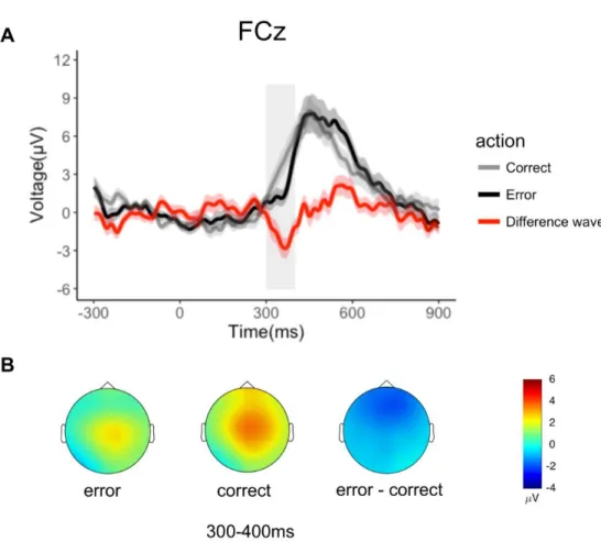
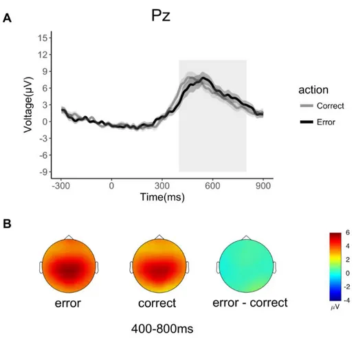
Analysis in the Time Domain: Action Observation-Related ERPs
In line with the literature, oERN and oPe/P300 were respectively analyzed on FCz and Pz electrodes, where they reached their maximum amplitude. The oERN was analyzed in the 100-ms time window following the end of the avatar’s action (300–400 ms) at FCz electrode. The oPe/P300 were measured in a preselected time window between 400 and 800 ms at the parietal sites (Pz), in line with previous results (Pavone et al. 2016). All ERPs analyses were based on mean amplitude (Luck 2005). Widely recognized in error literature, the ERN can be hidden from the massive contribution of error positivity components that propagate from parietal to frontal areas and that mask some of the frontal components (Luck 2005). For visualization purposes, we minimalized overlap
between different components (Fig. 2A) by computing the difference waves in which the average number of correct trials is subtracted from the average number of erroneous trial (Fischer et al. 2017; Koban et al. 2010; Maier and Steinhauser 2016).
Analysis in the Time-Frequency Domain
For the frequency analysis, we used a complex Morlet transformation to compute time-frequency decomposition. A mother wavelet with central time-frequency of 1 Hz and 3 s of time resolution (full width half maximum, FWHM) was designed as in Brainstorm software (Tadel et al. 2011) The other wavelets were computed from this mother wavelet and ranged from 1 to 30 Hz, with 0.5-Hz linear frequency steps. To normalize each signal and frequency bin separately with respect to a baseline, we computed the relative power change (in %) over the time-frequency decomposition as:
𝐹 = S(t, f) − Sbase (t, f) Sbase(t, f) ∗ 100
where S(t, f) is the signal spectrum at a certain given interval of time (t) and frequency (f), and Sbase(t,
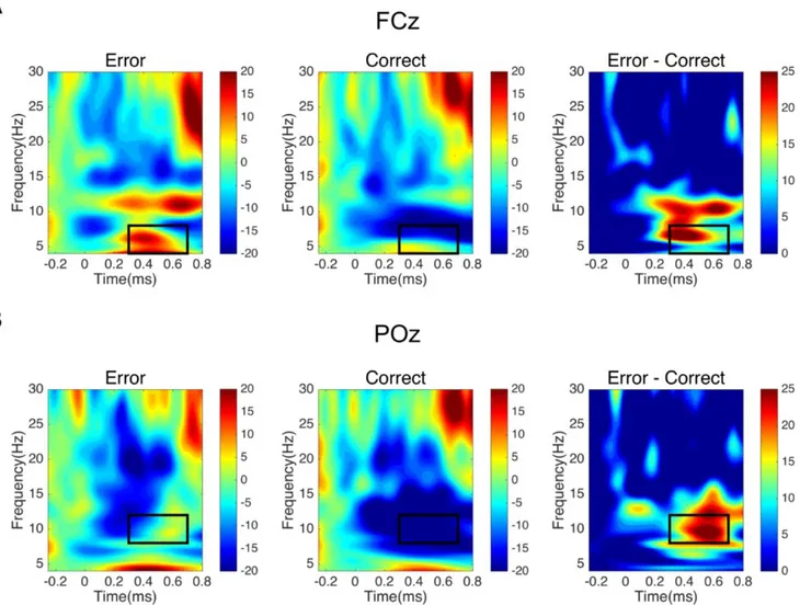
f) represents the signal power of the reference signal used as baseline. To avoid edge effects, the power activity from -250 to -50 ms, a window in which the avatar’s movement was identical in erroneous and correct conditions, was used as the baseline interval. Positive and negative values index a decrease or an increase in synchrony of the recorded neuronal population (Pfurtscheller and Lopes da Silva 1999) with respect to a given reference interval, where equal neural activity is expected between conditions. In our case, a relative power increase/decrease represents a modulation of power compared with the mean power activity during the baseline. As in Pavone et al. (2016), the main analyses were computed on FCz electrode, focusing on theta (4–8 Hz), alpha (8 –12 Hz), and beta (13–30 Hz) bands, and in the preselected time interval from 300 to 700 ms (i.e., in time windows of 400 ms from the end of the avatar’s action). On theta frequency, we also performed the analyses on POz electrode to rule out the possibility of a more general, rather than a mainly frontal, effect (Pavone
et al. 2016). Further exploratory analyses were also performed. To check whether the participants consciously perceived the error before the end of the action, we analyzed theta activity in the time range of action divergence before the outcome appeared (0–300 ms). It is widely held that evoked oscillations reflect a phase-locked activity to the stimulus in the time frequency activity. To investigate whether the reported theta effect is a different representation of the oERN in the time domain, we performed an additional analysis in which we removed the evoked response from each trial before computing the time-frequency decomposition for both experimental conditions.
Results
Time Domain Analysis
oERN
Analyses on oERN revealed a main effect of Condition [F(1,22)= 6.77, p=0.016, η2=0.24],
with erroneous actions showing less positive values than correct actions [mean value in erroneous condition (MERR) =1.76 µV; mean value in correct condition (MCORR) =3.54 µV] (Figure 2A). The
Figure 2 Electrophysiological results in the time domain for each condition (70% erroneous actions, 30% correct actions). A. Grand average waveforms of oERN at electrode FCz, over an interval of interest (300-400ms) chosen a priori. The onset of avatar’s limb-path deviation is at 0ms; the end of avatar’s action occurs at 300ms. Shaded areas denote the standard error around the mean. The grey rectangle highlights the time window considered for statistical analysis. For representation purposes we computed the difference wave on FCz, showing the negative amplitude of the oERN. B. Topographical voltage distribution for erroneous, correct and erroneous-minus correct action condition.
oPe
Analyses on oPe revealed that the main effect of Correctness between the two conditions was not significant [F(1,22)= 0.25, p= 0.62, η2= 0.01, MERR= 5.48µV; MCORR= 5.22µV; Fig. 3A]. Figure
3B shows the typical topographical distribution of oPe over centroparietal recording sites, for both correct and erroneous actions.
Figure 3. Electrophysiological results in the time domain for each condition (70% erroneous actions, 30% correct actions). A. Grand average waveforms of oPe at electrode Pz on an interval of interest (400-800ms) chosen a priori. The onset of avatar’s limb-path deviation is at 0ms; the end of avatar’s action occurs at 300ms. Shaded areas denote the standard error around the mean. The grey rectangle highlights the time window considered for statistical analysis. B. Topographical voltage distribution for erroneous, correct and erroneous-minus action condition.
Time-frequency results
Theta (4-8Hz)
ANOVA on the electrode and time interval of interest showed a significant effect of Condition [F(22,1)=18.35; p<0.005, η2=0.45] with higher theta power activity for observation of erroneous actions
compared to correct actions [MERR=7.18; MCORR=-8.11] (Figure 4; figure 5). A middle-frontal cluster
confirming the greater theta activity in the erroneous rather than correct actions [MERR= 8.33; MCORR=
-6.44]. The same ANOVA on POz showed no significant difference concerning the accuracy of the grasp [F(22,1)=3.34; p=0.08, η2=0.13]. The time interval between the zero-onset and the end of the
avatar’s movement (0-300ms) showed no significant difference [F(22,1)= 1.98; p=0.17, η2=0.08]. With
the removed evoked activity – obtained by removing trial by trial the evoked activity from the total activity before the grand average -, the analysis of the theta activity showed a significant effect of Condition [F(22,1)= 17.15; p<0.005, η2=0.44]: higher theta power activity was shown for observation
of erroneous actions compared to correct actions.
Alpha (8-12 Hz)
ANOVA on FCz in the 300-700ms showed a main significant effect of Condition [F(22,1)=11.
08; p<0.005, η2=0.34]. This effect was associated with increased alpha power for erroneous actions
and decreased alpha power for correct actions [MERR=5.36; MCORR=-12.92] (Fig.4). Analyses on POz
revealed a consistent alpha suppression [F(22,1)= 10.09; p<0.005, η2=0.31] in the posterior electrode
during the correct actions, rather than the erroneous [MERR= -3.48; MCORR= -23.70] (Fig.5).
Beta (12-30Hz)
No significant main effect or interactions were found for this band [F(22,1)= 0.47; p=0.50,
Figure 4. Time-frequency representation of Relative Power change (in %) with respect to the baseline for erroneous, correct, difference (error-correct) conditions. The onset of avatar’s limb-path deviation is at 0ms; the end of avatar’s action occurs at 300 ms. The black rectangles indicate the a priori chosen window of interest between 300-700ms. A. Erroneous, correct, difference plots at electrode FCz in the selected interval (300-700 ms) for the theta band (4-8 Hz). B. Erroneous, correct, difference plots at electrode POz in the selected interval (300-700 ms) for the alpha band (8-12 Hz). The black rectangles indicate the values that have been submitted to statistical analyses.
Figure 5. A. Time series representation of theta power (4-8Hz) in electrode FCz (left panel) and alpha power (8-12Hz) in electrode POz (right panel) plotted for the correct and erroneous action observation conditions. The onset of avatar’s limb-path deviation is at 0ms; the end of avatar’s action occurs at 300 ms. Light colors denote the standard error around the mean. Grey rectangles highlight the time window considered for statistical analysis. B. Topographical distribution of the ERS/D from baseline of theta (left) and alpha (right) averaged in the time window of interest (300-700 ms).
Embodiment ratings and relation with EEG signals
Preliminary application of the Shapiro-Wilk test showed that embodiment ratings were not normally distributed therefore a non-parametric analysis, including Friedman test, for within-group was used. In order to explore the link between sense of embodiment and electro-cortical indices of error processing, Spearman correlations between Embodiment ratings and error signatures (Theta and oERN) were conducted across subjects.
A significant difference in the avatar’s grasp accuracy (correct vs. erroneous) in terms of sense of Ownership (χ2(1)= 21, p<.05) and Agency (χ2(1) = 21, p<.05) was found (Figure 6). As expected,
there was a greater sense of Embodiment in the correct actions (Ownership: 0.59 ± 0.20; Agency: 0.48 ± 0.21) compared to erroneous actions (Ownership: 0.52 ± 0.20; Agency: 0.41 ± 0.20). The correlation analysis between theta power (range 300-700 on FCz) and Embodiment ratings revealed no significant association. Further analyses showed a negative correlation between the oERN and the sense of Embodiment. In particular, the negative correlation between the oERN and the sense of Embodiment was accounted for by the sense of Agency [r= -0.50, p= 0.02], i.e. greater negative values of the oERN were associated with stronger sense of Agency in the erroneous actions. The sense of Ownership showed a trend [r=-0.42, p=0.06] in the same direction.
Figure 6. Subjective reports of embodiment in correct and erroneous action observation conditions. On Y-axes embodiment ratings along a 1-100 points Visual Analogue Scale (VAS). Participants answered two questions: one concerning sense of Ownership (“How much did you feel the arm was yours”; 0 = no ownership to 100 = maximal ownership) and one concerning sense of Agency (“How much did you feel to control the arm” 0 = no control to 100 = maximal control). The order of the questions was randomized. The black diamond in the violin plots represents the mean value, while the grey lines connect individual subject observations between the two conditions. The rightmost panel shows a scatterplot representation of the correlation between oERN and sense of Agency.