RESEARCH ARTICLE
Evaluation of oxidative stress effects on
different macromolecules in adult growth
hormone deficiency
Antonio ManciniID1,2*, Carmine BrunoID1,2, Edoardo Vergani1,2, Francesco Guidi1,2, Flavia Angelini1,2, Elisabetta Meucci2,3, Andrea SilvestriniID2,3*
1 Dipartimento di Medicina e Chirurgia Traslazionale, UniversitàCattolica del Sacro Cuore, Rome, Italy, 2 Fondazione Policlinico Universitario A, Gemelli IRCCS, Rome, Italy, 3 Dipartimento di Scienze biotecnologiche di base, cliniche intensivologiche e peri-operatorie, UniversitàCattolica del Sacro Cuore, Rome, Italy
*[email protected](AM);[email protected](AS)
Abstract
Adult growth hormone deficiency (GHD) is being increasingly recognized to cause prema-ture mortality exacerbated by oxidative stress. A case-control observational study has been performed with the primary objective of evaluating new parameters of oxidative stress and macromolecular damage in adult GHD subjects: serum nitrotryptophan; Total Antioxidant Capacity expressed as LAG time; urinary hexanoil-lysine; urinary dityrosine and urinary 8-OH-deoxyguanosine. GHD was diagnosed using Growth Hormone-Releasing Hormone 50μg iv+arginine 0,5 g/Kg test, with a peak GH response<9μg /L when BMI was<30 kg/m2 or<4μg/L when BMI was>30 kg/m2. Patients affected by adult GHD were divided into three groups, total GHD (n = 26), partial GHD (n = 25), and controls (n = 29). Total Antioxidant Capacity, metabolic and hormonal parameters have been determined in separate plasma samples; nitrotryptophan in serum samples; hexanoil-lysine, dityrosine, 8-OH-deoxyguano-sine in urine samples. Assessment of hexanoil-ly8-OH-deoxyguano-sine exhibited a trend to increase in comparing total GHD vs partial and controls, although not significant. Values of 8-OH-deox-yguanosine did not significantly differ among the three groups. Significant lower levels of dityrosine in partial GHD vs total and controls were found. No significant difference in nitro-triptophan serum levels was found, while significantly greater values of Total Antioxidant Capacity were showed in total and partial GHD vs controls. Thus, our result confirm that oxi-dative stress is increased both in partial and total adult GHD. The lack of compensation by antioxidants in total GHD may be connected to the complications associated to this rare disorder.
Introduction
Adult growth hormone deficiency (GHD) is defined as a clinical expression of a reduced GH secretion caused by congenital or acquired diseases affecting the hypothalamus or pituitary
a1111111111 a1111111111 a1111111111 a1111111111 a1111111111 OPEN ACCESS
Citation: Mancini A, Bruno C, Vergani E, Guidi F,
Angelini F, Meucci E, et al. (2020) Evaluation of oxidative stress effects on different
macromolecules in adult growth hormone deficiency. PLoS ONE 15(7): e0236357.https://doi. org/10.1371/journal.pone.0236357
Editor: Nukhet Aykin-Burns, University of Arkansas
for Medical Sciences College of Pharmacy, UNITED STATES
Received: February 16, 2020 Accepted: July 3, 2020 Published: July 20, 2020
Copyright:© 2020 Mancini et al. This is an open access article distributed under the terms of the
Creative Commons Attribution License, which
permits unrestricted use, distribution, and reproduction in any medium, provided the original author and source are credited.
Data Availability Statement: All relevant data are
within the paper and its Supporting Information files.
Funding: This study was supported by a non-profit
grant (MS200104_0016) released by MerckTM (https://www.merckgroup.com/it-it) to A.M. The funders had no role in study design, data collection and analysis, decision to publish, or preparation of the manuscript.
gland. Adults who develop GHD may report central weight gain, difficulty to lose weight, dry skin, fatigue and reduced quality of life [1].
Interestingly, experimental evidences, both in childhood and adulthood, have associated the GHD condition to oxidative stress (OS) [2]. OS could be defined as the imbalance between the production of oxidants and antioxidants in favour of the former, leading to a disruption of redox signalling and/or molecular damage [3]. However, discordant data concern growth hor-mone (GH) effects on antioxidant levels are reported. GH, for instance, influences the function of immune cells, inhibiting apoptosis in neutrophils by downregulationFas expression and
increasing reactive oxygen species (ROS) production [4]. Furthermore, GH increases ROS production in monocytes, even though no data on apoptosis in these cells have been reported [5]. Moreover, results in experimental animal models are contradictory. In Wister rats on calo-ric restcalo-riction GH/insulin treatment decreases ROS production and DNA damage in heart mitochondria, whether in the liver the same treatment decreases ROS production, but enhances DNA damage, thus suggesting pleiotropic effects of GH depending on involved tis-sue [6]. Mice with knock-out for the receptor of Growth Hormone (GHR-KO) have increased antioxidant defences with different total antioxidant capacity (TAC) according to sex [7]. In Ames dwarf mice with congenital TSH, prolactin and GH deficiency the enzymatic antioxi-dants appear to be increased even though scavengers, on the contrary, are decreased [8]. In human being with pre-pubertal GHD, GH treatment seems to increase antioxidant capacity [9]. Moreover, Scacchi et al. show increased lipid peroxidation in adult GHD, counteracted by GH treatment [10]. Additionally, some papers report the relationship between OS and insulin resistance in GHD patients [11,12]. In adults, GHD, indeed, shares with metabolic syndrome clinical and biochemical features even though differential pattern of antioxidant response and inflammatory mediators may be involved [13,14].
OS can be related to proteins, nucleic acids and lipids damage [15], in turn associated to an augmented cardiovascular and oncological risk [16,17]. Confirming, adult GHD, a condition of increased OS, is characterized by hypercoagulability and faster atherosclerosis development [18] and increased incidence of cancer [19]. Also partial GHD in adulthood, defined as a sub-normal GH response to stimulation tests not so severe to require a replacement therapy, has been associated to altered cardiovascular and metabolic features [20–23].
Since the early identification of oxidative molecular damage may represent a possible strat-egy to contrast the appearance of the previous mentioned complications, we aimed to extend our findings with a specific focus on parameters of oxidative stress, not previously evaluated in adult GHD subjects, which assess damages on macromolecules (e.g. lipids, proteins, nucleic acids). Thus, in this case-control observational study, the primary objective was the evaluation of new parameters of oxidative stress in adult GHD. Specifically, we measured serum nitro-tryptophan (N-Try), as a biomarker of protein nitration, urinary hexanoil-lysine (HEL) as parameter of lipidic oxidation; urinary dityrosine (DT), as a parameter of protein oxidation, urinary 8-OH-deoxyguanosine (8-OHdG) as a parameter of DNA oxidation, and the evalua-tion of Total Antioxidant Capacity (TAC), while secondary objectives were the definievalua-tions of metabolic and lipid pictures in this clinical background.
Materials and methods
Study population
The patients involved in this study consisted of 80 subjects (36 males, 44 females), admitted to the University Hospital “Policlinico Gemelli” Dept. of Internal Medicine, and enrolled (from 2016 to 2018) after being given an explanation of purposes and nature of the study, conducted
Competing interests: The authors have declared
in accordance with the declaration of Helsinki, as revised in 2013. The study protocol was approved by “Fondazione Policlinico Gemelli” ethical committee (Protocol sf 39612/16).
After being given a written consent, GHD untreated patients and controls of both sexes were included in the study.
Exclusion criteria were: age under 18 or over 70; obesity of genetic origin or related to other endocrine diseases; history of cranial hypertension or active cranial hypertension, decompen-sated type 1 or 2 diabetes mellitus; autoimmune diseases under immunosuppressive treatment; corticosteroid treatment (except for topic, inhalatory and oral hydrocortisone as replacement regimen); other diseases characterized with low insulin-like growth factor-1 (IGF-1) such as liver disease, malabsorption and malnutrition; active malignancy. No patients received GH therapy at the study enrollment.
GHD was diagnosed with dynamic test, using Growth Hormone-Releasing Hormone (GHRH 50μg intravenous + arginine 0,5 g/Kg) (GHRH/ARG).
26 patients, 13 males and 13 females, presented a peak GH response < 9μg/L (or <4 μg/L when BMI � 30 kg/m2) and were classified as total GHD group; 25 patients, 12 males and 13 females, with GH peak between 9 and 16μg/L were considered as partial GHD group. Finally, 29 subjects, 11 males and 18 females, matched for age and sex, with GH peak > 16μg/L were included as control group.
Within the total GHD group, different etiologies were found: 10 cases of empty sella syn-drome, 11 cases of idiopathic isolated GHD, 1 case of Arnold-Chiari synsyn-drome, 1 case of Sheehan’s syndrome, 1 case of pituitary cist, 1 case of post-surgical hypopituitarism, 1 case of pituitary infarction.
Patients underwent urine and venous sampling at 8 a.m, after an overnight fasting, collect-ing blood samples by two pyrogen-free tubes, one containcollect-ing heparin as anticoagulant, and one without anticoagulant to obtain serum.
Serum evaluation for hormones and N-Try
The sample collected into vacutainer tube(s) containing no anticoagulant was incubated in an upright position at room temperature for 30–45 min (no longer than 60 min) to allow clotting and then centrifuged at 2500 rpm for 15’ at 4˚C without brake. Supernatant (serum) was col-lected and stored at -80˚C until use.
The following hormonal parameters were determined: freeT3 (fT3), freeT4 (fT4), thyroid-stimulating hormone (TSH) and adrenocorticotropic hormone (ACTH). The methods used in hormone assays were: electro-chemo-luminescent immunoassay for ACTH (normal range 10–55 pg/ml); chemo-luminescent immunoassay for insulin (n.r. 3–20μUI/ml) TSH (n.r. 0.35–2.80μUI/ml), fT3 (n.r. 2.4–4.2 ng/ml), fT4 (n.r. 8.5–16.5 pg/ml), IGF-1 (n.r. 80–330 ng/ ml). Serum concentrations of IGF-1 were measured by using immunochemiluminometric assays on a Roche Modular E170 analyzer (Roche Diagnostics, Indianapolis, IN, USA). The intra- and inter-assay CV for all hormones were, respectively, < 5.0% and < 7.0%.
Western Blot analysis was applied to quantify N-Try (formed by peroxynitrite and trypto-phan) as biomarker of protein nitration; nitration of tryptophan residues is an oxidative
modification of proteins connected with cellular signaling, vasorelaxation and, possibly, carcino-genic damage [24]; before the analysis albumin was removed from serum samples following the protocol reported by Chen et al [25]. Briefly, 100μl of serum was precipitated by addition of four volumes of ice-cold acetone containing 10% w/v TCA and incubated at -20˚C for 90 min. The mixture was centrifuged at 15000 g for 20 min at +4˚C and the pellet washed with an addition of 1ml of ice-cold acetone followed a centrifugation at 15000 g for 20 min at +4˚C. The pellet was re-suspended in 100μl of a buffer containing 25 mM HEPES, pH 7.4, 0.1% SDS, 4 M urea; the
mixture was hated at 55˚C for 10 min to permit a complete solubilisation. After albumin removal, 15μl of sample plus 5 μl of loading buffer was loaded into a 10% acrylamide/bis-acryl-amide gel. After running and transfer blot, the nitrocellulose was blocked for 1 hour at RT with 5% non-fat milk and then incubated with an anti-Nitrotryptophan monoclonal antibody (Japan Institute for the Control of Aging (JaICA), Nikken SEIL Co., Ltd.) (100ug/ml) diluted 1/500 or with anti-Transferrin antibody (0,7mg/ml) (ad109503, Abcam plc, USA) diluted 1/5000. As a secondary antibody, we used a biotinylated anti-mouse 1/1000 (ab97023, Abcam plc, USA) for nitro-tryptophan or an anti-rabbit (ab97051, Abcam plc, USA) 1/15000 for transferrin. Luminol reagent (XLS025.0000, Cynagen srl., Bologna, Italy) was added and quantified with Chemidoc instrument and ImageLab software (Bio-Rad California 94547, USA). Data were expressed as fold increase (protein investigated/protein positive control—nitro-tryptophan/transferrin).
Plasmatic evaluations
The sample collected into pyrogen-free tubes containing heparin was centrifuged within one hour at 4000 rpm for 15 minutes; separate plasma aliquots were stored at -80˚C until assayed. Assessment of biochemical and metabolic parameters was performed, including plasmatic glu-cose, total cholesterol, low-density lipoprotein (LDL), high-density lipoprotein (HDL), triglyc-erides and uric acid. Plasma concentrations of glucose, total cholesterol, HDL-cholesterol, triglycerides, uric acid were measured using enzymatic assays by an Olympus AU2700 chemis-try analyzer (Olympus America Inc., Center Valley, PA, USA). The intra-and inter-assay coef-ficients of variation (CV) for total cholesterol and triglycerides were < 1.5%, and < 2.5%, respectively. The intra-and inter-assay CV for HDL-cholesterol were < 2.5%, and < 3.0%, respectively. LDL cholesterol was calculated by Friedewald’s equation: LDL = total cholesterol —(HDL + triglycerides/5). HOMA index was calculated according to the formula {[fasting insulin (μU/ml)]�[fasting glucose (mmol/l)]}/22.5 [26].
TAC was evaluated by spectrophotometric method, with a modification of the method developed by Rice-Evans and Miller [27], as previously described [28]. The method is based on the antioxidants inhibition of the absorbance of the radical action 2,2I-azinobis (3-ethylben-zothiazoline-6 sulphonate) (ABTS.+) formed by interaction between ABTS (150μM) and fer-rylmyoglobin radical species, generated by activation of metamyoglobin (2.5μM) with H2O2
(75μM). Aliquots of the frozen plasma were thawed at room temperature and 10 μl of the sam-ples were tested immediately. The manual procedure was used with only minor modifications, i.e., temperature at 37˚ C to be in more physiological conditions and each sample assayed alone to carefully control timing and temperature. The reaction was started directly in cuvette through H2O2addition after 1 min equilibration of all other reagents (temperature control by
a thermocouple probe, model 1408 K thermocouple, Digitron Instrumentation Ltd, Scun-thorpe, United Kingdom) and followed for 10 min under continuous stirring, monitoring at 734 nm, typical of the spectroscopically detectable ABTS.+. The presence of chain-breaking antioxidants induces a lag time (the Lag phase) in the accumulation of ABTS.+whose duration is proportional to the concentration of this type of antioxidants. Antioxidant capacity afforded by chain-breaking antioxidants is expressed as length of Lag phase (LAG, sec). Trolox, a water-soluble tocopherol analog, was used as a reference standard and assayed in all experiments to control the system. Absorbance was measured with an Agilent 8453 UV/Vis spectropho-tometer (Palo Alto, CA, USA) equipped with a cuvette stirring apparatus and a constant temperature cell holder. Measurements of pH were made with a PHM84 Research pH meter (Radiometer, Copenhagen, Denmark); the electrode response was corrected for temperature. Unless otherwise stated, experiments were repeated two to three times; qualitatively similar results were obtained with individual values varying < 8%. In the Lag mode, the assay mainly
measures non-proteic and non-enzymatic antioxidants that are primarily extracellular chain-breaking antioxidants, such as ascorbate, urate and glutathione.
Urinary evaluations
After collected urine was immediately aliquoted and stored at− 80 ˚C. Before assayed, urine was centrifuged at 1500 g at 4 ˚C for 15 min and the supernatant was used for the measure-ment. In urine we evaluated the additional indexes of oxidative stress:
• HEL adduct, as biomarker of lipid peroxidation, by ELISA method; it is a novel lipid hydro-peroxide-modified lysine residue that is formed by oxidative modification by oxidized omega-6 fatty acid such as linoleic acid or arachidonic acid. HEL is therefore a biomarker for initial stage of lipid peroxidation [29].
• Urinary DT, as biomarker of protein oxidation, by ELISA method; DT is a tyrosine dimer derived from tyrosyl radicals formed by ROS, metal-catalyzed oxidation and peroxidases [30]. • Urinary 8-OHdG, by ELISA method; it is an oxidized nucleoside of DNA; upon DNA repair,
it is excreted in urine and is the most frequently detected parameter of DNA lesions [31]. For HEL and DT measurements, the sample was diluted in 3 parts of saline buffer (0.9% NaCl in distilled water) as declared in the datasheet of each kit. All the analyzes have been per-formed following the manufacturer’s instructions. All ELISA kits have been purchased from JaICA, Japan.
Statistical analysis
The statistical analysis was performed using Stata 13 software. The planned sample size of min-imum 25 cases for each group was not based on formal power estimates, due to the lack of any information on the size of the minimal difference worth detecting for each of the study param-eters. Instead, the sample size was established based on considerations of statistical practicality (with 30+30 subjects a normal distribution of the mean can be usually assumed) and feasibility, due to the rarity of GHD disease. With this sample size, the study has power >80% to detect any difference between cases and controls that is as large or greater than 75% of the standard deviation of that specific parameter. Considering that the comparison concerns differences between diseased subjects and healthy controls in physiologic parameters of oxidative stress, smaller differences are of little, if any, interest.
According to Kolmogorov-Smirnoff test, variables followed standard distribution. The sta-tistical analysis was carried out using the variance analysis (F test—ANOVA) to study the dif-ferences among groups and, when significant, the Student T test for unpaired data, in two groups comparison. Moreover, linear regression analysis was performed, with the calculation of the Pearson coefficient. Linear correlation analyses were executed to evaluate the correlation between variables. The level of significance has been set at 0.05.
Finally, Pearson’s coefficient was used to investigate the correlation between each parame-ter of oxidative stress (i.e. LAG, HEL, DT, 8-OH-dG, N-try) as independent variable with anthropometric, metabolic and hormonal parameters (i.e.BMI, glucose, insulin, HOMA-IR, total and HDl-cholesterol, triglycerides, basal GH, peak GH response and GH levels at 90 min after GHRH+arginine, fT3, fT4, TSH, IGF-1) as dependent variables.
Results
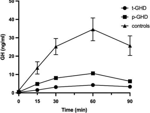
The GHRH/ARG combined test response in subjects enrolled in this study are depicted inFig 1as the average of the results. Mean± standard error of the mean (SEM) age of the three
groups were 50.28±2.22 years (total GHD group), 49.25±2.48 years (partial GHD group), 43.93±2.79 years (control group). Mean ± SEM BMI of the three groups were 31.05±2.47 kg/ m2(total GHD), 27.2±1.4 kg/m2(partial GHD), 25.5±1.1 kg/m2(controls).
Table 1shows mean and SEM of metabolic parameters in patients with total, partial GH deficiency and controls. As metabolic parameters are concerned, a significantly increased HOMA-index was observed both in total (3.4±0.47) and partial (2.65±0.33) adult GHD in
Fig 1. Average of the results of the GHRH/ARG test response in subjects enrolled in the study. Patients with
t-GHD have a peak of less than 9.0μg/L, those with p-GHD a peak lower than 16.0 μg/L and the controls have a response greater than 16.0μg/L. Abbreviations: GHRH/ARG test: growth hormone-releasing hormone/arginine test; t-GHD: total GHD; p-t-GHD: partial GHD; ctrl: control subjects.
https://doi.org/10.1371/journal.pone.0236357.g001
Table 1. Metabolic parameters.
TOTAL GHD PARTIAL GHD CONTROLS
Mean SEM Mean SEM Mean SEM
Glucose (mg/dl) 84.92 3.07 83.81 1.98 80.50 1.74 Insulin (μUI/ml) 15.86� 2.06 12.65� 1.53 7.49 0.89 HOMA-IR 3.40� 0.47 2.65� 0.33 1.51 0.19 Cholesterol total (mg/dl) 189.56 5.43 194.25 6.66 195.00 7.25 Cholesterol HDL (mg/dl) 53.76 3.52 58.15 3.24 60.23 2.56 Cholesterol LDL (mg/dl) 122.75 8.90 113.20 7.97 112.31 2.56 Triglycerides (mg/dl) 110.64 8.25 96.70 7.68 88.73 7.69 Uric acid (mg/dl) 6.09�˚ 0.26 5.13 0.34 4.44 0.19
Mean and SEM of metabolic parameters in subjects enrolled in this study: total GHD, partial GHD and controls. �p<0.05, total and partial GHD vs controls, using Student T test for unpaired data.
˚p<0.05, total GHD vs partial GHD, using Student T test for unpaired data.
comparison to controls (1.51±0.19), p<0.05 (T test for unpaired data). A trend to worse lipid pattern was shown in total GHD.
Table 2shows mean and SEM of hormonal parameters. IGF-1 levels did not differ among the three groups (111.4±6.8 in total GHD, 127.4±7.63 in partial GHD, 122.77±9.49 ng/ml in controls) p<0.05 (T test for unpaired data).
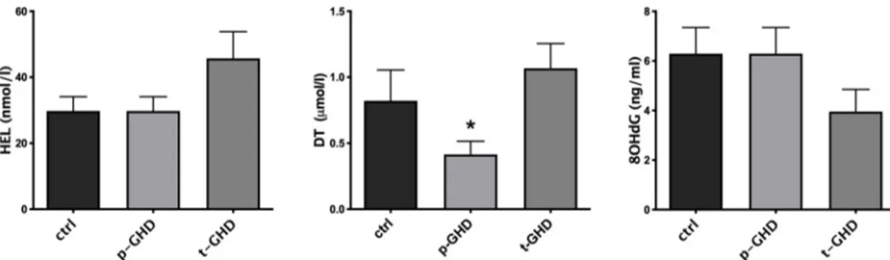
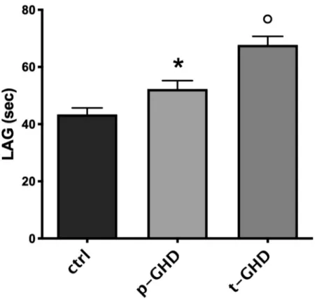
In the three groups the indexes of oxidative stress were also singularly compared in urine and serum of the subjects for each group.Fig 2shows mean± SEM of the values of urinary parameters (i.e. HEL, DT, 8-OHdG) in the three groups of patients. In evaluation of urine products of oxidation, a trend to increase in HEL in total GHD vs partial and controls was observed, although not significant. Values of 8-OHdG did not significantly differ among the three groups. On the contrary, significantly lower levels of DT in partial GHD vs total and con-trols were found (p<0.05- T test for unpaired data). A significant difference in N-Try was not found (Fig 3), while significantly greater values of TAC (p<0.05- T test for unpaired data) were observed in partial GHD vs controls, with a further significant increase in total GHD vs partial GHD (Fig 4).
Interestingly, data occurred in correlation analysis, depicting different patterns in control and partial GHD, on one side, and total GHD on the other one. For seek of clarity, in controls LAG values showed a positive correlation with BMI and insulin (r = 0.49 p = 0.03 and r = 0.51 p = 0.02, respectively) and an inverse correlation with GH stimulated levels (peak and 90 min) (p = -0.47 p = 0.02 and r = -0.44 p = 0.02, respectively); in partial GHD, LAG positively
Table 2. Hormonal parameters.
TOTAL GHD PARTIAL GHD CONTROLS
Mean SEM Mean SEM Mean SEM
IGF-1 (ng/ml) 111.44�˚ 6.80 127.41 7.63 122.77 9.49 fT3 (pg/ml) 3.02 0.08 3.18 0.07 3.04 0.09 fT4 (pg/ml) 11.19 0.45 11.43 0.28 11.14 0.29 TSH (μUI/ml) 1.13 0.09 1.64 0.17 1.51 0.28 ACTH (pg/ml) 20.16 1.82 22.16 3.31 22.81 2.79 Cortisol (ng/ml) 91.22 9.94 116.55 11.45 109.57 10.80
Mean and SEM of hormonal parameters in subjects enrolled in this study: total GHD, partial GHD and controls. �p<0.05, total and partial GHD vs controls, using Student T test for unpaired data.
˚p<0.05, total GHD vs partial GHD, using Student T test for unpaired data.
https://doi.org/10.1371/journal.pone.0236357.t002
Fig 2. Mean± SEM of the values of urinary Hexanoil-lysine (HEL), Dityrosine (DT) and 8-hydroxy-2 deoxyguanosine (8OHdG) evaluated in the three groups.�p<0.05 vs other two groups. Abbreviations: t-GHD: total GHD; p-GHD: partial GHD; ctrl: control subjects.
Fig 3. Western Blot and densitometric analysis of serum nitrotryptofan (NT) and transferrin (Tfr) levels in the three groups.
(A) Western blot analysis. Blots were acquired with aChemidoc XPS and the images were analyzed with the ImageLab software. Transferrin was used as an internal and normalization control. (B) Densitometric analysis from all the data analyzed. The relative densitometric expression of each patient was normalized with transferrin expression. Data are expressed as fold/increase (mean ±SEM). Abbreviations: t-GHD: total GHD; p-GHD: partial GHD; ctrl: control subjects.
https://doi.org/10.1371/journal.pone.0236357.g003
Fig 4. Mean±SEM of total antioxidant capacity, expressed by LAG (sec) in the three groups.�p<0.05 vs controls; ˚p<0.05 vs p-GHD and controls. Abbreviations: t-GHD: total GHD; p-GHD: partial GHD; ctrl: control subjects.
correlated with BMI and HOMA-IR (r = 0.53 p = 0.03 and r = 0.65 p = 0.008) and an inverse correlation with GH levels (90 min) (r = -0.43 p = 0.03). Finally, a negative correlation was found between HEL and GH at 90 min both in controls (r = -0.54 p = 0.006) and partial GHD (r = -0.60 p = 0.002), and between N-try and GH at 90 min in (r = -0.52 p = 0.03).
These correlations were not present in total GHD patients. On the contrary, an inverse cor-relation was observed between HEL and DT with HOMA-IR (r –0.42 p = 0.04 and r –0.42 p = 0.04, respectively), and a positive correlation between HEL and basal and peak GH (r = 0.79 p = 0.0004 and r = 0.43 p = 0.04, respectively), between 8-OH-dG with basal GH (r = 0.56 p = 0.03), and between N-Try and BMI (r = 0.59 p = 0.02).
Discussion
To the best knowledge of the authors, this is the first report that shows parameters of macro-molecular oxidative damage in adult GHD subjects. In serum, we evaluated N-Try, as index of protein oxidative damage; in urine samples, we evaluated HEL, DT and 8-OHdG, as parameters of oxidative damage of lipid, proteins and DNA, respectively. There were no sig-nificant changes among the three groups, except for DT that, unexpectedly, was sigsig-nificantly lower in partial GHD versus the other two groups (U-shaped pattern). This datum could be explained considering the antioxidant systemic response of the body, which was assessed in our study by TAC (expressed as Lag phase in sec). Indeed, already in partial GHD, TAC is significantly greater than controls, yet antioxidants are still able to compensate oxidative damage (as indicated by lower DT urinary levels). Oxidative stress is further increased in total GHD which presented the highest TAC levels (as a result of an increased production of antioxidants to balance the enhanced OS), a trend in increased HEL and increased urinary DT production (vs partial GHD), suggesting a partial compensation despite a remarkable antioxidants production.
Additionally, few studies pinpointed on the relationship between OS and adult GHD. Pre-pubertal GHD showed a significant reduced TAC and vitamin E correlated to the reduced lev-els of IGF-1/ IGF-BP3 while malondialdehyde (MDA), another marker of oxidative stress, was significantly increased and inversely correlated to IGF-1. The re-evaluation of these parame-ters after 1 year of recombinant GH (rGH) therapy showed an increase of TAC and a decrease of MDA, both returned to normal levels [9]. Higher peroxide levels and lower TAC was found in GHD patients by Scacchi et al, however no direct correlation with IGF-1 was highlighted. After 4 months of follow up with rhGH treatment, both peroxide levels and TAC were restored to control values [10]. On the contrary, the group of Gonzàlez-Duarte found higher TAC levels in adult GHD patients than controls [32]. The discrepancy between these results and those reported in the present study is probably due to different TAC evaluation method and the dif-ferent stages in which the disease was assessed.
Therefore, considering the complex picture and putting all data together, we can suggest that also partial GHD exhibits antioxidant systems reactivity, suggesting a condition of oxida-tive stress. We cannot establish if partial and/or total GHD represent different stages of the natural history of the syndrome (but longitudinal studies can clarify this point).
Other plasmatic protein damage indexes, such as nitro-tyrosine and carbonyls, have already been considered in other study, even though no difference was detected between adult GHD and healthy controls. Thus, data on protein serum damage indexes, represented here by N-Try, are congruent with that reported by Ozbey [33]. Furthermore, the same study report a significant increase in oxidized LDL, in turn associated to a worse atherosclerotic picture compared to healthy controls, in adult GHD, a result afterwards confirmed by several studies [10,32,33]. In striking contrast, Smith et al. reported in adult GHD reduced lipid peroxidation,
evaluated both as plasma lipid hydroperoxides and LDL susceptibility to peroxidation [34]. Despite, in this study lipid damage was not assessed by plasmatic indexes of lipid peroxidation, urinary HEL have been analysed, showing a trend in increased production, yet not significant, in total GHD group. A larger study population may support a better identification of this phe-nomenon. Importantly, Ozbey et al. report that oxidized LDL levels in adult GHD patients match with the duration of the disease in a group of patients with a median duration from the diagnosis of hypopituitarism of 96 months. This aspect could be decisive in the interpretation of data. Indeed, Gonzàlez-Duarte et al. pointed out that OS may be present since the early stage of the disease and may be influenced by time-course of the disease. The same study con-firmed the increase in oxidized lipoproteins production, highlighted the consumption of the antioxidant glutathione and the reduced/oxidized glutathione ratio and indicated, as previ-ously reported [35], a worse endothelial function in adult GHD [32].
Surprisingly, none of already cited paper has analyzed partial GHD condition, which, according to our results, seems to reveal an initial state of oxidative stress, although still com-pensated. Metabolic parameters, either, present some alterations both in total and in partial adult GHD, thus confirming by Colao et al. observations [20–22].
Correlation studies highlighted a differential pattern of association between macromolecu-lar damage and hormonal/metabolic parameters in the three groups studied, despite an unequivocal scenario was not already clear, very likely for the small number of subjects involved and therefore the difficulty to establish a direct cause-effect relation. While in con-trols and partial GHD subjects, OS seemed to be related to BMI and insulin-resistance and inversely related to GH levels, the correlations were different in total GHD, in which other mechanisms seems to be involved. The correlation with GH levels is difficult to elucidate, but we underline that the patients studied showed very low levels of the hormone. Moreover, evi-dence for such different patterns has been previously described [13].
The strength of this study lies in the novelty of the topic and in the parameters evaluated in such subjects, extending our previous reports on oxidative status in GHD [13]. Thus, we can suggest that oxidative stress is increased both in partial and total GHD, hypothesizing that lack of compensation by antioxidants can be associated to the recognized complications of this syndrome.
In spite of all precautions, our study may be still subject to certain biases, and some main potential restrictions should be considered. The number of subjects in the three groups is slightly small, so the statistical power of the study is limited; consequently, our findings will need to be confirmed in a larger population and will help to delineate the impact of OS in GHD condition. Therefore, the study design and the power analysis cannot draw a cause-effect relationship. In the present study, the only assessment of antioxidants is TAC, which is an expression of non-enzymatic antioxidants, even though enzymatic antioxidants (e.g CAT, GSHPx, and SOD) may be relevant in human being. The moment of the disease history in which the patient is evaluated may affect antioxidants production and macromolecular dam-age. Finally, more data and longitudinal studies are still needed in order to better characterize partial GHD and its possible evolution to total GHD. Anyhow, the relationships between OS and adult GHD merit further studies to develop targeted personalized therapy, including med-ical interventions with long-term hormonal replacement with GH.
Supporting information
S1 File. Blot results. Original unadjusted images underlying blot made for N-Try serum
eval-uation. (PDF)
Author Contributions
Conceptualization: Antonio Mancini, Andrea Silvestrini.
Data curation: Carmine Bruno, Edoardo Vergani, Francesco Guidi.
Formal analysis: Carmine Bruno, Edoardo Vergani, Francesco Guidi, Andrea Silvestrini. Funding acquisition: Antonio Mancini.
Investigation: Antonio Mancini, Carmine Bruno, Edoardo Vergani, Francesco Guidi, Flavia
Angelini, Elisabetta Meucci, Andrea Silvestrini.
Methodology: Antonio Mancini, Carmine Bruno, Edoardo Vergani, Andrea Silvestrini. Project administration: Antonio Mancini.
Resources: Carmine Bruno, Edoardo Vergani, Francesco Guidi, Flavia Angelini, Elisabetta
Meucci, Andrea Silvestrini.
Software: Carmine Bruno, Edoardo Vergani, Francesco Guidi. Supervision: Antonio Mancini, Elisabetta Meucci, Andrea Silvestrini.
Validation: Francesco Guidi, Flavia Angelini, Elisabetta Meucci, Andrea Silvestrini. Visualization: Edoardo Vergani.
Writing – original draft: Antonio Mancini, Carmine Bruno, Edoardo Vergani.
Writing – review & editing: Antonio Mancini, Carmine Bruno, Edoardo Vergani, Elisabetta
Meucci, Andrea Silvestrini.
References
1. Molitch ME, Clemmons DR, Malozowski S, Merriam GR, Vance ML, Shalet SM, et al. Evaluation and Treatment of Adult Growth Hormone Deficiency: An Endocrine Society Clinical Practice Guideline. J Clin Endocrinol Metab. 2011; 96(6):1587–609.https://doi.org/10.1210/jc.2011-0179PMID:21602453
2. Giovannini L, Tirabassi G, Muscogiuri G, Di Somma C, Colao A, Balercia G. Impact of adult growth hor-mone deficiency on metabolic profile and cardiovascular risk. Endocr J. 2015; 62(12):1037–48.https:// doi.org/10.1507/endocrj.EJ15-0337PMID:26300280
3. Halliwell B, Gutteridge JMC. Free Radicals in Biology and Medicine. Vol. 10, Free Radical Biology and Medicine. 1999. 449–450.
4. Matsuda T, Saito H, Inoue T, Fukatsu K, Han I, Furukawa S, et al. Growth hormone inhibits apoptosis and up-regulates reactive oxygen intermediates production by human polymorphonuclear neutrophils. J Parenter Enter Nutr. 1998; 22(6):368–74.
5. Warwick-Davies J, Lowrie DB, Cole PJ. Growth hormone activation of human monocytes for superoxide production but not tumor necrosis factor production, cell adherence, or action against Mycobacterium tuberculosis. Infect Immun. 1995.
6. Sanz A, Gredilla R, Pamplona R, Portero-Otı´n M, Vara E, Tresguerres JAF, et al. Effect of insulin and growth hormone on rat heart and liver oxidative stress in control and caloric restricted animals. Bioger-ontology. 2005; 6(1):15–26.https://doi.org/10.1007/s10522-004-7380-0PMID:15834660
7. Hauck SJ, Aaron JM, Wright C, Kopchick JJ, Bartke A. Antioxidant enzymes, free-radical damage, and response to paraquat in liver and kidney of long-living growth hormone receptor/binding protein gene-disrupted mice. Horm Metab Res. 2002; 34(9):481–6.https://doi.org/10.1055/s-2002-34787PMID:
12384824
8. Brown-Borg HM, Bode AM, Bartke A. Antioxidative mechanisms and plasma growth hormone levels: Potential relationship in the aging process. Endocrine. 1999; 11(1):41–8.https://doi.org/10.1385/ ENDO:11:1:41PMID:10668640
9. Mohn A, Di Marzio D, Giannini C, Capanna R, Marcovecchio M, Chiarelli F. Alterations in the oxidant-antioxidant status in prepubertal children with growth hormone deficiency: Effect of growth hormone replacement therapy. Clin Endocrinol (Oxf). 2005; 63(5):537–42.
10. Scacchi M, Valassi E, Pincelli AI, Fatti LM, Giraldi FP, Ascoli P, et al. Increased lipid peroxidation in adult GH-deficient patients: Effects of short-term GH administration. J Endocrinol Invest. 2006; 29 (10):899–904.https://doi.org/10.1007/BF03349194PMID:17185899
11. Yuen KCJ, Dunger DB. Therapeutic aspects of growth hormone and insulin-like growth factor-I treat-ment on visceral fat and insulin sensitivity in adults. Diabetes, Obesity and Metabolism. 2007. 9(1):11– 22.https://doi.org/10.1111/j.1463-1326.2006.00591.xPMID:17199714
12. Aguirre GA, Ita JR, Garza RG, Castilla-Cortazar I. Insulin-like growth factor-1 deficiency and metabolic syndrome. J Transl Med. 2016; 14:3.https://doi.org/10.1186/s12967-015-0762-zPMID:26733412
13. Mancini A, Di Segni C, Bruno C, Olivieri G, Guidi F, Silvestrini A, et al. Oxidative stress in adult growth hormone deficiency: different plasma antioxidant patterns in comparison with metabolic syndrome. Endocrine. 2017; 59(1):130–136.https://doi.org/10.1007/s12020-017-1468-1PMID:
29143180
14. Basile U, Bruno C, Napodano C, Vergani E, Pocino K, Brunetti A, et al. Plasmatic free light chains as inflammatory marker in insulin resistance: comparison of metabolic syndrome with adult growth hor-mone deficiency. BioFactors. 2018; 44(5).
15. Stadtman ER, Levine RL. Protein Oxidation. Ann N Y Acad Sci. 2006; 186:464–78.
16. Madamanchi NR, Vendrov A, Runge MS. Oxidative stress and vascular disease. Arteriosclerosis, Thrombosis, and Vascular Biology. 2005; 25(1):29–38.https://doi.org/10.1161/01.ATV.0000150649. 39934.13PMID:15539615
17. Sosa V, Moline´ T, Somoza R, Paciucci R, Kondoh H, LLeonart ME. Oxidative stress and cancer: An overview. Ageing Research Reviews. 2013; 12(1):376–90.https://doi.org/10.1016/j.arr.2012.10.004
PMID:23123177
18. Colao A. The GH-IGF-I axis and the cardiovascular system: Clinical implications. Clinical Endocrinol-ogy. 2008; 69(3):347–58.https://doi.org/10.1111/j.1365-2265.2008.03292.xPMID:18462260
19. Svensson J, Bengtsson BÅ, Rose´ n T, Ode´n A, Johannsson G. Malignant disease and cardiovascular morbidity in hypopituitary adults with or without growth hormone replacement therapy. J Clin Endocrinol Metab. 2004;
20. Colao A, Cerbone G, Pivonello R, Aimaretti G, Loche S, Di Somma C, et al. The growth hormone (GH) response to the arginine plus GH-releasing hormone test is correlated to the severity of lipid profile abnormalities in adult patients with GH deficiency. J Clin Endocrinol Metab. 1999; 84(4):1277–82.
https://doi.org/10.1210/jcem.84.4.5605PMID:10199767
21. Colao A, Di Somma C, Cuocolo A, Filippella M, Rota F, Acampa W, et al. The severity of growth hor-mone deficiency correlates with the severity of cardiac impairment in 100 adult patients with hypopituita-rism: An observational, case-control study. J Clin Endocrinol Metab. 2004; 89(12):5998–6004.https:// doi.org/10.1210/jc.2004-1042PMID:15579750
22. Colao A, Di Somma C, Spiezia S, Rota F, Pivonello R, Savastano S, et al. The Natural History of Partial Growth Hormone Deficiency in Adults: A Prospective Study on the Cardiovascular Risk and Atheroscle-rosis. J Clin Endocrinol Metab. 2006; 91(6):2191–200.https://doi.org/10.1210/jc.2005-2566PMID:
16537686
23. Murray RD, Adams JE, Shalet SM. Adults with partial growth hormone deficiency have an adverse body composition. J Clin Endocrinol Metab. 2004; 89(4):1586–91.https://doi.org/10.1210/jc.2003-030761
PMID:15070916
24. Nuriel T, Hansler A, Gross SS. Protein nitrotryptophan: Formation, significance and identification. J Pro-teomics 2011; 19; 74(11):2300–12.https://doi.org/10.1016/j.jprot.2011.05.032PMID:21679780
25. Chen YY, Lin SY, Yeh YY, Hsiao HH, Wu CY, Chen ST, et al. A modified protein precipitation procedure for efficient removal of albumin from serum. Electrophoresis. 2005; 26(11):2117–27.https://doi.org/10. 1002/elps.200410381PMID:15880626
26. Matthews DR, Hosker JP, Rudenski AS, Naylor BA, Treacher DF, Turner RC. Homeostasis model assessment: insulin resistance andβ-cell function from fasting plasma glucose and insulin concentra-tions in man. Diabetologia. 1985; 28(7):412–9.https://doi.org/10.1007/BF00280883PMID:3899825
27. Rice-Evans C, Miller NJ. Total antioxidant status in plasma and body fluids. Methods Enzymol. 1994; 234(C):279–93.
28. Mancini A, Milardi D, Bianchi A, Festa R, Silvestrini A, De Marinis L, et al. Increased total antioxidant capacity in seminal plasma of varicocele patients: A multivariate analysis. Syst Biol Reprod Med. 2007; 53(1):37–42.
29. Kato Y, Miyake Y, Yamamoto K, Shimomura Y, Ochi H, Mori Y, et al. Preparation of a monoclonal anti-body to N(epsilon)-(Hexanonyl)lysine: application to the evaluation of protective effects of flavonoid sup-plementation against exercise-induced oxidative stress in rat skeletal muscle. Biochem Biophys Res Commun. 2000; 274(2):389–93.https://doi.org/10.1006/bbrc.2000.3150PMID:10913348
30. Kato Y, Wu X, Naito M, Nomura H, Kitamoto N, Osawa T. Immunochemical detection of protein dityro-sine in atherosclerotic lesion of Apo-E-deficient mice using a novel monoclonal antibody. Biochem Bio-phys Res Commun. 2000; 18; 275(1):11–5.https://doi.org/10.1006/bbrc.2000.3265PMID:10944432
31. Saito S, Yamauchi H, Hasui Y, Kurashige J, Ochi H, Yoshida K. Quantitative determination of urinary 8-hydroxydeoxyguanosine (8-OH-dg) by using ELISA. Res Commun Mol Pathol Pharmacol. 2000; 107 (1–2):39–44. PMID:11334369
32. Gonza´ lez-Duarte D, Madrazo-Atutxa A, Soto-Moreno A, Leal-Cerro A. Measurement of oxidative stress and endothelial dysfunction in patients with hypopituitarism and severe deficiency adult growth hormone deficiency. Pituitary. 2012; 15(4):589–97.https://doi.org/10.1007/s11102-011-0374-4PMID:22228310
33. O¨ zbey N, Telci A, Cakatay U, Yurci A, Molvalilar S. Determination of oxidative protein and lipid damage in adult hypopituitary patients with GH deficiency. J Endocrinol Invest. 2003; 26(10):1001–7.https://doi. org/10.1007/BF03348199PMID:14759074
34. Smith JC, Lang D, McEneny J, Evans LM, Scanlon MF, Young I, et al. Effects of GH on lipid peroxida-tion and neutrophil superoxide anion-generating capacity in hypopituitary adults with GH deficiency. Clin Endocrinol (Oxf). 2002; 56(4):449–55.
35. Evans LM, Davies JS, Anderson RA, Ellis GR, Jackson SK, Lewis MJ, et al. The effect of GH replace-ment therapy on endothelial function and oxidative stress in adult growth hormone deficiency. Eur J Endocrinol. 2000; 142(3):254–62.https://doi.org/10.1530/eje.0.1420254PMID:10700719