UNIVERSITÀ DEGLI STUDI DI SASSARI
Ph.D SCHOOL IN BIOMOLECULAR AND BIOTECHNOLOGICAL SCIENCES CURRICULUM BIOCHEMISTRY AND MOLECULAR BIOLOGY
HEMOGLOBINOPATHIES IN MOUNTAINOUS REGION
OF THUA THIEN HUE, VIETNAM
Le Phan Minh Triet
Ph.D Thesis
Tutor:
Professor Bruno Masala
Professor Nguyen Viet Nhan
Director:
Professor Claudia Crosio
Dr. Le Phan Minh Triet: Hemoglobinopathies in mountainous region of Thua Thien Hue, Vietnam Ph.D thesis in Biochemistry and Molecular Biology
Ph.D School in Biomolecular and Biotechnological Sciences - University of Sassari INDEX
Summary 4
1. INTRODUCTION 6
1.1 Structure and function of human hemoglobins 6
1.2 Localization and organization of globin genes 11
1.2.1 Detailed chromosomal organization of the human globin genes 12
1.2.2 Structure of globin genes 13
1.2.3 Regulation of globin genes expression 15
1.3 Disorders of the synthesis of haemoglobin 19
1.3.1 Classification of hemoglobinopathies 20
1.3.2 Pathophysiology of hemoglobinopathies 23
1.4 Molecular basis of thalassemia 25
1.4.1 The -thalassemia 26
1.4.1.1 Nondeletion types of -thalassemia 26 1.4.1.2 Dominantly inherited -thalassemia (thalassemic structural variants) 37
1.4.2 The -thalassemia 42
1.5 Distribution and prevalence of thalassemic hemoglobinopathies 46 1.5.1 Distribution of hemoglobinopathies in Southeast Asia and in Vietnam 49
2. RESEARCH OBJECTIVES 53
3. MATERIALS AND METHODS 54
3.1. Study design 54
3.2. Study setting 54
3.2.1. Nam Dong district 54
3.2.2. A Luoi district 55
3.3. Population and sample 55
3.4. Methods 57
3.4.1. Socio-demographic data collection 57
3.4.2. Laboratory examination 57
3.4.2.1. Complete blood count (CBC) 57
3.4.2.2. Single tube osmotic fragility (OF) 57
3.4.2.3. Hb electrophoresis 58
Dr. Le Phan Minh Triet: Hemoglobinopathies in mountainous region of Thua Thien Hue, Vietnam Ph.D thesis in Biochemistry and Molecular Biology
Ph.D School in Biomolecular and Biotechnological Sciences - University of Sassari
3.4.3. Detection of mutations affecting globin genes 64 3.4.3.1. Amplification and isolation of α1 and α2 genes 65 3.4.3.2. Amplification and isolation of Gγ and Aγ genes 65 3.4.3.3. Amplification and isolation of β gene 65 3.4.3.4. Detection of Small Nucleotides Polymorphisms (SNPs) and
microsatellites on the β-cluster 66 3.4.3.5. PCR fragment purification and quantification 67
3.4.3.6. Sequence Reaction 67
3.4.3.7. Sequence analysis 68
3.4.3.8. Multiplex Ligation-dependent Probe Amplification (MLPA) 68
3.5. Ethical issue 68
3.6. Statistical method 69
4. RESULTS 70
4.1. Demographic characteristics of the studied population 70
4.2. CBC and OF characteristics 71
4.3. Electrophoresis and HPLC results 72
4.4. Distribution of hemoglobinopathies among the Kinh and the minorities populations 72 4.5. The molecular basis of the identified hemoglobinopathies 73
4.5.1. Hb E 73
4.5.2. β-thalassemia 75
4.5.3. Hb Constant Spring 77
4.5.4. Haplotyping of the -globin genes cluster 78
5. DISCUSSION 80
6. REFERENCES 86
Dr. Le Phan Minh Triet: Hemoglobinopathies in mountainous region of Thua Thien Hue, Vietnam Ph.D thesis in Biochemistry and Molecular Biology
Ph.D School in Biomolecular and Biotechnological Sciences - University of Sassari
Summary
Due to its high prevalence, -thalassemia in Southeast Asia is a major public health problem. Therefore, development of genetic counseling and prenatal diagnosis programs by the local government should be a priority. A few limited works have been done to provide the groundwork for such programs in North, South and Central Vietnam. In this Doctoral Thesis, I determined the spectrum of -thalassemia mutations in two Districs of the Central province of Thua Thien Hue (A Luoi and Nam Dong).
A community-based assessment of thalassemias and hemoglobinopathies was conducted to estimate the prevalence of hemoglobinopathies and to assess their molecular basis. 1100 participants were enrolled including 83.73% of the minorities (the Taoi, the Pako, the Cotu…) and 16.27% of the Kinh.
The blood samples were firstly screened by complete blood count and osmotic fragility test. Hemoglobinopathies were diagnosed by the combination of electrophoresis (Isoelectric focusing of native tetramers and Acid Urea Triton-polyacrylamide gel electrophoresis of the dissociated globin chains) and High Performance Liquid Chromatography (Cation Exchange HPLC and Reversed Phase HPLC). Mutations at the level of β and α globin genes were identified after DNA extraction, amplification by PCR of the affected gene, and DNA sequencing.
Four different mutations of the βo-thalassemia type (i.e. characterized by the absence of -globin synthesis cis to the mutation) were observed in five subjects:
- Two of these showed the AAG→TAG nonsense mutation at codon 17, which gives rise to a shortened, unstable, globin of 16 amino acid residues. One resulted a compound heterozygote in combination with the βE gene (genotype β0/βE) and one was β0/β.
- One showed the G→T substitution at the IVS-I nt 1, which completely prevents splicing of mRNA, combined with the βE gene (genotype β0/βE). Due to the presence of Hb F synthesis ameliorating the clinical severity, the globin genes were also sequenced. The C→T substitution in the promoter region, at position -158 with respect to the Cap site of the
G gene (also known as the XmnI polymorphism) was found in both chromosomes. This is a
further observation of an increased Hb F synthesis during erythropoietic stress, under the control of this mutation.
- One was the carrier of a four bp deletion (-TTCT) involving codons 41/42 (a frameshift mutation)(genotype β0/β).
Dr. Le Phan Minh Triet: Hemoglobinopathies in mountainous region of Thua Thien Hue, Vietnam Ph.D thesis in Biochemistry and Molecular Biology
Ph.D School in Biomolecular and Biotechnological Sciences - University of Sassari
- One was the carrier of the G insertion at codons 14/15 (a frameshift mutation) (genotype β0/β).
A total of six samples (three E/, one E/E, two E/o) were examined. Sequencing confirmed that the G→A missense substitution at codon 26 (GAG→AAG) of the β globin gene is responsible for Hb E.
The prevalence of the hemoglobinopathies appears to be higher within the minorities than the main Kinh population.
Dr. Le Phan Minh Triet: Hemoglobinopathies in mountainous region of Thua Thien Hue, Vietnam Ph.D thesis in Biochemistry and Molecular Biology
Ph.D School in Biomolecular and Biotechnological Sciences - University of Sassari
1. INTRODUCTION
1.1 Structure and function of human hemoglobins
Hemoglobins (Hb) are globular proteins having the fundamental role to carry oxygen (O2) molecules and carbon dioxide (CO2) molecules throughout the body, and it serves also to
destroy the physiologically important nitric oxide molecule (1). It has evolved to perform its transport functions in a highly efficient manner: a. the oxygen affinity of Hb allows nearly complete saturation with oxygen in the lungs, as well as efficient oxygen unloading in the tissues; b. its affinity increases with oxygenation, resulting in the sigmoid shape of the oxygen dissociation curve; and c. deoxy-Hb binds protons and oxy-Hb releases protons. This last property, which is known as the “alkaline Bohr effect”, also facilitates oxygen loading in the lungs and unloading in the tissues (see Fig. 1 and 2). The Perutz models of oxygenated and deoxygenated Hb provide important insights into the structural basis of these three major features of the equilibria of oxygen with Hb (2).
Figure 1. The sigmoid (cooperative) binding curve of human Hb. The sigmoid binding curve
can be analyzed as a hybrid curve reflecting the transition from a low-affinity to a high-affinity state. Because of its cooperative binding, as manifested by a sigmoid binding curve, Hb is more sensitive to the small differences in O2 concentration between the tissues and the
lungs, allowing it to bind oxygen in the lungs (where pO2 is high) and release it in the tissues
(where pO2 is low).
The roles of different parts of the Hb molecule in its equilibria have been deduced from its amino acid sequence, its helical conformation, models derived from x-ray crystallography
Dr. Le Phan Minh Triet: Hemoglobinopathies in mountainous region of Thua Thien Hue, Vietnam Ph.D thesis in Biochemistry and Molecular Biology
Ph.D School in Biomolecular and Biotechnological Sciences - University of Sassari
(3,4) studies of the kinetics of reactions of Hb with ligands (5) and observations utilizing nuclear magnetic resonance (6). The concentration of Hb within human red cells is extraordinarily high (14 g/dl), and its efficiency as an oxygen carrier is enhanced by its packaging in flexible cells of optimal shape for the diffusion of gases.
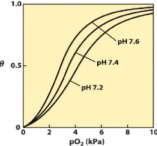
Figure 2. Effect of pH on O2 binding to Hb (the Bohr effect). The shift of pH from 7.4 to 7.6
(as it is in the lungs) increases O2 affinity, whereas the shift to 7.2 (as in the tissues) decreases O2 affinity thus facilitating its release.
As shown in Fig. 3, Hb molecule consists of four polypeptide subunits, two identical α-globin chains and two identical β-α-globin chains, covalently bound with a heme prostetic pigments, one in each of the subunits, which are held together by ionic bonds, hydrogen bonds, hydrophobic interactions, and van der Waals forces (1). The heme group contains a positively-charged iron (Fe2+) molecules (Fig. 4) which can reversibly bind to oxygen molecules to be transported to the various areas of the body. As the heme groups bind or release their oxygen loads, the overall Hb undergoes conformational changes which alters their affinity for oxygen (Fig. 1,2).
The heme group, which is typically hidden within the various subunits, is covalently bound, by means of the fifth coordination bond, to a different nitrogen atom belonging to a nearby histidine group, the “proximal His” at position 93 of the primary structure (8 of the F helix) (2). This histidine chain, together with other hydrophobic residues, stabilizes the heme group within each subunit. The O2 molecule binds to the side of the iron ion that is opposite
Dr. Le Phan Minh Triet: Hemoglobinopathies in mountainous region of Thua Thien Hue, Vietnam Ph.D thesis in Biochemistry and Molecular Biology
Ph.D School in Biomolecular and Biotechnological Sciences - University of Sassari
located at position 64 (E7). His E7 is not directly bound to the heme group nor to the O2
molecule (it is thus termed as the “distal His”) even though it has a very important function: O2 binds to heme with the axis at an angle because the perpendicular arrangement is sterically
blocked by His E7. This effect weakens the binding of O2 to Hb (2).
Figure 3. Quaternary structure of Hb. The four subunits are shown in different colors. The
heme groups are shown in dark gray. http://ucsdnews.ucsd.edu/newsrel/supercomputer/04-08ProteinDataBank.asp.
Figure 4. The heme group. The heme consists of a ring structure, protoporphyrin IX, which
binds an iron atom in its Fe2+ oxydation state. The Fe atom has six coordination bonds, four bonded to the flat porphyrin molecule and two perpendicular to it. Fifth and sixth coordination bonds are perpendicular to the flat cyclic ring structures; one is bound to a N atom of a His residue (the proximal His residue) whereas the other serves as the binding site for an O2 molecule. This group is found in myoglobin, Hb, cytochromes, and many other
heme proteins.
Because His is positively charged, its close proximity to the negatively charged iron ion prevents the iron ion from becoming too oxidized, which would inhibit the binding of oxygen molecules. This is critical to the Hb’s function of oxygen transport, since oxygen can bind to Fe2+, but not Fe3+. Also, the size, shape, and location of this distal histidine chain limits the
Dr. Le Phan Minh Triet: Hemoglobinopathies in mountainous region of Thua Thien Hue, Vietnam Ph.D thesis in Biochemistry and Molecular Biology
Ph.D School in Biomolecular and Biotechnological Sciences - University of Sassari
amount of CO2 molecules that will bind to the heme group (7). Because the heme group has a greater natural affinity for carbon monoxide than for oxygen, the lack of this distal histidine chain would allow heme groups to bind significantly more to carbon waste than to oxygen, preventing Hb proteins from providing cells with the necessary oxygen molecules for metabolic activities.
After an oxygen molecule binds to one of the heme groups of any subunit, other subunits undergo conformation changes exposing their own heme groups, thus giving the entire Hb structure greater oxygen affinity (8). The bond between oxygen the oxygen atom and the iron ion pulls the iron molecule closer to its heme group, which then pulls the proximal distal histidine chain backwards into the Hb molecule (8). This pull creates a strain on the other subunits, breaking ionic bonds in such a way that reveals their obscured individual heme groups. This positive cooperation allows binding at one subunit to increase the binding affinity at other subunits (9). As the result, the whole deoxyHb tetramer (T state) changes its conformation collapsing into the oxyHb (R state). A representation of this pulling is showed in Fig. 5.
Figure 5. The T → R transition. In these depictions of deoxyHb the α subunits are blue and
the β subunits are gray. Positively charged side chains and chain termini involved in ion pairs are shown in blue, their negatively charged partners in red. The transition from the T state to the R state shifts the subunit pairs substantially, affecting certain ion pairs. Most noticeably, the His HC3 residues at the carboxyl termini of the β subunits, which are involved in ion pairs in the T state, rotate in the R state toward the center of the molecule, where they are no longer in ion pairs. Another dramatic result of the T → R transition is a narrowing of the pocket between the β subunits.
The behaviour of Hb after binding of protons or CO2, which causes a conformational
change in the protein facilitating the release of oxygen indicates it is an allosteric protein. The sigmoidal (cooperative) curve which makes it efficient in binding (taking up O2 in lungs), and
Dr. Le Phan Minh Triet: Hemoglobinopathies in mountainous region of Thua Thien Hue, Vietnam Ph.D thesis in Biochemistry and Molecular Biology
Ph.D School in Biomolecular and Biotechnological Sciences - University of Sassari
efficient in unloading (unloading O2 in tissues) is a demonstration of an allosteric response to
the binding of molecules other than O2 in the heme pocket. In people acclimated to high
altitudes, the concentration of 2,3-Bisphosphoglycerate (2,3-BPG) in the blood is increased, which allows these individuals to deliver a larger amount of oxygen to tissues under conditions of lower oxygen tension. This phenomenon, where molecule Y affects the binding of molecule X to a transport molecule Z, is called a heterotropic allosteric effect.
Figure 6. Effect of BPG on oxygen binding to Hb. The 2,3-BPG concentration in normal
human blood is about 5 mM at sea level and about 8 mM at high altitudes. Hemoglobin binds to oxygen quite tightly when BPG is entirely absent, and the binding curve seems to be hyperbolic. At sea level, Hb is nearly saturated with O2 in the lungs, but just over 60%
saturated in the tissues, so the amount of O2 released in the tissues is about 38% of the
maximum that can be carried in the blood. At high altitudes, O2 delivery declines by about
one-fourth, to 30% of maximum. An increase in BPG concentration, however, decreases the affinity of Hb for O2, so approximately 37% of what can be carried is again delivered to the
tissues.
Fetal Hb (HbF, α2γ2), which is found in the developing fetus, binds oxygen with greater affinity than adult Hb. This means that the oxygen binding curve for HbF is left-shifted (i.e., a higher percentage of Hb has oxygen bound to it at lower oxygen tension), in comparison to that of adult Hb (HbA). As a result, fetal blood in the placenta is able to take oxygen from maternal blood.
Dr. Le Phan Minh Triet: Hemoglobinopathies in mountainous region of Thua Thien Hue, Vietnam Ph.D thesis in Biochemistry and Molecular Biology
Ph.D School in Biomolecular and Biotechnological Sciences - University of Sassari
Hemoglobin also carries nitric oxide (NO) in the globin part of the molecule. This improves O2 delivery in the periphery and contributes to the control of respiration. NO binds
reversibly to a specific Cys residue in globin; the binding depends on the state (R or T) of the Hb. The resulting S-nitrosylated Hb influences various NO-related activities such as the control of vascular resistance, blood pressure and respiration. NO is not released in the cytoplasm of erythrocytes but transported by an anion exchanger called AE1 out of them (10).
1.2. Localization and organization of globin genes
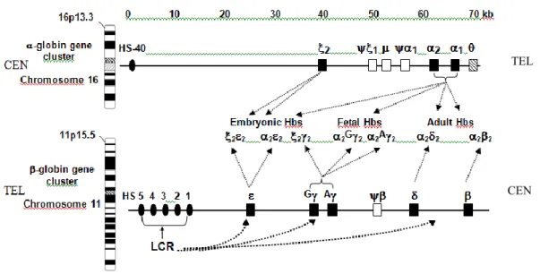
Human Hbs are tetrameric molecules encoded at two separate loci, the -like globin gene cluster located on chromosome 11p15.5 (close to the olfactory receptor genes) and the -like globin gene cluster on the terminus of chromosome 16p13.3 (close to heterochromatic gene encoding a putative RNA-binding protein) (11,12). As shown in Fig. 7 and 8, in each cluster the active genes are arrayed on the chromosome in the same order they are expressed developmentally: 5’-(embryonic)-G(fetal)-A(fetal)-(minor adult)-(major adult)-3’, and 5’-(embryonic)-2(fetal and adult)-1(fetal and adult)-3’, respectively. Hemoglobin
production is characterized by two switches: the production of embryonic Gower1 (22),
Gower2 (22) and Portland (22) Hbs switches around the first two months of gestation to
the production of two different fetal Hbs (HbF) (2G2 and 2A2) followed, just before birth,
to the major adult HbA (22) and minor adult HbA2 (22) tetramers. As the result of the
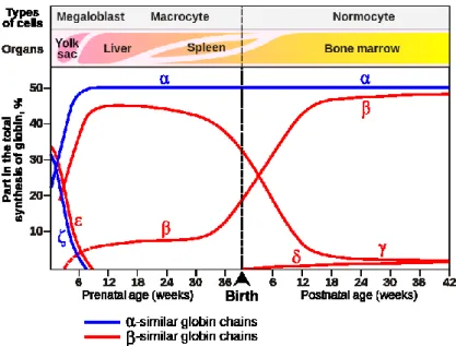
second switch, at birth the circulating Hb contains from 70 to 80% of HbF whereas 6 months later HbF covers less than 4-5 % of the total. The final adult Hb pattern is reached at 1 year of life (Fig. 8). At that time HbA comprises 97%, HbA2 2% and HbF 1%. It is at this stage
that mutations affecting the gene become clinically apparent. At birth, HbF contains G and A chains in the 70:30 ratio. The switch from fetal to adult Hb production is not complete, so that in the small amounts of HbF which persist in adult life the proportion of the two chains reverses to 40:60. The switch from fetal to adult Hb is not due to changes in stem cell populations but rather to changes in programs of gene expression occurring in the progeny of a single stem cell population. All adults have residual amounts of HbF, present in a subset of erythrocytes called F-cells which also contains HbA. The levels of HbF and F-cells in adults vary considerably, and are largely genetically controlled (13).
Dr. Le Phan Minh Triet: Hemoglobinopathies in mountainous region of Thua Thien Hue, Vietnam Ph.D thesis in Biochemistry and Molecular Biology
Ph.D School in Biomolecular and Biotechnological Sciences - University of Sassari
1.2.1 Detailed chromosomal organization of the human globin genes.
Two other α-like globin genes have been identified and characterized in the α-globin gene cluster, but their roles, if any, in encoding globin polypeptides are still uncertain. The θ-globin gene located to the 3’ or C-terminal side of the duplicated α-θ-globin genes (16). It is more closely related to the α-globin genes than to the ζ-globin genes and is expressed at low levels in erythroid cells (17,18). Clear homologs to the θ-globin gene are found in the homologous position in other mammalian α-like globin gene clusters.
Figure 7. Basic organization of human - and -globin gene complexes and expression of the
globin genes during ontogenesis. The locations of the -globin gene complex very close to the telomere of the short arm of chromosome 16 and the -globin gene complex on the short arm of chromosome 11 are shown. The genes are shown as boxes, named according to the globin polypeptide that is encoded. In both diagrams, the 5’→3’ transcriptional orientation is from left to right. The orientations with respect to the centromere (CEN) and telomere (TEL) are opposite; the -like globin genes are transcribed toward CEN whereas the -like globin genes are transcribed toward TEL. The composition of Hb produced at progressive developmental stages is given between the clusters. The stage specific interaction between the -LCR and the cluster is also schematized.
TEL
Dr. Le Phan Minh Triet: Hemoglobinopathies in mountainous region of Thua Thien Hue, Vietnam Ph.D thesis in Biochemistry and Molecular Biology
Ph.D School in Biomolecular and Biotechnological Sciences - University of Sassari
Figure 8. Globin chains production during ontigenesis. The production of embryonic Gower1
(22), Hb switches around the first two months of gestation to the production of HbF (22)
followed, just before birth, to the major adult HbA (22) and minor adult HbA2 (22)
tetramers.
The μ-globin gene is located just 3’ of the ψζ1-globin pseudogene (19, 20); it was initially called ψα2 (21) but with more accurate sequencing it is clear that this gene does not contain mutations that would render it inactive. It is a distant relative, being equally divergent from both α-globin and ζ-globin genes. Both the θ-globin gene and the μ-globin gene are transcribed and spliced in erythroid cells, albeit at much lower levels than the α-globin gene. Curiously, no Hb containing the θ-globin chain or the μ-globin chain has been identified, even by the sensitive mass spectrometry (20). Furthermore, the predicted structure of the θ-globin chain suggests that it would be unlikely to function normally as a Hb subunit (22). Thus these genes remain a puzzle. They tend to be retained over mammalian evolution, hence indicating constraint for some function. They are expressed at the RNA level but do not appear to be translated into a polypeptide. Perhaps they or their RNA transcripts play some role that has yet to be discovered.
1.2.2 Structure of globin genes
The coding region of each globin gene in humans (as well as in other vertebrates) is interrupted at two positions by tracts of noncoding DNA which were called intervening sequences (IVSs) or introns (23). In the β-like globin genes, the introns interrupt the sequence between codons 30 and 31 and between codons 104 and 105; in the α-globin gene family, the
Dr. Le Phan Minh Triet: Hemoglobinopathies in mountainous region of Thua Thien Hue, Vietnam Ph.D thesis in Biochemistry and Molecular Biology
Ph.D School in Biomolecular and Biotechnological Sciences - University of Sassari
IVSs interrupt the coding sequence between codons 31 and 32 and between codons 99 and 100 (Fig. 9). Interruptions due to introns occur at precisely the same position in the aligned primary sequence of the α- and β-globin chains. It has been hypothesized that the presence of the introns at these positions predates the separation of α-globin and β-globin genes, occurred in an ancestral vertebrate about 500 million years ago (24). The first IVS (IVS-1) is smaller than the IVS-2 in both α- and β-globin genes, but IVS-2 of the human β-globin gene is much larger than that of the α-globin gene.
The intron sizes of the ζ-like globin genes differs from that of the α-like globin genes since the introns in the α and ψα genes are small, fewer than 150 base pairs (bp), than those of the ζ and ψζ genes (25). The first introns of the ζ and ψζ genes are much larger than their second introns. The IVSs sequences interrupting the coding sequences of structural genes are removed during maturation of mRNA. Figure 9B shows that IVSs are transcribed into globin precursor mRNA (26), and next excised in such a way that the proper ends of the coding sequences joins to give rise the mature mRNA (27). This posttranscriptional processing of mRNA precursors has been termed “splicing”. A crucial prerequisite for the proper splicing of globin precursor mRNAs is the presence of specific nucleotide sequences at the exons-introns junctions.
Figure 9. Structure and expression pathway of globin genes. A. General structure of globin
genes. The coding sequences are separated by two IVSs (white boxes) into three exons. The first exon has a short 5’ untranslated region (UTR, gray box) followed by a coding region (black box). The central exon codes for protein, while the third ends with a 3’ UTR. The sizes of the boxes indicate the size of the tracts of the genes. Codon numbers are also given. Under the second intron of the -globin gene are indicated the “consensus sequence”. These are critical for the correct splicing process. Similar sequences are present in all introns. The vertical arrows indicate the splice site junctions within the consensus sequences where cleavage occurs during the process of cutting the introns and joining exons.
B. The pathway for expression of globin genes. The RNA transcript is shown with short boxes corresponding to the UTRs (gray), coding regions (black) and introns (white), with processing and splicing steps occurring in the nucleus to form mRNA (15).
Dr. Le Phan Minh Triet: Hemoglobinopathies in mountainous region of Thua Thien Hue, Vietnam Ph.D thesis in Biochemistry and Molecular Biology
Ph.D School in Biomolecular and Biotechnological Sciences - University of Sassari
Two different “consensus sequences” have been almost universally found at the 5’ (donor) and 3’ (acceptor) splice sites of introns (28). These are shown in Figure 9A, with the consensus surrounding the “branch point” A involved in the initiation of splicing. The doublets GT and AG (shown in boldface) at the 5’ and 3’ ends, respectively, of the intron, are invariant and thought to be absolutely required for proper splicing. This is referred to as the GT-AG rule. Mutations that alter those dinucleotide sequences or create similar consensus sequences at new sites in a globin gene are known determine abnormal processing of mRNA precursors thus representing the molecular basis for some types of thalassemia.
The globin genes nomenclature has been recently standardized following the Guideline for Human Gene Nomenclature. It is showed in Table 1.
Table 1. Nomenclature of the globin genes*
Gene Nomenclature HBZ HBA1 2 HBA2 θ HBQ HBE G HBG2 A HBG1 HBB HBD *Aguileta G., Bielawski J.P., Ziheng Y. Proposed nomenclature for the and globin gene family. Genes Genet. Syst. 81, 367-371, 2006.
1.2.3. Regulation of globin genes expression
Studies conducted during the past three decades have revealed much about the regulation of the human globin genes. These include information about DNA sequences needed in cis such as: promoters, upstream regulatory sequences, proximal enhancers and distal enhancers. The knowledge on these regulatory elements not only facilitated a better comprehension on the molecular basis of hemoglobinopathies but also was the most useful for attempts in improving the conditions of patients with hemoglobinopathies.
Dr. Le Phan Minh Triet: Hemoglobinopathies in mountainous region of Thua Thien Hue, Vietnam Ph.D thesis in Biochemistry and Molecular Biology
Ph.D School in Biomolecular and Biotechnological Sciences - University of Sassari
Promoters
A promoter is the DNA sequence needed for accurate initiation of transcription. It is a DNA segment that interacts with RNA polymerase II and its accessory factors (such as TFIID and TFIIB) to determine the start site of transcription; this is the basal promoter (29). Five motifs have been associated with basal promoters, and these are found in the promoters of human globin genes (Figure 10.A). They include the familiar TATA box to which TBP binds, along with the BRE to which TFIIB binds and the Inr and DPE motifs to which components of TFIID binds (29).
Efstratiadis et al. (30) revealed the presence of the ATAAA sequence (motif) about 25-30 bp 5’ to the start site of transcription of the globin genes, by far considered the most convincing evidence in its consensus role. More recent studies on other promoters revealed the presence and roles of additional motifs close to the start site of transcription. Matches to these motifs can be found readily at the appropriate positions in the human globin genes (Fig. 10.A). Except BRE, each of he motifs in Fig. 10A has been implicated in function by the discovery of a mutation in cases of β-thalassemia (from now on: -thal). Every base in the TATA box has been altered in one or another thalassemia, and mutations in Inr, MTE and DPE also are associated with this syndrome.
Upstream regulatory sequences
Adjacent to the basal promoter is the upstream regulatory region (29), which runs from about positions -40 to -250 (Figure 10). The CCAAT box appears to be the only one motif highly expressed in this region. Different proteins bind to this motif (30), and it has been implicated in promoter function because of its presence in many promoters and the results of mutagenesis and binding studies (31).
Two other motifs which are found to bind to proteins are the CACC box, which is bound by transcription factors in the Krüppel-like zinc finger class (KLF) and the WGATAR which is the binding site for GATA-1 and related proteins (32,33). Mutations in almost every position in the proximal CACC box have been associated with β-thal.
Proximal Enhancers
The enhancers are sequences that increase the activity of promoters; they can be located on either side of a gene or internal to it, and they can act at considerable distances from genes (34). Two enhancers have been found close to genes in the β-globin gene cluster, one that is 3’ to HBB and one that is 3’ to HBG1 (Figure 10.A). In both cases the enhancers are less than 1 kb downstream of the polyA additional signal for the respective genes. The HBG1 enhancer
Dr. Le Phan Minh Triet: Hemoglobinopathies in mountainous region of Thua Thien Hue, Vietnam Ph.D thesis in Biochemistry and Molecular Biology
Ph.D School in Biomolecular and Biotechnological Sciences - University of Sassari
was discovered as the only DNA segment in a 22kb region surrounding the γ-globin genes for DNA segments that boosted expression of a reporter gene driven by a γ-globin gene promoter in transfected erythroid cells (35).
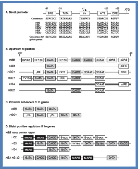
Figure 10. Motifs and binding sites in cis-regulatory modules of globin genes. (A) Motifs in
the basal promoter, as review by Maston et al. (29). Numbers along the top are relative to the transcription start site as +1, and ATG denotes the translation start site. Corresponding positions in the globin genes are given for each motif, followed by the consensus derived for the globin genes. Some ambiguous nucleotides are indicated. (B) Motifs in the regulatory regions immediately upstream of the basal promoters. Motifs are indicated by sequence (CCAAT, CACC, and GATA), by the name (βDRE, αIRE, γPE, OCT) or by the protein name followed by “bs” for “binding site” (BP2bs, NF1bs, BB1bs). Boxes found in several upstream regions are shaded. The boxes were placed in the correct order but spacing is not indicated. The thick line for the HBA upstream regions (both HBA1 and HBA2) denotes that it is a CpG island. (C ) Motifs in the proximal enhancers. (D) Motifs in distal positive regulators, including three hypersensitive sites of the β-globin LCR and HS-40 for the α-globin gene cluster (15).
Dr. Le Phan Minh Triet: Hemoglobinopathies in mountainous region of Thua Thien Hue, Vietnam Ph.D thesis in Biochemistry and Molecular Biology
Ph.D School in Biomolecular and Biotechnological Sciences - University of Sassari
Distal Enhancers
Both the α-like and β-like globin gene clusters are regulated by distal control regions. The β-like globin cluster is regulated by the distal LCR (36), and the α-like globin gene cluster is regulated by HS-40 (37). In both cases, deletion of the distal control region is associated with thalassemia. Without the LCR, erythroid expression of a β-globin transgene is not seen in all mouse lines (38), whereas with the LCR, the β-globin transgene is expressed at a high level in erythroid cells in almost all mouse lines, indicating strong enhancement and a reduction in position effects (39). A schematization of how an enhancer might affect gene transcription by bringing distal DNA is showed in Figures 11 and 12.
A clear picture on the mechanism of gene regulation was described in a Melanesian variant of the thal due to a single-nucleotide polymorphism located between the adult -globin genes and their enhancers. The finding that this mutation creates a novel promoter provides support for a mechanism of gene regulation by facilitated chromatin looping (40). The mechanism is showed in Fig. 12.
Figure 11. An enhancer might affect gene transcription by bringing distal DNA (perhaps
thousands of base pairs away from the start site of transcription) close to the promoter for a gene. The figure also explains how a silencer would work in an analogous fashion.
Dr. Le Phan Minh Triet: Hemoglobinopathies in mountainous region of Thua Thien Hue, Vietnam Ph.D thesis in Biochemistry and Molecular Biology
Ph.D School in Biomolecular and Biotechnological Sciences - University of Sassari
Figure 12. Possible mechanism for the downregulation of -globin gene expression in
hemoglobin H disease. (a) Schematic diagram of the human -globin locus. The -globin gene (light-blue oval) is expressed in the embryonic stage of development and is silenced at around 6 to 8 weeks of gestation. The -globin genes (dark-blue ovals) are activated in fetal liver, and then in bone marrow in the adult. The physiological levels of -like globin gene expression depend on the actions of upstream enhancers (HS-33 and HS-40), mainly HS-40, which is located 40 kb 5’ of the -gene. The single nucleotide polymorphism (SNP) 195 is shown as a green circle. (b,c) An interpretation for the SNP195 promoter-induced downregulation of the -globin genes. The effective interaction between proteins bound by the enhancer (depicted as a red circle) and the -globin genes is essential for their high-level expression, and is accomplished by chromatin looping. (b) In the normal locus the SNP195 region is lightly acetylated and chromatin flexibility favors interaction between the enhancer and the 1- and 2-genes. (c) When the SNP195 promoter site (green circle) is activated in HbH disease, histone acetylation is increased and the chromatin becomes more flexible as a consequence, resulting in a change in loop size. This change means that the enhancer now preferentially interacts with the new promoter, and no longer influences expression of the globin genes.
1.3. Disorders of the synthesis of hemoglobin
The study of the disorders of the synthesis of Hb is considered a paradigm for understanding insights into the cellular, molecular biology and pathophysiology of inherited genetic disorders. As many as 1570 disorders of Hb synthesis and of the structure have been so far identified and characterized. These are collected, and continuously updated, in the Globin Gene Server (42). Study of these disorders has established the principle of how a mutant genotype can alter the function of the encoded protein, which in turn can lead to a distinct clinical phenotype. Genotype/phenotype correlations have, in turn, provided important understanding of pathophysiological mechanisms of disease.
Dr. Le Phan Minh Triet: Hemoglobinopathies in mountainous region of Thua Thien Hue, Vietnam Ph.D thesis in Biochemistry and Molecular Biology
Ph.D School in Biomolecular and Biotechnological Sciences - University of Sassari
1.3.1. Classification of hemoglobinopathies
Disorders affecting the synthesis of the globin chains are classified into two categories: qualitative pathies (also known as hemoglobinopathies, pathies), and quantitative Hb-pathies (also known as thalassemias). Rarely, a single mutation affects both the structure and expression level of the affected gene, and these have been called quali-quantitative Hb-pathies or thalassemic hemoglobinoHb-pathies. Thus the latter were better classified as “dominant” β or -thal (41).
The hemoglobinopathies (or qualitative Hb-pathies) thus refer conditions characterized by the inheritance of mutated Hbs which contain or globin chains with changes at the level of the primary structure of globin which is synthesized at a normal rate. The affected Hb is also called “variant Hb” or “variant globin”. In most cases these Hb-pathies are due to a point mutation at the level of the structural gene which changes the meaning of the codon thus replacing the amino acid with a different one (missense mutations). Some sequence changes have little or no effects on Hb functions but are useful polymorphisms for genetic studies. A large number, however, are characterized by an altered Hb function (43). Variant Hbs are designated by letters of the alphabet or by the place names where the condition was first discovered. Even though researchers have identified more than 1100 structural hemoglobin variants, only three (Hb S, Hb C, and Hb E) are widespread. The homozygous state for the Hb S (sickle cell) gene results in sickle cell anemia, whereas the compound heterozygous state for the sickle cell and Hb C genes results in Hb SC disease. Hb SC disease, although milder, also has important public health implications. Hb E, the commonest variant globally, is innocuous in its heterozygous and homozygous states, but since it is synthesized less effectively than Hb A, it interacts with -thal to produce an extremely common condition called Hb E -thal, which is an increasingly important health burden in many parts of Asia.
As shown in Table 2, which is the updated summary of the 1578 Hb variants so far identified, all the and non- globin genes are subjected to mutations: 325 + 393 concern the globin genes, 828 the gene, 105 the , 58 the A and 69 the G genes, whereas 51 could be
considered due to mutations disturbing both the primary structure of the globin chain and the expression level of the affected gene (41).
Dr. Le Phan Minh Triet: Hemoglobinopathies in mountainous region of Thua Thien Hue, Vietnam Ph.D thesis in Biochemistry and Molecular Biology
Ph.D School in Biomolecular and Biotechnological Sciences - University of Sassari Table 2. Human Hemoglobin Variants
Summary of mutations categories (Updated August, 2013) *http://globin.cse.psu.edu/hbvar/menu.html
Table 3 shows the relatively few variants at the G and A globin genes having altered functional properties (44).
Table 3.
The thalassemias constitute a heterogeneous group of naturally occurring, inherited mutations characterized by abnormal globin gene expression resulting in total absence or quantitative reduction of - or -globin chain synthesis in human erythroid cells. -Thal is associated with absent or decreased production of -chains, whereas in the -thal, there is absent or decreased production of -chains. In those cases in which some of the affected
Dr. Le Phan Minh Triet: Hemoglobinopathies in mountainous region of Thua Thien Hue, Vietnam Ph.D thesis in Biochemistry and Molecular Biology
Ph.D School in Biomolecular and Biotechnological Sciences - University of Sassari
globin chain is synthesized, early studies demonstrated absence of an amino acid substitution. The thalassemia gene appears to be allelic with the structural gene encoding - or -globin (41).
Increase the knowledge of the molecular basis of -thal has followed and depended on progress and technical advances in the fields of biochemistry and molecular biology. In particular, recombinant DNA and polymerase chain reaction-based technologies (PCR) have contributed to a virtual explosion of new informations on the precise molecular basis of most
Table 4. Classification of hemoglobin disorders
I. QUANTITATIVE DISORDERS OF GLOBIN CHAIN SYNTHESIS/ACCUMULATION
The thalassemia syndromes
A. β-Thalassemia
Clinical classification: β-Thalassemia minor or trait
β-Thalassemia major β-Thalassemia intermedia Biochemical/genetic classification: β0-Thalassemia β+-Thalassemia δ-Thalassemia γ-Thalassemia Lepore fusion gene δβ-Thalassemia εγδβ-Thalassemia HPFH
“Dominant” thalassemia (structural variants with β-thalassemia phenotype)
β-Thalassemia with other variants:
HbS/β-thalassemia HbE/β-thalassemia Other
B. α-Thalassemia
Deletions of α-globin genes:
One gene: α+-thalassemia Two genes in cis: α0-thalassemia
Two genes in trans: homozygous α+-thalassemia
(phenotype of α0-thalassemia)
Three genes: HbH disease
Four genes: Hydrops fetalis with Hb Bart's
Nondeletion mutants:
Hb Constant Spring Other
Due to mutations of the ATRX transcription factor gene on chromosome X
α-Thalassemia associated with myelodysplastic syndromes (ATMDS):
Due to mutations of the ATRX gene
C. De novo and acquired α-thalassemia
α-Thalassemia with mental retardation syndrome (ATR):
Due to large deletions on chromosome 16 involving the α-globin genes
II. QUALITATIVE DISORDERS OF GLOBIN STRUCTURE: STRUCTURAL VARIANTS OF HEMOGLOBIN
A. Sickle cell disorders
SA, sickle cell trait
SS, sickle cell anemia/disease SC, HbSC disease
S/β thal, sickle β-thalassemia disease S with other Hb variants: D, O-Arab, other SF, Hb S/HPFH
B. Hemoglobins with decreased stability (unstable hemoglobin variants)
Mutants causing congenital Heinz body hemolytic anemia
Acquired instability-oxidant hemolysis: Drug-induced, G6PD deficiency
C. Hemoglobins with altered oxygen affinity
High/increased oxygen affinity states:
Fetal red cells
Decreased RBC 2,3-BPG Carboxyhemoglobinemia, HbCO Structural variants
Low/decreased oxygen affinity states:
Increased RBC 2,3-BPG Structural variants
D. Methemoglobinemia
Congenital methemoglobinemia:
Structural variants
Cytochrome b5 reductase deficiency
Acquired (toxic) methemoglobinemia
E. Posttranslational modifications
Nonenzymatic glycosylation Amino-terminal acetylation Amino-terminal carbamylation Deamidation
Ref. Forget and Bunn (41)
forms of thalassemia. Table 4 shows a comprehensive classification of the several, different, Hb disorders. The growth of this knowledge has matched the acquisition of detailed
Dr. Le Phan Minh Triet: Hemoglobinopathies in mountainous region of Thua Thien Hue, Vietnam Ph.D thesis in Biochemistry and Molecular Biology
Ph.D School in Biomolecular and Biotechnological Sciences - University of Sassari
information on the structure, organization, and function of the normal human globin genes. As a result, there have been a progressively clearer and increasingly complex picture of the molecular pathology of this group of genetic disorders. A major conclusion that was drawn was that a relatively limited number of phenotypes can result from a surprisingly large number of varied genotypes.
1.3.2. Pathophysiology of hemoglobinopathies
The causes for the anemia observed in the different forms of thalassemia are primary and secondary. In -thal, a reduced synthesis of -globin chains of Hb A (22) will result in an overall deficit of Hb accumulation in erythrocytes and cause a hypochromic, microcytic anemia with a low mean corpuscular hemoglobin concentration in affected erythrocytes. This is the case in both the heterozygous and homozygous states. In the homozygous state, however, another pathophysiological process worsens the anemia and is responsible for the major clinical manifestations in the syndrome referred to as -thal major or Cooley’s anemia. The continued synthesis in normal amounts of normal -globin chains results in the accumulation, within the erythroid cells, of excessive amounts of chains. These chains cannot find complementary globin chains with which to bind, these chains form insoluble aggregates and precipitate within the cell, causing membrane damage and premature destruction of the red cells. The -chain aggregates are called inclusion bodies or Heinz bodies. The process of inclusion body formation occurs not only in mature erythrocytes, but in particular in the erythroid precursor cells of the bone marrow. As a result, there is extensive intramedullary destruction of erythroid precursor cells, a process that is called “ineffective erythropoiesis”. The severity of the clinical manifestations in -thal generally correlates well with the amount of the free -chain pool and the degree of - to non--globin chain imbalance. Therefore, the fortuitous coinheritance of -thal together with homozygous -thal reduces the degree of - to non -globin chain imbalance and leads to a milder clinical course (1). Similarly, coinheritance of β-thal with conditions that are associated with increased levels of synthesis of -chains of HbF (22) leads to less imbalance between - and
non -globin chain synthesis, resulting in decreased formation of -chain inclusion bodies, increased effective production of red cells, and their prolonged survival in the circulation. The clinical course in most cases of homozygous -thal is severe. Although anemia is not evident at birth, severe hypochromic, microcytic, hemolytic anemia develops during the first year of
Dr. Le Phan Minh Triet: Hemoglobinopathies in mountainous region of Thua Thien Hue, Vietnam Ph.D thesis in Biochemistry and Molecular Biology
Ph.D School in Biomolecular and Biotechnological Sciences - University of Sassari
life and a regular transfusion program must be undertaken to maintain an adequate circulating hemoglobin level.
Owing to changes in the philosophy and practice of transfusion therapy, the clinical manifestations of homozygous -thal in childhood have changed considerably over the past decades. With modern transfusion therapy, most children will develop normally, with few or no skeletal abnormalities, and will have a reasonably good quality of life. To avoid iron overload from transfusional hemosiderosis, transfusion therapy is usually coupled with a vigorous program of iron chelation, typically using parenterally administered desferrioxamine and more recently, orally effective iron-chelating agents. Although it is possible to maintain iron balance with such a management program, compliance is often difficult to realize. Iron overload eventually develops in most patients and is the major cause of morbidity and mortality in young adults. The one therapy that is curative is bone marrow or stem cell transplantation, which is being increasingly practiced when feasible. The hope for the future is the development of even more effective oral iron-chelating agents and improved approaches to gene therapy.
Studies of the molecular basis of -thal have demonstrated that the gene defects responsible for the disorder are quite heterogeneous (41). In contrast to -thal, in which deletions in the -globin gene cluster account for most of the mutations, the molecular defects associated with -thal are usually point mutations involving only one (or a limited number of) nucleotide(s), but resulting in a major defect of -globin gene expression either at the transcriptional or posttranscriptional level, including translation.
Practically every conceivable type of defect in gene expression has been identified in one form or another of -thal. Over 200 point mutations have been identified. Some deletion types of -thal have also been described. In cases of -thal in which -globin gene expression is not totally absent (so-called +-thal), the -chain that is synthesized is usually structurally normal. There is a syndrome called dominant -thal in which a highly unstable, structurally abnormal -globin chain is synthesized, resulting in inclusion body formation in the heterozygous state. The coinheritance of HbE (22 GluLys) with -thal is very prevalent
in Southeast Asia and results in markedly variable and heterogeneous clinical manifestations, the basis for which is poorly understood. Finally, there are a number of -thal-like disorders, called -thal and hereditary persistence of fetal hemoglobin (HPFH), that are distinguished from the more typical forms of -thal by the presence of a substantial elevation of HbF in
Dr. Le Phan Minh Triet: Hemoglobinopathies in mountainous region of Thua Thien Hue, Vietnam Ph.D thesis in Biochemistry and Molecular Biology
Ph.D School in Biomolecular and Biotechnological Sciences - University of Sassari
heterozygotes, as well as in homozygotes and compound heterozygotes. These disorders are usually due to deletions of different sizes involving the -globin gene cluster, although nondeletion types of these disorders have also been identified.
The great heterogeneity of molecular lesions causing -thal may appear at first glance to create insurmountable problems in putting this knowledge to practical use in the form of prenatal diagnosis and genetic counseling.
The availability of rapid and accurate PCR reaction-based assays for the detection of specific mutations in small samples of DNA has resulted in a number of surveys for the detection of the prevalence of different -thal mutations in various population groups. The results of these surveys indicate that a given mutation is usually found only within one racial group and not another. Furthermore, a small number of different mutations, usually five or six, frequently accounts for 90% or more of the cases of -thal in a given population group. Thus, it is possible to devise efficient and precise prenatal diagnosis programs using DNA-based approaches. Such programs have led to a striking decrease in the number of births of infants with homozygous -thal in many countries where the disease is prevalent.
1.4. Molecular basis of thalassemia
As already mentioned, the -thal are characterized by a quantitative reduction in the production of -globin chains of HbA. More than 200 -thal alleles have now been characterized (42) involving mutations affecting any of the steps in the transcription of the -globin gene, posttranscriptional processing of its pre-mRNA, or the translation of its mRNA into protein. The vast majority of simple -thal are caused by point mutations within the gene or its immediate flanking sequences, although small deletions involving the gene may also occur. In the case of -chain production is totally abolished by the mutation, it is referred to as o-thal, whereas reduced output of -chains (of normal structure) produces +-thal, with the mildest forms sometimes referred to as ++- or “silent” thal. These common forms of -thal are inherited as mendelian recessives. Some structurally abnormal -chain variants are also associated with quantitative deficiencies of -globin chain production and have a phenotype of -thal, in which case they are referred to as “thalassemic hemoglobinopathies,”. This is the case, for example, of HbE (26 Glu→Lys). In others, the -globin variants are so unstable that they undergo very rapid postsynthetic degradation giving rise to a functional deficiency. These hyperunstable -chain variants act in a dominant negative fashion, causing
Dr. Le Phan Minh Triet: Hemoglobinopathies in mountainous region of Thua Thien Hue, Vietnam Ph.D thesis in Biochemistry and Molecular Biology
Ph.D School in Biomolecular and Biotechnological Sciences - University of Sassari
a disease phenotype even when present in the heterozygous state, and hence have been referred to as “dominantly inherited -thal.” (46).
The -thal and hereditary persistence of fetal hemoglobin (HPFH) are disorders related to the -thal that also involve down-regulation of -globin gene expression. In -thal the gene is also affected and there is variable compensation from increased HbF production. These disorders result from more extensive deletions within the -globin gene cluster, as do the deletion forms of HPFH in which HbF compensation is sufficient to minimize hematological abnormalities. Other forms of HPFH result from mutations in the promoters of the -globin genes that not only increase HbF production in adult life but are accompanied by reduced -chain production.
In this section, the molecular mechanisms underlying the different types of-thal are described.
The common denominator is absent or decreased synthesis of -globin chains, resulting in the accumulation of excess -globin chains that are responsible for the pathophysiology of the disorder. The severity of the phenotype is usually related to the degree of imbalance between - and non--globin chain synthesis, and the size of the free -chain pool. Hence severity is related to the type of allele (o, +, ++), ameliorated by an interacting -thal (by reducing the -chain excess) and any increased production of -chains (that decrease the excess of free a -chains by binding to them to form HbF).
1.4.1. The -thalassemia
1.4.1.1. Nondeletion types of -thalassemia
These defects account for the majority of the -thal alleles. They involve single base substitutions, small insertions or deletions within the gene or its immediate flanking sequences, and affect almost every known stage of gene expression. Allele frequency varies widely from one population to another though within any population there are usually a small number of common alleles together with many alleles that are rare for that population. These defects are listed in Table 5 according to the mechanism by which they affect gene function: transcription, RNA processing, or RNA translation. The mutations are continuously updated and the list is accessible electronically through the Globin Gene Server Website (42). Heterozygotes have minimal anemia but hypochromic (mean corpuscular haemoglobin, MCH, 18–24 pg), microcytic (mean corpuscular volume, MCV, 65–80 fL) red blood cells.
Dr. Le Phan Minh Triet: Hemoglobinopathies in mountainous region of Thua Thien Hue, Vietnam Ph.D thesis in Biochemistry and Molecular Biology
Ph.D School in Biomolecular and Biotechnological Sciences - University of Sassari
They are characterized by an increased proportion of HbA2 (normal <3.2%, -thal trait 3.5%–6.0%), and HbF levels that vary from normal (<1.0%) to slightly raised (1.0%–3.0%).
Transcriptional mutations
As shown in Table 5, as many as 26 mutations involve the conserved 100 bp DNA sequences that precede the site of the initiation of transcription and represent the -globin promoter (including the functionally important CACCC, CCAAT, and ATAA boxes), whereas 8 mutations are described in the stretch of 50 nucleotides in the 5’ untranslated region (UTR). These mutations affect transcription of the gene giving rise to mild +-thal. Mutations in all of the three conserved sequence motifs in the promoter, the two CACCC, CCAAT, and ATAA boxes, have been identified in different patients with -thal (47). These defects have been described in diverse ethnic groups, with ethnic variation in phenotype. Black individuals homozygous for the -29 A→G mutation have an extremely mild disease (48), whereas a Chinese individual homozygous for the same mutation had thalassemia major (49).The cause of this remarkable difference is not known but may be related to the different chromosomal backgrounds on which the apparently identical mutations have arisen or to the C-T polymorphism at position -158 upstream of the G-globin gene (Xmn1-G site), which is
associated with increased HbF production under erythropoietic stress. The Xmn1-G site is present in the chromosome carrying the −29 A→G mutation in Blacks but absent in that of the Chinese (50). The C-T mutation at position −101 to the-globin gene appears to cause an extremely mild deficit of -globin. In fact, heterozygotes are “silent” with borderline reduced/normal red cell indices (51,52).
Since the CAP +1 A-C mutation was described (53) 7 additional mutations in the 5’ UTR stretch have been characterized. The heterozygotes for this class of mutations are silent, with the hematological indices of thal carrier. It is possible that the CAP mutation decreases -globin gene transcription or decreases the efficiency of posttranscriptional addition of m7G
(capping) and mRNA translation.
Mutations of the splice site junction affecting RNA processing
The process of the IVSs excision from the precursor mRNA to produce functional mRNA (splicing) must be very accurate. Sequences critical in the splicing process are the invariant dinucleotides GT at the 5’ (Donor) and AG at the 3’ (Acceptor) splice junctions in the introns. Table 5 shows that 27 mutations affect either of the invariant dinucleotides in the splice junction completely abolish normal splicing and produce the phenotype of o-thal.
Dr. Le Phan Minh Triet: Hemoglobinopathies in mountainous region of Thua Thien Hue, Vietnam Ph.D thesis in Biochemistry and Molecular Biology
Ph.D School in Biomolecular and Biotechnological Sciences - University of Sassari
These mutations can be base substitutions that change one or the other of invariant dinucleotides or short deletions that remove them. Genes bearing these mutations appear to transcribe normally and, although some alternative splicing occurs using “cryptic” donor or acceptor sites, the misspliced mRNA do not translate into functional -globin.
Mutations of splice site consensus sequence affecting RNA processing
The invariant “donor” and “acceptor” dinucleotides essential for the splicing of introns are flanked by sequences that are rather well conserved and a consensus sequence can be recognized at the exon-intron boundaries. These encompass the last three nucleotides of the exon and the first six nucleotides of the intron for the 5’ donor site; and the last 10 nucleotides of the intron and the first nucleotide of the exon for the 3’ acceptor site. Thirteen different mutations (Table 5) within the consensus sequences at the splice site consensus sequence have been demonstrated to reduce the efficiency of normal splicing to varying degrees and produce a -thal phenotype ranging from mild to severe. Mutations at IVS1 position 5, G→C, T, or A considerably reduce splicing at the mutated donor site compared with normal. As shown in Fig. 13, the mutations appear to activate the use of three otherwise “cryptic” donor sites (i.e. sequences that mimic the consensus sequence for a splice site but are not normally used). Two of these are in exon 1, and one in IVS1 (54). On the other hand, the substitution TC in the adjacent nucleotide, IVS1 position 6, only mildly affects normal RNA splicing even though it activates the same three cryptic donor sites as seen in the IVS1– 5 mutants (54). Although the IVS1-6 TC mutation is generally associated with milder -thal, it has been shown that in some cases apparently identical mutations can be severe (55) perhaps because of the chromosomal background on which the mutations have arisen.
Dr. Le Phan Minh Triet: Hemoglobinopathies in mountainous region of Thua Thien Hue, Vietnam Ph.D thesis in Biochemistry and Molecular Biology
Ph.D School in Biomolecular and Biotechnological Sciences - University of Sassari
Figure 13. Alternative splicing of precursor -globin mRNA resulting in +-thal associated with the IVS1–110 G→A mutation. The G→A substitution at position 110 creates an AG dinucleotide within a potential 3’ acceptor splice site resulting in approximately 90% mRNA aberrantly spliced and 10% normally spliced (45).
Mutations creating new alternative splice sites in introns
Splicing mutation may be due also to base substitutions in IVS that generate new splicing signals, which are alternative to the normal splice sites. Six mutations have been identified in the -globin gene: two in IVS1 and four in IVS2 (Table 5)(45). Depending on the site and nature of the mutation, the phenotype may be either + or o-thal. The splicing mutation at position 110 of IVS1 was the first base substitution identified in a -thal gene (56,57). This mutation is one of the most common type of -thal in the Mediterranean population. The mutation is a G→A substitution that creates an acceptor AG in a positive consensus sequence environment, 19 bp 5’ to the normal acceptor AG of IVS1 (Fig. 13). This newly created alternative splice site is preferentially used in 80%–90% of the transcripts, whereas the normal splice site is used in only 10%-20% of the transcripts (58,59), thus giving the phenotype of + thal.
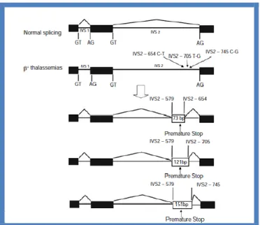
Another -thal gene with a T→G substitution in position 116 of IVS1, leads to a newly created 3’ acceptor site. In this case, the normal acceptor sequence, although intact, is not used, and little or no normal mRNA is produced resulting in a o-thal phenotype (60). Three other -thal genes are due to substitutions within IVS2 that generate new donor sites (Fig. 14). They include the IVS2 position 654 C→T, 705 T→G, and 745 C→G. In each case, an upstream acceptor site at position 579 is activated such that the normal 5’ donor site at
Dr. Le Phan Minh Triet: Hemoglobinopathies in mountainous region of Thua Thien Hue, Vietnam Ph.D thesis in Biochemistry and Molecular Biology
Ph.D School in Biomolecular and Biotechnological Sciences - University of Sassari
exon 2/IVS2 is spliced to the activated site at position 579 and the newly created donor site is spliced to the normal 3’ acceptor site at IVS1/exon 3. This two-stage splicing results in the retention of 73 bp of IVS2 in the misspliced mRNA for the IVS2 654 mutation. Variable amounts of splicing from the normal donor to the normal acceptor also occurs, resulting in phenotypes that range from + to o-thal. Several individuals heterozygous for the IVS2 654 C-T mutation with a severe phenotype have been described (50). The unusually severe disease in these individuals is thought to result from the accumulation of the misspliced mRNA, which was predicted to translate into a highly unstable chain variant with a dominant negative effect. Some normal splicing of IVS2 occurs; therefore, this mutation results in + rather than o-thal.
Figure 14. Alternative splicing of precursor -globin mRNA due to three base substitutions;
C→T at position 654, T→G at position 705, and C→G at position 745 in IVS2 of the -globin gene. Each of these mutations creates a 5’ donor site that is preferentially spliced to the normal 3’ acceptor site, whereas the same acceptor site is activated upstream at position 579 in IVS2 and spliced to the normal donor site at the exon 2–IVS2 junction. This results in the incorporation of 73, 121, and 151 bp of IVS2 into the aberrantly spliced mRNA associated with each of the different IVS2 mutations, respectively (45).
Mutations creating alternative splice sites in exons
Four mutations have been identified in exon 1 that are associated with activation of cryptic or alternative splice sites (50). Three modify the cryptic splice site spanning codons
Dr. Le Phan Minh Triet: Hemoglobinopathies in mountainous region of Thua Thien Hue, Vietnam Ph.D thesis in Biochemistry and Molecular Biology
Ph.D School in Biomolecular and Biotechnological Sciences - University of Sassari
24-27 in exon 1 so that it closely resembles the physiological consensus splice sequence AAGGTGAGT and activates it. The reduction in normal splicing is the molecular cause for the mild +-thal phenotype of these variants, including the E allele which is particularly common in Southeast Asia where it can reach a frequency of 75%. Interaction of the E allele with -thal genes is responsible for a large proportion of the thalassemia major observed in Southeast Asia. Similarly, the A→G mutation in codon 19 activates another cryptic donor site spanning codons 17–19 in exon 1 resulting in a reduced level of normally spliced mRNA that contains the codon 19 mutation encoding Hb Malay (61).
Mutations modifications causing abnormal posttranscriptional modifications
The nascent globin mRNA molecule is submitted to posttranslational modifications at both of its ends: a methylated ’m7G’ cap structure is added at the 5’ end, and a poly adenilic (A) tail is added at the 3’ end of the mRNA. Ten different -thal mutations (Table 5) have been associated with defective polyadenylation due to mutations involving the consensus sequence AATAAA required for the cleavage-polyadenylation reaction. Mutations involving the polyadenylation signal are: seven base substitutions at different positions of the consensus sequence, two short deletions of 2 and 5 bp each, and one deletion of the total AATAAA sequence. These mutations markedly decrease the efficiency of the cleavage-polyadenylation process but do not abolish it completely so that the associated phenotype is that of +-thal of moderate severity.
In the 3’ UTR region downstream of the termination codon, a C→G substitution at nucleotide 6, and a 13-bp deletion at nucleotides 90, also result in +-thal (50).
Mutations resulting in premature termination of translation
Approximately half the -thal alleles result from the introduction of premature termination codons, due to mutations creating a stop codon or to changes in the reading frame by insertion or deletion of a single or a few nucleotides. These frameshift defects cause premature termination further downstream when the next nonsense codon is reached (45,50). The AAG→TAG (Lys→Stop) at codon 17, and CAG→TAG (Gln→Stop) at codon 39 were the first nonsense mutations characterized and extensively studied. The former is was found in Chinese patients and the latter is the second most common cause of -thal in the Mediterranean population accounting for 95% of the cases of -thal in Sardinia.
The frameshift and nonsense mutations, inherited as a typical recessive -thal character, result in premature termination within exon 1 and 2 with a couple of exceptions