EXPERIMENTAL RESEARCH - FUNCTIONAL
Comparative analysis of explanted DBS electrodes
Michele Rizzi1,2&Alessandro De Benedictis3&Giuseppe Messina1&Roberto Cordella1&Davide Marchesi4&Raffaella Messina3&Federica Penner1&Angelo Franzini1&
Carlo Efisio Marras3
Received: 17 June 2015 / Accepted: 27 August 2015 / Published online: 7 September 2015 # Springer-Verlag Wien 2015
Abstract
Background Hardware-related complications frequently oc-cur in deep brain stimulation. Microscopy and spectroscopy techniques are effective methods for characterizing the mor-phological and chemical basis of malfunctioning DBS elec-trodes. A previous report by our team revealed the morpho-logical and chemical alterations on a malfunctioning explanted electrode when it was compared to a new device. The aim of this preliminary study was to verify whether these morphological and chemical alterations in the materials were a direct result of the hardware malfunctioning or if the failure was correlated to a degradation process over time.
Methods Two DBS electrodes were removed from two pa-tients for reasons other than DBS system impairment and were analyzed by a scanning electron microscope and by an energy-dispersive X-ray spectroscopy. The results were compared to a malfunctioning device and to a new device, previously ana-lyzed by our group.
Results The analysis revealed that the wear of the polyure-thane external part of all the electrodes was directly correlated with the duration of implantation period. Moreover, these al-terations were independent from the electrodes functioning and from parameters used during therapy.
Conclusions This is the first study done that demonstrates a time-related degradation in the external layer of DBS elec-trodes. The analyses of morphological and chemical proper-ties of the implanted devices are relevant for predicting the possibility of hardware’s impairment as well as to improve the bio-stability of DBS systems.
Keywords Scanning electron microscope . Energy dispersive X-ray . Environmental biodegradation . Electrodes analysis . Explanted electrodes . DBS
Introduction
Deep brain stimulation (DBS) is an effective treatment for different neurological and psycho-affective disorders [5,15]. Despite progressive technological advancement, hardware-related complications, including lead fracture or migration and infections, have been observed [1,12].
Currently, high-magnification microscopy (scanning elec-tron microscopy, SEM) and chemical characterization through spectroscopic techniques (energy-dispersive X-ray spectros-copy, EDS) are extensively used to perform failure analysis tasks in many medical fields [11,16], particularly in the case of defects arising from material failures. These methods could be implemented in the study of explanted DBS electrodes in order to analyze the structural basis of the impairment of im-planted systems.
In a previous report by our team [7], we revealed the mor-phological and chemical alterations on a malfunctioning explanted electrode in comparison with a new device, which was adopted as a control. Since a correlation between the observed changes and the electrode’s breakdown was not clearly demonstrated, a time-related influence was considered to be a cause of the device malfunction.
* Michele Rizzi [email protected]
1
Department of Neurosurgery, IRCCS Foundation Neurological
InstituteBCarlo Besta^, Milan, Italy
2
School of Neurosurgery, University of Milan, Milan, Italy
3
Department of Neuroscience and Neurorehabilitation Division of
Neurosurgery, Bambino Gesù Children’s Hospital, IRCCS,
Rome, Italy
Our report shows a preliminary comparative analysis be-tween a malfunctioning explanted electrode, two electrodes that were removed from two patients for reasons other than implanted system impairment and a new electrode.
Materials and methods
PatientsPatient 1
Patient 1 is a 25 years-old male with an onset of walking dys-tonia at the age of six. The patient was diagnosed with idio-pathic primary generalized dystonia at 11 years-old. DBS of the globus pallidus internus (GPi) was performed at the age of 16, in 2005. Consequently to stimulation, the patient remarkably improved and he was walking independently 1 year later (se-verity score at BFMDRS: 66). At the age of 19, a worsening of dystonia involving the lower limbs and the right upper limb appeared (BFMDRS: 71); the electrode’s impedance was higher than 2000 Ohm. In June 2010, the patient agreed to have the faulty electrodes removed and replaced with a new lead (3389; Medtronic) on the left side. The procedure was successful and without complications. Dur-ing the last follow-up (January 2014), DBS resulted to be e ff e c t i v e in t h e t r e at m e n t o f dy s t o n i c sy m p t o m s (BFMDRS: 60) (Table1).
Patient 2
This patient is a 30-year-old female who began experiencing seizures in 2003. Cortical and subcortical biopsies of the left frontal lobe suggested that Rasmussen encephalitis was the etiology. Four years after the onset, the patient developed a drug-resistant epilepsia partialis continua (EPC). Given that cognitive, motor, and language functions were good, a left caudal zona incerta (cZI) DBS was proposed in October 2007 (11) and significant improvements in the frequency of seizures was reported. In 2009, the patient had an EPC refrac-tory to the stimulation parameters arrangement to the AED dose augmentation and to the immunomodulatory treatment. The DBS system revealed correct impedance values and did not show any other signs of malfunctioning. Thus, the left motor cortex was surgically removed and the seizures were completely controlled. Moreover, the patient had requested for the DBS system to be switched off because it was ineffective. In March 2013, the entire DBS system was removed (Table1). Patient 3
Patient 3 is a 22-year-old male whose clinical history started at the age of 3 with a progressive spastic paraparesis associated to
dystonic movements of the right upper arm. The clinical picture progressively worsened and a generalized dystonia appeared. A diagnosis of primary DYT1- dystonia was posed (BFMDRS, severity score: 79.5; disability: 19). A drug-resistant status distonicus (SD) appeared at the age of 10. Bilateral GPi-DBS was performed in 2002; internal pulse generators (IPGs) were switched on and 2 days later the SD disappeared (BFMDRS, severity score: 63; disability: 10). In 2011, an infection of the connection cable occurred; antibiotic treatment was not effec-tive and removal of the cable became necessary, followed by dystonia worsening. Infection extended to the IPGs and succes-sively to the extracranial portion of the lead. The patient underwent the removal of the entire left DBS system in 2012 and a few months later a left pallidotomy was performed and resulted in improvement of the disease (Table1).
Electrode analysis
All of the electrodes were carefully removed from the patients, without any complications or damage to the hardware. The electrodes were preserved in three different surgical specimen boxes after rinsing them with saline solution. The study in-cluded both the explanted electrodes (3389; Medtronic, length 25 cm) and a new lead produced by the same company (3389; Medtronic, length 40 cm). Depending on the facilities avail-ability, the analysis through SEM-EDX techniques was not performed the same day of removal.
Scanning electron microscopy characterization of the electrode had been performed at the Micro and Nano-Fabrication Platform of Fondazione Filarete, Milan, Italy, using a Zeiss Sigma Field Emission Scanning Electron Mi-croscope (FE-SEM). The chemical composition analysis of the samples was performed using a Bruker Quantax 400 EDS detector (30 mm2XFlash silicon drift detector) installed on the SEM.
SEM was carried out to describe the morphological fea-tures of the electrodes at different magnifications, while EDX was performed to analyze the chemical properties of the materials.
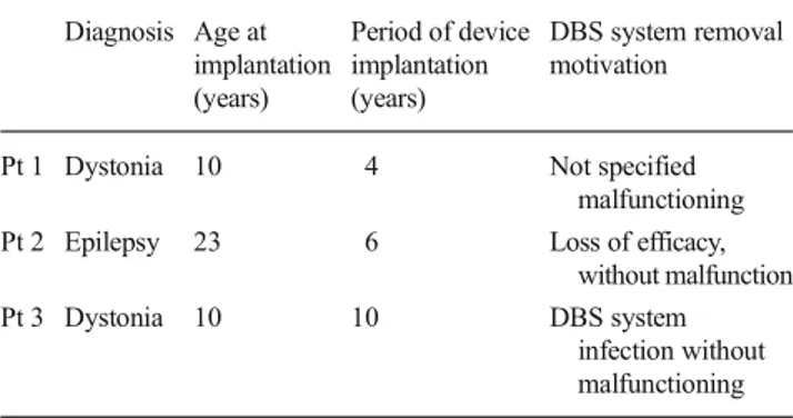
Table 1 General characteristics of the patients
Diagnosis Age at implantation (years) Period of device implantation (years) DBS system removal motivation
Pt 1 Dystonia 10 4 Not specified
malfunctioning
Pt 2 Epilepsy 23 6 Loss of efficacy,
without malfunction
Pt 3 Dystonia 10 10 DBS system
infection without malfunctioning
Results
The macroscopic inspection of the electrodes removed from patients 2 and 3 did not show any alterations. In a portion of the extracranial electrode of patient 1, we found brown-colored areas beneath the external layer of the electrode (i.e., outer jacket tubing). Moreover, the metallic coils appeared to be distorted and stretched throughout different parts of the electrode. The conductivity of this electrode reported zero (i.e., open circuit), while that of patients two and three was preserved [7].
The morphological analysis was performed through SEM, with different magnification on the outer jacket tubing and on the internal insulating jacket. The former was made of poly-u r e t h a n e ( 8 0 A poly-u r e t h a n e ) , w h i l e t h e l a t t e r w a s a fluoropolymeric structure, which isolates each single coil of the electrode which is composed by platinum and iridium.
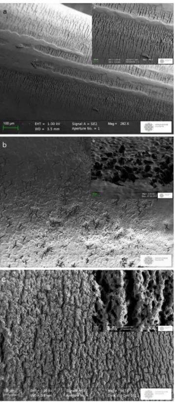
Figure1shows the comparison between each electrode at the level of the outer jacket tubing. In patient 1, crack-like forms, perpendicular to the lead axis, extended above the lead surface (282x) (Fig. 1a); in patient 2, star-like forms appeared to be more evident at the level of the jacket near the stimulating contact (347x) (Fig. 1b); in patient 3, crack-like forms, always perpendicular to lead direction, resulted more evident with the disappearance of the classical smooth surface (Fig. 1c). The right upper corner of Fig.1a, b and c shows a more detailed look at the outer jacket polymer surface, by using a higher mag-nification. In the same figure, the alterations extended to the whole surface, with a striking loss of the initial char-acteristics. Nevertheless, no communication between the inner part of the lead and the external lead was observed. As shown in Fig. 2, only a superficial part of the outer jacket tubing was severely deteriorated: the horizontal ar-row (letter b) shows the internal border of the outer jacket tubing, while the vertical arrow (letter a) points towards the external surface. The magnification of 14.66 Kilo-X detailed the above described alterations on the outer jack-et tubing in patients 2 and 3 (Fig.3a, b).
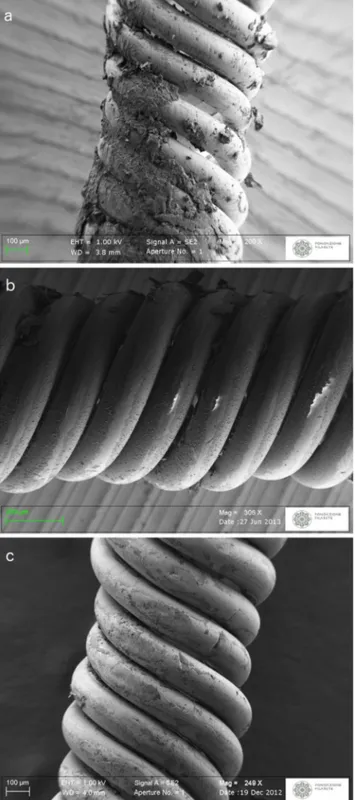
The internal insulating jacket was explored by careful-ly removing the outer jacket tubing. In patient 1, the electrode jacket was covered by remnants of degenerated material (Fig. 4a) [7]. The internal insulating jacket of patients 2 and 3 was found to be well preserved (Fig.4b and c).
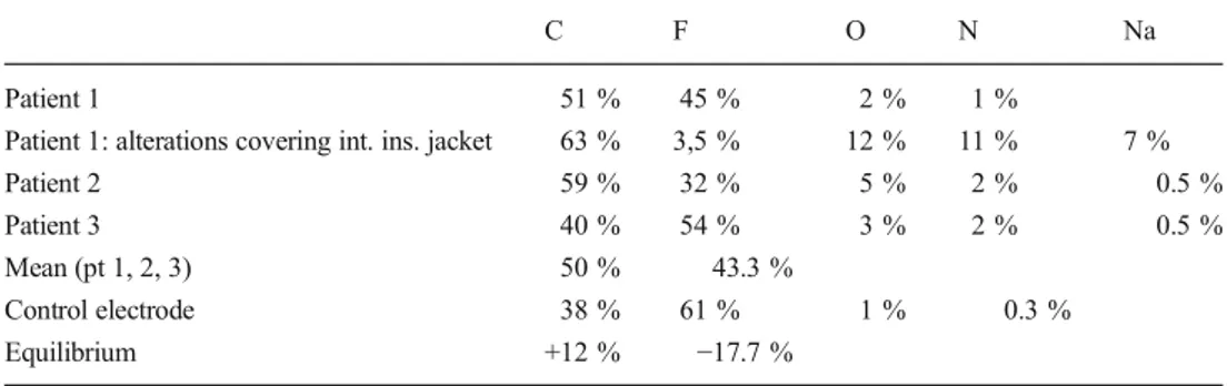
EDX analysis revealed that the outer jacket tubing of the four electrodes presented a chemical composition compatible with the typical composition of polyurethanes. A tendency of N/C ratio increase with the time to removal was observed (Table2). The internal insulating jacket re-vealed that the used electrodes had a prevalence of carbon (mean value of 50 % against 38 % of the control
electrode) and a minor percentage of fluorine (mean value 43.3 % against 61 % of the new electrode) (Table3). The chemical structure of the material partially covering the internal insulating jacket in patient 1 was composed of 63 % of carbon, 3.5 % fluorine, 12 % of oxygen, and 11 % nitrogen (Table 3) [7].
Discussion
D e s p i t e t h e w i d e s p r e a d u s e o f D B S a n d o t h e r neuromodulation procedures in clinical practice, clinicians are often faced with different adverse events related to the hardware that is implanted [1,12]. In order to investigate the morphological and chemical features of these devices, pre-clinical studies are required. Relevant information could also be collected by analyzing the DBS systems that are explanted because of complications such as lead fracture, migration or disconnection of the electrode, connection cable, or pulse gen-erator infection. SEM and EDX are effective methods supporting these studies.
Previously, our group reported a comparative evaluation between two electrodes, one explanted for malfunctioning and a new one [7]. We hypothesized that the observed modi-fications were secondary to the overheating of the lead that is associated with the partial degradation of the internal struc-ture. However, a direct cause for the malfunctioning was not found. In particular, it was not possible to determine if the alterations were truly relevant to the patient’s problem or if there were more frequent phenomenon occurring in the ma-jority of the DBS electrodes. For these reasons, we described two leads that were removed from two different patients for reasons other than malfunctioning. This is, to the best of our knowledge, the first study in which a similar analysis has been performed, allowing us to speculate about the effects of wear over time.
In all of the explanted leads, both the SEM morphological analysis and the EDX chemical study revealed alterations of the outer jacket tubing directly correlated with the duration of implantation period (Tables1,2and Figs.1,2,3). The most relevant alterations resulted in patient 3, where the electrodes were implanted for a period of 10 years (Table1). These con-siderations support the hypothesis that the polyurethane (PU) wears over time. The outer jacket tubing is composed of a PU, notably the 80A Urethane. Studies focusing on cerebral im-planted materials have never been performed, but previous studies on PUs implanted for other medical reasons, such as cardiac valves or breast implants, described the problem of long-term bio-stability of these materials [2,9,10,14]. A complete review revealed the Bmicro-fissure phenomenon^, also known as environmental stress cracking (ESC) or envi-ronmental biodegradation (EB). Different aspects appear to contribute to EB: residual polymer surface stress, introduced
during fabrication, hydrolytic degradation, and bulk oxidation catalyzed by corrosion of the metallic component. In our
Fig. 1 Morphology of the DBS leads analyzed by SEM: imaging of the
outer jacket tubing at similar magnification: a patient 1’s DBS lead,
showing crack-like alterations, at ×282 magnification. In the right upper corner, a particular at ×867 magnification (Marras et al. 2011); b patient 2’s DBS lead, showing star-like alterations, at ×347 magnification. In the right upper corner, a particular at ×3150 magnification; d patient 3’s DBS lead, showing crack-like alterations with the disappearance of the classical smooth surface, at ×347 magnification. In the right upper corner, a particular at ×3150 magnification
Fig. 2 Patient 3. Section of the outer jacket tubing: particular through SEM analysis at ×1010 magnification. The vertical white arrow and the
letterBa^ indicates the external part of the outer jacket tubing (close to the
degenerated surface); the horizontal white arrow and the letterBb^
indicates the internal part
Fig. 3 Morphology of the DBS leads analyzed by SEM: imaging of the
outer jacket tubing fissurations at ×14660 magnification: a patient 2’s
study, the oxidation process probably did not have a promi-nent role in the outer jacket tubing degradation: platinum-iridium wires were insulated by the internal insulating jacket and the internal layer of the outer jacket tubing was not affect-ed by the EB. Hydrolytic degradation [10,13] is carried out
through the immunogenic response of monocyte-mediated macrophages (MDM) and foreign body giant cells (FBGC), inducing enzymatic activation. In the human central nervous system, this role belongs to the microglia. As stated by Moss et al., DBS leads raise a FBGC reaction around them in case of brain implantation [8]. In patient 3, where the lead was im-planted for 10 years, EB resulted notable. In this case, multiple and prolonged extracranial DBS system infections caused by Serratia marcescens led to the removal of the electrode. So far, the enzymatic hydrolysis may have been supported by this germ. Our results suggest that the most likely relevant factor in polyurethane degradation is the biological response of MDM and FBGC on outer jacket tubing, which forms the brain-electrode interface. In our previous study, we considered EB as the result of a DBS electrode malfunctioning, whereas here it is shown how the material degradation progresses as the devices remain implanted in vivo, in functioning electrodes too. Overall, EB provokes evident alterations with regard to a lead implanted for a period of 10 years. In a patient with a long-term chronic implantation of a DBS system, such as a young dystonic patient, the outer jacket polymer degradation could deepen and cause a communication between the exter-nal biological space and the interexter-nal portion of the lead thus exposing the DBS system to a higher risk of malfunctioning, especially in young patients. It is also worth noting that the degenerative process involved only the most superficial part of the outer jacket tubing (Fig.2). These considerations indi-cate that the analyzed PU presents good short- and middle-term (10 years) bio-stability in the case of electrodes for DBS purpose. To avoid hardware-related complications, in life-time implanted electrodes, other materials could be taken into account.
The two classes of elastomers commonly used in implant-able medical devices are the polyurethanes and the silicone. In order to minimize their degradation, the modern generation of polyurethanes has been highly optimized by implementing the following properties: higher level of crystallinity, chemical purity, and aliphatic chains, which maximizes the hydropho-bicity. Even though an optimized polymer formulation and fabrication could minimize the polymer alteration, hydrolytic degradation cannot be completely eliminated, due to the
Fig. 4 Morphology of the DBS leads analyzed by SEM: imaging of
internal insulating jacket at similar magnifications: a patient 1’s DBS
lead (×200) (Marras et al. 2013); b patient 2’s DBS lead (×306); c
patient 3’s DBS lead (×249)
Table 2 Comparison between the chemical composition of the outer
jacket tubing of the analyzed electrodes
C O N Patient 1 84 % 9 % 7 % Patient 2 77 % 10 % 10 % Patient 3 75 % 14 % 9 % Mean (pt 1, 2, 3) 78.6 % 11 % 8.6 % Control electrode 81 % 13 % 6 % Equilibrium −2.4 % −2 % +2.6 %
Bin vivo^ intrinsic instability of the urethane-group. Silicones, on the other hand, are known to be more stable toward hydro-lysis [4]. Nevertheless, corrosion phenomena have been ob-served also on the silicones surface upon removal [6].
It is also worth mentioning that the described alterations are not directly related to the modality of brain stimulation. DBS parameters are different in dystonic and in epileptic patients: cyclic bipolar stimulation is generally used in the latter cases. In our series, each patient was treated by using continuous stimulation, while dystonic patients were managed through both higher monopolar current intensity (patient 1: 3.7 V; pa-tient 3: 3.2 on the right and 3.9 on the left; versus papa-tient 2: 2 V) and frequency (135 Hz and 185 Hz vs. 100 Hz).
Considering the internal insulating jacket, the comparison through SEM between the electrodes removed from patients 2 and 3 (Fig.4b, c) and the new electrode did not show any alterations [7]. On the contrary, considerable changes were observed in patient 1 (Fig.4a). In this case, fragments of material with a melted aspect partially covering the internal insulating jacket were found, which was similar to our previ-ous work [7]. These alterations were probably caused by a process that allows the detachment and the molecular rear-rangement of the lead material. The analysis of chemical com-position by EDX sustains this hypothesis (Tables2and3). In fact, the amount of each chemical element is coherent with the occurrence of a mixture between the polyurethane (forming the outer jacket tubing) and fluoropolymer (forming the inter-nal insulating jacket) (in particular see Table3:Balterations covering int. ins. Jacket^). In comparison to our previous findings, we can suggest that the degenerated materials found above the internal insulating jacket are probably correlated to the lead impairment, suggesting events, such as overheating with an open-circuit formation [7]. The other analyzed elec-trodes did not show alterations at this level.
In this preliminary study, neither an accurate statistical analysis nor a quantitative numerical quantification of the damage in SEM analysis was performed. This limitation was mainly due to the small number of cases that we considered.
In conclusion, environmental biodegradation (EB) is a phe-nomenon that occurs in each long-term implantable medical device. The morphological and chemical characterization of implanted hardware supports the design phase of DBS devices
by optimizing the bio-stability of the electrodes and also predicting the risk of impairment. Further data, coming from industrial and pre-clinical studies, are needed to test alterna-tive materials suitable for implantation and to verify the effi-cacy of new generation of devices [3].
Acknowledgments Daniel R. Bonner for language revision.
References
1. Benabid AL, Chabardes S, Mitrofanis J, Pollak P (2009) Deep brain
stimulation of the subthalamic nucleus for the treatment of
Parkinson’s disease. Lancet Neurol 8(1):67–81
2. Bucky LP, Ehrlich HP, Sohoni S, May JW Jr (1994) The capsule
quality of saline-filled smooth silicone, textured silicone, and poly-urethane implants in rabbits: a long-term study. Plast Reconstr Surg
93:1123–1131
3. Corbelli G, Ghisleri C, Marelli M, Milani P, Ravagnan L (2011)
Highly deformable nanostructured elastomeric electrodes with im-proving conductivity upon cyclical stretching. Adv Mater 23(39):
4504–4508
4. Coutry AJ (1996) Degradation of materials in biological
environ-ment. In: Ratner BD, Hoffman AS, Schoen FJ, Lemons JE (eds) Biomaterial science. An introduction to materials in medicine. Elsevier Academic Press, San Diego
5. Franzini A, Cordella R, Messina G, Marras CE, Romito LM,
Carella F, Albanese A, Rizzi M, Nardocci N, Zorzi G, Zekay E, Broggi G (2011) Deep brain stimulation for movement disorders. Considerations on 276 consecutive patients. J Neural Transm
118(10):1497–1510
6. Kołodzińska A, Kutarski A, Kozłowska M, Grabowski M, Marchel
H, Drela N, Opolski G (2013) Biodegradation of the outer silicone insulation of endocardial leads. Circ Arrhythm Electrophysiol 6(2):
279–286
7. Marras C, Rizzi M, Ravagnan L, De Benedictis A, Zorzi G,
Bongiorno G, Marchesi D, Messina G, Cordella R, Franzini A (2013) Morphological and chemical analysis of a deep brain stim-ulation electrode explanted from a dystonic patient. J Neural
Transm 120(10):1425–1431
8. Moss J, Ryder T, Aziz TZ, Graeber MB, Bain PG (2004) Electron
microscopy of tissue adherent to explanted electrodes in dystonia
and Parkinson’s disease. Brain 127(Pt 12):2755–2763
9. Paynter RW, Askill IN, Glick SH, Guidoin R (1988) The hydrolytic
stability of mitrathane (a polyurethane urea) an X-ray photoelectron
spectroscopy study. J Biomed Mater Res 22:687–698
10. Santerre JP, Duguay DG, Labow RS, Brash JL (1995) Interactions
of hydrolytic enzymes at an aqueous-polyurethane interface.
Proteins at interfaces II. J Am Chem Soc 602:352–70
Table 3 Comparison between the chemical composition of the internal insulating jacket of the analyzed electrodes
C F O N Na
Patient 1 51 % 45 % 2 % 1 %
Patient 1: alterations covering int. ins. jacket 63 % 3,5 % 12 % 11 % 7 %
Patient 2 59 % 32 % 5 % 2 % 0.5 %
Patient 3 40 % 54 % 3 % 2 % 0.5 %
Mean (pt 1, 2, 3) 50 % 43.3 %
Control electrode 38 % 61 % 1 % 0.3 %
11. Santerre JP, Woodhouse K, Laroche G, Labow RS (2005) Understanding the biodegradation of polyurethanes: from classical implants to tissue engineering materials. Biomaterials 26(35):
7457–7470
12. Sillay KA, Larson PS, Starr PA (2008) Deep brain stimulator
hardware-related infections: incidence and management in a large
series. Neurosurgery 62(2):360–366 (discussion 366–7)
13. Smith R, Oliver C, Williams DF (1987) The enzymatic degradation
of polymers in vitro. J Biomed Mater Res 21:991–1003
14. Wagner H, Beller FK, Pfautsch M (1977) Electron and light
micros-copy examination of capsules around breast implants. Plast
Reconstr Surg 60(1):49–54
15. Weaver FM, Follett K, Stern M, Hur K, Harris C, Marks WJ Jr,
Rothlind J, Sagher O, Reda D, Moy CS, Pahwa R, Burchiel K, Hogarth P, Lai EC, Duda JE, Holloway K, Samii A, Horn S, Bronstein J, Stoner G, Heemskerk J, Huang GD, CSP 468 Study Group (2009) Bilateral deep brain stimulation vs best medical ther-apy for patients with advanced Parkinson disease: a randomized
controlled trial. JAMA 301(1):63–73
16. Wiemer M, Butz T, Schmidt W, Schmitz KP, Horstkotte D, Langer C
(2010) Scanning electron microscopic analysis of different drug elut-ing stents after failed implantation: from nearly undamaged to major
damaged polymers. Catheter Cardiovasc Interv 75(6):905–911
All authors certify that they have no affiliations with or involvement in any organization or entity with any financial interest (such as honoraria; educational grants; participation in speakers’ bureaus; membership, em-ployment, consultancies, stock ownership, or other equity interest; and expert testimony or patent-licensing arrangements), or non-financial in-terest (such as personal or professional relationships, affiliations, knowl-edge or beliefs) in the subject matter or materials discussed in this manuscript.
All the patients have consented to the submission of this Experimental Research to the journal Acta Neurochirurgica.