PROCEEDINGS OF THE 1
stINTERNATIONAL CONFERENCE ON DESIGN AND
PROCESSES FOR MEDICAL DEVICES PROMED,
PADENGHE SUL GARDA (BRESCIA), ITALY
2 – 4 MAY 2012
Editors
Elisabetta Ceretti – Università di Brescia
Antonio Fiorentino– Università di Brescia
Luca Giorleo– Università di Brescia
Claudio Giardini– Università di Bergamo
Technology and Manufacturing System Group
copyright NEOS EDIZIONI srl
Via Genova 57, Rivoli (TO) - tel 011 9576450
e-mail [email protected] - http: www.neosedizioni.it
ISBN 978 88 6608 058 9
III
Table of Contents
Preface
IX
Committee Members
X
Gastroenterology
A micro fluidic in-vitro-system for the simulation of epithelial barriers
3
Monika Saumer, Carola Gress, Markus Jeziorsky, Karl-Herbert Schäfer
Myenteric plexus on small intestinal submucous membrane scaffolds supports intestinal barrier
growth
7
Lukas Schwarz, Karl-Herbert Schäfer
Design of multiple sample distal end of endoscope for use in biopsy procedure
11
Lorenzo Bianchetti, Nicolás J. Hendrichs, Elisabetta Caretti, Ciro A. Rodríguez, Karla Monroy
and Antonio Fiorentino
Pneumatic solution for multiple sample endoscope
17
Antonio Fiorentino, Gian Pietro Marenda, Santiago Di Nardo, Ciro Rodriguez, Claudio Giardini
and Elisabetta Ceretti
Cardiovascular Tissues
Dissection Properties of Ascending Thoracic Aortic Aneurysms Associated with Bicuspid and
Tricuspid Aortic Valves
23
Salvatore Pasta, Julie A. Phillippi, Livan Fratini, Fabrizio Micari, Thomas G. Gleason, David A.
Vorp
The Design and Manufacturing of Pulsatile Blood Vessel in Clinical Simulation Models
27
Scott F Miller, Jorge Sanz-Guerrero, Robert Dodde, Daniel D. Johnson, Hitinder Gurm, Albert
J Shih
Improving retinal artery and vein classification by means of a minimal path approach
31
Marc Saez, Sonia González-Vázquez, Manuel González-Penedo, Maria Antònia Barceló, Marta
Pena-Seijo, Antonio Pose-Reino, Gabriel Coll de Tuero G
Design and production of biodegradable cardiovascular stents in magnesium
35
A.G. Demir, B. Previtali,Q. Ge, M. Vedani, F. Migliavacca, L. Petrini, W. Wu, M. Bestetti, C.A.
Biffi
Computational approaches for the analisys of biological prosthetic heart valve
39
A. Avanzini, D. Battini, M. Berardi, G. Donzella
Microextrusion as a way for combining performance, functions and quality
43
Simone Maccagnan, Giuseppe Perale, Tiziano Capelletti, Gianni Pertici
Rapid Prototyping
Development of a extrusion head for biopolymers handling and application
49
P. Inforçatti Neto, F.D.A.S. Pereira, R.A. Rezende, J.V. Lopes da Silva
PROCEEDINGS OF THE 1st INTERNATIONAL CONFERENCE ON DESIGN AND PROCESSES FOR MEDICAL DEVICES – Ceretti et alii. PROMED, ISBN: 978 88 6608 058 9
____________________
* Corresponding author: Antonio Fiorentino
Via Branze 38, 25123 Brescia (Italy), ph: +39 0303715522, fax: +39 0303702448, [email protected]
PNEUMATIC SOLUTION FOR MULTIPLE SAMPLE ENDOSCOPE
A. Fiorentino
1*, L. Giorleo
1, E. Ceretti
1, M. Ghedi
2, R. Cestari
2, C. Giardini
3,
K. Monroy
4, N. Hendrics
41
DIMI – University of Brescia - Italy
2
School of Medicine – University of Brescia - Italy
3
DDT – University of Dalmine - Italy
4
CIDT – Tecnologico de Monterrey - Mexico
ABSTRACT: The paper presents an innovative and original solution of a pneumatic micro forceps for multiple sample
Gastro Intestinal (GI) endoscopy. The new device is based on the gastroenterologists requirements and wants to be a solution for shortening the operation time, for increasing the number and the quality of collected tissue samples by guaranteeing a good sample dimension and orientation, and for an overall cost reduction. In fact, the use of biopsy tests to sample tissue and investigate the presence of diseases in human body is considerably increased in the last years. The research is developed in close contact between medical doctors and engineers so to understand the GI endoscopy needs, identify new solutions, develop only feasible ideas and end up with a biopsy micro forceps prototype.
KEYWORDS: Biopsy micro forceps, pneumatic solution, multiple sampling
1 INTRODUCTION
Biopsy is a medical test based on the removal of tissue samples for laboratory and microscopic examinations. These samples of living tissue are necessary to investigate the presence of internal diseases in a human body. Biopsy can be performed in the Gastro Intestinal (GI) tract, in the respiratory tract, in the urinary tract and in female reproductive systems [1, 2]. Analyzed tissue samples can identify the presence of benign or malign tumors, bacteria (cause of gastritis and ulcers in the stomach) or microorganism and inflammations.
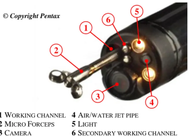
A standard biopsy is carried out by using an endoscope and inserting, through its working channel, a Biopsy Micro Forceps BMF (Figure 1). The endoscope is a tubular instrument used to examine the interior of a hollow organ. It is inserted directly through natural passages of body or through little incisions made in the patient’s body.
A modern standard endoscope, used in laparoscopic procedures, consists of a rigid or flexible tube with 11 mm diameter. In its distal end (head), the endoscope has a camera to capture images of the organ during the surgery. The head can be moved around 4 directions (up, down, left and right) by 4 controlling cables. Moreover, lighting system is present to illuminate the organ under inspection (Figure 1).
Figure 1: Endoscope distal end (head) and details
The endoscope has also 2 air and water jet pipes to inflate organs under operation and to clean the operation area. In a standard endoscope there is an additional working channel, usually with 2.2-3.2 mm in diameter, used for inserting and extracting additional accessories. By means of this channel, it is possible to extract tissue samples, to deflate/inflate organs or to extract polyps or tumor.
During a biopsy procedure the endoscope is inserted in the patient’s body until it reaches the point of operation where removing the desired tissue samples.
1 5 4 3 2 6 1 WORKING CHANNEL 2 MICRO FORCEPS 3 CAMERA
4 AIR/WATER JET PIPE
5 LIGHT
6 SECONDARY WORKING CHANNEL © Copyright Pentax
PROCEEDINGS OF THE 1st INTERNATIONAL CONFERENCE ON DESIGN AND PROCESSES FOR MEDICAL DEVICES – Ceretti et alii. PROMED, ISBN: 978 88 6608 058 9
Consequently, the biopsy micro forceps is inserted through the instrument channel till the distal end. By opening and closing the BMF jaws with a handle device it is possible to seize and collect the sample. After this, BMF is taken off from the working channel and the sample is released in a test-tube for the pathologist inspection. It takes about 2 minutes to collect the sample, to extract, insert and reposition the BMF. In many cases the number of desired samples for a good diagnosis is 6 or 7 but in case of tumor the samples can reach up to 15. On the market there are more than 100 available solutions depending on:
Mono or multisampling [3] Disposable or reusable BMF [4-6] BMF actuating system
BMF manufacturing processes specifications Control types.
The present research aims to identify an innovative BMF solution developed in close cooperation between medical doctors and engineers within the international cooperation project IREBID funded by EU.
2 PROBLEM DESCRIPTION
The study tries to solve unmet needs of GI endoscopy and, to identify these needs, interviews with gastroenterologists and pathologists and a detailed market analysis have been carried out. The main interest areas that have been identified for tissue sampling procedure improvement are the decrease in the overall surgery time, the reduction of endoscope working channel or micro forceps wear or breakage and the overall reduction of the device cost.
In particular, the most critical aspects emphasised by medical doctors have been:
Limits on sample dimension and depth [7].
Crushing of the tissue (which changes its morphology).
Necessity of collecting multiple samples from more places (i.e Stomach 4 8 samples, Colon 8 10 samples).
Commercial multisampling device are not effective. Breakage of opening/closing BMF mechanism. Wear of the working channel due to the BMF
sliding friction during in and out phases.
Risk of infections due to poor cleaning of reusable devices.
Long surgery time (2 minutes per sampling). High device cost.
Non-oriented extractions lead to a longer diagnosis process and criticalness in the pathology [8]. In particular, the effectiveness of an oriented sample is shown in Figure 2. The differences in the sample section are evident and it is easy to understand the request of pathologists for a oriented tissue so to facilitate the disease diagnosis.
Moreover, international standards are gradually introducing the adoption of single use BMFs and accessories. Recent studies demonstrate that disposable BMF are economically convenient [9,10] but other
Authors are in disagreement, especially when high number of biopsies are considered [11,12]. Therefore the device manufacturing needs to move to low cost disposal devices.
Consequently, the unmet doctor needs that an innovative device has to accomplish are:
The design a new multi-sample device with reduced wear of the endoscope working channel and of BMF.
An adequate tissue sampling procedure characterized by good sample dimension (> 2mm), and not crushed.
An oriented sample (location of the mucosa and submucosa) to facilitate the pathology diagnosis (see Figure 2).
A reasonable cost for a disposable device, to be applied with standard endoscopes having working channels with 2.0 2.8 mm diameter.
A disposable BMF device and accessories
Oriented Sample
Not oriented Sample
Figure 2: Sample extraction and influence of the sample orientation [8]
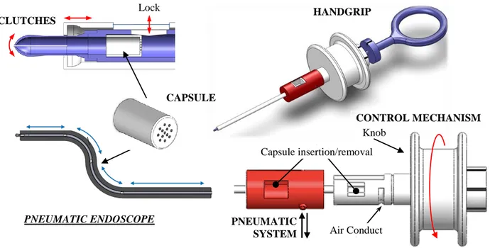
3 PNEUMATIC ENDOSCOPE
The identified solution was designed to substitute the traditional endoscopic BFM (Figure 3). It consists of elastic clutches (used for cutting the sample), a shuttle (a capsule for sample transportation), a pneumatic system for the shuttle movement and a secure control mechanism.
Clutches consist of elastic jaws which are closed or open
thanks to an external sliding cylinder. Their function is to cut the tissue sample and hold it. Since the actual trend towards oriented samples and to disposal solutions, the jaws have been designed to be formed from a tube, whose end is axially cut and formed. The other end is joined with a flexible tube (capsule channel) in which the shuttle can move.
The shuttle consists of a polymer capsule, holed on the bottom, and able to contain and transport the sample. The shuttle is inserted and removed through the control mechanism. Numbers or colours on the capsule are used to track the sampling order.
Thanks to a Pneumatic System connected to the working channel, it is possible to move the capsule back and forth, and a locking system allows to hold the capsule during the loading procedure. The holes on the bottom of
Figure 3: Pneumatic endoscope solution and details
the capsule allow to suck the tissue sample from the clutches into the capsule. The endoscope air inflation/deflation channel or the secondary working channel is used to compensate the pressure inside the patient (Figure 1).
Since the sampling procedure requires additional step with respect to traditional endoscopy sampling procedure, a secure control mechanism was introduced. It consists of a handgrip with a movable knob which controls the air mechanism and the capsule lock. In particular, the pneumatic system is connected to the capsule channel through air conducts in the handgrip, the knob opens and closes the connection. Its rotation controls an elastic retention mechanism that holds the capsule.
A total of 6 knob position were identified for the biopsy procedure and are reported in Table 1.
Table 1: Pneumatic endoscope handle settings.
Step Description Air Lock
1 Capsule insertion/removal off off 2 Distal end positioning IN off
3 Locking on IN ON
4 Biopsy sampling off ON 5 Tissue in the capsule OUT ON 6 Proximal end positioning OUT off
The proposed solution fulfils the requirements described in section 2. In fact, its shuttle allows collecting multiple samples without repositioning the head of the endoscope and loosing the sampling zone. Moreover, the capsule is designed to be used with standard endoscope (working channel diameter) and allows keeping the sample orientation and the collection order. In this way, the biopsy time is reduced and the pathologist needs are satisfied too.
With regards to traditional biopsy based on micro forceps, the procedure time is reduced since the pneumatic system allows a faster capsule movement with respect to traditional micro forceps insertion/extraction manual procedure. Moreover, the shuttle is made of polymer, therefore the working channel wear will be reduced with respect to the usage of steel forceps which are continuously moved in and out. The disposal bodies (shuttle and forceps) are cheap, in particular, because of the simpler forceps closing mechanism based on elastic clutches, their total cost is expected to be comparable or lower than the available disposal BMF.
Figure 4: Pneumatic endoscope prototype (scale 5:1). FDM Technology, ABS material.
Capsule insertion/removal Air Conduct PNEUMATIC SYSTEM CONTROL MECHANISM CAPSULE CLUTCHES Lock HANDGRIP Knob PNEUMATIC ENDOSCOPE
19
4 CONCLUSIONS AND FUTURE
WORKS
The development requirements for an endoscopic biopsy micro forceps are to reduce sampling time, to guarantee tissue sample orientation and to reduce the wear of the endoscope working channel. However, current solutions available do not meet all these requirements. In the present work, an innovative solution for endoscopic biopsies is proposed. Thanks to a pneumatic system for the sample collection and a control mechanism which guarantees the proper functionality, the proposed device is able to answer to the market requirements. Moreover, it has the advantages to be used with standard endoscopes and uses low cost disposable components. Actually, a functional prototype for movements testing (clutches, lock system and control mechanism) was fabricated using Fused Deposition Modelling technology (FDM) in an ABS resin showing good results (Figure 4). Further studies will investigate the device manufacturing feasibility and capability; therefore, Design For Manufacturing and Assembly (DFMA) and Strategic Manufacturing Cost Analyses are methodologies that will be applied to further the development of these endoscopic solutions.
ACKNOWLEDGEMENTS
The Authors would like to thank Dr. V. Villanacci for his support in the biopsy procedure understanding and A. Louki, A. Cappadona and L. Bianchetti the master students who collaborated in the development of several innovative BMF solutions.
This research was carried under the support of the IREBID project founded by FP7-PEOPLE-2009-IRSES-247476.
REFERENCES
[1] Klaus F.R. Shiller; Roy Cockel; Richard H. Hunt; Bryan F. Warren “Atlas of Gastrointestinal Endoscopy and Related Pathology”, Blackwell Science 2002, 2nd edition.
[2] Norton J Greenberger; Richard S. Blumberg, Robert Burakoff “Current Diagnosis & Treatment - Gastroenterology, Hepatology, & Endoscopy” McGraw Hill Lange 2009.
[3] Weinstein WM, Khatri N and Da Z. A prospective comparison of a multibite forceps with conventional-sized and large cup pinch biopsy forceps. Gastrointestinal Endoscopy , 45(4), 1997. [4] Rizzo J, David Bernstein D, Gress F. A
performance, safety and cost comparison of reusable and disposable endoscopic biopsy forceps: a prospective, randomized trial. Gastrointestinal Endoscopy, 51(3):257-61, 2000.
[5] Deprez PH, Horsmansm Y, Van Hassel M, Hoang P, Piessevaux H, Geubel A. Disposable versus reusable biopsy forceps: a prospective cost evaluation. Gastrointestinal Endoscopy, 51(3): 262- 265, 2000.
[6] Fireman Z. Biopsy forceps: Reusable or disposable? Journal of Gastroenterology and Hepatology 21(7):1089-92, 2006.
[7] Elmunzer BJ, Higgins PD, Kwon YM, Golembeski C, Greenson JK, Korsnes SJ, Elta GH. Jumbo forceps are superior to standard large-capacity forceps in obtaining diagnostically adequate inflammatory bowel disease surveillance biopsy specimens. Gastrointestinal Endoscopy, 68(2):273-278, 2008.
[8] Villanacci V., Baronchelli C., Ravelli P., Missale G., Williams C., Talbot I.C., Cestari R. The orientation of endoscopic biopsies of the gastrointestinal tract by the use of millipore filters: Endoscopical and pathological aspects. Giornale Italiano di Endoscopia Digestiva, 16(4):213-217, 1993.
[9] Yang R, Ng S, Nichol M, Laine L. A cost and performance evaluation of disposable and reusable biopsy forceps in GI endoscopy. Gastrointestinal Endoscopy, 51(3):266-70, 2000.
[10] Rizzo J, David Bernstein D, Gress F. A performance, safety and cost comparison of reusable and disposable endoscopic biopsy forceps: a prospective, randomized trial. Gastrointestinal Endoscopy, 51(3):257-61, 2000.
[11] Deprez PH, Horsmansm Y, Van Hassel M, Hoang P, Piessevaux H, Geubel A. Disposable versus reusable biopsy forceps: a prospective cost evaluation. Gastrointestinal Endoscopy, 51(3): 262-265, 2000. [12] Kozarek RA, Raltz SL, Merriam LD, Sumida SE.
Disposable versus reusable biopsy forceps: a prospective evaluation of costs. Gastrointestinal Endoscopy, 43(1):10-3, 1996.
![Figure 2: Sample extraction and influence of the sample orientation [8]](https://thumb-eu.123doks.com/thumbv2/123dokorg/5512092.63859/6.892.474.801.458.655/figure-sample-extraction-influence-sample-orientation.webp)