Full Terms & Conditions of access and use can be found at
http://www.tandfonline.com/action/journalInformation?journalCode=ycra20
CRANIO®
The Journal of Craniomandibular & Sleep Practice
ISSN: 0886-9634 (Print) 2151-0903 (Online) Journal homepage: http://www.tandfonline.com/loi/ycra20
Reliability of kinesiography vs magnetic resonance
in internal derangement of TMJ diagnosis: A
systematic review of the literature
Fulvia Costantinides, Silvia Parisi, Ingrid Tonni, Christiane Bodin, Erica
Vettori, Giuseppe Perinetti & Roberto Di Lenarda
To cite this article: Fulvia Costantinides, Silvia Parisi, Ingrid Tonni, Christiane Bodin, Erica Vettori, Giuseppe Perinetti & Roberto Di Lenarda (2018): Reliability of kinesiography vs magnetic resonance in internal derangement of TMJ diagnosis: A systematic review of the literature, CRANIO®, DOI: 10.1080/08869634.2018.1455433
To link to this article: https://doi.org/10.1080/08869634.2018.1455433
Published online: 08 Apr 2018.
Submit your article to this journal
View related articles
LITERATURE REVIEW
Reliability of kinesiography vs magnetic resonance in internal derangement of
TMJ diagnosis: A systematic review of the literature
Fulvia Costantinides DDS, MSca, Silvia Parisi DDSa, Ingrid Tonni DDS, MSc, PhDb, Christiane Bodin DDS, PhDc,
Erica Vettori DDS, MSca, Giuseppe Perinetti DDS, MSc, PhDa and Roberto Di Lenarda DDS, MSc, PhDa
aSchool of dental Sciences, university of Trieste, Trieste, Italy; bSchool of dental Sciences, university of brescia, brescia, Italy; cprivate practitioner,
brescia, Italy
ABSTRACT
Objective: The aim of this systematic review was to evaluate the accuracy and the diagnostic reliability of kinesiography and magnetic resonance imaging (MRI) in diagnosis of patients presenting temporomandibular disorders.
Methods: A literature survey carried out through PubMed, SCOPUS, LILIACS, and the Cochrane Library from the inceptions to the last access on August 18 2016 was performed to locate randomized clinical trials, controlled trials, prospective cohort studies, or retrospective studies (with or without a control group), that examined the diagnostic reliability of recording devices of mandibular movements in comparison to MRI.
Results: From the results, it was found that a significant correlation between these electronic devices and MR images could not be detected in case of disc displacement.
Discussion: The scientific evidence does not support the usefulness in clinical practice of the jaw-tracking devices to diagnose temporomandibular disorders because their diagnostic reliability is poor.
Abbreviations: MRI: magnetic resonance imaging; TMJ: temporomandibular joint; TMD: temporomandibular disorder; RCT: randomized clinical trial; CCT: controlled clinical trial; PCS: prospective cohort study; RS: retrospective study
Introduction
In recent years, several studies have questioned the diag-nostic reliability of electronic devices (kinesiographic or axiographic) as a diagnostic aid in temporomandibular disorders (TMDs), as they are not fully supported by scientific evidence [1,2]. The only exception so far is for magnetic resonance imaging (MRI), since it can depict temporomandibular joint (TMJ) disc position and the presence of joint effusion [3].
Also, recent investigations using more sophisticated instruments in an experimental setting have brought new insights into the assessment of jaw function and muscle activity, but clinicians may find it difficult to draw clinically useful information from studies using devices designed for research purposes [4].
Often kinesiography, axiography, and MRI have been used for dental diagnosis, particularly regarding the patho-logical conditions of the TMJ. Neff et al. [5] assessed post-operative functional outcomes regarding loss of vertical
height, disc mobility, relationship of condyle compared with disc and mandibular fossa, protrusive, and transla-tory movements in patients with condylar head fractures managed by osteosynthesis using MRI and axiography.
The reliability of instruments for making axiographic recordings in the diagnosis of internal derangements of the TMJ remains controversial, and the literature does not suggest that the sensitivity and specificity of jaw-tracking devices are reliable enough to be used for diagnosis and management of intra-articular TMDs [1,6].
For this reason, there is currently not an evident indi-cation for the use of axiographic devices for diagnosis and assessment of temporomandibular dysfunctions. To date, no systematic revision has dealt with the topic of diag-nostic reliability of jaw-tracking devices in comparison with MRI. In view of these considerations, the present systematic review was aided in the assessment of the diag-nostic accuracy and reliability of kinesiography or axiog-raphy, as compared to that from MRI in the diagnosis of internal derangements of the TMJ, to establish whether
KEYWORDS
Systematic review; kinesiography; magnetic resonance; Tmd; reliability; diagnosis; axiography; TmJ
© 2018 Informa uK limited, trading as Taylor & francis Group
2 F. COSTANTINIDES ET AL.
follow-up was included. References of all papers included were searched to identify any further relevant studies. Data items
Two reviewers extracted all data simultaneously but inde-pendently, using a standardized outline. The following data items were collected: study design; sample size; age and sex distribution of the sample; type of internal derange-ments of the TMJ; techniques of investigation; correla-tion between axiographic/kinesiographic parameters for MRI findings; accuracy of axiographic/kinesiographic such procedures may have a role in the management of
intra-articular TMDs. Materials and methods
Search strategy and study selection
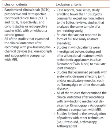
This systematic review was based on the guidelines of the Cochrane Handbook for Systematic Reviews of Interventions (The Cochrane Collaboration, 2011) and the reporting based on the PRISMA Statement [7]. All the articles that examined the diagnostic reliability of jaw-tracking devices in comparison to MRI were identified through a literature survey carried out through the following databases: (i) PubMed (www.ncbi.nlm.nih.gov/pubmed); (ii) SCOPUS (www.SCOPUS.com); (iii) Latin American and Caribbean Health Sciences (LILACS, www.lilacs.bvsalud.org) and (iv) The Cochrane Library (www.thecochranelibrary. com). The survey covered the period from inception to the last access on August 18 2016, without any limitation dic-tated by the language of the articles or publishing date [8]. The search algorithms used in each database are reported in Table 1. Finally, a manual search was also performed by scoring the references within the studies examined. The search was made to identify manuscripts meeting the fol-lowing inclusion criteria: studies that examined the clinical outcomes after recordings with jaw-tracking mechanical devices (i.e. kinesiograph, axiograph) in comparison with MRI. The studies had to be randomized clinical trials (RCTs), prospective and retrospective controlled clinical trials (pCCTs and rCCTs, respectively) and cohort studies or retrospective studies (rSs), regardless of the presence of a control group. Case series, case reports, studies enrolling fewer than 10 subjects, comments, expert opinion, letters to the editor, reviews, and studies that analyzed the same sample of a pre-existing study were excluded. Studies in which patients were investigated before, during, and after a functional treatment with orthodontic appliances (such as Bionator or Twin-Block) to evaluate joint changes were also excluded. In addition, studies that examined patients with systematic diseases affecting joint and/or masticatory muscles, such as fibromyalgia or other rheumatic diseases, were not collected. Full details of inclusion and exclusion criteria are listed in Table 2.
Two researchers (SP and FC) carried out the literature search independently (January 2016), by first analyzing titles and abstracts for relevance and presence of the selec-tion criteria listed above. The full text articles of included and uncertain records were obtained for further eligibility screening by the same two reviewers. In the event of an unsettled disagreement, the opinion of another co-author (GP) was consulted. In case of identification of redundant research in different papers, the paper with the highest
Table 1. Search strategy.
Note: * is the wildcard in the boolean research. If used after the root of a word, it will get results that contain variations of that root in the title or description.
PubMed (www.ncbi.nlm.nih.gov/pubmed)(diagnosis OR accuracy OR sensitivity OR specificity OR correlation OR comparison) ANd (Jaw Relation Record [meSh] OR axiography OR axiographic OR axiographia OR kinesiograph*) ANd (magnetic Resonance Imaging [meSh] OR mRI) ANd (tmj OR (temporomandibular and (disorder* OR dysfunction*)) OR internal derangement)
SCOPUS (www.scopus.com)((Axiography) OR (Jaw Relation Record)) ANd ((magnetic Resonance Imaging) OR (mRI)) ANd ((temporomandibular dis-order) OR (temporomandibular dysfunction) OR (internal derangement)) LILACS (www.lilacs.bvsalud.org)((Axiography) OR (Jaw Relation Record))
ANd ((magnetic Resonance Imaging) OR (mRI)) ANd ((temporoman-dibular disorder) OR (temporoman((temporoman-dibular dysfunction) OR (internal derangement))
Cochrane Library (Registered Controlled trials) (www.thecochranelibrary. com) ((Axiography) OR (Jaw Relation Record)) ANd ((magnetic Resonance Imaging) OR (mRI))
Table 2. details of inclusion and exclusion criteria. Inclusion criteria Exclusion criteria • Randomized clinical trials (RCTs);
prospective and retrospective controlled clinical trials (pCCTs and rCCTs, respectively); and cohort studies or retrospective studies (rSs). with or without a control group;
• All of the studies that examined the clinical outcomes after recordings with jaw-tracking me-chanical devices (i.e. kinesiograph and axiograph) in comparison with mRI.
• Case reports, case series, study enrolling fewer than 10 subjects, comments, expert opinion, letters to the editor, reviews, studies that analyzed the same sample of a pre-existing study;
• Studies that are not reported in form of full text (only abstract available);
• Studies in which patients were investigated before, during and after a functional treatment with orthodontic appliances (such as bionator or Twin-block) to evaluate joint changes;
• Studies that examined patients with systematic diseases affecting joint and/or masticatory muscles, such as fibromyalgia or other rheumatic diseases;
• All of the studies that examined the clinical outcomes after recordings with jaw-tracking mechanical de-vices (i.e. Kinesiograph, Axiograph) without a comparison with mRI; • Studies limited to the investigation
of patients with other techniques (i.e. ultrasound, Arthroscopy, Arthrography).
parameters for MRI findings; specificity of axiographic/ kinesiographic parameters and MRI; sensitivity of axio-graphic/kinesiographic parameters for MRI findings; and clinical implications according to authors’ conclusions about the diagnostic reliability of the methods based on the results obtained.
Assessment of risk of bias in individual studies Evaluation of the methodological quality of published studies is very important because it gives an indication of the strength of scientific evidence provided by these studies. However, no single approach in assessing method-ological soundness may be appropriate for all systematic reviews to evaluate the quality [9]. Therefore, contextual, pragmatic, and methodological considerations are fol-lowed when assessing study quality (Center for Reviews and Dissemination. Systematic Reviews, 2009). Herein, a custom risk of bias analysis has been used as follows: enrollment (prospective/retrospective), control group (yes/no), sample size calculation (yes/no), method error (yes/no), adequacy of statistical analysis (yes/no), and expert rates (yes/no). According to the retrieved infor-mation related to the single items, the overall risk of bias of the selected studies was defined as either Low or High. Results
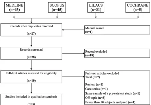
The results of the automatic and manual searches are shown in Figure 1. From the automatic search through
the main scientific databases, a total of 144 articles were found. Furthermore, a manual search was performed, and one article was added for the analysis. After having removed all double entries and applying the inclusion/ exclusion criteria, 10 studies were selected and analyzed in full-text. From the resulting 10 full-text articles assessed for eligibility, 7 studies were excluded for the reasons detailed in Table 3.
Three studies were judged to be relevant to the pres-ent study according to the inclusion/exclusion criteria [4,10,11]. The full details of the studies included in the review are summarized in Table 4.
Study design
The three selected studies comprised one cohort study [11] and one retrospective study (rS) [4]. The article by Lochmiller et al. [10] did not report any information about the study design.
Figure 1. flow diagram of the search strategy.
Table 3. full-text articles exclusion details.
Reason for exclusion Number of studies Study author
Review 1 badel et al. [17]
Same sample of a
pre-ex-istent study 1 beer et al. [18] Off topic 3 ettlin et al. [19]
hasegawa et al. [20] Neff et al. [5] Case series 1 dhanda et al. [21] fewer than 10 subjects
4 F. COSTANTINIDES ET AL.
Study population
The sample size of patients who were examined through the two methods of investigations (axiography or kinesi-ography and MRI) ranged from a minimum of 24 subjects [10] to a maximum of 90 subjects [11]. The mean sub-jects’ age was reported in two of the three studies, and it was included between 26.9 years [11] and 43.1 years [4]. In two studies, both sexes were monitored [4,11]. Only the study by Lochmiller et al. [10] did not provide any information as regards both age and sex. Only one study considered a real control group [11]. In contrast, the study by Manfredini et al. [4] considered a sample of patients with internal derangements of the TMJ in which the sub-jects served as cases and controls. In all of the studies, the subjects were systemically healthy.
Temporomandibular disorders
The study by Lochmiller et al. [10] analyzed subjects with bilateral or unilateral reciprocal articular disorders, but the type of intra-articular TMD was not specified. The article by Neff et al. [11] investigated 57 healthy subjects who did not present any internal derangement of the TMJ, and a group of 33 patients as control. This last group com-prised 6 patients with clinically manifest dysfunctions, which were so far conservatively treated, and 27 patients with condylar fractures that were previously surgically treated (21 unilateral fractures, 6 bilateral fractures, at least 12 months after the end of the treatment, with slight or moderate dysfunctions). The article by Manfredini et al. [4] considered a group of patients in the attempt to get deeper into the assessment of TMJ symptoms and/or differentiate them from other muscle disorders, and at the end of the investigation, found only unilateral disorders. In particular, the frequency of diagnosis was: (i) disc dis-placement with reduction (DDR): 35.5% (right) and 54.8% (left); (ii) disc displacement without reduction (DDNR): 3.2% for both the articulations; (iii) effusion (Eff): 29% (right) and 19.4% (left).
Techniques of investigations
The methods of investigation used for the analysis were well described in each article. Axiography or kinesiogra-phy and MRI were adopted in all studies. One study used both conventional static MRI and dynamic MRI (CINE-MRI) and arthrosonography, a further procedure to exam-ine the TMJ [11]. Only one study carried out a “manual investigation” that included dynamic compression and translation, in addition to electronic axiography and MRI (computer-assisted methods), for a differential diagnosis of joint sounds [10]. Manfredini et al. [4] performed MRI
Table 4.
Summariz
ed da
ta of the thr
ee studies included in the r
eview
.
Not
es: AIC: angle of c
ondylar inclina tion; pr : pr otrusion; v: v arianc e; NS: not sig nifican t; NA: not a vailable; S -m RI: sta tic m RI; CIN e-m RI: cinema tic m RI; K G: k inesiog raphic; A X: axiog raphic; rS: r etr ospec tiv e study ; d ef : deflec tion a t the end of ja w mo vemen t; I nc: incisur es; d ev : devia
tion during mandibular opening;
dd R: disc displac emen t with r educ tion; dd NR: disc displac emen t without r educ tion; eff : effusion. Study Sample size Se x and age TMJ disor ders Techniques of in ves -tiga tion used Cor rela tion A X/ KG par amet ers and MRI (n [%]) Ac cur ac y of A X/K G par amet ers f or MRI findings (n [%]) Specificit y of A X/ KG par amet ers and MRI (n [%]) Sensitivit y of A X/ KG par amet ers f or MRI findings(n [%]) Clinical implica -tions Neff et al . [ 11 ]; 57 m : 26; f: 31 h ealth y T m J, No T m J disor der Axiog raph y, S -m RI, CIN e-m R, Ar -thr osonog raph y pr : 91% ( v = 0.038) with CIN e-m R; NA CIN e-m RI: 56/57 (98%); NA CIN e-m RI and S -m RI ar e superior t o axiog raph y 26.9 y ears S-m RI: 54/57 (97%) AXIOGR Aph Y: NA AIC: 94% with (v = 0.00336) CIN e-m R Cohor t 6 m : 1; f: 5 Clinically manif est dy sfunc tions , so far c onser va tiv ely tr ea ted CIN e-m RI: 25/33 (76%): 41.4 y ears S-m RI: 28/33 (85%); AXIOGR Aph Y: NA 27 m : 19; f: 8 Sligh t or moder at e dy sfunc tions; 21: unila ter al c ondylar fr ac tur es , sur gically tr ea ted; 6: bila ter al fr ac tur es , sur gically tr ea ted 31.3 y ears lochmiller et al . [ 10 ] 24 NA bila ter al or unila ter al r ecipr ocal ar ticular disor ders Axiog raph y, m RI, C om -pr ession t ests and Tr ansla tion t ests 88% 49% NA NA Axiog raph y does not add an y impor tan t finding NA manfr edini et al . [ 4 ] 31 m : 13%; f: 87% u nila ter al disor ders ( dd R: 35.5–54.8%; dd NR: 6,5%; eff : 19.4–29%) Kinesiog raph y and m RI NS KG par amet ers: KG par amet ers: KG par amet ers: m RI is superior t o kinesiog raph y d ef : 38.7–54.8% d ef : 40–55.1% d ef : 38.1–50% 43.1 y ears d ev : 30–50% d ev : 47.6–100% d ev : 42–54.8% Inc: 3.4–10% Inc: 100% Inc: 9.6–71% rS
3.4 to 10%. The study by Neff et al. [11] considered only the specificity of CINE-MRI and static MRI in the group with TMJ dysfunctions (33 of 90 total subjects) and in the group of subjects with healthy TMJs (57 of 90 total sub-jects). The specificity values were very different between the two considered groups of patients. The specificity in the first group coincided with 76% (25/33) for CINE-MRI and with 85% (28/33) for static MRI; for the healthy group, the percentages were changed: 98% (56/57) for CINE-MRI and 97% (54/57) for static CINE-MRI. Only in the article by Manfredini et al. [4], the percentages of sensitivity of various kinesiographic parameters for MRI findings were provided (deflection: 38.1–50.0%; deviation: 47.6–100%; incisure: 100%). The other two articles did not provide any information about the sensitivity of the techniques [10,11]. All of the clinical outcomes of the three included studies are summarized in Table 4.
Main reported results and clinical outcomes
In their conclusions, Lochmiller et al. [10] reported that the electronic jaw-tracking devices are essential for diag-nosis position in patients with intra-articular dysfunc-tions, and the study revealed a good correlation between MRI and axiography; but axiographic recordings do not give any additional diagnostically relevant findings that go beyond the manual examination. Neff et al. [11] concluded that concerning metric sensitivity, both MRI techniques (CINE-MRI and Static-MRI) are sufficiently able to match axiography; in particular, Static-MRI can be considered the method of choice, due to the better representation of morphological details. In the last study, Manfredini et al. [4] affirmed that MRI is superior to kinesiographic devices, and kinesiography cannot be used in dental prac-tices as a method of diagnosis and management of internal derangements of the TMJ.
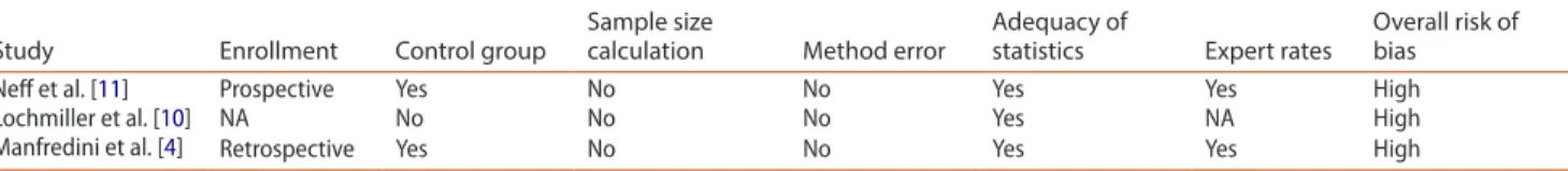
Risk of bias in individual studies
The results of the quality analysis of the included stud-ies are summarized in Table 5. The overall risk of bias was judged to be high in all three studies [4,10,11]. The enrollment of the patients was prospective for the study by Neff et al. [11], retrospective for the study by Manfredini et al. [4], while in the last study [10], this information was not available. A real control group was considered only in the study by Neff et al. [11] (33 subjects with tempo-romandibular dysfunctions). In the study by Manfredini et al. [4], the same sample of patients (31 subjects) was considered as case group and control group. None of the three included studies used an estimation of the sample size [4,10,11]. Method error was not evaluated in any of the included studies for either recording procedure and kinesiographic recordings to detect internal
derange-ments of the TMJ.
Correlation between kinesiographic or axiographic recordings and magnetic resonance imaging
Two of the three studies reported a statistically significant correlation between MRI findings and kinesiographic/ axiographic parameters [10,11]. In the article by Neff et al. [11], axiography and MRI (specifically CINE-MRI) showed a correlation equal to 91% in the jaw movement of protrusion and about the angle of condylar inclination measured during protrusion’s movement (94%, with a variance of 0.036). Lochmiller et al. [10] found a corre-lation equal to 88% between electronic axiography and MRI findings; the remaining 12% was because clinically relevant joint sounds were not attributable to the dislo-cation of the articular disc, and therefore, they were not represented in the axiographic tracings. Only in the study by Manfredini et al. [4] the correlation between MRI find-ings and kinesiographic parameters was defined as not significant because there was not more than one kinesio-graphic variable that showed a p-value below p = 0.10 with any MRI finding. In detail, DDR showed p-values ranging from 0.62 to 0.999; DDNR p-values ranging from 0.063 to 0.999, and MRI-depicted joint effusion showed p-values from 0.09 to 0.999.
Clinical outcomes
In only one study, the value of accuracy of axiographic/ kinesiographic parameters for MRI findings was reported [10]. In the study by Manfredini et al. [4], because of the poor revealed relationship between kinesiographic and MRI findings, the accuracy of the various kinesiographic findings to predict the presence of any specific MRI sign at the patient level was poor. In particular, the accuracy value of kinesiographic deflection for diagnosing signs detected on MRI ranged from 38.7 to 54.8%, that of kine-siographic deviation spanned from 42 to 54.8%, and that of kinesiographic incisures from 9.6 to 71%. In the study by Lochmiller et al. [10], the accuracy of the registered axiographic tracings with the use of dynamic tests of com-pression and translation in relation with results of MRI corresponded to 49%. This means that half of all the axio-graphic tracings coincided with MRI findings. The article by Neff et al. [11] did not determine the level of accuracy of electronic axiography for MRI results. The value of speci-ficity of the different kinesiographic parameters for MRI findings was considered only in the study by Manfredini et al. [4]. Specifically, kinesiographic deflection’s values ranged from 40 to 55%, that of kinesiographic deviation from 30 to 50%, and that of kinesiographic incisures from
6 F. COSTANTINIDES ET AL.
other method adopted for the comparison with kinesiog-raphy and axiogkinesiog-raphy, for several years was considered the gold standard in diagnosis of internal derangements of the TMJ. Moreover, over the years, the indications for the routine use of this technique in TMJ diseases are losing for the high costs and the evidence of the benign natural course of most TMJ disc displacements; but in the new DC/TMD, MRI is returned as an indication [14]. In this respect, Manfredini et al. [4] suggested that a thorough clinical assessment is often enough for managing the majority of patients’ intra-articular TMDs, and so, the use of electronic devices for analyzing patients with internal derangements of the TMJ should stand comparison with less expensive diagnostic approaches. In line with this opinion, Lochmiller et al. [10] found that the recorded condylar movements give no additional, diagnostically relevant insights that go beyond the manual examination. In the systematic review, mandibular movements during the axiographic recordings were examined independently, without always considering the single types of internal derangement dysfunction or influencing factors of jaw motions. Among these, muscle disorders could have an important role on the accuracy of jaw-tracking devices, and they could change the findings. If permanent disc dis-placements are present, it should be pointed out that if this disturbance persists for longer, it does not need to show limited tracks anymore. In the course of time, the initial short tracks become long and regular, and they cannot be distinguished from normal [15]. Furthermore, there is a large variety of systems used to record mandibular movements. As supported by Lueckerath et al. [16], their recordings could not be offhand with each other. For this reason, there are currently no generally accepted guide-lines for the interpretation of the recording’s devices. Only the study by Neff et al. [11] involves a cohort/case-control study, and they investigated separately the healthy patients and the subjects with intra-articular TMD. In the study by Manfredini et al. [4], a convenience sample of patients with internal derangement of the TMJ was considered, in which subjects and joints with or without specific signs in MRI images served as cases and controls. The last study [10] investigated a small sample of patients without a control group. It is necessary to consider a larger sample size and to examine two different large pure control and case groups. It is important to observe that the evaluations [4,10,11]. To avoid interpretation bias related to the
dif-ferent radiologists or clinicians assessing the images, the evaluations were made by expert clinicians with exper-tise in the interpretation of MRI images and axiographic tracings [4]. The expert rates were reported in two of the three studies [4,11]. In the study by Lochmiller et al. [10], the evaluation of the outcomes of MRI and axiography by expert clinicians was not mentioned. The modalities of statistical analysis were appropriate in all three studies included in the review [4,10,11].
Discussion
The present systematic revision was conducted with the aim to evaluate diagnostic reliability and accuracy of kinesiography and axiography as compared with MRI in diagnosis of patients presenting internal derangements of the TMJ. There is a great controversy about the use of a jaw-tracking device in the diagnosis of intra-articular TMDs because the external validity of commercially avail-able devices has not yet been assessed. In contrast, despite a cautionary statement by the research community [12], the use of kinesiographic and axiographic instruments have been accepted by several clinical practitioners, with-out appraising their validity, based only on claims and opinions of the users of these instruments [4,13]. From the analysis of the articles, it is possible to observe that the three studies [4,10,11] present a high risk of bias, due to the persons who collected data and the persons who interpreted images and tracings not being blinded (Table
5). It should be emphasized that the scientific data avail-able in the literature are very poor, and this fact repre-sents a limit for the research of a diagnostic value of these instruments in a clinical setting. From the 144 articles obtained by the first automatic search in the literature, at the end of the selection’s procedure, only three studies that satisfied all the inclusion and exclusion criteria were selected for the analysis. The majority of the articles did not deal with the theme of the accuracy of axiography in comparison with MRI but either considered the value of two methods independently or used them for monitoring functional and clinical outcomes in patients treated for intra-articular TMDs without explaining if a technique of investigation was superior to another. It should be con-sidered that, in the field of TMJ dysfunctions, MRI, the
Table 5. Analysis of the risk of bias for the 3 included studies.
Note: NA: not available.
Study Enrollment Control group Sample size calculation Method error Adequacy of statistics Expert rates Overall risk of bias
Neff et al. [11] prospective Yes No No Yes Yes high
lochmiller et al. [10] NA No No No Yes NA high
Disclosure statement
No potential conflict of interest was reported by the authors. ORCID
Giuseppe Perinetti http://orcid.org/0000-0002-7226-5134 References
[1] Theusner J, Plesh O, Curtis DA, et al. Axiographic tracings of temporomandibular joint movements. J Prosthet Dent. 1993;69:209–215.
[2] Tsolka P, Preiskel HW. Kinesiographic and electromyographic assessment of the effects of occlusal adjustment therapy on craniomandibular disorders by a double-blind method. J Prosthet Dent. 1993;69:85–92. [3] Tasaki MM, Westesson PL. Temporomandibular joint:
diagnostic accuracy with sagittal and coronal MR imaging. Radiol. 1993;186:723–729.
[4] Manfredini D, Favero L, Federzoni E, et al. Kinesiographic recordings of jaw movements are not accurate to detect magnetic resonance-diagnosed temporomandibular joint (TMJ) effusion and disk displacement: findings from a validation study. Oral Surg Oral Med Oral Pathol Oral Radiol. 2012;114:457–463.
[5] Neff A, Kolk A, Meschke F, et al. Kleinfragmentschrauben vs. Plattenosteosynthese bei Gelenkwalzenfrakturen [Small fragment screws vs. plate osteosynthesis in condylar head fractures]. Mund- Kiefer- Gesichtschirurgie MKG. 2005;9:80–88. German.
[6] Okeson JP. Management of temporomandibular disorders and occlusion. 7th ed. Amsterdam: Elsevier Health Sciences; 2014. p.483.
[7] Liberati A, Altman DG, Tetzlaff J, et al. The PRISMA statement for reporting systematic reviews and meta-analyses of studies that evaluate healthcare interventions: explanation and elaboration. BMJ. 2009;339:b2700. [8] Robinson KA, Dickersin K. Development of a highly
sensitive search strategy for the retrieval of reports of controlled trials using PubMed. Int J Epidemiol. 2002;31(1):150–153.
[9] Antczaic AA, Tang J, Chalmeks TC. Quality assessment of randomized control trials in dental research. II. Results: periodontal research. J. Periodontal Res. 1986;21:315–321. [10] Lochmiller W, Bumann A, Landeweer GG. Zur Wertigkeit
der elektronischen axiographie in der klinischen funktionsdiagnostik [On the validity of the electronic axiography in clinical functional diagnostics]. Fortschritte Kieferorthopädie. 1991;52:268–273. German.
[11] Neff A, Kolk A, Beer A, et al. Stellenwert des statischen MRT im Vergleich mit CINE-MRT, Achsiographie und arthrosonographie [Importance of static MRI compared with CINE-MRI, axiography and arthrosonography]. Dtsch Zahnärztliche Z. 2002;57:353–357. German. [12] Manfredini D, Bucci MB, Montagna F, et al.
Temporomandibular disorders assessment: medicolegal considerations in the evidence-based era. J Oral Rehabil. 2011;38:101–119.
[13] Cooper BC. Parameters of an optimal physiological state of the masticatory system: the results of a survey of were made by expert clinicians with expertise in the
inter-pretation of MRI images and axiographic tracings in two studies analyzed in full-text [11,12], but the lack of blind-ness of these experts and the persons who performed the techniques results in a low studies’ quality. Moreover, a strong limit for a methodological study is the lack of the analysis of the repeatability. This aspect could provide a further stimulus for future research on a larger sample of patients with an effective follow-up.
The results demonstrate that axiographic or kinesio-graphic instruments cannot represent useful jaw-tracking devices to detect intraarticular disorders in the clinical setting, although limited available data should be con-sidered. Neff et al. [11], in their study, concluded that both MRI techniques (static MRI and CINE-MRI) are superior to electronic axiography, mainly for the diagnosis process, documentation, and therapeutic decisions. They considered static MRI to be the method of choice, due to a superior representation of morphological details, but the study presents a high risk of bias. Manfredini et al. [4] reported that the correlation between kinesiographic parameters and MRI findings was not significant because none of the chosen parameters were correlated with any of the MRI findings. These authors also reported that the accuracy of kinesiographic findings to predict MRI diag-noses was not acceptable and too low to support the use of kinesiography in the clinical setting. In the last study [10], a high correlation between axiography and MRI (88%) was reported; furthermore, the authors showed that the remaining 12% was due to disc dislocations. The accuracy reported in this study was of 49%; approximately half of all axiographic interpretations corresponded to MRI results. According to the authors’ opinion, this high percentage derives from the techniques of manual examinations, which have enhanced the accuracy of electronic jaw-track-ing devices, with regard to the visualization of internal derangement. According to this evidence, instrumental analysis does not add any more important, further infor-mation, as compared to manual investigation.
Conclusion
A significant correlation between jaw-tracking systems and MR images in cases of TMJ disc displacement could not be detected. The scientific evidence does not support the usefulness of jaw-tracking devices in clinical practice to diagnose internal derangements of the TMJ, because those instruments’ accuracy and reliability to diagnose TMJ disease is poor. It is necessary to perform further studies on this topic, less subjected to risk of bias, in par-ticular, having larger samples and a control group to better elucidate the role of these TMJ recording’s instruments.
8 F. COSTANTINIDES ET AL.
[18] Beer A, Kolk A, Neff A, et al. Cine-MRT des Kiefergelenks im Vergleich zur konventionellen MRT und achsiographie [Cine MRI of the temporomandibular joint in comparison to static MRI and axiography]. RöFo Fortschritte Auf Dem Geb. Röntgenstrahlen Nukl. 2004;176:506–512. German.
[19] Ettlin DA, Mang H, Colombo V, et al. Stereometric assessment of TMJ space variation by occlusal splints. J Dent Res. 2008;87:877–881.
[20] Hasegawa Y, Kakimoto N, Tomita S, et al. Movement of the mandibular condyle and articular disc on placement of an occlusal splint. Oral Surg Oral Med Oral Pathol Oral Radiol Endod. 2011;112:640–647.
[21] Dhanda J, Cooper C, Ellis D, et al. Technique of temporomandibular joint replacement using a patient-specific reconstruction system in the edentulous patient. Br J Oral Maxillofac Surg. 2011;49:618–622.
[22] Braun S, Hicken JS. Ultrasound imaging of condylar motion: a preliminary report. Angle Orthod. 2000;70:383–386.
practitioners using computerized measurement devices. CRANIO®. 2004;22:220–233.
[14] Schiffman E, Ohrbach R, Truelove E, et al. Diagnostic criteria for temporomandibular disorders (dc/tmd) for clinical and research applications: recommendations of the International RDC/TMD Consortium Network and Orofacial Pain Special Interest Group. J Oral Facial Pain Headache. 2014;28:6–27.
[15] Kobs G, Bernhardt O, Meyer G. Accuracy of computerized axiography controlled by MRI in detecting internal derangements of the TMJ. Stomatologija, Baltic Dental Maxillofac J. 2004;6:7–10.
[16] Lückerath W. Zur Differentialdiagnostik elektronischer Aufzeichnungen der Gelenkbahnen funktionsgestörter Patienten [Differential diagnosis of electronic TMJ tracings in dysfunction patients]. Dtsch. Zahnärztliche Z. 1991;46:22–726. German.
[17] Badel T, Ćimić S, Munitić M, et al. Clinical view of the temporomandibular joint disorder. Acta Clin Croat. 2014 Dec;53(4):462–470.