1
Università degli Studi del Piemonte Orientale
“Amedeo Avogadro”
DEPARTMENT OF HEALTH SCIENCESPHD PROGRAM IN
BIOTECHNOLOGY FOR HUMAN HEALTH
Cycle XXVIII
PhD Thesis
iPSC-based strategy to correct the bleeding
phenotype in Hemophilia A
Tutor: Prof. Antonia Follenzi
Coordinator: Prof. Claudio Santoro
PhD candidate: Maria Talmon
2 Summary
Hemophilia A (HA) is an X-linked bleeding disorder caused by mutations in the coagulation factor VIII (FVIII) gene. The current therapy is based on the administration of recombinant or plasma-derived FVIII. However, this treatment is not a definitive cure and the 20-40% of patients develops neutralizing antibodies. In this context, gene and cell therapy represent attractive alternative therapeutic strategies. Reprogramming of somatic cells can be used to generate induced Pluripotent Stem Cells (iPSCs), which can be differentiated into progenitor cells relevant for gene and cell therapy applications. iPSCs offer the potential to generate patient-specific cells, reducing the risk of immune rejection by offering an autologous source of cells for transplantation. Moreover, for monogenic disease treatment iPSCs technology could be associated with gene therapy allowing the genetic correction of the disease defect. Since Yamanaka’s pivotal work, iPSCs were generated starting from several cell types. Human iPSCs are most frequently derived from dermal fibroblasts because of their accessibility and relatively high reprogramming efficiency. However, in hemophilic patients, to harvest fibroblasts from skin biopsies is dangerous because of the risk of bleeding. In this context, we utilized patient-derived peripheral blood cells as an easy-to-access source of cells to generate iPSCs. Then we genetically corrected and differentiated them into functional endothelial cells (ECs) secreting FVIII. Reprogrammed healthy and hemophilic CD34+ cells gave rise to bona fide iPSCs colonies showing embryonic stem cells-like morphology. iPSCs were positive at alkaline phosphates staining, expressed both nuclear and surface pluripotency markers at RNA and protein levels. Moreover, the CpG islands at iPSCs NANOG core promoter showed a demethylated profile, confirming that obtained colonies were well reprogrammed at morphological, transcriptional and epigenetic levels. iPSCs-derived embryoid bodies expressed the three germ layers markers and were able to differentiate in adipose, osteogenic and chondrogenic cells. Then, both healthy and hemophilic iPSCs were differentiated into ECs. The obtained ECs expressed endothelial specific markers (KDR, Tie2, CD31, VEC) and formed tubules when cultured in matrigel. HA-iPSCs corrected by transduction with a lentiviral vector carrying the human B domain deleted form of FVIII (BDD-FVIII) under the endothelial specific promoter of VE-cadherin efficiently expressed FVIII after differentiation. Moreover, when transplanted by portal vein injection in the liver of our immunocompromised mouse model of HA, corrected HA-ECs were able to engrafted, proliferate and functionally secreted FVIII up to 12 weeks post transplantation. Therapeutic correction was also reached when corrected ECs were injected in association with microcarrier beads in the peritoneum of the mice. Together these data showed the possibility to obtained functionally FVIII secreting endothelial cells, starting from patient specific gene corrected iPSCs confirming the suitability of this approach for HA gene-cell-therapy.
3 INDEX OF CONTENTS
INTRODUCTION 4
Hemophilia A and coagulation factor VIII 5 Gene and cell therapy for hemophilia A 9 Induced pluripotent stem cells (iPSCs) 15
Generation of iPSCs 18
Viral delivery systems 21 Non-viral delivery systems 23 Molecular challenges of reprogramming 25
Application of iPSCs 29
iPSCs in hematological diseases 31
iPSC and hemophilia A 32
MATERIALS AND METHODS 35
RESULTS 44
Generation of iPSCs from MNCs of healthy and hemophilic donors 45 Differentiation of healthy iPSCs into endothelial cells 46 Genetic correction of hemophilic iPSCs and differentiation into endothelial cells 46 Generation of iPSCs from different cell sources of healthy and hemophilic donors 47 Differentiation of CD34+-iPSCs into endothelial cells 48 Hemophilic CD34+-iPSCs were genetically corrected for FVIII expression and
differentiated into endothelial cells 49 In vivo FVIII expression and hemophilic phenotype correction after transplantation of
genetically corrected iPSCs-derived ECs in a mouse model of HA 50
Figures legend 65
DISCUSSION 68
BIBLIOGRAPHY 75
4
5 Hemophilia A and coagulation factor VIII
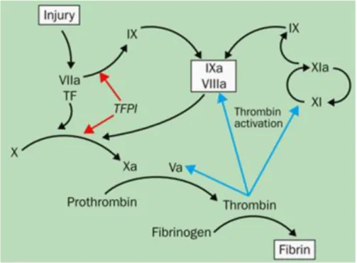
Hemophilia A (HA) is a recessive X-linked bleeding disorder that occurs in 1:5000 male live births. It is due to the lack or reduced activity of coagulation factor VIII (FVIII) [1-3], a non-enzymatic cofactor part of the coagulation cascade. When activated by thrombin FVIII is able to bind the factor IX (FIX) in a complex that activates factor X (FX), which converges in the common pathway of the coagulation cascade converting fibrinogen to fibrin and generating clot formation (Fig. 1).
Figure 1.Schematic representation of coagulation cascade. Activated FVIII binds
FIX forming a complex activating FX and converging in the common pathway of coagulation cascade.
The reduction of FVIII in the plasma causes hemophilic patients to have a lifelong bleeding tendency of clinical severity proportional to the degree of FVIII reduction. Indeed, based on the residual FVIII activity, there are three forms of HA: the severe form, in which the levels of FVIII are below 1%, the moderate form, between 1 and 5%, and the mild form, from 5 to 40% of FVIII activity [1]. Several mutations in coagulation FVIII gene (F8) cause hemophilia A. The most frequent mutation that affects approximately 45% of patients with severe hemophilia is the inversion of the first 22 exons caused by intrachromosomal recombination between the intron 22 (int22h1) with two homologous sequences that are distant 500kb from the gene (int22h3 and int22h3) [4]. Other mutations causing the severe form are small deletions or insertions, missense and non-sense mutations that taken together occur in 45% of severe HA patients. Less frequent
6
mutations are large deletions, splice site mutations and intron 1 inversion [5]. In contrast, missense mutations are mainly associated with moderate and mild form [2]. Disease clinical manifestations range from spontaneous bleeding, with frequent haemarthroses in the most severe form, to secondary bleeding with rare haemarthroses in milder form [4]. Diagnosis is made based on family history or following the first haemarthrosic episodes that occur at different ages according to the clinical form of hemophilia (0-3 years for severe, 2-7 for moderate, 5-14 for mild) [1]. The main diagnostic technique used in laboratory is the evaluation of activated partial thromboplastin time (aPTT), which estimates the ability of patients plasma to shorten the time required for clot formation in a standard FVIII-free plasma [6]. However, this method can sometimes be inaccurate to diagnose the mild form and provides in 5-10% of cases false negative results. This implies the use of other more sensitive techniques such as two-stages clotting assay, chromogenic assay and genetic investigation may be more informative [6]. Moreover, the analysis of F8 mutation is useful to further characterize the gene defect and the risk of inhibitor development, one of the major concern of hemophilia A actual treatment [7]. It was reported that patients who had severe hemophilia A and mutations predicting a null allele developed inhibitors more frequently (22% to 67%) than patients with missense mutations (5%) [8]. The current therapeutic practice consists in the administration of recombinant FVIII or blood products and is carried out based on the disease severity. Patients with severe hemophilia receive prophylaxis 2-3 times a week, while patients with mild and moderate form receive replacement therapy on demand [9]. Primary prophylaxis is defined as regular continuous treatment initiated in the absence of documented osteochondral joint disease, determined by physical examination and/or imaging studies, and started before the second clinically evident large joint bleed and age of 3 years. Continuous prophylaxis means the intention of treating for 52 weeks/year and at least 45 weeks/year (85%) [10]. Patients receive 25-40 IU kg-1 per dose administered intravenously three times a week [10].
The main complication is the development of neutralizing antibodies (inhibitors) to FVIII that occurs within the first 50 exposure days and is common in 20-40% of patients with the severe form. This worsens the clinical aspect because it makes ineffective the treatment and further reduces the residual activity of the endogenous FVIII and expose patients to an increased risk of morbidity and mortality [11]. The treatment of patients with inhibitors is more problematic and the therapeutic strategies are mainly focused on the treatment or prevention of bleeding episodes using agents that bypass the inhibitor (e.g. plasma-derived activated prothrombin complex concentrates and recombinant activated FVII). Eradication of the inhibitor through long-term intensive treatment with large doses of FVIII (immune tolerance induction; ITI) is effective in approximately two third of cases. Despite the good outcomes the high costs of ITI reduced its feasibility to few patients
7
requiring the development of other strategies such as blockade of costimulation, oral tolerance, immunosuppressive treatment and antigen-specific regulatory T cell [12].
F8 was firstly characterized and cloned in 1984 [13]. F8 maps to band Zq28 at the tip of the long arm of the X chromosome. It measure 186 kb in length and is constituted by 26 exons encoding for a mature protein of 2332 amino acids (263 KDa) plus 19 aa signal oligopeptide at the N-terminal [14]. FVIII is synthesized as an inactive single chain and it is organized in 6 domains: A1, A2, B forming the heavy chain and A3, C1, C2 that constitute the light chain. Between A1/A2, A2/B and B/A3 domains are present three acidic regions (a1, a2, a3) that contain the thrombin binding sites (Arg372, Arg740, Arg1689) and are crucial for FVIII activation [15] (Fig. 2).
Figure 2. Domain structure and processing of FVIII.FVIII is organized in 6 domains:
A1, A2, B, A3, C1, C2. After cleavage at the C-terminal region of B domain, FVIII consists in a heterodimer formed by the heavy chain and the light chain linked by a divalent metal ion between the A1 and A3 domains.
The overall structure of FVIII is similar to FV, in particular in the A domains they share approximately 40% amino acid identity also with the copper binding protein ceruloplasmin [16]. Moreover C domain share some homologies between FVIII, FV and proteins that bind negatively charged phospholipids (e.g., fat globular protein and the lipid-binding lectin discoidin I) [17, 18]. In contrast, FVIII B domain is unique and do not show significant similarity with FV or other proteins [19]. Once synthesized, FVIII enters in the endoplasmic reticulum (ER) in which undergoes two important modifications: the elimination of the signal peptide and the introduction of oligosaccharide chains on asparagines residues predominantly arranged on the B domain. The N-glycosylation is fundamental to ensure the correct folding of the protein, to prevent the aggregation of intermediate forms and to allow the interaction of FVIII with enzymes and chaperon proteins essential to the correct intracellular processing, vesicular trafficking, exocytosis and secretion of
8
FVIII [20]. The ER-Golgi transition is mediated by the interaction between FVIII B domain with specific protein complex, in particular lectin-mannose binding 1 (LMAN1) also known as endoplasmatic reticulum-Golgi intermediate compartment 53 kDa protein (ERGIC53) and multiple coagulation factor deficiency 2 protein (MCFD2) [19]. Defects in these molecules cause the combined deficiency of circulating FVIII and FV, which share in part the same intracellular processing of FVIII [21]. In the Golgi, FVIII is cleaved close to the C-terminal region of B domain (after the aa 1313 and aa 1648) producing an heterodimer consisting in the heavy chain (200 kDa) and the light chain (80kDa) that are not covalently linked by a divalent metal ion (mainly Cu2+) between the A1 and A3 domains. Finally, the processes that complete the intracellular maturation of FVIII are the modification of saccharide groups introduced in ER and the sulfurization of some tyrosines located in the acidic regions target of thrombin proteolytic activity [22]. In physiological conditions the FVIII concentration in plasma is 200-300 ng/ml and is associated with high affinity with the vWF, which is 50-folds in excess compared to the FVIII [23]. The role of this interaction is to increase the half-life of FVIII by reducing the clearance and avoiding the inactivation by protein C. Moreover, the vWF prevents the premature association of FVIII with other coagulation factors before its activation mediated by thrombin. The regions involved in the binding between FVIII and vWF are located in correspondence of the acidic sequence a3, at the C-terminal of the B domain and in the C2 domain [24]. Not surprisingly, therefore, those mutations in the gene of vWF or FVIII impairing the ability of interaction between the two proteins can produce very similar pathological phenotypes [24]. The FVIII B domain (40% of the protein) is not essential for hemostatic cofactor activity. Indeed, early biochemical studies demonstrated that deletion of the B domain resulted in a functional molecule [25]. This observation was used to develop a shorter version of FVIII, called B-domain-deleted (BDD)-FVIII, characterized by the removal of most of the B domain, with only 14 residual amino acids. The BDD-FVIII was expressed more efficiently compared with wild-type FVIII resulting in a 17-fold increase in mRNA levels over full-length FVIII and in a concomitant increase in the amount of synthesized primary translation product [25]. Moreover, no immunologic differences were detected between BDD- and wild-type FVIII [26, 27]. The first generation of these recombinant FVIII products was synthesized by gene-transfection in mammalian cells (Chinese Hamster Ovary cells) [27]. Currently, it is produced in absence of any animal or human proteins reaching the highest level of safety and it is extensively used in clinics [26]. Although FVIII mRNA is detected in different human and mouse organs such as liver, spleen, lymph nodes, kidney [28-31] and hematopoietic cells [32, 33], transplantation studies in hemophilic animal models and patients demonstrated that liver is the primary source of FVIII [34, 35]. However, the identity of liver cells expressing FVIII was controversial. Both hepatocytes and liver sinusoidal endothelial cells (LSECs)
9
were proposed to be the main sources of FVIII but their role in FVIII production is still debated. Initially and for a long time hepatocytes were considered the FVIII expressing cells both at mRNA and protein level by in vivo and in vitro experiments [28, 36-38]. Although in early years the presence of FVIII was reported mainly in LSECs rather than hepatocytes. Several studies reported the presence of the FVIII antigen in LSECs, but not in the parenchyma of adult human liver [39-41]. Subsequently, Kumaran et al. showed that hemophilia A mice transplanted with unfractionated liver cells, (mixture of hepatocytes, LSECs, Kupffer cells, and hepatic stellate cells) or of the cell fraction enriched in LSEC survived a bleeding injury challenge, whereas purified hepatocytes transplantation did not prevent deadly bleeding[42]. Then, it was reported that the only cells producing FVIII mRNA and protein in the fetal and adult human liver are LSECs [43, 44]. Recent works confirmed FVIII expression in endothelial cells (EC) by using a Cre/Lox strategy to selective knocking out FVIII expression in several cell types and concluding that FVIII is not produced in hepatocytes but mainly secreted by EC [45, 46]. Fahs et al. used promoters to target defects in endothelial cells and hepatocytes and demonstrated that a severe hemophilic phenotype was associated with lack of FVIII expression in endothelial cells [46]. Likewise, Everett et al. reached the same conclusion knocking out, in hepatocytes or LSEC, Lman1, a cargo receptor in the early secretory pathway that is responsible for the efficient secretion of FVIII to the plasma [45].All together these studies pointed out LSECs as the main source of FVIII in the liver, bringing them to the forefront in the design of alternative therapeutic approach for hemophilia A, such as gene and cell therapy. Gene and cell therapy approaches could allow the continuous FVIII production and secretion in the blood stream achieving sustained levels of FVIII rather than the peaks in circulation of the replacement therapy due to the short half-life of infused FVIII protein [43].
Gene and cell therapy for hemophilia A
Gene and cell therapy could constitute a powerful therapeutic approach for many pathologies, in particular for monogenic diseases. They can be applied alone or combined depending on therapeutic needed (Fig.3).Gene therapy is a form of molecular medicine that has developed since the early nineties and is still evolving: the main purpose is to introduce into the target cells (but also in tissues or organs) DNA sequences in safe and efficient way, with the ultimate goal to obtain a therapeutic effect, or at least slow the disease progression.
10
Figure 3: Strategy of delivery of a therapeutic transgene in “pure” gene therapy (direct delivery) or in a
combined cell-gene therapy strategy. In gene therapy the therapeutic transgene packaged into a delivery vehicle was directly injected into the patient. When gene therapy was combined to cell therapy, the transgene in inserted into delivery cells expanded in culture and then re-infused into the patient. (Terese Winslow2001)
Hemophilia A represent an ideal target for gene therapy since it is a monogenic disease and to restore FVIII activity al level superior than 2% is sufficient to ameliorate the bleeding phenotypes of patients with an overall increase of quality of life. Good clinical trials results were already obtained from Hemophilia B gene therapy using adeno associated-viral vector (AAV) to deliver FIX into the patients [47]. In particular, the best results were obtained in a clinical trial using an AAV pseudotyped with AAV8 capsid protein. It has been reported that the AAV8 capsid has stronger liver tropism and lower seroprevalence in human, in comparison with the AAV2 used in the previous trial. Moreover, it provide less virus uptake by antigen presenting cells and is able to mediate effective transduction in animals with pre-existing immunity to AAV2 [48]. Regarding the expression cassette, an artificial liver specific promoter (LSP), based on the hAAT promoter and an ApoE enhancer, was used to drive the expression of a codon optimized FIX cDNA [49]. Finally, the AAV8.LSP.FIX was administered by peripheral vein injection in 10 patients enrolled in the study. By these strategies FIX activity was restored at level between 2-6% up to a median of 3.2 years. However 4 patients were treated with prednisolone following the increase of liver enzyme and all of them have received at least one recombinant FIX infusion [47]. Nevertheless, this clinical trial showed the feasibility of gene therapy for hemophilia B, increasing the quality of patients' life and encouraged new efforts to improve this approach making it a suitable alternative to replacement therapy. Despite the relevant results obtained for hemophilia B, gene therapy for hemophilia A has
11
seen significantly less progress into the clinic due to some factors that complicates FVIII expression in comparison with FIX: i) the size and complexity of FVIII (9 kb) make it too large for some vector system, such as AAV; ii) using a comparable vector delivery, transduced cells express 100 fold less FVIII level than FIX [50];iii) FVIII is naturally 5-6 fold more immunogenic than FIX, making the transgene mediated immune response a big concern. Moreover, the AAV‐FIX clinical trials detected an anti-AAV capsid immune response at high vector doses suggesting a limit to the administered vector dose [47]. This limit is very challenging for FVIII gene therapy. Indeed, it is established that the necessary dose to achieve therapeutic levels of FVIII are higher than for FIX [51]. However, several approaches for hemophilia A gene therapy using different vector systems were attempted. AAV vectors are impaired by their limited capacity to packaged genome larger than 5 kb. To circumvent this problem FVIII light and heavy chain were split in two distinct AAV and upon co-injection in mice, biologically active FVIII was detected in circulation [52, 53]. Nevertheless, since the interaction between the two chains occurs inside the cells, it is necessary that both vectors co-transduce the same cell to allow the production of functional FVIII, reducing the efficiency of the strategy and increasing the overall dose of vector to use. Despite these difficulties other studies reported that delivering the heavy and the light chains in two separate AAV resulted in a dose-response with sustained expression of FVIII at therapeutic levels both in dog and mouse models of hemophilia A [54, 55]. Another option to overcome the size limit of AAV vectors was to use a B domain deleted FVIII (BDD-FVIII) that reduced by one third the final size of cDNA without compromising the coagulation biological activity of the protein. Even though a minimum promoter is required to not exceed the package capacity of AAV. By this attempt, several studies have showed sustained FVIII levels in mice [56, 57]. However, they reported in most of treated mice anti-FVIII antibodies formation that was overcome by using the serotype AAV8 instead the AAV1 [57]. AAV vectors containing the BDD-FVIII have demonstrated to be suitable to induce FVIII expression also in hemophilic dogs [58-61]. Even so the doses needed to reach therapeutic correction were significantly higher than the maximum doses of AAV-FIX administered to human in clinical trials and given the dose-dependence of immune response to capsid, the use of this vector could be not feasible in humans [51]. A recent study has demonstrated to induce remarkable therapeutic expression of FVIII in non-human primate by targeting FVIII expression in hepatocytes combined with the use of a codon optimized FVIII [62]. However, the macaques were injected with a non-species specific transgene developing anti-FVIII antibodies requiring transient immunosuppression to reduce anti-FVIII antibodies titer, thus the long term follow up was not possible [62]. Among the other viral vectors lentiviral and retroviral were more feasible to be employed for the treatment of hemophilia A. The first proof of concept for in vivo gene therapy
12
using a -retroviral vector was assessed in neonatal hemophilic mice. Approximately 50% of injected mice expressed physiological or even higher FVIII levels that were sustained up to 14 months. In this study, the remaining animals that showed only transient or undetectable FVIII expression developed anti-FVIII specific antibodies [63]. This approach was also successful in a canine model of hemophilia A by targeting transgene expression in the liver without antibodies formation [64]. However, when retrovirus was used for human gene therapy only low circulating FVIII was detected in patients [65, 66]. In 2000, Park and colleagues demonstrated that also LV could be use in vivo to induce human FVIII expression in wild type mice. By intraportal injection they direct FVIII expression predominantly to the liver with an ubiquitous promoter (EF1α) and reaching human FVIII levels of about 15%. Unfortunately, hFVIII expression was only transient due to antibodies formation despite the fact that mice were not hemophilic and normally expressed murine FVIII [67]. Later, similar results (5% of activity) was obtained in hemophilia A mice by intraperitoneal (IP) injection or ex vivo bone marrow transduction using a LV carrying the BDD-FVIII under the control of an ubiquitous promoter. However also in this case neutralizing antibodies were developed, with higher frequency in IP group rather than in ex vivo bone marrow transduction [68]. Notably, the ubiquitous transgene expression mediated by LV may be detrimental because of the adverse reaction given by proteins ectopically expressed [69] or the innate or adaptive immune response against the transgene once it is expressed by antigen presenting cells (APC) [70, 71]. To overcome this issues the use of cell specific promoter to de-targeted transgene expression in APC allowed the sustained expression of therapeutic gene in the selected cells by reducing immune response [72, 73]. Over the years many authors have used several tissue specific promoters to drive transgene expression in cells of interest including endothelial cells, hepatocytes, dendritic cells, hematopoietic stem cells, megakaryocytes, B cells [74-76]. Moreover, another degree of cell targeting is represented by the post-transcriptional regulation based on micro RNAs (miRNAs). For gene transfer purpose, the insertion of complementary sequences to a specific miRNA (miRNA target sequence, mirT) to the 3' of the expression cassette, offers the possibility to reduce selectively the transgene synthesis in the cell types in which that particular miRNA is expressed. For example, in 2007, Brown and colleagues showed that the presence of miRT142-3p,complementary to miRNA 142-3p which is selectively expressed in hematopoietic cells avoided transgene expression (FIX) in APC limiting immune response against FIX by preventing transgene expression in those cells allowing FIX expression only in hepatocytes using a liver specific promoter [77].Despite its advantages, “pure” gene therapy approaches face some challenges in hemophilia A treatment. In light of these findings, an approach combining gene and cell therapy deserves further consideration to determine if an effective and safe cell-based treatment can be developed. Cell therapy consists in
13
the transplantation in a patient of cells from external sources in order to achieve the treatment of a specific disease. For genetic disease, cell therapy approaches involving the gene transfer of the correcting gene into the cells to be transplant can represent an alternative strategy to gene therapy alone. To design a cell-based therapy approach it is fundamental to identify the best cell sources to treat the disease (Fig. 4). To cure hemophilia A cell sources could include cells competent for FVIII production, such as liver sinusoidal endothelial cells (LSECs), stem cells derived from bone marrow, blood-outgrowth endothelial cells (BOECs), endothelial progenitor cells derived from the differentiation of induced pluripotent stem cells (iPSCs) and embryonic stem cells (ESCs). Transplantation of LSECs was demonstrated in several animal models to be able to correct hemophilia A bleeding phenotype [42, 43, 78]. Several liver-derived cells were studied to be applied in the therapy of hemophilia A.
Figure 4. Potential cell sources for hemophilia A treatment [79].
In mice, in 2005, it was demonstrated that the presence of endothelial cells in different liver-cell mixtures was fundamental to correct clotting dysfunction in hemophilic transplanted mice [42]. Subsequently, Follenzi et al. transplanted purified mature LSECs into the portal vein of hemophilic mouse model and demonstrated the engraftment in the liver and the therapeutic correction of hemophilia A with plasma FVIII activity more than 10% of normal plasma levels [78]. Additionally, successful transplantation of human LSECs isolated from adult liver was recently reported [43, 80]. Filali et al. isolated liver endothelial cells and transplant them into
monocrotaline-14
treated immunodeficient mice yielding LSEC engraftment. Interestingly, although culture conditions caused loss of LSEC fenestration, normal phenotype was re-acquired after transplantation, pointing out the role of microenvironmental signals in supporting some of the unique features of the LSEC phenotype. Another important observation was that macrovascular endothelial cells obtained from human umbilical vein and microvascular endothelial cells obtained from adult adipose tissue failed to repopulate liver. These findings indicate irreversible differentiation of these endothelial cell compartments that cannot be overcome by the signals provided by the liver environment [80]. Accordingly, cellular therapy for hemophilia A could need to rely on LSECs or immature endothelial progenitors as sources of transplantable cells. Cell therapy with adult LSECs is limited by access to living donor tissue or cadaverous livers. Cryopreservation could be used to bank adult liver cells as LSECs were shown to engraft into uPA-NOG mice liver after thawing [43]. Ex vivo expansion of LSECs could be used to increase the number of adult LSECs available for transplantation, but the effects of in vitro culture on the viability, proliferation and function of these cells needs to be further evaluated. Due to the restricted proliferative capacity of adult cells, an immortalized cell line was recently developed from adult human LSECs using lentiviral transduction with Htert [81]. These cells expressed some LSEC specific markers and demonstrated endocytic properties. However, the safety and potential of this cell line to engraft and produce FVIII remains to be tested.
An alternative cell source is represented by BOECs, clinically-promising peripheral blood-derived cell type. Most of the freshly isolated BOECs originate from vascular walls, however 5% are estimated to come from the bone marrow [82]. These bone marrow precursors have a much higher proliferative potential and are responsible for the majority of cell outgrowth in culture and are, thus, referred to as BOECs. When intravenously injected, BOECs primarily engrafted the spleen and bone marrow. Moreover, BOECs transfected with FVIII and transplanted into NOD/SCID mice were able to secrete human FVIII into the plasma [83]. FVIII-transduced BOECs have also been grown into sheets and then transplanted subcutaneously resulting in partial correction of the disease phenotype in HA mice [84].However, BOECs are present at a low frequency in adult peripheral blood and can be limited expanded in culture (PD=20;[85]). Thus, other stem cell and progenitor populations were evaluated for their potential for in vivo FVIII production either by acting as carrier cells for ectopic FVIII expression or differentiation into LSECs after transplantation. For instance, rat bone marrow-derived CD133+CD45+CD31+ cells have been shown to differentiate into LSECs after transplantation [86-88], but the potential of these cells as a source of FVIII still remains to be evaluated. Even though LSECs and stem cells derived from various adult and fetal sources may play a great role in the therapy of hemophilia A, there are
15
potential limitations that may hinder their clinical utility such as: i) lack of availability of a suitable donor tissue; ii) low yield of LSECs from liver homogenates, iii) limited expansion potential of stem cells or LSECs due to loss of functional or proliferative capacity in vitro. The extent of these potential limitations is not fully known and requires further studies. Nonetheless, to provide a sufficient number of cells to treat hemophilia A, investigation into alternative LSECs sources is prudent. The discovery of embryonic stem cells (ESCs) and induced pluripotent stem cells (iPSCs) has revolutionized the study of regenerative medicine due to their extensive potential for self-renewal, growth and pluripotency allowing creation of cells belonging to all three primary germ layers and, thus, including LSECs. Wang et al. showed that mouse chimeras created by injecting ESCs into FVIII-deficient mouse blastocysts were able to correct the bleeding disorder as FVIII was stably expressed in chimeras [89]. Although this study offers a basic proof of principle that ESCs can differentiate into FVIII-producing cells, much work is required to optimize the culture conditions required to generate transplantable and functional FVIII-producing cells such as LSECs or their precursors from pluripotent stem cells. There are also safety concerns that must be addressed, such as the risk of teratoma formation or genetic mutation of the extensively cultured and selected cells, before pluripotent stem cells can be considered for human transplantation. Although ESCs are viewed as the gold-standard of pluripotent stem cells, many current studies focus on iPSCs as they offer several advantages over ESCs. In particular, iPSCs offer the potential to generate patient-specific cells, reducing the risk of immune rejection by offering an autologous source of cells for transplantation.
Induced pluripotent stem cells (iPSCs)
Induced pluripotent stem cells are ESC-like cells obtained by the reprogramming of adult somatic cells. In 2006, Takahashi and Yamanaka demonstrated that the introduction into mature cells of transcription factors relevant for the maintenance of ESCs identity could induce pluripotency. In particular, they demonstrated that the ectopic expression of the transcription factors Oct4, Sox2, Klf4 and c-Myc was sufficient to revert the phenotype of mouse and human somatic cells to a pluripotent state [90, 91]. Oct4, also known as Pou5f1, belongs to the Octamer binding protein family. In humans, the OCT4 gene generate three isoforms but in most reports Oct4 mainly refers to OCT4A that has been found to maintain stemness in pluripotent stem cells [92]. Oct4 null embryos die in uterus during the peri-implantation stages of development [93, 94]. Although these embryos are able to reach the blastocyststage, in vitro culture of the inner cells mass (ICM) of homozygous mutantblastocysts produces only trophoblast lineages. ESCs cannot be derived from Oct4 null blastocysts. Suppression of Oct4 resulted in spontaneous differentiation into the
16
trophoblast lineages in both mouse [95] and human ESCs [96]. Oct4 acts in concert with other regulatory factors as Sox2 and Nanog (Fig. 5). Sox2 is a part of family DNA binding protein known as sex-determining region Y (SRY) related high mobility group (HMG-box) proteins expressed in ESCs [97]. Like Oct4, Sox2 marks the pluripotent lineage of the early mouse embryo; it is expressed in the ICM, epiblast, and germ cells. Sox2 null embryos die at the time of implantation due to a failure of epiblast (primitive ectoderm) development [98]. Homozygous mutant blastocysts appear morphologically normal, but undifferentiated cells fail to proliferate when blastocysts are cultured in vitro, and only trophectoderm and primitive endoderm-like cells are produced. Sox2 forms a heterodimer with Oct4 and synergistically regulates Fgf4 [97], UTF1 [99], and Fbx15 [100]. In addition, similar co-regulation by Sox2 and Oct4 has been reported in the regulation of the two transcription factors themselves [99, 101, 102], as well as Nanog[103, 104]. Genome-wide chromatin immunoprecipitation analyses demonstrated that Oct4, Sox2, and Nanog share many target genes in both mouse and human ESCs [105, 106]. Surprisingly,Sox2 deletion in mouse ESCs is rescued by the cDNA introduction of not only Sox2 but also Oct4, suggesting that one of the primary functions of Sox2 might be to maintain Oct4 expression [107]. Klf4 or Krüppel like factor 4 is a zinc finger transcription factor that regulates cell proliferation and differentiation [108]. Klf4 is highly expressed in undifferentiated mouse ESCs [109]. Klf4 can function both as a tumor suppressor and an oncogene. In cultured cells, the forced expression ofKlf4 results in the inhibition of DNA synthesis and cell cycle progression [110, 111].Klf4 null embryos develop normally, but newborn mice die within 15 hours and show an impaired differentiation in the skin [112] and in the colon [113], thus indicating that it plays a crucial role as a switch from proliferation to differentiation. KLF4 interacts as transactivator with Oct4-Sox2 in the synergistic activation of Nanog [114, 115]. Nanog is a homeodomain transcription factor expressed in pluripotent cell lines and in the ICM and maintain undifferentiated state by inhibiting and regulating the activity of pro-differentiation Bone Morphogenic Protein (BMP) [116, 117]. Nanog have been shown to be dispensable for reprogramming, indeed its expression can be triggered by the exogenous introduction of Sox2 and Oct4 that tightly regulate Nanog transcription. However, since 2006, several transcription factors cocktail were used to reprogramming somatic cells involving also Nanog and Lin 28 [118, 119], to avoid the use of c-Myc. It is a proto-oncogene required for cell growth and proliferation and in stem cell maintenance [120]. Although it improves the efficiency of the reprogramming process, the removal of c-Myc from the reprogramming factors paved the way for the development of a safer system [121] to generate iPSCs.
17
Figure 5: Core network of pluripotency regulation. Oct4 and Sox2 form a heterodimer (dotted lines)
that activates Oct4, Sox2 and Nanog transcription. Nanog regulates its own transcription and activates Oct4 and Sox2. An external signal, termed B, suppresses Nanog expression[122].
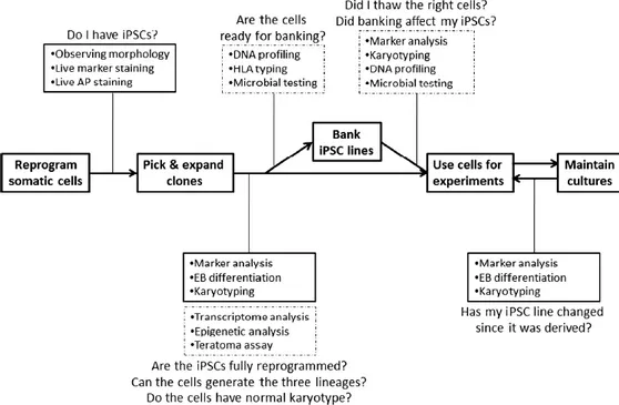
The established iPSCs showed ESCs-like phenotype, could differentiate into cell types of all three germ layers and possessed the ability of unlimited self-renewal in all cell types (Fig. 6). iPSCs grow on feeders, exhibit high nucleus to cytoplasm ratio, large nucleoli and form compact and uniform colonies with well-defined borders [123]. They show the same proliferation rate and feeder dependence of ESCs. Colonies stain positive for alkaline phosphatase (AP). iPSCs express pluripotency markers such as surface markers (SSEA-3, SSEA-4, tumor-related antigen (TRA)-1-60, TRA-1-81) as well transcription factors Oct4, Sox2, Klf4 and Nanog at comparable levels to those of ESCs. iPSCs have high telomerase activities, that is typical of pluripotent cells and fundamental to prevent cells aging [124]. Moreover, iPSCs remodel their epigenome during the reversion of differentiated cells into a stem-like state [125]. For example, they show unmethylated state of CpG islands at core promoter of pluripotent cells specific transcription factors, such as Oct4 and Nanog. Finally, iPSCs could differentiate into cell types of all three germ layers (ectoderm, mesoderm and endoderm). This ability can be assessed in vivo by teratomas formation assay [126] and in vitro evaluating the embryoid bodies (EB) formation capability and then the expression of the three germ layers markers. Although teratoma formations is one of the assay to be done for a complete characterization, iPSCs that form teratomas might not be the best choice for in vivo use [127, 128].
18
Figure 6: Scheme of iPSCs characterization to assess they acquired ESC-like phenotype, gene
expression and epigenetic patterns. Common characterization studies are found in solid boxes, while less common in dotted boxes [129].
Generation of iPSCs
The generation of patient-specific iPSCs is a critical step in cell therapy and other clinical applications. Since Yamanaka pivotal work, scientists investigated several strategies to generate iPSCs, considering several cells sources for the reprogramming and a variety of methods for the reprogramming factors delivery in cells. Started from fibroblasts iPSCs were obtained from several cell types: keratinocytes [130], melanocytes [119], hepatocytes [131], gastric epithelial cells [132], neural stem cells [133], adipose stem cells [134], pancreatic cells [135], cord blood cells [136], blood progenitor cells [137, 138], peripheral blood cells [139, 140] (Table 1).
Human iPSCs are most frequently derived from dermal fibroblasts because of their accessibility and relatively high reprogramming efficiency. However, to obtain fibroblasts to be reprogrammed skin biopsy and a prolonged period of cells expansion in culture are required. It cannot be ignored that patients would experience the pain and the risk of infection when obtaining dermal fibroblasts. These issues limit the application of iPSCs, in particular for patients with coagulopathies that risk uncontrolled bleeding in consequence of biopsy. In this perspective, blood cells are most easily accessible source of patient’s tissues for reprogramming because it is not need to maintain cell cultures extensively prior to reprogramming experiments. Furthermore, the venipuncture is safer than skin biopsy. In 2008, Hanna et al. used four retroviral vectors carrying
19
20
Oct3/4, Klf4, Sox2, and c-Myc to reprogram mouse B lymphocytes [142]. Then, one year later, iPSCs where generated from mouse T lymphocytes by the introduction of Oct4, Sox2, Klf4, and c-Myc in a p53-null background [143]. In the same year, Haase et al. generated human iPSCs from cord blood (CB) [136]. It is an advantage that CB can be obtained from public and commercial banks without any risk to donors and iPSCs were efficiently derived from frozen sample [144]. However, the use of CB is still limited in the perspective of therapeutic applications because it is not an autologous source of cells. Interestingly, Loh et al. [139], Seki et al. [145], and Staerk et al. [140] independently derived iPSCs from human peripheral blood cells. Loh et al. separated mononuclear cells (MNCs) and granulocyte colony stimulating factor (G-CSF)-mobilized CD34+ cells from peripheral blood samples, which were collected through venipuncture and Ficoll density centrifugation[139]. After infection with lentiviruses expressing Klf4, Sox2, Oct4, and c-Myc, CD34+ cells showed a reprogramming efficiency of 0.002%, whereas MNCs showed relatively low values of 0.0008% to 0.001%. Staerk et al. utilized a doxycycline-inducible lentivirus construct to derive iPSCs from T lymphocytes and myeloid cells [140]. This lentivirus construct encoded four the reprogramming factors into a polycistronic expression cassette (pHAGE2-TetOminiCMV-hSTEMCCA). Their results showed that the reprogramming efficiency of T lymphocytes was higher than that of myeloid cells. Because T lymphocytes exhibited a higher proliferation rate and had a better long-term growth potential in vitro than myeloid cells, Seki et al. induced T lymphocytes into iPSCs by a temperature-sensitive mutant SeV vector encoding human Oct4, Sox2, Klf4, and c-Myc with an efficiency of 0.1%. This SeV vector is a non-integrating type, and it could not proliferate at standard culture temperatures [145]. So these characteristics significantly increase the safety for the generation of iPSCs. Interestingly, in 2013 iPSCs were efficiently generated from non-mobilized CD34+ cells reprogrammed using a policystronic lentiviral vector carrying the four factors (STEMCCA vector). A mean of 5.3±2.8 iPSC colonies per 20 mL of non-mobilized peripheral blood were obtained [146]. All together these works provided the evidence that iPSCs from peripheral blood cells can be considered reliable and safe. Therefore, methods to generate iPSCs from human peripheral blood cells improving the efficiency of the reprogramming process could accelerate research and promote clinical applications of iPSCs in the future.
Other than the identification of the best source of cells to be reprogrammed, a key point in iPSCs generation is the choice of the strategy to reprogram somatic cells (Fig. 7). Actually, gene-delivery of transcription factors crucial for the induction and the maintenance of the pluripotency is the preferred method to induce cell reprogramming [147]. Scientists have used either viral or
non-21
viral methods to deliver reprogramming factors into cells [148, 149]. Lately, new reprogramming methods using RNA, protein and microRNA delivery have been used to generate iPSCs [150-152].
Figure 7. Schematic illustration of the reprogramming methods. iPSCs can be obtained both by viral
or non-viral systems. Viral methods include the use of integrating (Retroviral and Lentiviral) and non-integrating (Adenovirus and Sendai virus) vectors. Non-viral methods consist of the use of DNA-based (lipid or cationic polymers and mini-circle vectors) and non-DNA-based methods (protein, mRNA and the alteration of culture conditions). (Talmon et al., Submitted).
Viral delivery systems. Viral reprogramming methods can be summed up in the use of
retroviral, lentiviral and non-integrating viral vectors. Initially, iPSCs were obtained by retroviral vector transduction to successfully introduce the four Yamanaka factors (Oct4, Sox2, Klf4 and c-Myc) into mouse or human somatic cells [90, 91]. Four different retroviral vectors were used with each one carrying a transcription factor. Mouse iPSCs created chimeric mice that were competent for germline transmission [153]. However, both the chimeras and progeny derived from mouse iPSCs had an increased incidence of tumor formation, due primarily to reactivation of the endogenous c-Myc [153]. The removal of c-Myc from Yamanaka’s transcription factor cocktail has been a pivotal action in the generation of safer iPSCs, reducing the tumorigenicity of these cells [121]. Despite a reduced efficiency, murine and human iPSCs were successfully generated with only Oct4, Sox2 and Klf4.Moreover, chimeric mice generated from iPSCs obtained omitting c-Myc, did not form tumors after 100 days; when c-Myc was included, 20% of chimeric mice developed tumors [121]. An advantage of retroviral vectors is their capability to be spontaneously silenced after reprogramming induction and activation of the endogenous transcription factors. However,
22
these vectors transduce only dividing cells and the use of high doses of retroviral particles from integrating viruses increased the risk for insertional mutagenesis [154].
An alternative approach is the use of lentiviral vectors (LVs). LVs have several advantages: the high transduction efficiency to a wide variety of dividing and non-dividing cells, the stable and reproducible transgene expression and the possibility to generate a single polycistronic LV carrying all the reprogramming factors reducing the insertional mutagenesis risk [155]. Moreover, temporal expression control of the reprogramming factors was permitted by the use of doxycycline-inducible LVs [156]. However, given that the reprogramming cassettes integrate into the host genome, the risk of eventual reactivation exists. Thus excision strategies to remove LV expression cassette, including Cre-loxP recombination system and PiggyBac (PB) transposition, have been used. When the endogenous reprogramming machinery is activated, the exogenous sequences may be removed for safer reprogrammed cells [157]. In Cre-excisable LV the reprogramming cassette is flanked by two loxP sites in the LTR sequences. After the induction of pluripotent state, the recombinase Cre could be transiently expressed resulting with removal of the expression cassette. PB transposition is host-factor independent, and has been demonstrated to be functional in various human and mouse cell lines [157]. The PB transposon/transposase system requires only the inverted terminal repeats flanking a transgene and transient expression of the transposase enzyme to catalyze insertion or excision events. Murine and human embryonic fibroblasts were efficiently reprogrammed using doxycycline-inducible transcription factors delivered by PB transposition [157]. Importantly, PB transposons are completely removable from their integration site without any modification of the original DNA sequence, making this reprogramming system the ideal method to generate non-genetically modified human iPSCs for regenerative medicine.
Moreover, to obtain more clinically applicable iPSCs, non-integrating vectors were used [158, 159]. These vectors have a mutated integrase coding sequence, so they do not integrate in the host genome, remaining present in an episomal form in the nucleus [160] without losing the transduction efficiency of the integrating counterpart [161]. For these reasons, adenoviral and Sendai viral vectors, derived from non-integrating viruses, were used to reprogram somatic cells. Stadfeldtet al. [159] reprogrammed mouse fetal liver cells and finally adult hepatocytes with adenoviral vectors containing the four reprogramming factors. Although the efficiency was lower than integrating vectors, they obtained bona fide iPSCs. Nevertheless, they were not able to generate iPSCs from adult fibroblasts, probably because hepatocytes required lower expression of reprogramming factors than fibroblasts. One year later, adenoviral vectors were used to generate iPSCs from human embryonic fibroblasts using a higher multiplicity of infection. The obtained cells were pluripotent, able to differentiate and, most important, free from integrated viral DNA in
23
the host chromosomes [162]. This work demonstrated that reprogramming with adenoviral vectors generated bona fide iPSCs, but with a low rate comparing the number of transduced to reprogrammed cells. Conversely, using Sendai Virus (SeV)-based vectors, an RNA virus that replicates in the cytoplasm and does not integrate in the host genome, iPSCs were originated with an higher efficiency compared to the other methods [158]. One advantage of SeV is that RNA viruses are diluted during cell passage, reducing the number of viral particles at each passage; however, some residual viruses can still be present after several passages. Regardless, it is possible to eliminate the cells still containing the virus by a negative selection using a specific antibody that recognizes the hemagglutinin-neuraminidase (HN), the major protein expressed in SeV-infected cells, resulting in the maintenance of only virus-free reprogrammed cells [158]. Integration of reprogramming transcription factors into the cell genome is not necessary for maintenance of pluripotency [163]. The silencing of reprogramming factors expressed from a retroviral vector, the possibility to excise transgenes in LVs, and the efficient use of non-integrating vectors indicate that sustained expression of exogenous factors is not required. Indeed, the exogenous transgenes reactivate the endogenous transcriptional machinery of stem cells factors that are able to self-maintain the pluripotent status.
Non-viral delivery systems. The alternative safe and cost-effective approaches are
viral delivery systems of reprogramming factors, which can be divided into DNA-based and non-DNA-based methods.
DNA-based methods. Plasmids carrying reprogramming factors are encapsulated into lipid
or cationic polymers and subsequently transfected into cells. Plasmids remain in an episomal form and are allowed a short-term transgene expression [164, 165]. In 2008, Okita et al.[164]described the successful reprogramming of mouse embryonic fibroblast to iPSCs obtained by lipofectamine transfection of two plasmids: one containing c-Myc, and the other containing the three factors: Oct4, Sox2 and Klf4. Later, it was demonstrated that the nucleofection of a polycistronic plasmid co-expressing Oct4, Sox2, Klf4, and c-Myc was equally efficient [166]. iPSCs from human foreskin fibroblasts were successfully generated by sequential transfections of non-episomal plasmids that independently encoded the four factors Oct4, Nanog, Sox2, and Lin28 [167]. iPSCs obtained by these methods did not show any plasmid integration but were able to give rise to teratomas when injected into mice and contributed to adult chimeras. However, cells were reprogrammed with 100-1000x lower efficiency compared with viral methods probably due to lower transgene expression levels. Thus, further studies are required to optimize these systems [158]. To this purpose, a reprogramming strategy with the use of mini-circle DNA was developed. Mini-circle DNA lacks
24
plasmid backbone sequences and confers higher levels of sustained transgene expression upon delivery. Transgene-free iPSCs were generated from human adipose stem cells [168]using a mini-circle vector carrying a single cassette containing Oct4, Sox2, Lin28, and Nanog. Mini-mini-circle DNA provides higher transfection efficiency and longer ectopic expression compared with regular plasmids and could be useful in translational studies because adult cells can be reprogrammed without genomic modification. Therefore, the production of these virus-free iPSCs addressed a critical safety concern for potential use of iPSCs in regenerative medicine.
Non-DNA methods. Up to now, the methods described involved the use of genetic materials,
which could cause unexpected genetic alterations. Thus, alternative strategies have been investigated, such as the delivery of reprogramming proteins or mRNA directly into the cells and the manipulation of cell culture conditions parameters. In 2009, the first successful generation of protein-induced iPSCs (piPSCs) was described. In this system, the purified Oct4, Sox2, Klf4 and c-Myc proteins were fused to polyarginine peptide tags, which allowed the recombinant proteins to cross the plasma membrane. The first colonies appeared after four rounds of protein delivery and subsequent 30-35 days of culture. However, this reprogramming method is not as efficient as gene-delivery systems; indeed, multiple protein transductions are required [152]. An alternative strategy is the generation of iPSCs by direct mRNA transfection [169, 170]. Synthetic mRNA of the classic reprogramming factors and LIN28 were manufactured and modified to overcome an antiviral response [169]. Daily transfection gave rise to colonies after only 18 days, showing a higher efficiency and kinetic rate. This method eliminates the risk of genomic integration and insertional mutagenesis and allows the regulation of protein stoichiometry in culture [169]. Another level of reprogramming regulation can be through the manipulation of cell culture conditions [171]. The Oct4, Sox2 and Nanog genes are not completely dormant so they can be activated by altering culture conditions through exposing the cells to a lower amount of atmospheric oxygen or altering the culture media by adding different chemical compounds, such as valproic acid or histone deacetylase, which modifies the chromatin state [172, 173]. For example, histone deacetylase (HDAC) inhibitors such as Valproic Acid and sodium butyrate, improved reprogramming efficiency, resulting in upregulation of epigenetic remodeling of pluripotency-associated genes [172, 174, 175]. Moreover, it was reported that cocktails containing inhibitors of the MAPK/ERK kinase, GSK3β, transforming growth factor β (TGF-β)/Activin/Nodal receptor in addition to the human leukemia inhibitory factor (hLIF), facilitates iPSC generation [176, 177]. Furthermore, work from Ding’s laboratory showed the additional use of the histone methyltransferase (HMT) inhibitor, BIX-01294, activating calcium channels in the plasma membrane improved the reprogramming efficiency using the four Yamanaka factors [178, 179]. Very recently, Li et al., [180] reported that a
25
compound cocktail containing cyclic pifithrin-a (a P53 inhibitor), A-83-01, CHIR99021, thiazovivin, NaB and PD0325901 significantly improves the reprogramming efficiency (170-fold more) for human urine-derived cells. However, this short-term induction is not always self-sufficient to induce and maintain a genuine pluripotency state, but it can help the reprogramming, preventing the use of c-Myc, Klf4 or other potential oncogenes [172].
Molecular challenges of reprogramming
Reprogramming is a stochastic event with variable efficiency [181]. This characteristic is due to the molecular barriers that must be overcome to reach pluripotency. Somatic cells, to return to ESC-like state, have to remodel their gene expression, their transcriptome, their miRNAs pattern and their epigenome (Fig. 8).
Figure 8. Molecular challenges for reprogramming. Somatic cells reprogrammed to iPSCs will
remodel their transcriptome, miRNA pattern and epigenome to return to a stem cell state (Talmon et al., Submitted).
Genome-wide expression analysis identified a large set of ESC-specific or enriched genes. Among them there were included he extensively studied pluripotency-promoting transcription factors (TFs) Oct4, Sox2, and Nanog [90, 107]. Genome-wide profiling of Oct4, Sox2 and Nanog binding sites, showed that they bind the promoters of several hundred genes [105, 106], acting both as transcriptional activators and repressors. Indeed, they enhance gene expression to maintain
26
pluripotency and, at the same time, they downregulate lineage-specific genes to prevent differentiation. Oct4, Sox2 and Nanog are involved in a complex network of gene regulation that also includes positive/negative feedback-loops that balance the expression level of the pluripotency TFs. Moreover, many of the target genes are transcriptional factors and chromatin modifiers acting in early embryogenesis to maintain the pluripotent state in vivo or in vitro [182, 183].
In addition to TF regulation, miRNAs have emerged as a novel class of gene expression regulators. The stemness of pluripotent cells is also sustained by specific miRNAs that are enriched in ESCs, which regulate genes involved in cell cycle, cell signaling and epigenetics. In ESCs and iPSCs, several stem cell-specific miRNAs were identified as being highly related to each other as they are grouped in clusters on the same chromosome and are transcribed in a single primary transcript. These miRNAs maintain ESCs properties by promoting the G1-S transition of the cell cycle and that aberrant miRNA biogenesis impairs the proliferation of ESCs, which accumulate in the G1 phase. The key miRNAs for these functions are the 290 cluster in mouse and miR-302/367 cluster in humans, both of which are abundantly expressed in pluripotent cells and absent in somatic cells [184, 185]. These two clusters have similar targets and/or regulatory networks and are master regulators of the ESC cell cycle, which promotes self-renewal and pluripotency. For example, in human ESCs, Oct4/Sox2 regulates the miR-302 cluster to post-transcriptionally modulate cyclin D1, a key controller of cell cycle progression [186]. In mouse ESCs, the miR-290 cluster controls de novo methylation through Retinoblastoma-like Protein 2 (Rbl2)-dependent regulation of DNA methyltransferase (Dnmts) [187] . Based on these findings, it appears that the main function of the miR-290/302 seed family is to shorten the G1 phase of cell cycle to support self-renewal, and to secure the epigenetic status to maintain ESCs pluripotency. Moreover, Oct4, which binds to the miR302 promoter in a vitamin C-dependent manner, transcriptionally regulates miR-302/367 expression [188]. Thus, there is a tight interplay between ESC-specific TFs and miRNAs. The biogenesis of miRNAs is critical to achieving efficient reprogramming [189]. Indeed, depletion of the miR-302/367 family reduces reprogramming efficiency in response to transduction with Oct4, Sox2, Klf4 with or without c-Myc [190, 191], suggesting that the miR-302/367 family plays essential roles in the process. On the contrary, the ectopic expression of the 290 or miR-302 clusters has been shown to improve the reprogramming efficiency [192]. Other miRNAs also have been identified to favor the achievement of a pluripotent state. For example, 17/92, miR-106b/25, and miR-106a/363 clusters enhance reprogramming by targeting and inhibiting the transforming growth factor beta-receptor 2 (TGFBR2), which is strictly involved in the regulation of cell proliferation. Notably, miR-17,miR-29, miR-93, and miR-106a have also been highly induced in the early stages of reprogramming [189, 193]. Moreover, the miR-130/301/721 family
27
downregulate the homeobox transcription factor Meox2 to achieve ~2-fold increase in reprogramming [194]. All these miRNAs share a similar seed region with the miR-290/302 family, suggesting that an abundance of miRNAs containing the miR-290/302 seed region play significant roles in various biological functions regulation of cell cycle, cell proliferation and self renewal and intrinsically act as positive regulator of reprogramming [195].
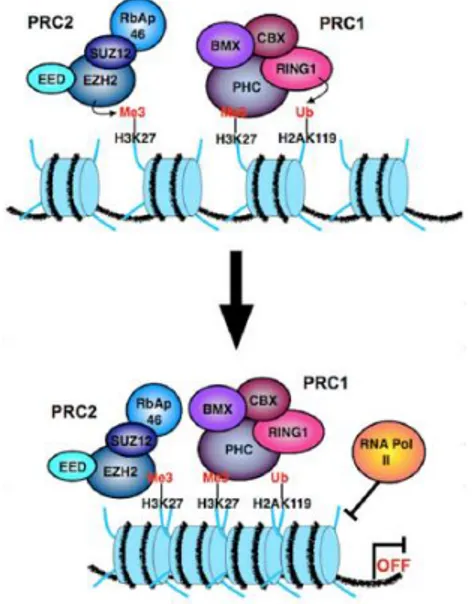
Finally, another important level of regulation for iPSC generation is the epigenome. Chromatin status and histone modifications are crucial in the regulation of transcription mechanisms. Currently, for the improvement of iPSCs generation and differentiation, it is very important to understand the epigenetic marks and mechanisms that are involved in the induction and maintenance of the pluripotent state, and if epigenetic memory can influence these processes. It is well known that cells of the early mammalian embryo, including pluripotent ESCs and primordial germ cells (PGCs), are epigenetically dynamic and heterogeneous. The histological analysis of the nuclei of stem cells, progenitors and differentiated progeny show that several cellular types, like neoblast cells in planaria and hematopoietic stem cells in mammals, are characterized by chromatin open state [196]. This particular state implements the transcription program and allows a rapid switch upon induction of differentiation [196]. During development and differentiation, regulation of gene transcription is governed at an epigenetic level by the balance between activating and repressing modifications, such as the trimethylation of lysine 4 of histone 3 (H3K4me3) and the trimethylation of lysine 27 of histone 3 (H3K27me3), respectively, in the nucleosomes of the chromatin [197]. H3K27 methylation is mediated by polycomb group (PcG) proteins [105] (Fig. 9). It is interesting to note that some targets of PcG proteins tend to be co-occupied by the TFs Nanog, Oct4 and Sox2 [198].
Figure 9:Polycomb group (PcG) proteins mediated H3K27 methylation. PRC2 induces EZH2-mediated
28
Another histone mark commonly associated with gene repression is histone 3 lysine methylation (H3K9). One enzyme associated with this mark is the euchromatic histone-lysine N-methyltransferase 2 (EHMT2), which is notably required to silence Oct4 during differentiation [183]. The low level of H3K9 methylation in undifferentiated ESCs is maintained by H3K9 histone demethylases (HDMs),jumonji domain-containing 1A (jMjD1A) and jumonji domain-containing 2C (jMjD2C). These enzymes regulate global levels of the repressive marks H3K9 and maintain the pluripotent state by directly demethylating H3K9 in the promoter regions of ESC TFs, allowing their expression. Interestingly, Oct4regulates the genes encoding jMjD1A and jMjD2C,which represents a positive feedback-loop that integrates the action of the TFs and histone modifiers to maintain the undifferentiated ESCs state [106].
The epigenetic control of the undifferentiated-differentiated state transition and the way that the epigenetic barriers are overcome are critical issues in the generation of iPSCs. At the present, molecular mechanisms that underlie epigenetic chromatin remodeling during reprogramming is still unclear, however, several proteins are known to regulate chromatin marks and are associated with the distinct epigenetic states of cells before and after reprogramming [197]. New insights have been gained by treating the cells during reprogramming with agents that promote the chromatin open state. For example, the DNMT inhibitor 5-aza-cytidine, the histone deacetylase (HDAC) inhibitor, valproic acid (VA), and EMHT2 inhibitor lead to increased efficiency of iPSC generation [172, 178] (Figure 10).
Figure 10: Model of inhibition of transcription directed by methylation of CpG islands in gene
29
Indeed, Huangfu et al. [172] demonstrated that iPSCs could be generated from primary human fibroblasts by transducing the cells only with Oct4 and Sox2 and adding VA in the culture medium. These results have opened the possibility to reprogram cells without c-Myc and Klf4. More recently, several epigenetic studies focus on enzymes that regulate this process. For example, Onder and collaborators, using shRNA approach, demonstrated that inhibition of the core components of polycomb repressive complex 1 and 2reduced reprogramming efficiency, whereas suppression of the H3K79 histone methyltransferase accelerates reprogramming and increase the yield of iPSC colonies [182]. These studies demonstrated that the knowledge of epigenetic mechanisms in order to act on the chromatin and histones status to be able to trigger the iPSC reprogramming process is crucial to improve reprogramming efficiency and to develop new strategies to avoid oncogenes employment.
Applications of iPSCs
iPSC technology can lead to several clinical applications. iPSCs and iPSC-derived differentiated cells can be useful to study disease biology, to model diseases in vitro and to develop new drugs, being a tool for both screening and toxicity tests. iPSCs, because of their versatility, can be differentiated into any of several cell types, which can be used as a model to study molecular mechanisms underlying disease development in a target cell type. Moreover, these cells can be studied in a developmental stage biologically relevant to phenotype analysis of diseases such as in trisomy 21 [201] or Alzheimer’s disease [202] studies, or in the observation of multistage oncogenesis, cellular transformation [203] or in hematological malignancies [204]. These models allow a better understanding of disease pathogenesis and the comparison between affected and healthy cells.
iPSCs can also be used as platform for drug discovery, affording advantage to the high cost of generating new drugs. For example, hepatotoxicity and cardiotoxicity are two principal causes of drug failure during preclinical testing. iPSCs could be differentiated into cell targets having a primary tissue-like phenotype and unlimited availability; then it could be possible to assess multi-parameter readouts of general and mechanism-specific hepatotoxicity or cardiotoxicity [205]. Moreover, the variability in individual responses to potential therapeutic agents is another problem in effective drug development. The use of human iPSCs would also allow the study of single nucleotide polymorphisms that influence the ability of an individual to metabolize and clear drugs and toxins. The accurate prediction of human drug toxicity is a vital element of the drug discovery process. iPSC technology allows the screening of a library of human cell lines that may represent


![Figure 4. Potential cell sources for hemophilia A treatment [79].](https://thumb-eu.123doks.com/thumbv2/123dokorg/4806856.49639/13.892.223.732.469.870/figure-potential-cell-sources-hemophilia-treatment.webp)

![Table 1. iPSCs derived from different species and somatic cell types[141].](https://thumb-eu.123doks.com/thumbv2/123dokorg/4806856.49639/19.892.107.818.109.1061/table-ipscs-derived-different-species-somatic-cell-types.webp)