Abstract.
–
OBJECTIVE: This review aimed at examining efficacy of interventional radiothera-py (brachytheraradiothera-py-IRT) alone or combined with external beam radiotherapy (EBRT) in stage I esophageal cancer as exclusive treatment.MATERIALS AND METHODS: A systematic research using PubMed, Scopus, and Cochrane library was performed. ClinicalTrials.gov was searched for ongoing or recently completed tri-als, and PROSPERO was searched for ongoing or recently completed systematic reviews. We analyzed only clinical study as full-text publica-tion, reporting on patients with stage I esopha-geal cancer treated with IRT alone or in combi-nation with other treatments (e.g., EBRT). Con-ference paper, survey, letter, editorial, book chapter, and review were excluded. Patients who underwent previous surgery were exclud-ed. Time restriction (1990-2018) was applied for years of the publication.
RESULTS: Twelve studies have been selected. The number of evaluated patients was 514; the median age was 69 years. In the IRT group, the median: local control (LC) was 77% (range 63%-100%), disease-free survival (DFS) was 68.4% (range 49%-86.3%), the overall survival (OS) was
60% (range 31%-84%), the cancer specific sur-vival (CSS) was 80% (range 55-100%), and grade 3-4 toxicity range was 0%-26%.
CONCLUSIONS: IRT alone or combined to EBRT is an effective and safe treatment option for patients with stage I esophageal cancer. De-finitive radiation therapy could be an alternative to surgery in patients with superficial cancer.
Key Words:
Interventional radiotherapy, Brachytherapy, Esoph-ageal cancer.
Introduction
Advances in endoscopic techniques have led
to an increase in early diagnoses of esophageal
cancer: it is nowadays possible to observe tumors
located within the mucosa or submucosa only
1-3.
The distinction among the aforementioned layers
is relevant as it modifies the indication of surgery.
There is a different risk of lymph nodes metastases
(0-6%) in the presence of mucosal involvement or
European Review for Medical and Pharmacological Sciences
2020; 24: 7589-7597
V. LANCELLOTTA
1, F. CELLINI
1, B. FIONDA
1, V. DE SANCTIS
2, C. VIDALI
3,
V. FUSCO
4, F. FRASSINE
5, D. TOMASINI
5, A. VAVASSORI
6, M.A. GAMBACORTA
1,7,
P. FRANCO
8, D. GENOVESI
9, R. CORVÒ
10, L. TAGLIAFERRI
11
UOC Radioterapia Oncologica, Dipartimento di Diagnostica per Immagini, Radioterapia
Oncologica ed Ematologia, Fondazione Policlinico Universitario A. Gemelli IRCCS, Rome, Italy
2Department of Radiation Oncology, Faculty of Medicina e Psicologia, Sant’Andrea Hospital,
University of Rome “La Sapienza”, Rome, Italy
3
Department of Radiation Oncology, Azienda Sanitaria Universitaria Integrata di Trieste (ASUITS),
Trieste, Italy
4
Department of Radiation Oncology, Centro di Riferimento Oncologico Regionale, Rionero in
Vulture, Potenza, Italy
5
Radiation Oncology Department, Ospedali Civili Hospital and Brescia University, Brescia, Italy
6Division of Radiation Oncology, European Institute of Oncology IRCCS, Milan, Italy
7
Università Cattolica del Sacro Cuore, Rome, Italy
8
Department of Oncology, Radiation Oncology, University of Turin, Turin, Italy
9
Department of Radiotherapy, “SS Annunziata” Hospital “G. D’Annunzio” University, Chieti, Italy
10Radiation Oncology, IRCCS Ospedale Policlinico San Martino and Department of Health Science,
University of Genoa, Genoa, Italy
The role of interventional radiotherapy
(brachytherapy) in stage I esophageal cancer:
an AIRO (Italian Association of Radiotherapy
and Clinical Oncology) systematic review
V. Lancellotta, F. Cellini, B. Fionda, V. De Sanctis, C. Vidali, et al.
in case the submucosa is also involved (38-53%)
4-9
. Usually, endoscopic resection is reserved for
patients with involvement of the lamina propria
mucosa, while esophagectomy, with lymph nodes
sampling, is indicated for patients with tumors
invading the muscolaris mucosa
4-9. Despite the
improvements of surgical techniques (e.g.:
robot-ic esophagectomy), some complrobot-ications are still
observed
10-12. Eligibility for surgery depends, not
only on the disease’s stage, but also on patient’s
performance status, age, comorbidities and
possi-bility to preserve the quality of life. Hence,
radio-therapy (RT) could replace surgery as a curative
treatment for patients with stage I disease since its
efficacy is comparable with surgery
13-15. Moreover,
interventional radiotherapy (IRT) also indicated as
brachytherapy (BT) may play an important role in
this scenario because of its ability to provide an
excellent dose distribution, short treatment time
and organ at risk preservation.
According to international guidelines, good
candidates for IRT include patients with tumors
<10 cm in length and confined to the esophagus
wall, those with thoracic locations, and patients
without regional node involvement
16.
However, there is still no standardization
among the different centers in terms of IRT
tech-nique used and prescribed dose. Thus, the role of
IRT on local control (LC) and overall survival
(OS) in patients with early esophagus cancer is
still controversial
17-22.
The aim of this review is to examine the
ef-ficacy of IRT after external beam radiotherapy
(EBRT) in stage I esophagus cancer in terms of
LC, disease-free survival (DFS), cancer-specific
survival (CSS), OS and safety.
The project was conceived and developed
with-in the frame of the Brachytherapy study group,
Interventional Radiotherapy, and intra-operative
radiotherapy (IORT) of the Italian Association
of Radiotherapy and Clinical Oncology (AIRO).
Material and Methods
A systematic research using PubMed,
Sco-pus, and Cochrane library was performed to
identify full articles evaluating the efficacy of
IRT in patients with stage I esophagus
can-cer. ClinicalTrials.gov was searched for ongoing
or recently completed trials, and PROSPERO
was searched for ongoing or recently completed
systematic reviews. The studies were identified
using the following medical subject headings
(MeSH) and keywords including: “esophageal
neoplasms”, “brachytherapy”, “intraluminal
ra-diotherapy”. The search was restricted to the
English language. The Medline search strategy
was: (“Brachytherapy” [Mesh] OR
“Brachythera-py” [All fields]) AND (“Esophageal Neoplasms”
[Mesh] OR “Esophageal Cancer” [All fields]).
To avoid missing relevant studies we chose this
strategy with high sensitivity but low specificity.
We analyzed only clinical study as full-text
publication, reporting on patients with stage I
esophagus cancer treated with IRT alone or in
combination with other treatments (e.g.,
exter-nal beam radiation therapy). Conference papers,
surveys, letters, editorials, book chapters, and
reviews were excluded. Patients who underwent
previous treatment were excluded. Regarding the
years of publication, time restriction (1990-2018)
was considered.
Three independent authors expert in
esopha-gus cancer with respect to IRT (VL – Rome, BF
– Rome) and EBRT (FC – Rome) screened
cita-tions in titles and abstracts to identify appropriate
papers. In addition, two radiation oncologists
of another institution performed an independent
check of the data (FF – Brescia, DT – Brescia).
Eligible citations were retrieved for full-text
re-view. Uncertainties about their inclusion in the
review were considered by a multicenter expert
team from 4 different institutions and involved
in the AIRO Interventional Radiotherapy study
group (VDS – Rome, VF – Rionero in Vulture,
CV – Trieste, AV – Milan). Finally, a committee
composed by the Chair of the
“Brachythera-py, Interventional Radiotherapy and IORT Study
Group” (LT), a member of AIRO committee
expert in Gastroenteric cancer (MAG), two
mem-bers of the scientific commission of AIRO expert
in Gastroenteric cancer (DG, PF), Chair of the
scientific commission of AIRO (RC) performed
a definitive check and the approval of the review.
The primary outcome was the LC after IRT
while the secondary outcomes included: DFS,
CSS, OS and the rate of adverse events rate.
A summary table was created, including
sam-ple size, median age, LC, DFS, CSS, OS and
Toxicity.
Results
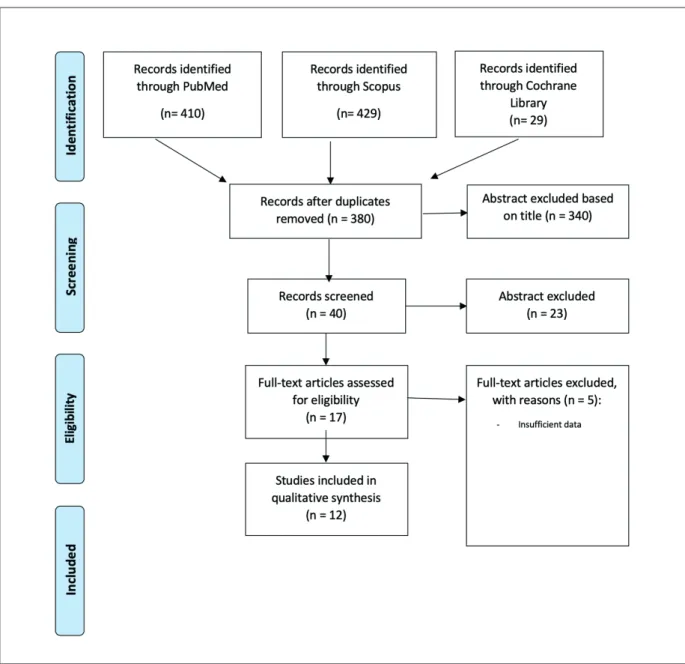
The literature search allowed us to retrieve
429 articles. After exclusion of papers (based on
abstracts) and after exclusion of conference
pa-Interventional radiotherapy in stage I esophageal cancer
pers, surveys, letters, editorials, book chapters,
reviews, and papers not using the English
lan-guage, 17 papers were included. Among these,
5 articles were excluded because of insufficient
data, leaving 12 studies assessing the clinical
efficacy of IRT in DFS and LC, as reported in
Figure 1.
All studies were retrospective and
monocen-tric
15,22-32. In accordance with the selection
crite-ria, only data from the IRT and EBRT treatment
arms were extracted and considered for the
anal-yses. The global number of evaluated patients
was 514, and the median age was 69 years. The
median LC was 77% (range 63-100%), DFS was
68.4% (range 49-86.3%), the OS was 60% (range
31-84%), the CSS was 80% (range 55-100%), and
the grade 3-4 toxicity range was 0%-26%.
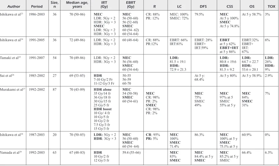
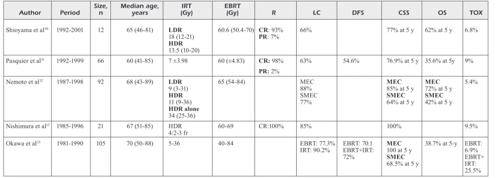
Table I lists the characteristics of the included
studies.
Discussion
The present systematic review of 12 studies
showed that IRT, in combination with EBRT in
early esophagus cancer patients, is comparable
in terms of outcome to surgery. Although
ra-diotherapy has shown favourable outcomes for
Table I. Characteristics of the included studies.
Size, Median age, IRT EBRT
Author Period n years (Gy) (Gy) R LC DFS CSS OS TOX
Ishikawa et al23 1986-2003 36 70 (50-86) MEC MEC CR: 88% MEC: 100% 79.5% MEC At 5 y 58.7% 3%
LDR: 5Gy × 2 56 (50-60) PR: 12% SMEC: 72% At 5 y 100%
HDR: 3Gy × 3 56 (52-60) SMEC
SMEC SMEC At 5 y 74.8%
LDR: 5Gy × 2 60 (54–62) HDR: 3Gy × 3 60 (54-64)
Ishikawa et al24 1991-2005 36 72 (49-86) LDR: 5Gy × 2 60 (48-64) CR: 88% EBRT: 66% EBRT: 28% EBRT EBRT:32% 6%
HDR: 3Gy × 3 PR:12% IRT:81% EBRT+ at 5 y 62% EBRT+
IRT:59% EBRT+IRT IRT:
at 5 y 86% 67%
Tamaki et al25 1991-2007 54 70 (49-86) LDR: 5Gy × 2 MEC LDR: LDR: LDR: LDR:
HDR: 3Gy × 3 56 (56-60) 81.5 ± 19.1 80.8 ± 19.6 64.7 ± 22.7 26% SMEC HDR: HDR: HDR: HDR: 60 (54-64) 72.9 ± 21.3 81.5 ± 9.2 53.6 ± 20.1 9% Sai et al26 1985-2002 27 69 (53-85) HDR 50-55 At 5 y: At 5 y 80% At 5 y 58.9% 2.9% 7-10 Gy/2 Fr 56-59 68.4% 11-12 Gy/3 Fr 60-70
Murakami et al27 1992-2002 87 70 (43-89) HDR alone MEC
35 Gy/14 fr 54 (50-58) MEC MEC MEC MEC 7%
36 Gy/18 fr SMEC CR: 98% 75% 97% at 5 84%
30 Gy/15 fr 60 (54-61) PR: 2% SMEC SMEC SMEC
25 Gy/5 fr SMEC 49% 55% at 5 y 31% HDR boost CR: 98% 10 Gy/ 4 fr PR: 2% 10 Gy/5 fr 10 Gy/2 fr 7.5 Gy/3 fr 15 Gy/3 fr
Ishikawa et al28 1987-2003 20 70 (50-85) LDR: 5Gy × 2 MEC CR: 95% MEC 86.3% MEC 60.9% 0%
HDR: 3Gy × 3 56 (50-60) PR: 5% 100% 100% at 5 y
SMEC SMEC SMEC
60 (54–64) 71.4% 75.1% at 5 y
Yamada et al29 1992-2003 63 67 (48-83) HDR 59.4 (55-66) MEC MEC MEC 66.4% 6.3%
10 Gy/2 fr 83% 84.4% at 5 y 85.2% at 5 y
12 Gy/3 fr SMEC SMEC SMEC
Table I (Continued). Characteristics of the included studies.
Size, Median age, IRT EBRT
Author Period n years (Gy) (Gy) R LC DFS CSS OS TOX
Shioyama et al30 1992-2001 12 65 (46-81) LDR 60.6 (50.4-70) CR: 93% 66% 77% at 5 y 62% at 5 y 6.8% 18 (12-21) PR: 7% HDR 13.5 (10-20) Pasquier et al31 1992-1999 66 60 (41-85) 7 ±3.98 60 (±4.83) CR: 98% 63% 54.6% 76.9% at 5 y 35.6% at 5y 9% PR: 2%
Nemoto et al32 1987-1998 92 68 (43-89) LDR 65 (54-84) MEC MEC MEC 5.4%
9 (3-31) 88% 85% at 5 y 72% at 5 y
HDR SMEC SMEC SMEC
11 (9-36) 77% 64% at 5 y 42% at 5 y
HDR alone
34 (25-36)
Nishimura et al15 1985-1996 21 67 (51-85) HDR 60-69 CR:100% 85% 100% 9.5%
4/2-3 fr
Okawa et al33 1981-1990 105 70 (50-88) 5-36 40-84 EBRT: 77.3% EBRT: 70.1 MEC 38.7% at 5-y EBRT:
IRT: 90.2% EBRT+IRT: 100 at 5 y 6.9%
72% SMEC EBRT+
68.5% at 5 y IRT:
25.5%
Abbreviation: IRT: interventional radiotherapy; EBRT: external beam radiotherapy; Gy: gray; MEC: mucosal esophageal cancer; SMEC, submucosal esophageal cancer; DFS:
disease free survival; LC: local control; OS: overall survival; CSS: cancer specific survival; TOX: toxicity; R: response to treatment; CR: complete remission; PR: partial remission; fr: fractions; y: years; HDR: high-dose rate; LDR: low-dose rate.
V. Lancellotta, F. Cellini, B. Fionda, V. De Sanctis, C. Vidali, et al.
early esophageal cancer, the treatment has not
been standardized according to the published
reports
13,15,33,34.
Surgical resection is the primary treatment for
stage I esophagus cancer with a 5-year OS rate
of 100% for cases with mucosal involvement and
65-90% for patients with submucosal cancer
4,34,35.
The present review showed a median 5-year OS
rate of 60% (range 31%-84%). The 5-year OS rate
for presentation limited to mucosal involvement
only is 84%
27. Therefore, a comparison of
treat-ment methods should be carried out separately
for mucosal and submucosal cancers, because
the depth of invasion is one of the most
im-portant prognostic factors for the choice of the
treatment
36-40. The discrepancy in 5-years OS
between surgery and radiotherapy could be due
to patient’s features.
In fact, patients who underwent exclusive
ra-diotherapy for early esophageal cancer usually
presented a higher rate of comorbidities
com-pared to those who underwent surgery, such as
cardiac or pulmonary disease, which may explain
the reduction of OS rates in the non-surgical
group. Probably, if radiotherapy were used as an
exclusive treatment in early esophagus cancer
in patients without several comorbidities, the
5-years OS rate might be improved. To endorse
this consideration, the present review has shown
that median 5-year CSS rate was 80% (range
55-100%). These values are better than median
5-years OS and are comparable to those obtained
with surgery.
A particularly controversial issue is whether
the use of IRT improves outcomes of early
esoph-agus cancer patients. IRT may be characterized
by certain level of variability due to
institution-al-based approaches, different dose fractionation
schedules and equipment availability, potentially
limiting to define the effect of the IRT after EBRT.
In the present review, only 5 studies reported an
improvement of LC and CSS
15,23,24,28,33. Okawa
treated with EBRT alone 58 (55%) patients and
both EBRT and IRT 47 patients (45%) with stage
I esophagus cancer. The authors showed that LC
rate in patients treated with radiation therapy was
excellent, especially in the group treated with
EBRT and IRT (EBRT: 77.3% vs. EBRT+IRT:
90.2%)
33. Nishimura and Ishikawa demonstrated
that both LC and CSS after EBRT+IRT were
superior to those after EBRT alone
15,23,24,28. On
the other hand, 7 out of 12 papers showed that
delivering IRT after EBRT doesn’t improve LC
and CSS
25-27,29-32. The retrospective setting of the
evaluated papers probably limits the strength of
the evidence: new prospective trials will
hopeful-ly provide more clear data. The risk to develop
high toxicity is correlated to IRT dose per
frac-tion that should not exceed 5Gy, particularly to
prevent esophageal ulcers
32-36. The present review
reported an acceptable G3-G4 late toxicity rate
(range 0-26%).
Despite these positive results, IRT is not
rou-tinely considered as a treatment option in patients
with early-stage esophageal cancer.
Suntharal-ingam et al
37reported that IRT was
implement-ed in only 6% of cases and two recent
sur-veys confirmed that 17.5% of the Italian centers
considered it for the treatment of esophageal
cancer
38,39. Probably the lack of experience, the
inadequate educational level in this field and
the complexity of such treatment, that requires
a multidisciplinary and multi-professional team,
do not permit the widespread use of IRT in
clin-ical routine
40. Having regard to the rarity of this
disease, discussion of clinical cases in expert
multidisciplinary team may provide more
homo-geneous treatment approaches and improvement
of clinical outcomes
41-48. The presented results
also emphasize the need to combine analysis of
treatment results from different centres in order
to create predictive models to define a
“personal-ized medicine”
49-52.
Conclusions
We provided support that EBRT+IRT is an
effective and safe treatment option for patients
with stage I esophagus cancer. Definitive
radi-ation therapy could be an alternative to surgery
in patients with superficial cancer. Further
ran-domized controlled studies should investigate the
optimal radiation dose and number of fractions to
obtain the highest outcomes rates and the lowest
risk of severe adverse events.
Conflict of Interest
The Authors declare that they have no conflict of interests.
References
1) Mori M, AdAchi Y, MAtsushiMA t, MAtsudA h, KuwA -no h, sugiMAchi K. Lugol staining pattern and his-tology of esophageal lesions. Am J Gastroenterol 1993; 88: 701-705.
Interventional radiotherapy in stage I esophageal cancer
2) tAKubo K, AidA J, sAwAbe M, KurosuMi M, AriMA M,FuJishiro M, ArAi t. Early squamous cell carcino-ma of the oesophagus: The Japanese viewpoint. Histopathology 2007; 51: 733-742.
3) inoue h, hondA t, nAgAi K, KAwAno t, Yoshino K, tAKeshitA K, endo M. Ultrahigh magnification en-doscopic observation of carcinoma in situ of the esophagus. Dig Endosc 1997; 9: 16-18.
4) KodAMA M, KAKegAwA t. Treatment of superficial cancer of the esophagus: a summary of respons-es to a qurespons-estionnaire on superficial cancer of the esophagus in Japan. Surgery 1998; 123: 432-439. 5) endo M, Yoshino K, KAwAno t, nAgAi K, inoue h.
Clinicopathologic analysis of lymph node metas-tasis in surgically resectedsuperficial cancer of the thoracic esophagus. Dis Esophagus 2000; 13: 125-129.
6) bollschweiler e, bAldus se, schröder w, Prenzel K, gutschow c, schneider PM, hölscher Ah. High rate of lymph-node metastasis in submucosal esoph-ageal squamous-cell carcinomas and adenocar-cinomas. Endoscopy 2006; 38: 149-156.
7) MAKuuchi h, shiMAdA h, MizutAni K, chino o, nishi t, tAnAKA h, MAchiMurA t, MitoMi t, osAMurA Y. Clini-cal pathologiClini-cal analysis of surgiClini-cally resected superficial esophageal carcinoma to determine criteria for deciding on treatment strategy. Diagn Ther Endosc 1997; 3: 211-220.
8) nishiMAKi t, tAnAKA o, suzuKi t, AizAwA K, wAtAnAbe h, Muto t. Tumor spread in superficial esophageal cancer: histopathologic basis for rational surgical treatment. World J Surg 1993; 17: 766-772. 9) tAchibAnA M, YoshiMurA h, KinugAsA s, hAshiMoto n,
dhAr dK, Abe s, Monden n, nAgAsue n. Clinico-pathological features of superficial squamous cell carcinoma of the esophagus. Am J Surg 1997; 174: 49-53.
10) MosKovitz Ah, rizK nP, venKAtrAMAn e, bAins Ms, Flores rM, PArK bJ, rusch vw. Mortality increases for octogenarians undergoing esophagogastrec-tomy for esophageal cancer. Ann Thorac Surg 2006; 82: 2031-2036.
11) KinugAsA s, tAchibAnA M, YoshiMurA h, dhAr dK, shibAKitA M, ohno s, KubotA h, MAsunAgA r, nAg -Asue n. Esophageal resection in elderly esopha-geal carcinoma patients: improvement in postop-erative complications. Ann Thorac Surg 2001; 71: 414-418.
12) Ferguson MK, MArtin tr, reeder lb, olAK J. Mor-tality after esophagectomy: risk factor analysis. World J Surg 1997; 21: 599-603.
13) oKAwA t, doKiYA t, nishio M, hishiKAwA Y, Mori -tA K. Multi-institutional randomized trial of exter-nal radiotherapy with and without intralumiexter-nal brachytherapy for esophageal cancer in Japan. Japanese Society of Therapeutic Radiology and Oncology (JASTRO) Study Group. Int J Radiat Oncol Biol Phys 1999; 45: 623-628.
14) MurAKAMi M, KurodA Y, nAKAJiMA t, oKAMoto Y, MizowAKi t, KusuMi F, hAJiro K, nishiMurA s, MAtsusue s, tAKedA h. Comparison between chemoradiation
protocol intended for organ preservation and con-ventional surgery for clinical T1-T2 esophageal carcinoma. Int J Radiat Oncol Biol Phys 1999; 45: 277-284.
15) nishiMurA Y, oKuno Y, ono K, MitsuMori M, nA -gAtA Y, hirAoKA M. External beam radiation ther-apy with or without high-dose-rate intraluminal brachytherapy for patients with superficial esoph-ageal carcinoma. Cancer 1999; 86: 220-228. 16) gAsPAr le, nAg s, hersKovic A, MAntrAvAdi r, sPeis
-er b. American brachytherapy society consensus guidelines for brachytherapy of esophageal can-cer. Int J Radiat Oncol Biol Phys 1997; 38: 127-132.
17) KAto h, tAchiMori Y, wAtAnAbe h, YAMAguchi h, ishi -KAwA t, itAbAshi M. Superficial esophageal carci-noma. Surgical treatment and the results. Cancer 1990; 66: 2319-2323.
18) hölscher Ah, bollschweiler e, schneider PM, siewert Jr. Prognosis of early esophageal cancer: com-parison between adeno- and squamous cell car-cinoma. Cancer 1995; 76: 178-186.
19) sugiMAchi K, wAtAnAbe M, sAdAnAgA n, iKebe M, Kit -AMurA K, Mori M, KuwAno h. Recent advances in the diagnosis and surgical treatment of patients with carcinoma of the esophagus. J Am Coll Surg 1994; 178: 363-368.
20) cellini F, MorgAnti Ag, di MAtteo FM, MAttiucci gc, vAlentini v. Clinical management of gastroesoph-ageal junction tumors: past and recent evidences for the role of radiotherapy in the multidisciplinary approach. Radiat Oncol 2014; 9: 45.
21) cellini F, vAlentini v. Targeted therapies in combi-nation with radiotherapy in oesophageal and gas-troesophageal carcinoma. Curr Med Chem 2014; 21: 990-1004.
22) cellini F, rAMellA s, ciresA M, PorziellA v, MeAcci e, Fiore M, trodellA l, d’Angelillo r. Role of induc-tion therapy in esophageal cancer. Rays 2005; 30: 329-33.
23) ishiKAwA h, sAKurAi h, tAMAKi Y, nonAKA t, YAMAKAwA M, sAito Y, KitAMoto Y, higuchi K, hAsegAwA M, nA -KAno t. Radiation therapy alone for stage I (UICC T1N0M0) squamous cell carcinoma of the esoph-agus: indications for surgery or combined chemo-radiotherapy. J Gastroenterol Hepatol 2006; 21: 1290-1296.
24) ishiKAwA h, nonAKA t, sAKurAi h, tAMAKi Y, KitAMo -to Y, ebArA t, shioYA M, nodA se, shirAi K, suzuKi Y, tAKAhAshi t, nAKAno t. Usefulness of intraluminal brachytherapy combined with external beam radi-ation therapy for submucosal esophageal cancer: long-term follow-up results. Int J Radiat Oncol Bi-ol Phys 2010; 76: 452-459.
25) tAMAKi t, ishiKAwA h, tAKAhAshi t, tAMAKi Y, KitAMo -to Y, oKAMoto M, nodA se, KAtoh h, shirAi K, sAKu -rAi h, nAKAno t. Comparison of efficacy and safe-ty of low-dose-rate vs. high-dose-rate intralumi-nal brachytherapy boost in patients with superfi-cial esophageal cancer. Brachytherapy 2012; 11: 130-136.
V. Lancellotta, F. Cellini, B. Fionda, V. De Sanctis, C. Vidali, et al.
26) sAi h, MitsuMori M, ArAKi n, MizowAKi t, nAgAtA Y,nishiMurA Y, hirAoKA M. Long-term results of de-finitive radiotherapy for stage I esophageal can-cer. Int J Radiat Oncol Biol Phys 2005; 62: 1339-1344.
27) MurAKAMi Y, nAgAtA Y, nishibuchi i, KiMurA t, KenJo M, KAneYAsu Y, oKAbe t, hAshiMoto Y, AKAgi Y. Long-term outcomes of intraluminal brachytherapy in combination with external beam radiotherapy for superficial esophageal cancer. Int J Clin Oncol 2012; 17: 263-271.
28) ishiKAwA h, sAKurAi h, YAMAKAwA M, sAito Y, nAKAYA -MA Y, KitAMoto Y, oKAMoto M, hArAdA K, hAsegAwA M, nAKAno t. Clinical outcomes and prognostic factors for patients with early esophageal squa-mous cell carcinoma treated with definitive radia-tion therapy alone. J Clin Gastroenterol 2005; 39: 495-500.
29) YAMAdA K, MurAKAMi M, oKAMoto Y, oKuno Y, nAKA -JiMA t, KusuMi F, tAKAKuwA h, MAtsusue s. Treatment results of chemoradiotherapy for clinical stage I (T1N0M0) esophageal carcinoma. Int J Radiat Oncol Biol Phys 2006; 64: 1106-1111.
30) shioYAMA Y, nAKAMurA K, sAsAKi t, oogA s, urAshiMA Y, KiMurA M, uehArA s, terAshiMA h, hondA h. Clini-cal results of radiation therapy for stage I esopha-geal cancer: a single institutional experience. Am J Clin Oncol 2005; 28: 75-80.
31) PAsquier d, MirAbel X, Adenis A, rezvoY n, hecquet g, Fournier c, coche-dequeAnt b, Prevost b, cAs -telAin b, lArtigAu e. External beam radiation ther-apy followed by high-dose-rate brachytherther-apy for inoperable superficial esophageal carcino-ma. Int J Radiat Oncol Biol Phys 2006; 65: 1456-1461.
32) neMoto K, YAMAdA s, hAreYAMA M, nAgAKurA h, hi -roKAwA Y. Radiation therapy for superficial esoph-ageal cancer: a comparison of radiotherapy meth-ods. Int J Radiat Oncol Biol Phys 2001; 50: 639-644.
33) oKAwA t, tAnAKA M, KitA-oKAwA M, nishio M, KiKu -chi Y, shirAto h, YAMAdA s, AsAKAwA h, niibe h, do -KiYA t. Superficial esophageal cancer: multicenter analysis of results of definitive radiation therapy in Japan. Radiology 1995; 196: 271-274.
34) isono K, sAto h, nAKAYAMA K. Results of a nation-wide study on the three-field lymph node dissec-tion of esophageal cancer. Oncology 1991; 48: 411-420.
35) KAto h, tAchiMori Y, wAtAnAbe h, YAMAguchi h, ishi -KAwA t, itAbAshi M. Superficial esophageal carci-noma. Surgical treatment and the results. Cancer 1990; 66: 2319 -2323.
36) AKAgi Y, hiroKAwA Y, KAgeMoto M, MAtsuurA K, ito A, FuJitA K, KenJo M, Kiriu h, ito K. Optimum fractionation for high-dose-rate endoesophageal brachytherapy following external irradiation of early stage esophageal cancer. Int J Radiat On-col Biol Phys 1999; 43: 525-530.
37) sunthArAlingAM M, MoughAn J, coiA lr, KrAsnA MJ, KAchnic l, hAller dg, willett cg, John MJ, MinsKY
bd, owen Jb. 1996-1999 Patterns of Care Study. The national practice for patients receiving radia-tion therapy for carcinoma of the esophagus: re-sults of the 1996-1999 Patterns of Care Study. Int J Radiat Oncol Biol Phys 2003; 56: 981-987. 38) neMoto K, MAtsuMoto Y, YAMAKAwA M, Jo s, ito Y,
oguchi M, KoKubo n, nishiMurA Y, YAMAdA s, oKA -wA t. Treatment of superficial esophageal cancer by external radiation therapy alone: results of a multi-institutional experience. Int J Radiat Oncol Biol Phys 2000; 46: 921-925.
39) nAbeYA K, nAKAtA Y. Extent of resection and lymph-adenectomy in early squamous cell esophageal cancer. Dis Esophagus 1997; 10: 159-161. 40) tAgliAFerri l, vAvAssori l, lAncellottA v, de sAnc
-tis v, bArberA F, Fusco v, vidAli c, FiondA b, collo -cA g, gAMbAcortA MA, Aristei c, corvò r, MAgrini sM. Can brachytherapy be properly considered in the clinical practice? Trilogy project: the vision of the AIRO (Italian Association of Radiotherapy and Clinical Oncology) Interventional Radiothera-py study group. J Contemp BrachytheraRadiothera-py 2020; 12, 1: 84-90
41) MoreirA lF, KAMiKAwA Y, nAoMoto Y, hAisA M, or -itA K. Endoscopic mucosal resection for superfi-cial carcinoma and high-grade dysplasia of the esophagus. Surg Laparosc Endosc 1995; 5: 171-175.
42) FuKudA M, hirAtA K, nAtori h. Endoscopic ultraso-nography of the esophagus. World J Surg 2000; 24: 216 -226.
43) tAgliAFerri l, Kovács g, Aristei c, de sAnctis v, bAr -berA F, MorgAnti Ag, cAsà c, Pieters br, russi e, livi l, corvò r, giovAgnoni A, ricArdi u, vAlenti -ni v, MAgrini sM. Directors of the Italian Radiation Oncology Schools. Current state of intervention-al radiotherapy (brachytherapy) education in Ita-ly: results of the INTERACTS survey. J Contemp Brachytherapy 2019; 11: 48-53.
44) Autorino r, vicenzi l, tAgliAFerri l, soAtti c, KovAcs Pg, Aristei c. A national survey of AIRO (Italian Association of Radiation Oncology) brachyther-apy (Interventional Radiotherbrachyther-apy) study group. J Contemp Brachytherapy 2018; 10: 254-259. 45) Koëter M, vAn steenbergen ln, leMMens ve, rutten
hJ, rouKeMA JA, wiJnhoven bP, nieuwenhuiJzen gA. Hospital of diagnosis and probability to receive a curative treatment for oesophageal cancer. Eur J Surg Oncol 2014; 40: 1338-1345.
46) Kovács g, tAgliAFerri l, vAlentini v. Is an Interven-tional Oncology Center an advantage in the ser-vice of cancer patients or in the education? The Gemelli Hospital and INTERACTS experience. J Contemp Brachytherapy 2017; 9: 497-498. 47) MorgAnti Ag, PAsquArelli l, deodAto F, digesù c,
di FAlco c, dinAPoli n. Videoconferencing to en-hance the integration between clinical medicine and teaching: a feasibility study. Tumori 2008; 94: 822-829.
48) KovAcs g, tAgliAFerri l, lAncellotA v, KovAcs A, iez -zi r, gAMbAcortA MA. Interventional Oncology:
Interventional radiotherapy in stage I esophageal cancer
should interventional radiotherapy(brachythera-py) be integrated into modern treatment proce-dures? Turk J Oncol 2019; 34: 16-22.
49) tAgliAFerri l, budruKKAr A, lenKowicz J, cAMbeiro M, bussu F, guinot Jl, hildebrAndt g, JohAnsson b, MeYer Je, niehoFF P, rovirosA A, tAKácsi-nAgY z, boldrini l, dinAPoli n, lAnzotti v, dAMiAni A, gAt -tA r, FiondA b, lAncellottA v, soror t, Monge rM, vAlentini v, Kovács g. ENT COBRA Ontology: the covariates classification system proposed by the Head & Neck and Skin GEC-ESTRO Working Group for interdisciplinary standardized data col-lection in head and neck patient cohorts treated with interventional radiotherapy (brachytherapy). J Contemp Brachytherapy 2018; 10: 260-266. 50) tAgliAFerri l, gobitti c, collocA gF, boldrini l, FA
-rinA e, FurlAn c, PAiAr F, viAnello F, bAsso M, ceriz -zA l, MonAri F, siMontAcchi g, gAMbAcortA MA, len
-Kowicz J, dinAPoli n, lAnzotti v, MAzzArotto r, rus -si e, MAngoni M. A new standardized data collec-tion system for interdisciplinary thyroid cancer management: Thyroid COBRA. Eur J Intern Med 2018; 53: 73-78.
51) tAgliAFerri l, PAgliArA MM, MAsciocchi c, scuPolA A, AzArio l, griMAldi g, Autorino r, gAMbAcortA MA, lAricchiutA A, boldrini l, vAlentini v, blAsi MA. Nomogram for predicting radiation maculopathy in patients treated with Ruthenium-106 plaque brachytherapy for uveal melanoma. J Contemp Brachytherapy 2017; 9: 540-547.
52) Meldolesi e, soest Jv, Alitto Ar, Autorino r, dinAP -oli n, deKKer A, gAMbAcortA MA, gAttA r, tAgli -AFerri l, dAMiAni A, vAlentini v. VATE: VAlidation of high TEchnology based on large database analy-sis by learning machine. Colorectal Cancer 2014; 3: 435-450. Doi: 10.2217/crc.14.34.