Università degli Studi di Ferrara
DOTTORATO DI RICERCA IN"FARMACOLOGIA E ONCOLOGIA MOLECOLARE" CICLO XXVI
COORDINATORE Prof. ANTONIO CUNEO
Novel perspectives in the physio-pharmacological
regulation of opioid receptors.
Settore Scientifico Disciplinare BIO/14
Dottorand o
Tutore
Dott.ssa Calcagno Mariangela Prof. Morari Michele
Tutti abbiamo una ferita segreta per riscattare la quale combattiamo.
Table of content
List of abbreviations 4
Introduction 6
The opioid system 6
MOP receptors 8
DOP receptors 9
KOP receptors 11
NOP receptors 12
Opioids and dopamine system 13
Dopamine and DOP receptor transmission 14
Dopamine and N/OFQ-NOP receptor system 15
Targeting protein-protein interaction 16
RGS proteins 16
RGS4 as a new drug target 19
RGS4 inhibitors 21
Purpose 24
Materials and methods 25
Animals 25
Lesion of the DA system 25
Behavioral studies 26
Data presentation and statistical analysis 27
Drugs 27
Results 28
Part 1. Role of endogenous DA in the motor action exerted by NOP receptor ligands 28 Part 2. Influence of dopamine D2 receptors in the motor effects of DOP receptor ligand 35 Part 3. Targeting RGS4 as possible approach to restore motor deficits 37
Discussion 46
Concluding remarks 50
Acknowledgments 51
References 52
List of abbreviations
6-OHDA 6-hydroxydopamine
ANOVA analysis of variance
BG basal ganglia
BNTX 7-benzyldenenaltrexone
CCG Center of Chemical Genomics, University of Michigan
CNS central nervous system
CPu caudate putamen
DA dopamine
DAMGO [D-Ala2,N-MePhe4,Gly-ol]-enkephalin
DELT [D-Ala2, D-Glu4]-deltorphin
DOP δ-opioid peptide
DPDPE [D-Pen2,D-Pen5]-enkephalin
DYN dynorphin
EM endomorphin
ENK enkephalin
GABA γ-amino butyric acid
GAPs GTPase-accelerating proteins
GDP guanosine-5’ diphosphate
GEF guanine-nucleotide-exchange factor
GF-4 1-[1-Cyclooctylmethyl-5-(1-hydroxy-1-methyl-ethyl)-1,2,3,6-tetrahydro-pyridin-4-yl]-3-ethyl-1,3-dihydro-benzoimidazol-2-one GIRK G-protein inwardly rectifying potassium channel
GP globus pallidus
GPCR G-protein coupled receptor
GRK G-protein coupled receptor kinase
GTP guanosine-5’ triphosphate
GTPase guanosine triphosphatase
GP globus pallidus
HEK human embryonic kidney cells
i.c.v. intracerebroventricular
i.p. intraperitoneal
J-113397 1-[(3R,4R)-1-cyclooctylmethyl1-3-hydroxymethyl1-4-piperidyl]-3- ethyl-1,3-dihydro-2H-benzimidazol-2-one
KOP κ-opioid peptide
L-DOPA 3,4-dihydroxyphenylalanine
MOP μ-opioid peptide
MPTP 1-methyl-4-phenyl-1,2,3,6-tetrahydropyridine
N/OFQ nociceptin/orphanin FQ
NAcc nucleus accumbens
NOP nociceptin/orphanin FQ peptide
NTB naltriben NTI niltrindole PDYN pro-dynorphin PENK pro-enkephalin PD Parkinson’s disease PDZ PSD95/Dlg/Z0-1 domain POMC pro-opiomelanocortin
PPIs protein-protein interactions
PPIIs protein-protein interactions inhibitors
PTX pertussis toxin
RGS regulator of G-protein
RGS4 regulator of G-protein 4
RM repeated measures
SNc substantia nigra compacta
SNr substantia nigra reticulata
SNC-80 (+)-4-[(αR)-α-(2S,5R)-allyl-2,5-dimethyl-1-piperazinyl)-3-methoxy- benzyl]-N,N-diethylbenzamide TM trans-membrane Trap-101 1-[1-(Cyclooctylmethyl)-1,2,3,6-tetrahydro-5-(hydroxymethyl)-4- pyridinyl]-3-ethyl-1,3-dihydro-2H-benzimidazol-2-one hydrochloride UFP-101 [Nphe1,Arg14,Lys15]Nociceptin-NH2
UFP-512 H-Dmt-Tic-NH-CH(CH2-COOH)-Bid
Introduction
The opioid system
The term “opioid” was coined by Acheson referring to all compounds with a morphine-like action and distinct chemical structures ranging widely from alkaloids to peptides. The use of opioid analgesics has a long history, dating back over five millennia. Despite the flow of time and the energies of scientists around the world, however, the functions of opioid receptors in the brain is not well understood until these days.
The opioid receptor system plays a central role in pain control and is a key regulator of hedonic homeostasis, mood and wellbeing. It is now understood that morphine and other opioid drugs are not only involved in setting pain (nociceptive) threshold and in controlling nociceptive processing but also participate in modulation of gastrointestinal, endocrine and autonomic function, and play a possible role in cognition. In the past two decades, the refinement of pharmacological tools and the generation of genetic models have helped clarify the specific role of each opioid receptor subtypes in many aspects of opioid-related physio-pathology.
The existence of receptors for opiate drugs was proposed by Beckett and Casy in 1954 based on their studies on structure-activity relationships for antinociceptive activity (e.g. the ability to relieve pain) in a series of synthetic opiates. They proposed a unique opioid receptor that followed the lock and key mechanism to interact with opioids1, but there were several anomalies that did not fit the postulated structurally rigid receptor, which allowed the binding of diverse molecules with a morphine-like structure. As early as 1965, based on structure-activity analysis studies, Portoghese proposed the existence of more than one opioid receptor type or the possibility of multiple modes of interaction between ligands and opioid receptors2. Evidence for the existence of multiple opioid receptor subtypes arose from work identifying the different anatomical location and pharmacological profiles of compounds that were eventually used to name them, i.e. morphine (mu), ketocyclazocine (kappa)3 and vas deferens (delta)4. Recently, a fourth opioid-like receptor has been included in the opioid receptor family and has been termed opioid-like receptor 1 (ORL-1)5. This receptor was originally placed within the opioid family due to sequence and functional homologies, it produces downstream effects that are similar to those of the other three opioid receptors, but it does not interact with high affinity with classical opioid ligands. Receptor nomenclature has varied several times over the years and the most recent classification proposed by the International Union of Pharmacology (IUPHAR) is mu-opioid peptide (MOP) receptor, kappa-opioid
peptide (KOP) receptor, delta-opioid peptide (DOP) receptor and nociceptin/orphanin FQ (N/OFQ) peptide (NOP) receptor.
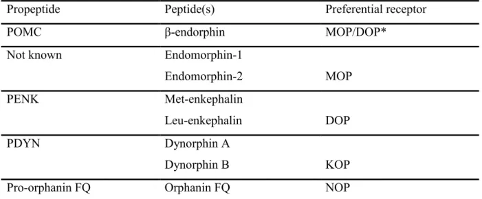
Opioid receptors are the endogenous targets of neuropeptides derived from three distinct protein precursors encoded by pro-opiomelanocortin (POMC), pro-enkephalin (PENK), and pro-dynorphin (PDYN) genes6. POMC encodes the peptide β-endorphin, which has agonist activity at both MOP and DOP receptors. Differently from the other endogenous opioids, the biosynthetic pathway of endomorphin-1 (EM-1) and endomorphin-2 (EM-2), which are both selective for the MOP receptor, is still unclear. The endogenous DOP receptor peptides are met-enkephalin (met-ENK) and leu-enkephalin (leu-ENK), cleaved from PENK. PDYN gives rise to the KOP receptor agonists dynorphin A (DYN-A) and dynorphin B (DYN-B)7,8, whilst N/OFQ is from the polypeptide precursor pro-N/OFQ (Tab. 1).
Propeptide Peptide(s) Preferential receptor
POMC β-endorphin MOP/DOP*
Not known Endomorphin-1
Endomorphin-2 MOP
PENK Met-enkephalin
Leu-enkephalin DOP
PDYN Dynorphin A
Dynorphin B KOP
Pro-orphanin FQ Orphanin FQ NOP
Table 1. Brain opioid peptides, their precursor molecules and preferential binding sites. * β-endorphin binds with rather similar affinity to both MOP and DOP receptor9.
Each type of opioid receptors is differentially localized in the central nervous system (CNS), and the striatum has the highest levels of endogenous opioid peptides and receptors8,10.
Opioid receptors are G-protein coupled receptors (GPCR), and share a similar seven transmembrane structure, the highest homology occurring in the trans-membrane (TM) domains, intracellular loops and C-terminus6. However, it is surprising that all the cloning studies pointed just to three different gene products when the literature at the time suggested the existence of three MOP (µ1, µ2, µ3), two DOP (δ1 and δ2), and three KOP (κ1, κ2, κ3) receptor subtypes, based on distinct pharmacological properties.
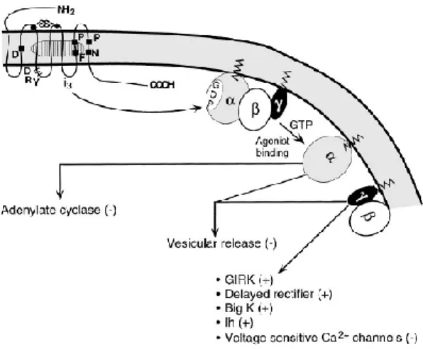
Opioids have predominantly direct inhibitory effects on cells in the CNS, which are mediated through the activation of pertussis toxin (PTX) sensitive Gi/0-proteins. This leads to inhibition
K+ channels, leading to reduced excitability and inhibition of neurotransmitter release11 (Fig. 1). Despite these inhibitory effects at the cellular level, opioids have excitatory actions in multiple regions of the nervous system in vivo. Excitation by opioids is generally attributed to inhibition of inhibitory pathways (disinhibition). However, recent studies indicate that opioids can directly excite individual cells. These effects may occur when opioid receptors interact with other GPCR such as receptor tyrosine kinases12.
Figure 1. The best characterized pathway of effector activation by opioids. Three primary classes of effectors include the inhibition of adenyl cyclase, inhibition of vescicular release, and interactions with a number of ion channels. These effectors are affected by the GTP-bound form of α-subunit as well as by free β/γ-subunits of pertussis toxin-sensitive G protein. GIRK, G protein inwardly rectifying potassium channel.
MOP receptors
The MOP receptor was the last of the classical opioid receptors to be cloned13. It is located throughout the central nervous system in areas involved in sensory and motor function including regions involved in the integration and perception of the senses. The highest density of MOP receptors is found in the CPu (of the BG). High density is also found in the neocortex, thalamus, nucleus accumbens (NAcc), hippocampus and amygdala14. MOP receptors are located pre-synaptically on primary afferent neurons within the dorsal horn of the spinal cord where they inhibit glutamate release and hence transmission of nociceptive stimuli15. MOP receptors mediate positive reinforcement following direct (morphine) or indirect (alcohol, cannabinoids, nicotine) stimulants, and the understanding of MOP receptor function is central to the development of therapies for addiction.
The endogenous ligands for the MOP receptor are EM-1 and EM-216, and the finding that the analgesic and addictive properties of morphine are abolished in mice lacking the MOP receptor has unambiguously demonstrated that MOP receptors mediate both the therapeutic and the adverse activities of this compound17.
Based on binding and pharmacological studies, the existence of various subtypes of the MOP receptor has been postulated, but only one receptor has been cloned18,19. At least 10 variants have been identified, some of which express truncated forms of the receptor, or variations in the intracellular tip of the C-terminus of the receptor20,21. These have been defined using knockout mice, antisense mapping studies, and studies showing subtype differences in agonist affinity and analgesia.
MOP receptor agonists are known to stimulate locomotion when given at low doses, and sedation at higher ones22. As a matter of facts, selective MOP receptor stimulation in the substantia nigra reticulata (SNr) facilitates spontaneous locomotion and turning behavior in rats23. The effect on locomotion can be correlated with the MOP receptor-dependent dopamine (DA) release in striatal areas24 and the control of GABAergic nigro-thalamic output neurons25.
Whilst the analgesic effect of opioids is elicited by central activation of opioid receptors, a number of the common side-effects, including reduced gastrointestinal motility, leading to constipation, urinary retention and pruritus, are regulated by activation of peripherally located opioid receptors. Major side-effects associated with MOP agonists include respiratory depression through a reduction in the sensitivity of central and peripheral chemoreceptors to hypercapnia. MOP opioids also have effects on the cardiovascular system, thermoregulation, hormone secretion and immune function.
Studies using MOP receptor knockout mice have defined the role that the MOP receptor plays tonically and when stimulated by exogenously applied ligands. MOP receptor knockout mice show increased sensitivity to thermal pain, implicating the receptor in this mode of nociception. However, no change in threshold of mechanic pain was seen26. None of the predicted effects or side-effects of morphine were seen in mice lacking the MOP receptor, suggesting that both the wanted and unwanted effects of morphine are attributable to action at the MOP receptor27.
DOP receptors
The DOP receptor was the first to be cloned28 and is less widely distributed compared to the other opioid receptors. Highest densities are found in the olfactory bulb, cerebral cortex, NAcc and CPu. DOP receptors are located mostly pre-synaptically on primary afferents where they inhibit the release of neurotransmitters.
The DOP receptor mediates the antinociceptive/analgesic actions of some opioids through both spinal and supraspinal sites. Indeed, DOP receptor agonists induced clear antinociceptive responses in several acute29 and chronic30 models of pain in rodents, although some of these
effects might be partially due to a cross-reactivity with MOP receptor31.
The DOP receptor has high affinity for leu/met-ENKs which are its endogenous ligands. Although only one DOP gene has been cloned thus far, recent evidence regarding the direct antinociceptive effects of DOP receptor agonists has suggested that at least two DOP subtypes are expressed: the putative δ1 subtype (DOP1) and the putative δ2 subtype (DOP2). The
putative DOP1 subtype is preferentially activated by [D-Pen2, D-Pen5] enkephalin (DPDPE) and antagonized by 7-benzylidenenaltrexone (BNTX), while the putative DOP2 subtype is preferentially activated by [D-Ala2, D-Glu4] deltorphin (DELT) and blocked by naltriben (NTB)32,33. This was also confirmed by the lack of cross-tolerance between DPDPE and DELT34. Several studies on the antinociceptive actions of combined DOP receptor subtype agonists and antagonists support these findings35-38. On the other hand, some of the pharmacological effects of DOP agonists may appear through partial activation of other opioid receptors39 or heterodimer forms of the DOP receptor40,41.
The generation of mice lacking either MOP or DOP receptors has allowed to revisit the selectivity of prototypical DOP receptor agonists under in vivo experimental conditions. In DOP knockout animals, the analgesia induced by the agonists DPDPE and deltorphin was either abolished, reduced, or maintained, depending on the nociceptive assay and route of administration42; the observation of residual activity of DOP receptor agonists in DOP receptor-deficient animals strongly supports the cross-reactivity of DOP at MOP receptors in vivo43. MOP and DOP receptors are usually considered to act similarly in most opioid-controlled behaviors. In fact, both MOP receptor agonists and DOP receptor agonists have been shown to reduce gastrointestinal tract motility and cause respiratory depression6. It was, therefore, surprisingly the finding of opposing phenotypes in DOP and MOP deficient mice in several behavioral models, when the two mutants strains were studied in parallel (e.g., DOP receptor knockout mice displayed anxiogenic and depressive-like responses 44, while MOP receptor mutants showed opposite responses).
A great deal of data suggests a significant but complex role of DOP receptor in the regulation of motor activity; the DOP receptor is strongly expressed in the striatum, and ENKs act as co-transmitters in striatal GABAergic neurons projecting to globus pallidus (GP)45,46. Here, they inhibit GABA release from striato-pallidal terminals47, thereby opposing the inhibitory post-synaptic influence produced by striato-pallidal neurons48. This facet of DOP receptor function is of potential interest in diseases involving impaired motor control such as Parkinson’s disease (PD). Indeed, striatopallidal neurons become pathogenically overactive following DA depletion, leading to overinhibition of pallido-subthalamic GABAergic and disinhibition of subthalamo-nigral glutamatergic projections49. Consequently, PENK-A expression in
striatopallidal neurons rises50,51, possibly to attenuate exaggerated GABA release and compensate for motor deficit47. Consistently, the DOP agonist SNC-80 reversed akinesia and showed locomotor-stimulating properties in reserpinized or haloperidol-treated rats as well as MPTP-treated marmosets40,30,47,52-55. In 2009, Mabrouk and collegues56 showed that the DOP receptor agonist UFP-512 increased locomotor coordination in a hemiparkinsonian rat model at low doses and had opposite effects at higher ones. Also, the DOP receptor antagonist NTI diminished abnormal movements classically described in the 6-OHDA model57. More recently, the most recently developed DOP receptor agonists do not show locomotor-activating properties54,55,58-60. Therefore, agonist-biased activity for different DOP receptor agonists may occur.
KOP receptors
The KOP receptor was the second of the opioid receptor family to be cloned61. Its endogenous ligand is DYN A and the prototypical exogenous agonist at KOP receptor is the non-peptide benzomorphan ketocyclazocine, the actions of which have been shown to be distinct from those elicited by stimulation of the MOP receptor (e.g. sedation without marked effects on heart rate). The side-effects elicited by KOP receptor agonists have, to date, limited their clinical use. However, it has been shown that KOP agonists (e.g. enadoline) may have neuroprotective actions via their ability to inhibit post-ischemic glutamate release62. Enadoline has also been reported to increase locomotion in monoamine-depleted rats63; the same group demonstrated a synergistic effect between enadoline and L-DOPA, suggesting that KOP receptor agonists might be used as adjuncts to L-DOPA therapy in PD. However, clinical studies using the KOP receptor agonist spiradoline (U-62066) failed to prove its efficacy alone or in combination with L-DOPA in the treatment of PD64. Indeed, KOP receptors are localized in the rat SNr but are not detectable in SNc65, placing this peptide receptor system in a strategic position to modulate the output of BG and motor function. Nonetheless, KOP receptor agonists have been shown to produce contrasting effects compared to MOP and DOP receptor agonists in a number of behavioral paradigms. For instance, KOP receptor stimulation reduced locomotor activity in naïve mice while DOP66 and MOP22 stimulation enhanced it. Additionally, KOP receptor agonists have been shown to produce dysphoria in contrast to the euphoria brought about by MOP receptor agonists67. The advantage of KOP receptor agonists over MOP or DOP receptor agonists is that they do not cause respiratory depression. It should also be mentioned that KOP agonists also exert an anti-opioid action, attenuating analgesia elicited by endogenously released or exogenously administered MOP agonists.
NOP receptors
In 1995, the endogenous agonist for the NOP receptor was isolated by two independent groups. This neuropeptide was termed nociceptin by Meunier et al.68, and orphanin FQ by Reinscheid et al.5. It is now commonly referred to as nociceptin/orphanin FQ (N/OFQ). There is a significant similarity between the amino acid sequences of N/OFQ and those of classical opioid peptides; this resemblance is particularly stricking in the case of DYN-A. Also, at the cellular level, N/OFQ produces actions similar to those of classical opioids, resulting in reduced neuronal excitability and inhibition of transmitter release. Despite these analogies with the opioid system, N/OFQ does not bind with high affinity to the classic opioid receptors5. Accordingly, the N/OFQ system has evolved into a distinct and independent system69-71.
Initial studies concentrated on the role N/OFQ in pain. However, exogenous N/OFQ administration has also been shown to modulate locomotion, stress and anxiety, feeding72,73, learning and memory74-76, reward/addiction, and neuroendocrine response to stress77,78.
Under laboratory conditions, N/OFQ has been shown to have a pronociceptive, anti-analgesic effect when applied supraspinally, whilst causing analgesia when applied spinally. N/OFQ anti-opioid action is the most likely cause for the supraspinal pronociceptive effect. N/OFQ might inhibit either endogenous opioid tone or stress-induced analgesia produced during testing procedures in laboratory animals. It is believed that endogenous levels of N/OFQ may act to set threshold to pain, as NOP receptor antagonists have been shown to produce a long lasting analgesia of similar extent to that of morphine. Therefore, NOP receptor antagonists have been proposed as novel analgesics or adjuvants to classical therapies, to reduce the amount of opioid analgesics and the side-effects correlated.
The generation of useful research tools, particularly transgenic animals and selective receptor ligands (especially antagonists) facilitated the understanding of the complex biological roles played by N/OFQ. Knockout mice for the N/OFQ precursor (ppN/OFQ-/-)79 or the NOP receptor (NOP-/-)80 are available; more recently NOP-/- rats were also generated81.
NOP receptor knockout mice show a partial loss of tolerance to morphine, which is consistent with the up-regulation of N/OFQ production in chronic morphine tolerant mice82. Studies in knockout mice confirmed that morphine tolerance to analgesia, but not acute response to morphine, was markedly attenuated. This action has also been confirmed through the actions of potent selective NOP antagonists, which also attenuate morphine tolerance. These findings suggest the N/OFQ–NOP system contributes to neuronal plasticity involved in the development of tolerance seen with chronic morphine exposure.
Opioids and dopamine system
Previous reports have shown that the involvement of opioid receptors in behavioral or rewarding effects depends on the central DA system, although contradictory reports also exist. Indeed, there is a body of evidence indicating that the rewarding or motivational effects of exogenously administered opioids are purely secondary to activation of the mesolimbic DA system83-86, although the involvement of D1 versus D2 receptors is controversial86,87. On the other hand, the absolute requirement of an intact DA system for the opioid action has been challenged, and DA-independent mechanism of opiate reward has been proposed88-93.
Major depressive disorder was shown to be associated with a reduction in response to rewarding stimuli in the dopaminergic mesolimbic pathway in a recent neuroimaging study94. This neuronal network is modulated by opioids, typically by MOP and DOP receptors, at the level of DA neurons and afferent structures. Therefore, a deficit in endogenous opioids, mainly ENKs, in the NAcc and ventral tegmental area (VTA), may lead to a decrease in the neurobiological control of mood states and reward.
A plethora of studies supports the cross-talk between the opioid and the DA systems; for example, the conditioned place preference induced by morphine or heroin is attenuated by either pretreatment with DA receptor antagonist85,95 or 6-hydroxydopamine (6-OHDA) lesion of the NAcc, the terminal area of the mesolimbic DA projections85. Moreover, chronic administration of the DA D1 receptor antagonist SCH23390 during conditioning sessions also
attenuates both the morphine-induced place preference96-98 and the KOP receptor agonist induced place aversion96,97.
DOP and MOP receptors are highly concentrated in the striosomes, one of the functional subdivisions of the mosaic structure of the mammalian striatum99. Striosome compartment has been implicated in motor and behavioral brain functions100 and their disorders101-103. Striosomal opioid signaling has emerged as a potent regulator of striatal activity104, whereas its functional significance in the pathophysiology of movement disorders remains to be elucidated.
An enhancement of opioid transmission is thought to play a compensatory role in altered functions of the BG under the conditions of striatal DA depletion in PD8,105. However the precise mechanism by which the increased opioid signaling modulates the BG activity is still under debate. One of the most recognized models of functional organization of the BG indicates a key role for balance in the activity of the two major striatal output pathways, i.e., the direct and indirect pathways106.DA depletion is known to enhance opioid transmission in the striatum107; strikingly, this is found to occur in medium spiny neurons (MSNs) that form the indirect pathway. The level of expression of ENKs and PPE-A mRNA is increased in
striato-pallidal MSNs108-110, whereas that of DYN and PPE-B mRNA was unaltered or decreased in striato-nigral MSNs110,111. The pronounced upregulation of ENKs in striato-pallidal MSNs can cause a compensatory down-regulation of both MOP and DOP receptors in their target cells112. This is consistent with the finding that prolonged activation of MOP and DOP receptors by opioid ligands (i.e., ENKs) promotes their proteolytic degradation, a process that contributes to homeostatic regulation of cellular responsiveness to opioids113.
Dopamine and DOP receptor transmission
DOP receptor agonists have many stimulant-like properties in vivo. Indeed, it has been shown that DOP receptor agonists induce hyperlocomotion114 and place preference115. Despite these stimulant-like properties, the nonpeptidic DOP receptor agonist SNC-80 is not self-administred by monkeys37 and does not facilitate intracranial self-stimulation116. Consistent with these behavioral findings, systemic administration of SNC-80 fails to promote DA efflux directly from rat striatum117 and does not increase extracellular DA levels in the CPu or NAcc assessed by microdialysis118. In addition to direct stimulant-like actions of DOP receptor agonists there is considerable evidence that DOP receptor activity can influence the actions of psychomotor stimulants in a variety of behavioral paradigms. For example, blocking DOP receptors with the antagonist naltrindole (NTI) attenuates some of the behavioral effects of amphetamine and cocaine, suggesting that endogenous DOP receptors signaling may modulate stimulant activity119-122. In addition, DOP receptor activation with agonists such as SNC-80 and TAN-67 can enhance the discriminative effects of stimulants in rats and monkeys37,123-125, increase methamphetamine-induced injurious behavior126 and significantly enhance amphetamine and cocaine-stimulated locomotor activity117. Consistent with the latter, DOP receptor agonists augment amphetamine-stimulated DA release from rat striatal slices117. The mechanisms by which DOP receptor activation with endogenous or exogenous ligands influences psychostimulant function is unknown.
The disruption of DOP receptor function modifies learning and memory abilities in mice, suggesting a key role for DOP receptors in modulating hippocampal- and striatum-dependent behaviors, and further revealing potential neural substrates engaging DOP receptors in these processes 127. In the striatum DOP receptors are prominently expressed in cholinergic interneurons and inhibit their activity128-130.
A small proportion of these receptors is also detected in GABAergic interneurons130 or pre-synaptic glutamatergic terminals131. Consequently DOP receptors have multiple potential effects on striatal function. DOP receptor knockdown in cholinergic interneurons should facilitate their depolarization and subsequent acetylcholine release. Disinhibiting cholinergic
interneurons, however, was demonstrated to bias striatal networks towards increased striatopallidal activity132; removal of DOP receptor tone in GABAergic interneurons and/or glutamatergic terminals may, therefore, underlie facilitated striatal function. Up-regulation of ENK transmission along the striato-pallidal pathway is thought to play a crucial role in maintaining motor function under parkinsonian conditions. To support the view that such up-regulation is compensatory in nature47, DOP receptor agonists promoted movement and attenuated parkinsonian-like motor deficits in rodent and non-human primate models of PD47,52,133,134.
Finally, the notion that DOP receptors have neuroprotective activity is currently being examined135; indeed, deprivation of oxygen and blood supply induced neuronal death, and DOP opioid receptor activation seem to be beneficial in situations of ischemia or hipoxia136. In addition, in 1999 Borlongan and colleagues137 reported that DADLE could be used as a supplement factor for improving the cell viability of fetal mesencephalic cells and as a protective agent against neurotoxicity in a cell PD model.
Dopamine and N/OFQ-NOP receptor system
The widespread anatomic distribution of the N/OFQ-NOP receptor system68,138-143 suggests its involvement in a broad array of neurologic functions. N/OFQ and the NOP receptor are moderately to heavily expressed in DA-rich areas such as the VTA, substantia nigra pars compacta (SNc), and prefrontal cortex (PFC)143,144. N/OFQ is, therefore, in a position to influence DA neuronal activity and several studies have shown a functional interaction between these two systems. N/OFQergic transmission was found to be upregulated in the SNr
145, following DA receptor blockade or loss of nigral DA neurons (6-OHDA lesioning). At the
same time, loss of nigro-striatal DA inputs causes striatal N/OFQ expression to drop.
NOP receptor antagonists have been shown to increase motor performance in naïve and parkinsonian rats146-148 and mice147,149. Preliminary evidence has pointed to the involvement of mesencephalic DA neurons in the motor responses to NOP receptor ligands. Indeed, exogenous N/OFQ-induced hyperlocomotion was prevented by haloperidol150 or DA synthesis inhibitor151, whereas N/OFQ-induced hypolocomotion was accompanied by a inhibition of striatal DA release146. Likewise, hyperlocomotion induced by low J-113397 was accompanied by a facilitation of striatal DA release146 while hypolocomotion induced by high doses of J-113397 was reversed by the D2/D3 receptor antagonist amisulpride152. N/OFQ was shown to suppress striatal DA release from VTA153 and SNc146 as well as locomotor activity5,150,154,155. On the other hand, MOP receptor agonists stimulate DA release indirectly, through inhibition of intranigral156 or intra-VTA157 GABAergic interneurons.
Targeting protein- protein interaction
One of the most essential components of cellular processes are protein- protein interactions (PPIs). The binding between two or more proteins in a cell can have a wide array of effects: modulation or initiation of signal transduction, regulation of patterns of gene transcription, stabilization of cytoskeletal structures, and stimulation of cellular replication or death. The cellular network of PPIs could contain many potential sites for drug targeting. Indeed, in the past years, much effort has been focused on the identification of specific inhibitors of PPIs. Currently, there are a number of clinically relevant therapies that target PPI interfaces. Most currently used PPI inhibitors (PPIIs) in the clinic are based upon humanized monoclonal antibodies. While this class of therapeutics possesses some very desirable properties (e.g. high specificity, low toxicity), it also shows several limitations that make this approach less promising for the widespread development of PPIIs (e.g. lack of cell/blood-brain barrier permeability, poor oral bioavailability, high cost of manufacture).
The CNS is, in particular, ripe for pharmacological targeting of protein-protein interactions. This is due, in part, to the fact that the highly organized nature of CNS signal transduction relies heavily on localization and compartimentalization of signaling functions. Blocking the protein-protein interactions underlying this compartimentalization might provide more subtle tissue-specific therapeutic actions that would be achieved by blocking the signal pathway itself. Furthermore, highly specific neural transcriptional patterns of regulatory molecules, e.g. Regulators of G Protein Signaling (RGS) proteins, provide great opportunities for cell-type selective modulation of signaling.
RGS proteins
In the conventional model of GPCR activation, the interaction between the ligand and the 7-TM receptor catalyzes guanine nucleotide exchange on the Gβγ-complexed (and GDP-bound) Gα subunit. The α-GTP and the βγ of the heterodimer dissociate, and are thus free to propagate the signal forward via separate (and sometimes converging) interactions with adenyl cyclases, phospholipase-C isoforms, potassium and calcium ion channels, guanine-nucleotide exchange factors (GEFs) for the GTPase RhoA (“RhoGEFs”), and other effector enzymes. The Gα subunit resets the cycle by forming Gα-GDP which has low affinity for effectors but high affinity for Gβγ; thus, the inactive GDP-bound heterodimer is capable once again of interacting with the activated receptor. In this cycle, the duration of heteromeric G-protein signaling is controlled by the lifetime of the Gα subunit in its GTP-bound state.
The G protein pathways are regulated by a number of proteins including scaffolding proteins such as RGS proteins, G protein coupled receptor kinases (GRKs), and arrestins. These
proteins are critical for the proper temporal and spatial regulation of GPCR signaling, and allow for a more finely tuned GPCR signaling for therapeutic purposes.
The importance of RGS proteins in GPCR signal was first appreciated in studies on yeast (Saccharomyces cerevisiae)158 and nematode worms (Coenorhabditis elegans)159. The yeast RGS gene Sst2 was identified in the 1980s following an arrest in the G1 to S phase transition in the cell cycle during a genetic screen for mutants; similarly, the C. elegans RGS gene
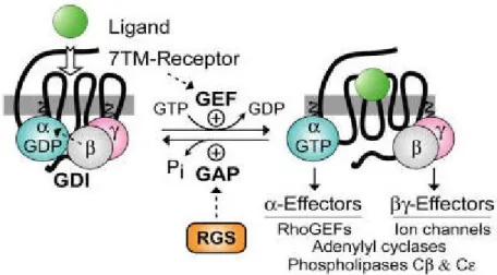
EGL-10 was identified in a genetic screen. Analysis of the polypeptide sequence of EGL-EGL-10 and Sst2159,160 revealed a shared region of ~120 amino acids that was also present in several mammalian proteins with previously unrecognized biochemical functions, for example, the T-cell activation immediate-early gene G0S8, now known as RGS2161. This ~120 amino acids region (the RGS domain or RGS-box), which is present in EGL-10, Sst2, RGS2 and other members of the RGS-protein superfamily, binds directly to the GTP-bound Gα subunit to markedly accelerate its rate of GTP hydrolysis and hence the rate of inactivation of GPCR signaling. Indeed, the RGS proteins are best known as GTPase-accelerating proteins (GAPs) for Gα subunits (Fig. 2). The discovery of these proteins resolved the timing paradox of how rapid regulation of GPCR signaling could occur given the slow rate of GTP hydrolysis by purified Gα subunits.
Figure 2. Standard model of the guanine nucleotide cycle governing 7TM receptor-mediated activation of heterotrimeric G protein-coupled signaling. The Gβγ heterodimer serves to couple Gα to the receptor and also to inhibit its spontaneous release of GDP (i.e., acting as a guanine nucleotide dissociation inhibitor or “GDI” for Gα162,163). Ligand-occupied, 7TM cell-surface receptors stimulate signal onset by acting as guanine nucleotide
exchange factors (GEFs) for Gα subunits, facilitating GDP release, subsequent binding of GTP, and release of the Gβγ dimer. Both the GTP-bound Gα and liberated Gβγ moieties are then able to modulate the activity of various enzymes, ion channels, and other effectors. Regulator of G-protein signaling (RGS) proteins stimulate signal termination by acting as GTPase-accelerating proteins (GAPs) for Gα, dramatically enhancing their intrinsic rate of GTP hydrolysis (taken from Siderovski and Willard164).
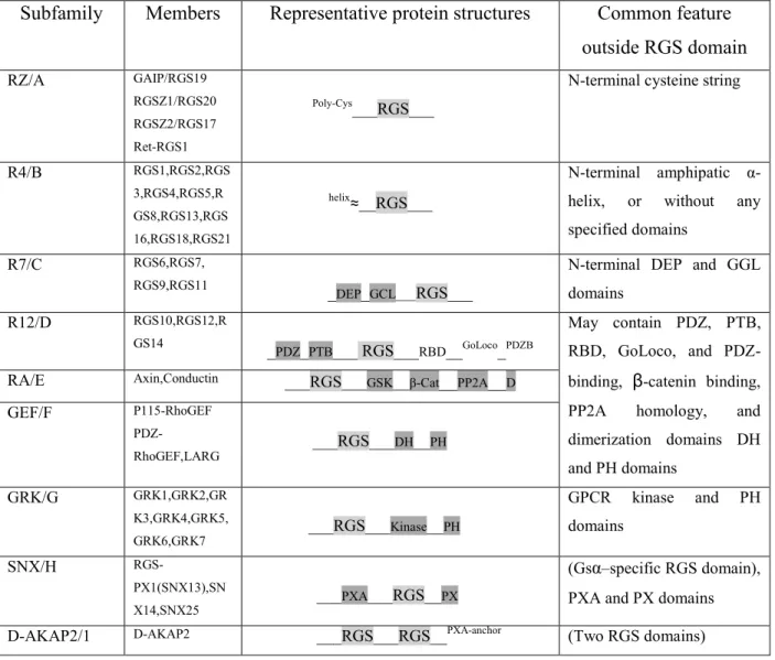
There are over twenty identified proteins in the mammalian RGS family, which share a common catalytic “RGS domain” but differ in structure, substrate specificities and tissue distribution165,166. They interact with limited selectivity for most Gα subtypes, the only exception being Gαs, for which no RGS interaction has been confirmed. According to the
similarity in sequence and features of structural domains, RGS proteins have been classified into nine subfamilies (Tab. 2). These molecules are more than just Gα GAPs167,168. For example, there is an RGS-box in the amino terminus of p115-RhoGEF, a known GEF for the monomeric G protein RhoA, which functions as a signaling bridge between GPCRs and RhoA169. Small-molecule inhibitors of the RhoGEF RGS-box that block G13α binding should reduce GPCR-mediated activation of RhoA and its downstream signaling pathways, which are involved in cellular transformation and metastatic progression.
There are at least three ways in which specificity of RGS-protein action can arise. First, at the molecular level, RGS proteins have specificity for discrete Gα subfamilies, and there is an increasing evidence for selective RGS-protein modulation of particular receptor actions. Second, each tissue expresses a distinct repertoire of RGS proteins. Finally, RGS proteins can be differentially up- or downregulated by physiological signals or pathological conditions, which can provide state-dependent specificity170.
There are increasing reports of RGS selectivity for specific GPCRs, suggesting that targeting a RGS may provide a mechanism to selectively regulate signaling through a particular GPCR171,172.
Genetic ablation of RGS activity either by deletion of a particular RGS gene or by expression of RGS-insensitive Gα subunits has dramatic physiological consequences173. For example, RGS4-deficient mice display increased sensivity to carbachol-potentiated, glucose-stimulated insulin release. Deletion of RGS9 produces a variety of neurological effects, including sensitization to morphine analgesia and reward, with decreased tolerance deficits in working memory, and motor coordination defects174,175. Knock-in mice, expressing a mutant Gαi
subunit which was unable to interact with RGS proteins176, show dramatic phenotypes, including spontaneous antidepressant-like effects as well as resistance to diet-induced obesity177,178. Thus, targeting a specific RGS protein and inhibiting protein-protein interactions, such as that between RGS and Gα subunit, may open new therapeutic strategies. In particular, molecules that are able to disrupt the RGS-Gα interaction should increase the magnitude and/or duration of G-protein signaling responses, leading to pronounced physiological effects.
Subfamily Members Representative protein structures Common feature outside RGS domain RZ/A GAIP/RGS19 RGSZ1/RGS20 RGSZ2/RGS17 Ret-RGS1 Poly-Cys___RGS___
N-terminal cysteine string
R4/B RGS1,RGS2,RGS 3,RGS4,RGS5,R GS8,RGS13,RGS 16,RGS18,RGS21 helix≈__RGS___ N-terminal amphipatic α-helix, or without any specified domains
R7/C RGS6,RGS7,
RGS9,RGS11 _DEP_GCL___RGS___
N-terminal DEP and GGL domains R12/D RGS10,RGS12,R GS14 _ PDZ_PTB___ RGS___RBD__GoLoco_PDZB May contain PDZ, PTB, RBD, GoLoco, and PDZ-binding, β-catenin binding, PP2A homology, and dimerization domains DH and PH domains
RA/E Axin,Conductin ___RGS___GSK__β-Cat__PP2A__D
GEF/F P115-RhoGEF PDZ-RhoGEF,LARG ___RGS___DH__PH GRK/G GRK1,GRK2,GR K3,GRK4,GRK5, GRK6,GRK7 ___RGS___Kinase__PH GPCR kinase and PH domains SNX/H RGS-PX1(SNX13),SN X14,SNX25 ___PXA___RGS__PX (Gsα–specific RGS domain), PXA and PX domains D-AKAP2/1 D-AKAP2 ___RGS___RGS__PXA-anchor (Two RGS domains)
β-Cat, β-catenin-binding; D, dimerization domain: D-AKAP, dual-specifocoty A-kinase anchoring protein; DEP, disheverlled/EGL-10/pleckstrin; DH, double homology; GEF, guanine nucleotide exchange factor; GGL, Gγ-like; GoLoco, Gαi/o-Loco; GRK, G protein-coupled receptor kinase; GSK. Glycogen synthase kinase 3β-binding; PDZ, PSD95/Dlg/Z0-1/2; PH, pleckstrin homology; PP2A, protein phosphatase 2A; PTB, phosphotyrosine-binding; PX, phosphatidylinositol-binding; PXA, PX-associated; RBD, Ras-binding domain; SNX, sortin nexin.
Tab. 2. Classification of RGS proteins subfamilies and their structural features (taken from Xie and Palmer179).
RGS4 as a new drug target
RGS4 is one of the most extensively studied RGS proteins. RGS4 is a relatively small protein of simple structure, in which the N-terminal domain discriminates among receptor signaling complexes. Accordingly, deletion of the N-terminal domain of RGS4 eliminated receptor selectivity and reduced potency by 104-fold180. It attenuates the intensity and duration of Gαi/0
and Gαq/11 subunits-coupled receptor signaling 181,182 and is involved in many clinical
diseases. Genetic studies indicate that RGS4 is a vulnerability factor for schizophrenia183,184. In addition, RGS4 plays important roles for dopaminergic control of striatal long-term depression, susceptibility to Parkinson’s disease185 and neural plasticity186.
RGS4 is highly expressed in the brain and robustly distributed in regions that are involved in the response to drugs of abuse and cognition processes. These regions include the prefrontal cortex, striatum, hippocampus and locus coeruleus165,187. In vitro and genetic studies have linked RGS4 to the regulation of µ-opioid receptor signaling and supported its role in morphine reward and physical dependence188-190. Indeed, GPCR and their downstream signaling partners play a crucial role in the activation of gene expression in the striatum after acute exposure to psychostimulants like amphetamine or cocaine. Not surprisingly, G-protein signaling itself is subject to tight regulation that may be disrupted in drug addiction and neuropsychiatric disorders183,191-194. Several studies suggested a potential role of RGS proteins in long-term adaptation processes observed in response to pharmacological treatment195,196 or occurring during the development of neurodegenerative diseases 193. RGS4 is known to regulate the signal of several Gαi-coupled receptors, such as metabotropic glutamate
receptors197, opioid receptors198,199, and 5-HT1 receptors but probably not dopamine D2 receptors200. On the other hand, D1 and D2 receptors regulate RGS4 gene expression; either pharmacological blockade of D1 receptors or selective stimulation of D2 receptors increases RGS4 gene expression in the striatum201,202. The tight transcriptional control to which RGS4 is submitted pleads in favour of its potential contribution to the fine tuning of D2 signaling cascade. Particularly striking is that D2 agents act on pre-synaptic D2 receptors to regulate RGS4. This observation, together with the colocalization patterns of RGS2 or RGS4 with D1 or D2 receptors, confirmed the roles of these RGS proteins in D1 and D2 signaling. Moreover, the rapid transient regulation of RGS2 mRNA and delayed transient regulation of RGS4 mRNA suggest that synergistic compensations in DA signaling potentially mediated by this RGS are temporally additive203.
The effects of RGS proteins on opioid receptors have been examined in several systems. The findings are not always consistent, probably due to the different methodologies used. It has been shown that members of the RZ, R4 and R7 subfamilies204 of RGS proteins play crucial roles not only in terminating acute opioid agonist action but also in opioid receptor desensitization, internalization, recycling, and degradation199,205, thereby affecting opioid tolerance and dependence206,207. Much work has been performed with RGS4, because of its small structure and its wide distribution in the brain, especially in brain regions important for opioid actions. In vitro, RGS4 has been shown to reverse DOP receptor agonist induced inhibition of cAMP synthesis in membranes prepared from NG108-15 cells182, and overexpression of RGS4 in HEK293 cells also attenuates morphine-, DAMGO, and DPDPE-induced inhibition of adenylyl cyclase208,209. These and other previous studies have provided evidence that RGS4 can negatively regulate opioid receptor signaling in transfected systems;
moreover Wang and colleagues210, using a short hairpin RNA (shRNA) to reduce the expression level of RGS4, showed that changes in DOP but not MOP receptor-mediated signaling occur. This finding argued in favor of a selective interaction of RGS4 with the DOP receptor. Indeed, co-immunoprecipitation studies by the same group indicated that the DOP but not the MOP receptor is closely associated with RGS4, providing further evidence for a selective interaction between RGS4 and the DOP receptor. Indeed, RGS4 interacts directly, probably through its N-terminal region211, with the GST fusion proteins of the C-tail and the third intracellular loop of the DOP receptor but only interacts with the GST-fusion C-terminal tail peptides of the MOP receptor198. On the other hand, it was hypothesized also an indirect interaction between the DOP receptor and RGS4212, perhaps mediated by an intermediate scaffold, such as spinophillin. Spinophillin is known to bind to several GPCR at the third intracellular loop and to RGS4213. Given that RGS4 is widely expressed in many brain regions165, including the amygdala, NAcc, and caudate-putamen, where DOP receptors are also highly expressed10, the selective RGS4 modulation of DOP receptor signaling may play a significant role in modulation of DOP receptor-mediated behaviors.
RGS4 inhibitors
The development of small molecule inhibitors of RGS proteins has been pursued due to their strong modulatory role in GPCR signaling214-216 and possibly therapeutic potential. The localized expression of these proteins allows to achieve tissue specificity for GPCR agonist actions170,216,217; furthermore, the rationale for targeting RGS4 relies on the up-regulation of this protein in various diseases, for example, in neuropathic pain models208.
The GTPase accelerating activity of RGS4 is regulated by phosphatidylinositol 3,4,5-triphosphate at a site far away from the Gα interaction interface218,219 (“B” site). Targeting this allosteric site217, might be a more tractable approach for inhibiting the RGS-Gα protein-protein interaction than attempting to orthosterically occlude the protein-protein-protein-protein interaction220. Developing small molecules or peptide modulators of RGS proteins is a booming field170,215,217,221-223. RGS inhibitor could act as GPCR signaling potentiator. Given alone, it would be expected to potentiate the effects of endogenous ligands, and given with a GPCR agonist, it would be expected to increase its potency or selectivity.
Most diseases resulted from changes in a complex set of signaling pathways, in which different RGS proteins are involved; on the other hand, the activity profile that would provide the best effect for one disease, may not be suitable for another disease. So while still hypothetical, it is possible to imagine that an inhibitor with a specific set of activity against
different RGS proteins or a combination of two or more inhibitors with specific targets might be valuable in the treatment of certain disease.
Two different groups published independent series of peptide inhibitors of RGS4 functions. The first series was designed to mimic the switch I region of Gαi224,225; the second series was
developed by a random yeast-two hybrid screening campaign226. The peptides derived by these two series had modest (mid-low micromolar) activity on both binding and functional assays. The first small molecule inhibitor of RGS4 was published in 2007227. This compound, CCG-4986 (4-chloro-N-[N-4-nitrophenyl)methoxysulfanyl]benzene-1-sulfonamide), was identified through a flow-cytometry protein interaction assay (FCPIA)-based high throughput screen on a diverse compound library. CCG-4986 has a 4 μM IC50 value for the inhibition of
RGS4 binding to Gα0, and shows significant selectivity for RGS4 over RGS8, its closest
relative based upon sequence homology. The GTP hydrolysis assay also confirmed the activity of this compound: CCG-4986 blocked GAP activity of RGS4. It is able to form irreversible covalent adducts with the RGS in both orthosteric (i.e. at the site of Gα binding)228,229 and allosteric interaction sites229. There is a limit in the use of CCG-4986 as a pharmacological tool, because it does not function in a cellular environment, but the development of this compound and the evidence that it is able to inhibit RGS and also to produce allosteric modulation, may provide greater specificity among RGS proteins.
Other limits related to the use of CCG-4986 regard its irreversibility and its inactivation in presence of reducing agents228,229; for these reasons several studies attempted to identify new compounds that act reversibly and retain substantial function in the presence of glutathione, a predominant intracellular reductant. In 2010, Blazer and colleagues described the first set of compounds that can reversibly inhibit RGS4. The prototypical compound of this class, CCG-63802, has an in vitro IC50 value of 10 μM in the FCPIA assay. Specifically, mutagenesis
studies predict that the binding of CCG-63802 to the “B” site of RGS4 is able to induce a conformational change; the “B” site of RGS4 is an important location for the binding of calmodulin and acidic phospholipids, which reciprocally regulate RGS GAP activity, whereby phospholipid binding inhibits the RGS function and this effect can be displaced by calmodulin218,230. Despite this family of compounds provided the first proof-of-concept that RGS proteins can be inhibited by small molecules in a reversible fashion, they do not appear to possess significant cellular activity. For this reason, even if the “B” site hypothesis has been studied from different functional angles, there are no data that specifically address how this allosteric binding results in altered GAP activity. A suitable compound to examine in depth this hypothesis needs to have a greater potency at RGS4 (~2 Log more potent than CCG-63802) and to show a smaller polar surface area than CCG-63802 to improve membrane
permeability. The CCG-50014 class of compounds includes molecules with documented cellular activity. CCG-50014 is the most potent small molecule RGS inhibitor, it irreversibly inhibits RGS4 over RGS proteins (including RGS7, RGS8, RGS16 and RGS19) with a 30 nM IC50 value. While irreversible, this compound provides the basis for studying the molecular
mechanisms of RGS allosteric inhibition.
In the present study, we used CCG-203769, which is a member of the class of compounds derived from CCG-50014, and has been synthetized by Dr. Neubig and his coworkers at the University of Michigan. CCG-203769 and correlated molecules act by inhibiting both the RGS/Gα PPI and RGS activity in a living cell and have a greater aqueous solubility than CCG-50014; moreover, their action has a measurable effect upon GPCR signaling.
Previous studies demonstrated that in β cells, M3 receptor activation potentiates glucose-stimulated insulin release231 and that this event is under RGS4-control232. Using isolated mouse islets, Blazer and colleagues attempted to determine if CCG-50014 and CCG-203769 were able to enhance the M3 activity on glucose-stimulated insulin release with promising preliminary data.
Both the CCG-63802 and the CCG-50014 classes of molecules could help provide further information on the location and the geometry of small molecules binding sites on RGS4 and to study the effects of RGS protein in vivo.
New compounds derived from these classes could be useful research tools and may potentially have relevant therapeutic roles.
Purpose
This study is based on the evidence that the opioidergic system markedly contributes to the physio-pharmacological regulation of the motor function. The overall goal of the study was to investigate the mechanisms underlying the motor responses to opioid receptor ligands, in particular the contribution of endogenous DA, and of intra-cellular modulators of the opioid receptors.
Essentially, we investigated whether different DA receptor subtypes, more specifically, pre- and post-synaptic D2 receptors, were differentially recruited in motor responses to NOP and DOP receptors agonists and antagonists, and tested the hypothesis that inhibition of RGS proteins, could amplify the motor actions of DOP receptor agonists.
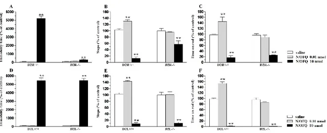
To achieve these aims, in the first part of this work, we undertook a combined pharmacological and genetic approach, using selective DA receptor antagonists, and mice carrying genetic deletions of the D2 receptor (D2R-/-)233, or its long (post-synaptic) isoform (D2L-/-)234. These data point to the involvement of D2 receptors in the motor actions of NOP receptor ligands, further suggesting that post-synaptic and pre-synaptic D2 receptor subpopulations may mediate motor facilitation and motor inhibition induced by low and high doses of NOP receptor ligands, respectively (part 1).
Using the same genetic approach, D2R-/- and D2L-/- mice were next used to dissect out the role of pre- and posts-ynaptic D2 receptors in the motor actions of DOP receptor ligands. The results indicate that genetic removal or pharmacological blockade of post-synaptic D2 receptors disclose a motor promoting action of DOP receptor ligands, suggesting the existence of a negative D2/DOP receptor interaction both at the membrane and network level. As D2R-/- mice have been considered a model of PD, this data suggested that the antiparkinsonian actions of DOP receptor agonists are DA-independent (part 2).
In the third part of this study, we used a selective small molecule inhibitor, CCG-203769 (provided by Dr R Neubig, University of Michigan), to disclose the role of RGS4 on motor activity in parkinsonian conditions, and investigate whether RGS4 blockade could amplify the antiparkinsonian response to DOP receptor ligands. We found that CCG-203769 reversed neuroleptic-induced parkinsonism, and rescued stepping activity in 6-OHDA hemilesioned mice and 6-OHDA hemilesioned rats, also producing a synergistic beneficial symptomatic response when given in combination with a DOP agonists. These data suggest that RGS4 might be a novel therapeutic target in PD (part 3).
Materials and methods
Animals
All animals used in this study were housed with free access to food and water and kept under environmentally controlled conditions (12-h light/dark cycle with light on between 07:00 and 19:00). The experimental protocols were approved by the Italian Ministry of Health (licenses n. 94/2007B and 194/2008B) and Ethical Committee of the University of Ferrara. Adequate measures were taken to minimize animal pain and discomfort.
Mice
Young male (20-25 g; 8-9 weeks old) C57BL/6J mice, 129/Sv/C57BL6J D2R+/+ and D2R-/-233 and 129/Sv/C57BL6J D2L+/+ and D2L-/-234 were used in this study. C57BL/6J mice were purchased from Harlan Italy (S. Pietro al Natisone, Italy), while genetically modified mice were provided by Emiliana Borrelli (University of California, Irvine).
Rats
Young adult male (120-125 g; 12-13 weeks old) Sprague-Dawley were used in this study. Rats were purchased from Harlan Italy (S. Pietro al Natisone, Italy).
Lesion of the DA system
In order to lesion the DA neurons located in SNc and deplete striatum of DA, different protocols were used. All lesion procedures led to an unilateral massive destruction of the nigrostriatal DA projection.
6-OHDA lesion in C57BL/6J mice
Mice were anaesthetized with a mixture of isoflurane and air, and secured in a stereotaxic frame. Unilateral lesion of nigral DA neurons was induced by injecting 6-OHDA (Tocris Bioscience, Bristol UK), dissolved in 0.02% ascorbate saline at the concentration of 3.0 μg/μl freebase 6-OHDA. Mice received 2 injections X 2 μl 6-OHDA into the striatum at the following coordinates from bregma: (i) AP= +1.0, ML =−2.1, DV= −2.9; (ii) AP= +0.3, ML = −2.3, DV= −2.9235, as previously described236. In order to assess the efficacy of the lesion, all mice were tested for spontaneous rotation and for akinesia/bradykinesia (bar and drag tests) 10 days after lesion.
6-OHDA lesion in rats
Unilateral lesion of nigro-striatal DA neurons was induced in isoflurane-anaesthetised rats145 by stereotaxically injecting 8 µg of 6-OHDA (in 4 µl of saline containing 0.02% ascorbic acid) in the right medial forebrain bundle (MFB) according to the following coordinates from bregma: AP= -4.4 mm, ML= -1.2 mm, DV= -7.8 mm below dura237.
Drug-induced rotation
The rotational model238 was used to select the rats which had been successfully lesioned with 6-OHDA. Two weeks after lesion, rats were injected with amphetamine (5 mg/Kg i.p., dissolved in saline) and only those rats performing >7 ipsilateral turns/min were enrolled in the study. This behavior has been associated with >95% loss of striatal extracellular DA levels239.
Behavioral studies
Motor activity in rodents was evaluated by means of different behavioral tests (bar, drag and rotarod test) specific for different motor abilities, as previously described145. The different tests are useful to evaluate motor functions under static or dynamic conditions. Two features that are analyzed are akinesia and bradykinesia. Akinesia appears as an abnormal absence or poverty of movements, that is associated in hemilesioned mice and rats to the loss of the ability to move the forepaw when placed on blocks at different heights (bar test). Bradykinesia is referred to slowness of movement and in particular to difficulties of adjusting its position in response to backwards dragging (drag test). The battery of tests described below, can be used to assess the degree of bradykinesia and akinesia of the animals, representing important behavioral correlates of parkinsonian symptoms. We performed these tests in a fixed sequence (bar test, drag test, and rotarod test).
Bar test
This test, also known as the catalepsy test240, measures the ability of the animal to respond to an externally imposed static posture. Each rodent was placed gently on a table and the right and left forepaws were placed alternatively on blocks of increasing heights (1.5, 3 and 6 for mice and 3, 6 and 9 for rats). The immobility time (in sec) of each forepaw on the blocks was recorded (cut-off time 20 sec per step, 60 sec maximum). Akinesia was calculated as total time spent on the blocks by each forepaw.
Drag test
The test (modification of the “wheelbarrow” test241), measures the ability of the animal to balance its body posture using forelimbs in response to an externally imposed dynamic stimulus (backward dragging145). Each rodent was gently lifted by the tail (allowing the forepaws on the table) and dragged backwards at a constant speed (about 20 cm/sec) for a fixed distance (100 cm). The number of touches made by each forepaw was counted by two separate observers (mean between the two forepaws).
Rotarod test
This test analyzes the ability of the rodents to run on a rotating cylinder (diameter 8 cm) and provides information on different motor parameters such as coordination, gait, balance, muscle tone and motivation to run242. The fixed-speed rotarod test was employed according to a previously described protocol146,149,152. Briefly, animals were tested at stepwise increasing speeds (180 sec each) and time spent on the rod calculated (in sec).
Data presentation and statistical analysis
Data are expressed as means ± SEM of n determinations per group or as percentages of the control sessions. Different statistical analyses were performed, as appropriate, using the Student’s t test, one-way repeated measures (RM) ANOVA followed by the Newman-Keuls test or the Bonferroni’s multiple comparisons test. P values <0.05 were considered to be statistically significant.
Drugs
6-OHDA hydrobromide, amisulpride, raclopride, SCH23390 and SNC-80 were purchased from Tocris Bioscience (Bristol, UK). S33084 was provided by Institut de Recherches Servier (Croissy-sur-Seine, France). N/OFQ and J-113397 were synthetized in the laboratories of the Department of Pharmaceutical Chemistry at the University of Ferrara. CCG-203769 was provided by Dr. Richard Neubig (University of Michigan). All drugs were freshly dissolved in the vehicle just prior to use.
Results
Part 1. Role of endogenous DA in the motor action exerted by NOP receptor ligands.
In the first part of the present study we investigated the motor profiles of NOP receptor ligands in C57BL/6J mice, using static and dynamic tests providing complementary information on motor parameters: the bar, drag and rotarod test. Then, to evaluate the contribution of the various DA receptor subtypes in the motor action of NOP receptor ligands, we used selective DA receptor antagonists and analysed the impact of sub-threshold doses of these antagonists on the motor effects of NOP receptor ligands. These experiments revealed the involvement of D2 receptors in the motor responses to NOP receptor ligands, and suggested that different D2 receptor subpopulations might mediate motor facilitation and inhibition observed with the lower and high doses of the NOP antagonist J-113397. To confirm pharmacological data we adopted a genetic approach, by testing N/OFQ and the NOP receptor antagonist J-113397 in D2 knockout mice.
1.1 The NOP receptor antagonist J-113397, and exogenous N/OFQ dually modulated motor activity.
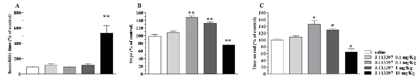
Basal activity of C57BL/6J mice was 0.8 ± 0.1 sec (immobility time in the bar test), 16.5 ± 0.9 steps (drag test) and 937 ± 62.1 sec (time on rod). J-113397 increased the immobility time (i.e. inhibited movement initiation) in the bar test at the highest dose tested (10 mg/Kg) (Fig. 1A) and dually modulated stepping activity and rotarod performance (Fig. 1B and C), causing facilitation at low doses (0.3-1 mg/Kg) and inhibition at higher doses.
Since data obtained with the NOP antagonist suggested a dual regulation of motor activity by endogenous N/OFQ, we investigated whether exogenous N/OFQ could replicate this pattern. N/OFQ, given i.c.v., monotonically increased immobility time from 0.1 nmol onwards (Fig. 2A). Conversely, it dually regulated stepping activity and rotarod performance (Fig. 2B and C), causing motor facilitation at 0.01 nmol and motor inhibition at 0.1-10 nmol.
Fig. 1. J-113397 dually modulated motor activity in C57BL/6J mice. Systemic administration of J-113397 (0.1-10 mg/Kg, i.p.) affected motor performance in the bar (A), drag (B) and rotarod (C) tests. All tests were performed before (control session) and after (10 min) drug injection. Data are means ± SEM of 6 determinations per group and were expressed as percentage of the control session. *p<0.05, p**<0.01 different from saline (one-way ANOVA followed by the Bonferroni test).
Fig. 2. N/OFQ dually modulated motor activity in C57BL/6J mice. I.c.v. injection of N/OFQ (0.1-30 nmol) affected motor performance in the bar (A), drag (B) and rotarod (C) tests. All tests were performed before (control session) and after (10 min) N/OFQ injection. Data are means ± SEM of 6 determinations per group and were calculated as percentage of the control session. *p<0.05, p**<0.01 different from saline (one-way ANOVA followed by the Bonferroni test).
1.2 Dopamine receptor antagonists differentially modulated motor actions of NOP receptor ligands.
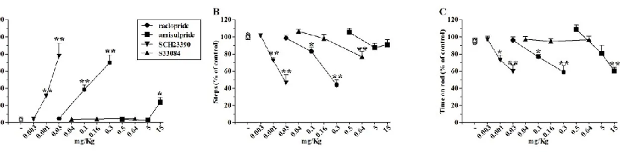
The D1/D5 (SCH23390), D2/D3 (raclopride and amisulpride) and D3 (S33084) receptor antagonists were administered to C57BL/6J mice to evaluate the contribution of various DA receptor subtypes in the motor action exerted by NOP receptor ligands. The D2/D3 receptor antagonist raclopride, given alone (0.03-0.3 mg/Kg, i.p.), caused a dose-dependent and long-lasting elevation of immobility time, and reductions in the number of steps and time on rotarod (Fig. 3A-C). Amisulpride replicated the motor inhibiting action of raclopride in the bar and rotarod test (5-15 mg/Kg, i.p.) but did not affect the stepping activity in the drag test (Fig. 3A-C). The D1/D5 antagonist SCH23390 produced consistent motor inhibition in all three tests, being effective at 0.01 mg/Kg (i.p.) (Fig. 3A-C), whereas the D3 antagonist S33084 did not produce any marked changes in motor activity; the only effect observed was a mild inhibition of stepping at 0.64 mg/Kg (i.p., Fig. 3B).
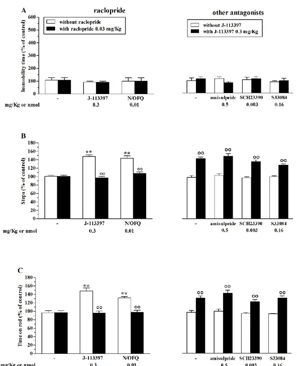
We then selected doses of DA receptor antagonists that did not produce effect on motor activity, and analysed their impact on the motor effects of NOP receptor ligands. Low doses of N/OFQ or the NOP receptor antagonist J-113397 were ineffective in the bar test, both in the absence and in the presence of DA receptor antagonists (Fig. 4A). Raclopride (0.03 mg/Kg) prevented the increase in stepping activity (Fig. 4B) and rotarod performance (Fig. 4C) induced by J-113397 (0.3 mg/Kg), Whereas amisulpride (0.5 mg/Kg), SCH23390 (0.003 mg/Kg) and S33084 (0.16 mg/Kg) were ineffective (Fig. 4B-C). Raclopride also prevented the motor facilitation induced by the low dose of N/OFQ (0.01 nmol) (Fig. 4A-C). On the other hand, motor inhibition induced by high doses of the NOP receptor antagonist J-113397 (10 mg/Kg) in the bar (Fig. 5A), drag (Fig. 5B) and rotarod (Fig. 5C) tests was prevented by amisulpride but not by raclopride (Fig. 5A-C). In the drag and rotarod tests, amisulpride reversed motor inhibition induced by J-113397, resulting in long-lasting stimulation (Fig. 5B and C). These data suggest the involvement of D2 receptors in the motor actions exerted by the NOP receptor antagonist J-113397. Considering the different responses to raclopride and amisulpride, we also speculate that different D2 receptor subpopulations might mediate motor facilitation and motor inhibition observed with lower and high doses the NOP antagonist. To dissect out the contribution of pre- and post-synaptic D2 receptors on the motor profile of NOP receptor ligands we undertook a genetic approach.
Fig. 3. DA receptor antagonists impaired motor activity in C57BL/6J mice. Administration of the D2/D3 receptor antagonists raclopride (0.03-0.3 mg/Kg, i.p.) and amisulpride (0.5-15 mg/Kg, i.p.), the D1/D5 receptor antagonist SCH23390 (0.003-0.03 mg/Kg, i.p.) and the D3 receptor antagonist S33084 (0.04-0.64 mg/Kg, i.p.) produced dose-dependent motor inhibition. Motor activity has been evaluated as immobility time in the bar test (A), number of steps in the drag test (B) and time spent on rod in the rotarod test (C). All tests were performed before (control session) and 30 min after drug administration. Data are means ± SEM of 6 determinations per group and are expressed as percentage of the control session. *p<0.05, **p<0.01 different from saline or vehicle (ANOVA followed by Newman Keuls test for multiple comparisons).
Fig. 4. Motor facilitation induced by NOP receptor ligands was selectively prevented by raclopride. Pretreatment with D2/D3 receptor antagonist raclopride (0.03 mg/Kg, i.p.) prevented motor facilitaton caused by low doses of N/OFQ (0.01 nmol, i.c.v.) or the NOP receptor antagonist J-113397 (0.3 mg/Kg, i.p.). Conversely, pretreatment with the D2/D3 receptor antagonist amisulpride (0.5 mg/Kg, i.p.), the D1/D5 receptor antagonist SCH23390 (0.003 mg/Kg, i.p.) and the D3 receptor antagonist S33084 (0.16 mg/Kg, i.p.) did not affect motor facilitation induced by J-113397 (0.3 mg/Kg, i.p.). Motor activity has been evaluated as immobility time in the bar test (A), number of steps in the drag test (B) and time on rod in the rotarod test (C). All tests were performed before (control session) and 10 min after NOP receptor ligands administration. Data are means ± SEM of 6 determinations per group and are expressed as percentage of the control session. **p<0.01 different from saline or vehicle; °°p<0.01 different from the same group in the absence of raclopride or J-113397 (ANOVA followed by Newman Keuls test for multiple comparisons).
Fig. 5. Motor inhibition induced by the NOP receptor antagonist J-113397 was selectively prevented by amisulpride. Pretreatment with the D2/D3 receptor antagonist amisulpride (0.5 mg/Kg, i.p.) prevented motor inhibition caused by high doses of the NOP receptor antagonist J-113397 (10 mg/Kg, i.p.). Pretreatment with the D2/D3 receptor antagonist raclopride (0.03 mg/Kg, i.p.) did not affect motor inhibition induced by J-113397 (10 mg/Kg, i.p.). Motor activity has been evaluated as immobility time in the bar test (A), number of steps in the drag test (B) and time spent on the rod in the rotarod test (C). All tests were performed before (control session) and 10 min after J-113397 injection. Data are means ± SEM of 6 determinations per group and are expressed as percentage of the control session. *p<0.05, **p<0.01 different from saline or vehicle; °°p<0.01 different from the same group in the absence of DA receptor antagonists (ANOVA followed by Newman Keuls test for multiple comparisons).